Mapping the Human Memory B Cell and
Serum Neutralizing Antibody Responses
to Dengue Virus Serotype 4 Infection
and Vaccination
Usha K. Nivarthi,
aNurgun Kose,
bGopal Sapparapu,
b,cDouglas Widman,
aEmily Gallichotte,
aJennifer M. Pfaff,
eBenjamin J. Doranz,
eDaniela Weiskopf,
fAlessandro Sette,
fAnna P. Durbin,
g,hSteve S. Whitehead,
iRalph Baric,
aJames E. Crowe, Jr.,
b,c,dAravinda M. de Silva
aDepartment of Microbiology and Immunology, University of North Carolina School of Medicine, Chapel Hill, North Carolina, USAa; The Vanderbilt Vaccine Center, Vanderbilt University Medical Center, Nashville, Tennessee, USAb; Department of Pediatrics, Vanderbilt University Medical Center, Nashville, Tennessee, USAc; Department of Pathology, Microbiology and Immunology, Vanderbilt University Medical Center, Nashville, Tennessee, USAd; Integral Molecular Inc., Philadelphia, Pennsylvania, USAe; La Jolla Institute for Allergy and Immunology, La Jolla, California, USAf; Johns Hopkins Bloomberg School of Public Health, Department of International Health, Baltimore, Maryland, USAg; Center for Immunization Research, Johns Hopkins Bloomberg School of Public Health, Baltimore, Maryland, USAh; Laboratory of Infectious Diseases, NIAID, National Institutes of Health, Bethesda, Maryland, USAi
ABSTRACT
The four dengue virus (DENV) serotypes are mosquito-borne flaviviruses
responsible for dengue fever and dengue hemorrhagic fever. People exposed to
DENV develop antibodies (Abs) that strongly neutralize the serotype responsible for
infection. Historically, infection with DENV serotype 4 (DENV4) has been less
com-mon and less studied than infections with the other three serotypes. However,
DENV4 has been responsible for recent large and sustained epidemics in Asia and
Latin America. The neutralizing antibody responses and the epitopes targeted
against DENV4 have not been characterized in human infection. In this study, we
mapped and characterized epitopes on DENV4 recognized by neutralizing antibodies
in people previously exposed to DENV4 infections or to a live attenuated DENV4
vaccine. To study the fine specificity of DENV4 neutralizing human antibodies, B cells
from two people exposed to DENV4 were immortalized and screened to identify
DENV-specific clones. Two human monoclonal antibodies (MAbs) that neutralized
DENV4 were isolated, and their epitopes were finely mapped using recombinant
vi-ruses and alanine scan mutation array techniques. Both antibodies bound to
quater-nary structure epitopes near the hinge region between envelope protein domain I
(EDI) and EDII. In parallel, to characterize the serum neutralizing antibody responses,
convalescence-phase serum samples from people previously exposed to primary
DENV4 natural infections or a monovalent DENV4 vaccine were analyzed. Natural
in-fection and vaccination also induced serum-neutralizing antibodies that targeted
similar epitope domains at the EDI/II hinge region. These studies defined a target of
neutralizing antigenic site on DENV4 targeted by human antibodies following
natu-ral infection or vaccination.
IMPORTANCE
The four serotypes of dengue virus are the causative agents of
den-gue fever and denden-gue hemorrhagic fever. People exposed to primary DENV
infec-tions develop long-term neutralizing antibody responses, but these principally
rec-ognize only the infecting serotype. An effective vaccine against dengue should elicit
long-lasting protective antibody responses to all four serotypes simultaneously. We
and others have defined antigenic sites on the envelope (E) protein of viruses of
dengue virus serotypes 1, 2, and 3 targeted by human neutralizing antibodies. The
Received18 October 2016Accepted21 December 2016
Accepted manuscript posted online28 December 2016
CitationNivarthi UK, Kose N, Sapparapu G, Widman D, Gallichotte E, Pfaff JM, Doranz BJ, Weiskopf D, Sette A, Durbin AP, Whitehead SS, Baric R, Crowe JE, Jr, de Silva AM. 2017. Mapping the human memory B cell and serum neutralizing antibody responses to dengue virus serotype 4 infection and vaccination. J Virol 91:e02041-16.https://doi.org/10.1128/ JVI.02041-16.
EditorDouglas S. Lyles, Wake Forest University
Copyright© 2017 American Society for Microbiology.All Rights Reserved.
Address correspondence to James E. Crowe, Jr., [email protected], or Aravinda M. de Silva, [email protected].
epitopes on DENV4 E protein targeted by the human neutralizing antibodies and
the mechanisms of serotype 4 neutralization are poorly understood. Here, we report
the properties of human antibodies that neutralize dengue virus serotype 4. People
exposed to serotype 4 infections or a live attenuated serotype 4 vaccine developed
neutralizing antibodies that bound to similar sites on the viral E protein. These
stud-ies have provided a foundation for developing and evaluating DENV4 vaccines.
KEYWORDS
Dengue virus serotype 4, human, neutralization, antibody responses,
epitope, infection, memory B cells, vaccination
D
engue virus (DENV) is a mosquito-borne positive-sense RNA virus belonging to the
Flaviviridae
family (1). Dengue is transmitted to people by
Aedes aegypti
or
Aedes
albopictus
mosquitoes (2, 3). Recent estimates indicate that nearly 400 million people
are infected worldwide each year, which makes dengue the most common and serious
vector-borne disease of humans (4).
While the majority of DENV infections are asymptomatic, symptomatic infections
can cause disease in a spectrum ranging from mild dengue fever to severe dengue
hemorrhagic fever and dengue shock syndrome (5). A primary DENV infection provides
lifelong protection against disease caused by the infecting homologous serotype in
most subjects (6). A secondary infection with virus of a heterologous serotype increases
the risk of developing severe dengue hemorrhagic fever (7). To understand the
molecular basis of a protective DENV antibody response, it is critical not only to map
the epitopes of strongly neutralizing human monoclonal antibodies (hMAbs) but also
to characterize the polyclonal neutralizing antibody responses to viruses of all the four
serotypes after natural infection. This knowledge is critical for evaluating antibody
responses to vaccination and improved second-generation vaccine design.
The DENV envelope (E) glycoprotein is required for viral binding and entry into cells
(8, 9). E protein is also the main target of neutralizing antibodies (10). The four serotypes
(DENV1 to DENV4) have variations of 25% to 40% in the amino acid sequence of the E
protein (11, 12). The E protein monomer consists of three domains (envelope protein
domain I [EDI], EDII, and EDIII), and two of these protomers form head-to-tail dimers in
viral particles. Three dimers lie parallel to each other in the particles, forming a raft (13,
14), and 30 of these rafts are arranged in a herringbone pattern on the mature virion.
Unlike the DENV neutralizing antibody response in mice, which principally targets
simple epitopes in EDIII (15, 16), nearly all human neutralizing antibodies target
complex quaternary structure E protein epitopes that are displayed on intact dengue
virions but not on soluble forms of E protein after natural infections (17, 18). Epitopes
of human type-specific neutralizing antibodies against DENV1, DENV2, and DENV3 have
been mapped to quaternary structure epitopes that span different E protein molecules
(18–21).
DENV4 has been less studied than the other serotypes. Kostyuchenko and
col-leagues recently solved a near-atomic-resolution structure of DENV4 (22). DENV4
surface E molecules are tightly packed compared to those of other serotypes, which
may explain the more rigid and stable nature of DENV4. DENV4 type-specific and
neutralizing MAb 5H2 was isolated from a chimpanzee infected with DENV4 (23). 5H2
binds to an epitope on the lateral ridge of EDI (23, 24). However, the epitopes targeted
by DENV4-specific antibodies in humans have not been characterized. The goal of the
current study was to map the antigenic sites on DENV4 recognized by antibodies in
people exposed to primary DENV4 infections or a monovalent live attenuated DENV4
vaccine.
RESULTS
infections or people who had received a monovalent live attenuated DENV4 vaccine
(Table 1) (25, 26).
Individuals exposed to DENV have specific antibodies in circulation as well as
DENV-specific memory B cells (MBCs). Some of the DENV-specific antibodies in
circu-lation bind only to viruses of the serotype of the previous infection (type specific), while
others cross-react with two or more serotypes. Using serum samples from people
exposed to DENV4 natural infections or a monovalent live attenuated DENV4 vaccine,
we performed antibody depletion studies to determine the relative contributions of
serotype cross-reactive or type-specific antibodies to DENV4 neutralization. Polystyrene
beads coated with the homotypic (DENV4) or heterotypic (DENV2) DENV serotypes
were incubated with the immune serum samples to deplete specific populations of
antibodies. Depletion of antibodies was confirmed by enzyme-linked immunosorbent
assay (ELISA) before using the samples in DENV4 neutralization assays. Depleting the
DENV4 immune serum samples with the homotypic DENV4 antigen led to the removal
of nearly all the DENV-specific (serotype cross-reactive and DENV4 type-specific)
anti-bodies in the samples. As anticipated, depletions performed with DENV4 antigen led to
a large drop in levels of DENV4 neutralizing antibodies in both DENV4 infection and
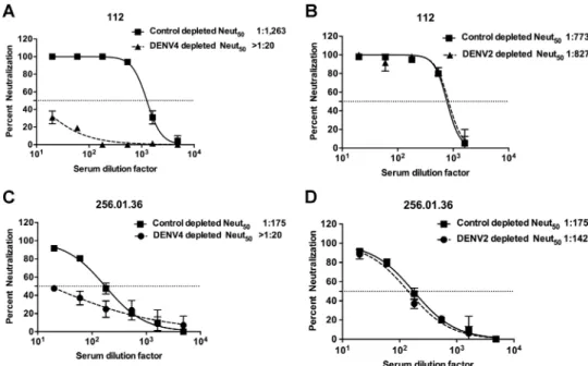
vaccine sera (Fig. 1A and C and Table 2). Depleting DENV4 immune sera with
hetero-typic DENV2 antigen led to the removal of serotype cross-reactive but not of DENV4
type-specific antibodies. There was minimal to no reduction in neutralization of DENV4
in sera depleted with DENV2 antigen (Fig. 1B and D and Table 2). The control depletions
for subject 112 yielded different neutralization titers in the homotypic and heterotypic
depletions (1,263 versus 773). This could have been because these control depletions
were performed in separate experiments using different lots of the serum aliquots from
the same individual. However, the trends remained the same for the neutralization
titers against all four serotypes, with DENV4 titers being the highest in both cases.
Overall, these results demonstrate that type-specific antibodies were primarily
responsible for DENV4 neutralization in serum samples collected following natural
infection or monovalent DENV4 vaccination.
Isolation of DENV4 neutralizing human monoclonal antibodies.
To further
characterize the B cell response to DENV4 infection, we transformed B cells from two
DENV4-immune individuals (subjects 002 and 112) and isolated hMAbs, as previously
described (27). The transformed B cell culture supernatants were screened for binding
to DENV4. On the basis of the number of positive wells and the number of transformed
TABLE 1Panel of DENV immune seraSerum type Serum Yr of infection
Location of infection
Time between infection and blood draw (yrs)
Reciprocal of Neut50titers against DENV1 to DENV4a
DENV1 DENV2 DENV3 DENV4 DENV4 immune
sera
002 1994 Guatemala 15 ⬍20 ⬍20 29 77
102 2007 Honduras 2 ⬍20 ⬍20 41 159
112 2001 Nicaragua 2 128 346 175 1,639
07/333 Unknown Thailand Unknown ⬍20 153 367 >1,280 06/302 Unknown Thailand Unknown ⬍20 32 60 >1,280
06/105 Unknown Thailand Unknown ⬍20 26 91 685
DENV2 immune serum
08/90 Unknown Thailand Unknown 20 >1,280 60 32
DENV3 immune serum
118 2009 Nicaragua 1 60 32 980 76
DENV4 vaccine immune serab
256.03.36 ⬍20 ⬍20 ⬍20 142
256.03.38 ⬍20 ⬍20 ⬍20 148
256.03.57 ⬍20 ⬍20 ⬍20 144
256.03.68 ⬍20 ⬍20 ⬍20 988
aThe Neut
50titer values in bold signify the highest Neut50reciprocal titers for each sample. Each value depicts the mean Neut50titer from two replicates.
B cells tested (determined by average colony counts in transformed wells), the
fre-quencies of DENV-specific B cells in circulation were estimated to be 0.19% and 0.20%
of transformable B cells for subjects 002 and 112, respectively. Previously, it was
reported that there may be a long-term set point frequency of about 0.1% to 0.2%
DENV-specific B cells in the circulating memory B cell pool following DENV infection
(28), and the frequencies of 0.19% and 0.20% in the two subjects studied here are
consistent with those previous estimates. Note that, even though the frequencies of
DENV4-specific B cells determined for subjects 002 and 112 were similar, their serum
titers were different. As mentioned above, the B cell response to primary DENV
infection is a mix of DENV serotype cross-reactive and type-specific antibodies.
More-FIG 1DENV4 is neutralized by type-specific antibodies in human primary infection immune sera or vaccine immune sera. Primary DENV4-immune serum sample 112 (A and B) and DENV4 NIH monovalent vaccine serum sample 256.01.36 (C and D) were depleted of antibodies binding to DENV4 antigen (A and C) or DENV2 antigen (B and D). Control depletions were performed using bovine serum albumin as an antigen. Results presented here for antibody depletions are representative of data obtained with three primary DENV4 immune sera and four DENV4 monovalent vaccine sera (Table 2). Results are from 2 technical replicates in one experiment. Each point represents the mean neutralization value from the two replicates, and the error bars depict the standard deviations.TABLE 2DENV4 neutralization by human immune sera depleted of DENV binding antibodiesa
Serum type Sample ID
Homotypic depletionb Heterotypic depletionc
Control depleted (Neut50)
DENV4 depleted (Neut50)
% loss of neutralization (meanⴞSD)
Control depleted (Neut50)
DENV2 depleted (Neut50)
% loss of neutralization (meanⴞSD) DENV4 immune
sera
002 58 ⬍20 100 82 84 0
102 112 43 62 113 98 13
112 1,263 ⬍20 100 773 827 0
DENV4 vaccine immune sera
256.03.36 175 ⬍20 100 175 142 19
256.03.38 61 ⬍20 100 61 83 0
256.03.57 53 ⬍20 100 98 75 23
256.03.68 425 160 62 409 271 34
aA U937⫹DC-SIGN flow cytometry-based neutralization test was performed on human DENV4 immune sera depleted of DENV4 (homotypic) or DENV2 (heterotypic)
binding antibodies, and Neut50(i.e., the dilution factor required to neutralize 50% of infection) values against DENV4 were calculated in these depleted immune sera. Percent loss of neutralization was calculated as follows: % loss of neutralization⫽100⫺[(DENV-depleted Neut50/control-depleted Neut50)⫻100]. The mean percentages of loss in neutralization between homotypic and heterotypic depletions in both naturally infected and vaccine immune sera were significant (P⫽0.01 andP⫽0.001, respectively) by two-samplettests performed to compare the percentages of loss of neutralization between homotypic and heterotypic depletions in both naturally immune and vaccinated serum samples. Each value represents the mean Neut50titer from two replicates.
over, a relatively small fraction of the virus-specific B cells produce neutralizing Abs
(29–31). The serum neutralizing Abs are derived from long-lived plasma cells (LLPCs) in
the bone marrow and not the memory B cell compartment. Previous studies have also
reported a weak and inconsistent correlation between DENV-specific or other viral
antigen-specific antibody titers in the serum and the frequency of antigen-specific
memory B cells in the blood (32–34).
We also determined the DENV serotype specificity of all the positive B cell culture
supernatants from subject 112. Of the 34 DENV antigen-reactive supernatants,
anti-bodies in 32% bound only to DENV4 (type-specific) and in 68% bound to two or more
serotypes (cross-reactive). From the EBV-transformed B cell lines secreting DENV
antigen-reactive antibodies, we isolated 8 human B cell hybridoma cell lines, as
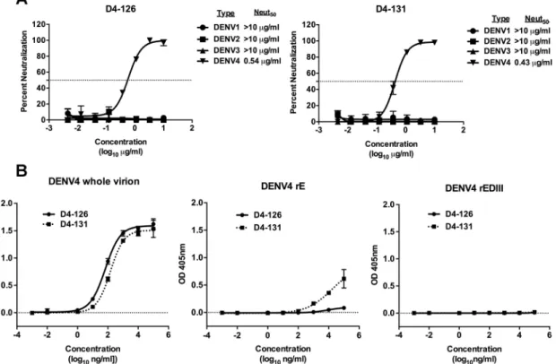
previously described (28). Two cell lines, designated D4-126 and D4-131, secreted
DENV4 type-specific and neutralizing hMAbs with Neut
50values (defined as the dilution
factor required to neutralize 50% of infection) of 0.54
g/ml and 0.43
g/ml,
respec-tively (Fig. 2A). Both of these antibodies were isolated from subject 002. To characterize
the binding properties of D4-126 or D4-131 hMAbs further, we performed binding
assays with whole DENV4 virions, recombinant E (rE) or rEDIII proteins of DENV4, and
increasing concentrations of hMAb D4-126 or D4-131. The two hMAbs bound to whole
DENV4 particles similarly (Fig. 2B). HMAb D4-126 did not bind to rE protein, whereas
D4-131 exhibited low levels of binding to rE protein at high concentrations (
⬎
10
g/ml) (Fig. 2B). The hMAbs did not bind to rEDIII protein (Fig. 2B). These studies
revealed that neutralizing hMAb D4-126 or D4-131 bound best to intact DENV4 virions.
However, it is also possible that MAb D4-126 or D4-131 could bind to E dimers formed
from rE monomers at higher concentrations as demonstrated by the D4-131 MAb
binding to rE at higher concentrations.
Additionally, these antibodies were not cross-reactive to the Zika virus (H/PF/2013
and MR 766 strains) and remained type specific to DENV4.
hMAb neutralization of different DENV4 strains.
To determine if hMAbs D4-126
and D4-131 neutralized diverse strains of DENV4, we used a panel of recombinant
isogenic DENV4 expressing the E protein from different DENV4 genotypes and
labora-tory strains (Table 3). The hMAbs equally neutralized all variants tested, except for a
Cambodia 2010 genotype I (GI) strain, which was not neutralized by D4-126 and was
neutralized less efficiently by D4-131 than by other strains tested (Fig. 3).
Mapping the epitopes of DENV4 neutralizing hMAbs.
Human DENV1, DENV2,
and DENV3 type-specific neutralizing antibodies often bind to quaternary structure
epitopes centered on the EDI/II hinge (17–19, 21, 38) and/or the EDIII region (2, 20, 21,
39, 40). Recently, we demonstrated that it is possible to recover recombinant chimeric
DENVs displaying E protein domains or epitopes from viruses of two different serotypes
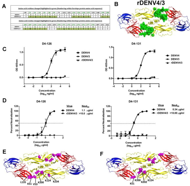
(38, 39). We used a rDENV4 strain with a mutated EDI/II hinge region and EDII region
(rDENV4/3) to map the binding sites of hMAbs D4-126 and D4-131 (Fig. 4A and B). The
recombinant virus has 25 amino acid replacements at different positions that are
highlighted in green (Fig. 4A and B) (unpublished data). We did not detect any binding
or neutralizing activity for the hMAb D4-126 or D4-131 with the rDENV4/3, indicating
that the DENV4 EDI/II hinge residues are part of the epitope recognized by these MAbs
(Fig. 4C and D).
As an alternative approach to mapping the epitopes of D4-126 and D4-131, both
hMAbs were screened by shotgun mutagenesis against a comprehensive mutation
library in which nearly every residue within the precursor membrane (prM) and E was
individually mutated to alanine, as described previously (35). Residues were identified
as critical to binding of the DENV4 hMAb if they did not bind to the DENV4 hMAb but
did bind to other conformation-dependent MAbs. Six amino acids (K51, V53, K124,
L135, K200, and K234) in the EDI/II hinge and EDII regions were critical for binding of
D4-126 (Fig. 4E). K51, V53, K124, and K200 were among the residues that were altered
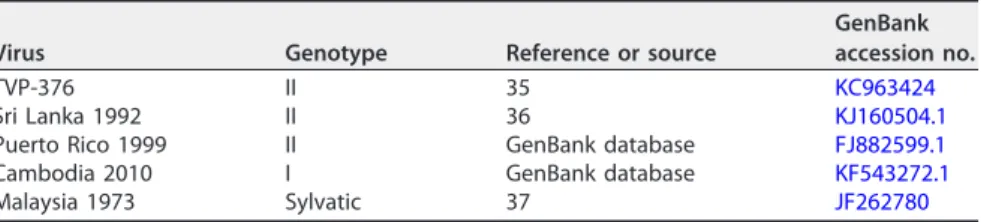
TABLE 3Characteristics of DENV4 strains used in this studyVirus Genotype Reference or source
GenBank accession no.
TVP-376 II 35 KC963424
Sri Lanka 1992 II 36 KJ160504.1
Puerto Rico 1999 II GenBank database FJ882599.1
Cambodia 2010 I GenBank database KF543272.1
Malaysia 1973 Sylvatic 37 JF262780
in rDENV4/3. Four amino acids (K51, K124, K200, and K202) within the EDI/II hinge and
EDII region were critical for binding of D4-131 (Fig. 4F). K51, K124, and K200 were
among the residues that were altered in rDENV4/3. These data validated our
observa-tions that the EDI/II hinge region residues are critical for binding of D4-126 and D4-131
and also indicated that the epitopes differ slightly between the two hMAbs.
DENV4 neutralizing hMAbs define epitopes targeted by serum antibodies in
DENV4-immune individuals.
HMAbs D4-126 and D4-131 were isolated from
circulat-ing memory B cells. Serum antibodies are generally thought to derive from secretion of
long-lived plasma cells (LLPCs) residing in the bone marrow. To determine if DENV4
polyclonal serum neutralizing antibodies in immune sera also targeted the D4-131 and
D4-126 epitopes, we performed competition-binding assays with DENV4 immune
serum samples and labeled DENV4-specific MAbs. As depicted in Fig. 5, DENV4 immune
sera effectively blocked the binding of each of the MAbs, whereas DENV2 or DENV3
immune sera had marginal effects on MAb binding. The magnitude of the DENV4
immune responses (based on their neutralization titers for DENV4) in serum samples
correlated with the ability to block 50% of the binding of D4-126 or D4-131. These
findings indicated that there are antibodies in DENV4 polyclonal immune serum
samples that bind to sites similar to those that overlap tightly with the epitopes
recognized by hMAbs D4-126 and D4-131.
To further assess the epitope specificity of functionally neutralizing antibodies in
DENV4 sera collected following infection or experimental vaccination, we performed
neutralization assays with rDENV4/3, which had lost the epitopes recognized by hMAbs
D4-126 and D4-131. Neutralization assays using the rDENV4/3 strain were performed
with sera from six subjects with prior natural DENV4 infection and sera from 4 subjects
previously immunized with a DENV4 live attenuated vaccine. The percent loss of
neutralization was calculated as follows: percent loss of neutralization
⫽
100
⫺
[(rDENV4/3 Neut
50/DENV4 Neut
50)
⫻
100] (Table 4). Sera from both types of immune
donors neutralized the rDENV4/3 strain less efficiently than sera from wild-type (WT)
DENV4, although to differing extents. The mean percentages of loss in neutralization
with rDENV4/3 in both naturally infected immune sera and vaccine sera were significant
(P
⫽
0.005 and
P
⫽
0.002, respectively) by one-sample
t
test. Of the six DENV4
postinfection sera tested, those from three subjects (002, 102, and 07/333) showed a
⬎
60% loss in neutralization against rDENV4/3, while the remaining infection sera
displayed a more modest or no detectable reduction in neutralization (Fig. 6A). All of
the vaccine sera tested also showed a
⬎
60% loss in neutralization against the rDENV4/3
strain (Fig. 6B). These results suggested that the EDI/II hinge region is a target of
polyclonal type-specific DENV4 neutralizing antibodies.
DISCUSSION
Sustained humoral immunity depends on LLPCs to maintain protective levels of
antibody in serum and on MBCs, which comprise a subset of cells poised to expand and
adapt in response to subsequent exposure to the infecting pathogen (41). In this study,
we characterized the properties of MBC- and LLPC-derived human antibodies that
neutralize DENV4. Although people exposed to DENV4 infections developed serotype
cross-reactive and type-specific antibodies, our results show that the type-specific
antibodies were the principal determinants of neutralization of DENV4. Using MAbs
isolated from the MBCs of people exposed to DENV4, we identified epitopes centered
on the EDI/II hinge as the target of DENV4 neutralizing antibodies. In people exposed
to DENV4 infections or a live attenuated vaccine candidate, both MBC- and
LLPC-derived polyclonal neutralizing antibodies also recognized epitopes centered on the
EDI/II hinge of DENV4.
Substantial progress has been made in understanding the epitopes targeted by
human antibodies that neutralize DENV serotypes 1, 2, and 3 (2, 18–21, 30), whereas
serotype 4 is relatively less studied. The two DENV4 neutralizing MAbs whose results are
reported in this study were sensitive to changes in or near the EDI/II hinge region. The
hinge region plays a critical role in the conformational change that the E protein
undergoes at low pH to fuse to the endosomal membrane allowing viral uncoating and
the release of viral RNA into the cellular cytoplasm (42). Because these epitopes are
located in this region, it is reasonable to hypothesize that these DENV4 hMAbs act by
FIG 6The DENV4 EDI/II hinge region is a target of neutralizing antibodies in people exposed to DENV4 infection or to an experimental vaccine. A U937⫹DC-SIGN flow cytometry-based neutralization assay was performed with primary DENV4 infection sera (A) or DENV4 monovalent vaccine sera (B) and WT DENV4 or rDENV4/3. The 50% neutralization (Neut50) titers were calculated and plotted. Values corresponding to loss of neutralization against rDENV4/3
for each sample are indicated. Samples that did not block 50% of infection at the highest concentration were assigned a value of 5. Results are from 2 technical replicates in one experiment. Each point represents the mean neutralization value from the two replicates.
TABLE 4Percent loss in neutralization of polyclonal DENV4 immune and vaccine sera against rDENV4/3a
Serum type
Serum sample
Reciprocal of Neut50titers against DENV4 and rDENV4/3
% loss in neutralization DENV4 rDENV4/3
DENV4 immune sera
002 115 44 62
102 153 31 80
112 1,746 1,609 8
07/333 1,256 399 69
06/302 1,660 1,058 37
06/105 399 204 49
DENV4 vaccine immune sera
256.03.36 562 178 69
256.03.38 255 5 98
256.03.57 153 5 97
256.03.68 1,167 276 76
aA U937⫹DC-SIGN flow cytometry-based neutralization assay was performed with primary DENV4 infection
preventing conformational changes in the E protein required for fusion, entry, and
initiation of a productive viral infection.
As reported previously with DENV3 natural isolates (43), some DENV4 epitopes
targeted by neutralizing mouse MAbs differ in potency depending on the DENV4
genotype (35). We evaluated if hMAb D4-126 and D4-131 effectively neutralized
different strains of DENV4. All strains studied here were neutralized well, except for one
GI strain (Cambodia 2010) that was resistant and partially resistant to hMAb D4-126 and
D4-131, respectively. Three of the 16 amino acids differing between the E proteins of
Sri Lanka 1992 (GII) and the Cambodian 2010 (GI) in EDII (L122S, T203K, and H233Y)
overlapped with the region identified by shotgun mutagenesis as representing the
binding sites of D4-126 and D4-131. We propose that natural variation between DENV4
strains leads to poor or altered binding of D4-126 and D4-131 and neutralization
escape. Moreover, recent studies demonstrated that the virion particles of some DENV
strains flex and “breathe” more than those of other strains, which can also lead to better
exposure of partially hidden epitopes (35, 44, 45). Differences in amino acid sequences
outside the main footprints of D4-126 and D4-131 also may alter epitope exposure
indirectly and contribute to strain-specific differences in neutralization sensitivity.
The LLPC-derived polyclonal serum antibodies likely provide the first line of defense
against reinfection
in vivo. Our studies using blockade of antibody binding
demon-strated that the DENV4 polyclonal immune sera contained antibodies that specifically
blocked the binding of MAbs D4-126 and D4-131 to their epitopes. Additionally, a
recombinant DENV4 strain missing the D4-126 and D4-131 epitopes was less sensitive
than WT DENV4 to neutralization by DENV4 infection or vaccine immune sera. These
results establish that the regions/epitopes defined using MAbs are targets of the
LLPC-derived polyclonal serum antibody response as well. In some individuals, a
fraction or most of the serum DENV4 neutralizing antibody response was unaffected by
EDI/II hinge mutations, indicating that other regions and epitopes are likely involved in
DENV4 neutralization. Chimpanzee DENV4 type-specific strongly neutralizing MAb 5H2
is directed to the EDI region (23). Cockburn et al. demonstrated that at least a portion
of the antibodies in DENV4 convalescent patient sera bound to epitopes on DI that
overlapped with that of the MAb 5H2 footprint (24). Hence, our findings, while
providing good insights into the DENV4 neutralizing antibody responses, clearly
em-phasize the need for further analyses of the polyclonal serum neutralizing antibody
responses using a larger panel of DENV4 immune sera. There is also a need to finely
map the epitopes of the other neutralizing antibodies against DENV4 to draw broader
and more generalizable conclusions about the DENV4 serum neutralizing antibody
responses.
In summary, we propose that the EDI/II hinge region is a target of DENV4
neutral-izing human antibodies in both the MBC and LLPC compartments. The EDI/II hinge
region is also a target of human type-specific antibodies that neutralize DENV1 and
DENV3. Our observations have implications for understanding the mechanisms of
DENV4 neutralization, evaluating new vaccine candidates, and developing
next-generation vaccines.
MATERIALS AND METHODS
Cells.Aedes albopictusC6/36 cells (American Type Culture Collection; CRL-1660) were maintained in minimal essential medium (MEM; Gibco) at 32°C. Vero cells (American Type Culture Collection; CCL-81) were maintained in Dulbecco’s modified Eagle’s medium-F12 (DMEM-F12) at 37°C. A human monocyte lymphoma cell line (U937) (American Type Culture Collection; CRL-1593.2) ectopically expressing den-dritic cell (DC)-specific intercellular adhesion molecule-3-grabbing nonintegrin DC-SIGN (U937⫹ DC-SIGN) (46, 47) was maintained in RPMI 1640 (Gibco) medium supplemented with 50 mM beta-mercaptoethanol at 37°C. U937⫹DC-SIGN cells were kindly provided by the laboratory of Mark Heise at the University of North Carolina, Chapel Hill. All growth and maintenance media used were supple-mented with 5% fetal bovine serum (FBS), 100 U/ml penicillin, 100 mg/ml streptomycin, 0.1 mM nonessential amino acids (Gibco), and 2 mM glutamine. All cells were incubated in the presence of 5% CO2. The 5% FBS was reduced to 2% to make infection medium for each cell line.
approved by the Institutional Review Board of the University of North Carolina at Chapel Hill (protocol 08-0895). Written informed consent was obtained from all subjects before their participation in the study.
DENV immune sera.DENV serum samples used in this study were obtained from the previously existing dengue traveler collections as mentioned above. The samples were not from acute clinical patient samples and hence were not PCR confirmed. DENV4 immune sera were also obtained from people who had received a live attenuated monovalent DENV4 vaccine (25, 26) developed by the US National Institutes of Health (NIH) and were provided by Anna Durbin and Stephen Whitehead. All samples were coded and analyzed anonymously.
Viruses, rE, and rEDIII.DENV1 (American genotype; strain West Pac74), DENV2 (Asian genotype; strain S-16803), DENV3 (Asian genotype; strain CH-53489), and DENV4 (American genotype; strain TVP-376) (provided by Robert Putnak, Walter Reed Army Institute of Research, Silver Spring, MD) were used for both binding enzyme-linked immunosorbent assays (ELISAs) and neutralization assays. All viruses used in the neutralization assays were grown in C6/36Aedes albopictusmosquito cells at 32°C, as previously described (48). DENV4 was purified in our laboratory as previously described (16). Purified live DENV2 was purchased from Microbix Biosystems, Inc. (Mississauga, Ontario, Canada). Recombinant envelope (rE) proteins (80% of E protein) from each of the four serotypes were produced within our laboratory or purchased from Hawaii Biotech, Inc. (49). Recombinant EDIII proteins were produced within our laboratory as described previously (50).
Depletion of DENV4-specific antibodies from human immune sera collected from subjects with prior DENV4 infection or vaccination.Purified DENV was absorbed onto 4.5-m-diameter Polybead polystyrene microspheres (Polysciences, Inc.) at a bead (microliters) to ligand (micrograms) ratio of 5:2. Polystyrene beads were washed three times with 0.1 M borate buffer (pH 8.5) and incubated with the relevant purified DENV (DENV4 for homotypic depletions and DENV2 for heterotypic depletions) over-night at room temperature (RT). Control beads were incubated overover-night with an equivalent amount of bovine serum albumin (BSA). The control and virus-adsorbed beads were blocked with BSA (10 mg/ml)– borate buffer for 30 min at RT three times and washed four times with phosphate-buffered saline (PBS). DENV4 immune sera from naturally infected individuals or NIH vaccine candidate recipients were depleted of virus-specific antibodies by incubating the samples with virus-adsorbed beads for 1 h at 37°C with end-over-end mixing. Samples were subjected to at least three sequential rounds of depletions before successful removal of the respective antibodies was confirmed by ELISA. The ability of the depleted samples to neutralize viruses of all of the four serotypes was tested after the confirmation ELISA.
Generation of DENV4-specific MAbs.Previously cryopreserved peripheral blood mononuclear cells (PBMCs) isolated from blood samples collected as part of the dengue traveler studies were thawed rapidly in a 37°C water bath and washed prior to transformation with Epstein-Barr virus (EBV) contained in the clarified supernatants from cultures of B95.8 cells (ATCC VR-1492) and incubated with CpG and additional supplements, as described previously (27, 51). Cultures were incubated at 37°C with 5% CO2
for 10 days prior to screening for DENV4-reactive cell lines with ELISA. The minimal frequency of DENV4-reactive B cells was estimated on the basis of the number of wells with DENV4-reactive supernatants compared to the total number of lymphoblastoid cell line (LCL) colonies in the transfor-mation plates as follows: (number of wells with DENV4-reactive supernatants)/(number of LCL colonies in the plate). Cells from wells with supernatants reacting in the DENV4 capture ELISA were subjected to cytofusion, as previously described (27, 51). Following cytofusion, hybridomas were selected for growth in hypoxanthine-aminopterin-thymidine (HAT) medium containing ouabain. Wells containing hybrid-omas producing DENV4-reactive antibodies were cloned biologically by 3 rounds of limiting dilution plating. Once clonal, the cell lines were used to produce MAb immunoglobulin G (IgG) in cell superna-tants, using serum-free medium, followed by protein G column purification.
Virus, rE, and rEDIII ELISA.Equivalent quantities of DENV (as previously titrated by ELISA) were captured by anti-E mouse MAb 4G2, or rE proteins were directly coated (rE, 100 ng/well; rEDIII, 200 ng/well) on ELISA plates overnight at 4°C (39). Plates were blocked with 3% (vol/vol) normal goat serum (Gibco, Thermo Fisher, USA)–Tris-buffered saline (TBS)– 0.05% (vol/vol) Tween 20 (blocking buffer). Primary antibodies were diluted serially to generate a range of concentrations. Alkaline phosphatase-conjugated secondary antibodies were used to detect binding of primary antibodies with p-nitrophenyl phosphate substrate, and reaction color changes were quantified by spectrophotometry.
Blockade of binding assays.Assays for blockade of binding were performed as described previously (52, 53). Briefly, DENV4 was captured using mouse anti-E MAb 4G2 and was blocked as described above. Serial dilutions of DENV serum were added to the DENV4-coated plates and incubated at 37°C for 1 h. The plates were washed, and alkaline phosphatase-conjugated DENV4 hMAbs were added and incubated at 37°C for 1 h. P-nitrophenyl phosphate substrate was added, and reaction color changes were quantified by spectrophotometry. Percentages of blockade of binding were calculated as follows: [100⫺ (optical density of sample/optical density of control)⫻100].
Flow cytometry-based U937ⴙDC-SIGN neutralization assay. Neutralization potentials of the DENV4 immune sera and hMAbs were measured using a flow cytometry-based neutralization assay with U937⫹DC-SIGN cells as previously described (17). Briefly, virus and antibody mixtures or sera were incubated for 1 h at 37°C, prior to the addition of U937⫹DC-SIGN cells. After 2 h of incubation, cells were washed twice with infection media. Cells then were fixed and permeabilized 24 h after infection and probed with 2H2 (anti-prM MAb) conjugated to Alexa-Fluor 488, and the percentage of infected cells was determined using a Guava flow cytometer (FC) (EMD Millipore). Stained cells were analyzed to calculate 50% neutralization titers.
plasmids (plasmids A, B, C, and D), allowing production of genomic cDNA. Plasmids were digested, and genome fragments were ligated together into a full-length cDNA genome from which RNA transcripts were derived. These transcripts were electroporated into cells, and cell culture supernatant containing viable virus was harvested. Virus was passaged two times on C6/36 cell monolayer cultures and stored at⫺80°C. To generate rDENV, the nucleotide sequence of the E glycoprotein was changed to alter the amino acid residues. rDENV4/3 contains EDI/II hinge residues (with 25 amino acids altered) from DENV3 (Fig. 4A) (Sri Lanka 1989, designated UNC3001; NCBI accession no.JQ411814.1) (43).
Generation of DENV4 strains displaying diverse E glycoproteins.In order to examine genetic diversity within a serotype, a panel of near-isogenic rDENV4 was generated by replacing the E gene of WT genotype II infectious clone virus (Sri Lanka 1992; accession no. KJ160504.1) (36) with E glycoprotein genes representing diverse strains within DENV4. Subgenomic A plasmids encoding E protein genes only were synthesized (all other proteins were encoded by genes based on DENV Sri Lanka 1992) representing genotype I (GI) (Cambodia 2010; NCBI accession no. KF543272.1) (V. Duong, S. Lay, and P. Buchy, unpublished data) or genotype II (GII) (Puerto Rico 1999; NCBI accession no.FJ882599.1), available in GenBank database (http://www.ncbi.nlm.nih.gov/nuccore/228539113) or a sylvatic E protein sequence (Malaysia 1973; NCBI accession no.JF262780) (37). Recombinant subgenomic A plasmids were synthe-sized, and then viral assembly and rescue were performed as described above for generation of rDENV4.
Shotgun mutagenesis epitope mapping.Shotgun mutagenesis epitope mapping was performed as described previously (35). Briefly, a DENV4 prM-E protein expression construct was subjected to high-throughput alanine scanning mutagenesis to generate a comprehensive mutation library (with each residue mutated to alanine and alanine residues mutated to serine). Expression plasmids encoding the mutant proteins (97% coverage) were generated and arrayed into 384-well plates. Mutants were transfected into HEK-293T cells (American Type Culture Collection CRL-3216) and allowed to express for 22 h. Cells were fixed in 4% (vol/vol) paraformaldehyde (Electron Microscopy Sciences) and permeabil-ized with 0.1% (wt/vol) saponin (Sigma)–PBS containing calcium and magnesium. Cells were stained with purified anti-DENV4 hMAbs (D4-126 and D4-131) diluted in 10% normal goat serum (NGS; Sigma)– 0.1% saponin (pH 9.0). Antibody binding was detected using Alexa Fluor 488-conjugated secondary antibody (Jackson ImmunoResearch Laboratories)–10% NGS (Sigma)– 0.1% saponin. Cells were washed three times with PBS supplemented with 0.1% saponin, 1 mM MgCl2, and CaCl2followed by two washes in PBS. The
mean cellular fluorescence was detected using a high-throughput flow cytometer (HTFC; Intellicyt). Mutations were identified as critical to the hMAb epitope if they did not bind the test hMAb but did bind other conformation-dependent MAbs. This counter-screen strategy facilitated the exclusion of E mutants that were locally misfolded or had expression defects (54).
ACKNOWLEDGMENTS
We thank all the dengue-immune travelers who participated in the study. We also
thank members of the de Silva laboratory for their assistance.
This research was supported by funding from U.S. National Institutes of Health
grants R01 AI107731, (principal investigator [PI], Aravinda M. de Silva), R01 AI125198 (PI,
Aravinda M. de Silva), U19 AI109761 (PI, Ralph S. Baric), and P01 AI106695 (PI, Eva Harris)
and the Bill and Melinda Gates Foundation (PI, Anna P. Durbin).
REFERENCES
1. Bhatt S, Gething PW, Brady OJ, Messina JP, Farlow AW, Moyes CL, Drake JM, Brownstein JS, Hoen AG, Sankoh O, Myers MF, George DB, Jaenisch T, Wint GR, Simmons CP, Scott TW, Farrar JJ, Hay SI. 2013. The global distribution and burden of dengue. Nature 496:504 –507.https://doi.org/ 10.1038/nature12060.
2. Rouvinski A, Guardado-Calvo P, Barba-Spaeth G, Duquerroy S, Vaney MC, Kikuti CM, Navarro Sanchez ME, Dejnirattisai W, Wongwiwat W, Haouz A, Girard-Blanc C, Petres S, Shepard WE, Despres P, Arenzana-Seisdedos F, Dussart P, Mongkolsapaya J, Screaton GR, Rey FA. 2015. Recognition deter-minants of broadly neutralizing human antibodies against dengue viruses. Nature 520:109 –113.https://doi.org/10.1038/nature14130.
3. Simmons CP, Farrar JJ, Nguyen VV, Wills B. 2012. Dengue. N Engl J Med 366:1423–1432.https://doi.org/10.1056/NEJMra1110265.
4. Thomas SJ, Endy TP. 2011. Critical issues in dengue vaccine develop-ment. Curr Opin Infect Dis 24:442– 450. https://doi.org/10.1097/ QCO.0b013e32834a1b0b.
5. Halstead SB. 2007. Dengue. Lancet 370:1644 –1652. https://doi.org/ 10.1016/S0140-6736(07)61687-0.
6. Imrie A, Meeks J, Gurary A, Sukhbaatar M, Truong TT, Cropp CB, Effler P. 2007. Antibody to dengue 1 detected more than 60 years after infection. Viral Immunol 20:672– 675.https://doi.org/10.1089/vim.2007.0050. 7. Halstead SB. 2003. Neutralization and antibody-dependent
enhance-ment of dengue viruses. Adv Virus Res 60:421– 467.https://doi.org/ 10.1016/S0065-3527(03)60011-4.
8. Modis Y, Ogata S, Clements D, Harrison SC. 2004. Structure of the
dengue virus envelope protein after membrane fusion. Nature 427: 313–319.https://doi.org/10.1038/nature02165.
9. Klein DE, Choi JL, Harrison SC. 2013. Structure of a dengue virus enve-lope protein late-stage fusion intermediate. J Virol 87:2287–2293. https://doi.org/10.1128/JVI.02957-12.
10. Roehrig JT. 2003. Antigenic structure of flavivirus proteins. Adv Virus Res 59:141–175.https://doi.org/10.1016/S0065-3527(03)59005-4.
11. Holmes EC, Twiddy SS. 2003. The origin, emergence and evolutionary genetics of dengue virus. Infect Genet Evol 3:19 –28.https://doi.org/ 10.1016/S1567-1348(03)00004-2.
12. Vasilakis N, Weaver SC. 2008. The history and evolution of human dengue emergence. Adv Virus Res 72:1–76.https://doi.org/10.1016/ S0065-3527(08)00401-6.
13. Zhang X, Ge P, Yu X, Brannan JM, Bi G, Zhang Q, Schein S, Zhou ZH. 2013. Cryo-EM structure of the mature dengue virus at 3.5-A resolution. Nat Struct Mol Biol 20:105–110.
14. Kuhn RJ, Zhang W, Rossmann MG, Pletnev SV, Corver J, Lenches E, Jones CT, Mukhopadhyay S, Chipman PR, Strauss EG, Baker TS, Strauss JH. 2002. Structure of dengue virus: implications for flavivirus organization, maturation, and fusion. Cell 108:717–725.https://doi.org/10.1016/S0092 -8674(02)00660-8.
16. Wahala WM, Kraus AA, Haymore LB, Accavitti-Loper MA, de Silva AM. 2009. Dengue virus neutralization by human immune sera: role of envelope protein domain III-reactive antibody. Virology 392:103–113. https://doi.org/10.1016/j.virol.2009.06.037.
17. de Alwis R, Smith SA, Olivarez NP, Messer WB, Huynh JP, Wahala WM, White LJ, Diamond MS, Baric RS, Crowe JE, Jr, de Silva AM. 2012. Identification of human neutralizing antibodies that bind to complex epitopes on dengue virions. Proc Natl Acad Sci U S A 109:7439 –7444. https://doi.org/10.1073/pnas.1200566109.
18. Teoh EP, Kukkaro P, Teo EW, Lim AP, Tan TT, Yip A, Schul W, Aung M, Kostyuchenko VA, Leo YS, Chan SH, Smith KG, Chan AH, Zou G, Ooi EE, Kemeny DM, Tan GK, Ng JK, Ng ML, Alonso S, Fisher D, Shi PY, Hanson BJ, Lok SM, MacAry PA. 2012. The structural basis for serotype-specific neutralization of dengue virus by a human antibody. Sci Transl Med 4:139ra83.https://doi.org/10.1126/scitranslmed.3003888.
19. Fibriansah G, Tan JL, Smith SA, de Alwis AR, Ng TS, Kostyuchenko VA, Ibarra KD, Wang J, Harris E, de Silva A, Crowe JE, Jr, Lok SM. 2014. A potent anti-dengue human antibody preferentially recognizes the con-formation of E protein monomers assembled on the virus surface. EMBO Mol Med 6:358 –371.
20. Fibriansah G, Ibarra KD, Ng TS, Smith SA, Tan JL, Lim XN, Ooi JS, Kostyuchenko VA, Wang J, de Silva AM, Harris E, Crowe JE, Jr, Lok SM. 2015. Dengue virus. Cryo-EM structure of an antibody that neutralizes dengue virus type 2 by locking E protein dimers. Science 349:88 –91. https://doi.org/10.1126/science.aaa8651.
21. Fibriansah G, Tan JL, Smith SA, de Alwis R, Ng TS, Kostyuchenko VA, Jadi RS, Kukkaro P, de Silva AM, Crowe JE, Lok SM. 2015. A highly potent human antibody neutralizes dengue virus serotype 3 by binding across three surface proteins. Nat Commun 6:6341.https://doi.org/10.1038/ ncomms7341.
22. Kostyuchenko VA, Chew PL, Ng TS, Lok SM. 2014. Near-atomic resolution cryo-electron microscopic structure of dengue serotype 4 virus. J Virol 88:477– 482.https://doi.org/10.1128/JVI.02641-13.
23. Lai CJ, Goncalvez AP, Men R, Wernly C, Donau O, Engle RE, Purcell RH. 2007. Epitope determinants of a chimpanzee dengue virus type 4 (DENV-4)-neutralizing antibody and protection against DENV-4 chal-lenge in mice and rhesus monkeys by passively transferred humanized antibody. J Virol 81:12766 –12774.https://doi.org/10.1128/JVI.01420-07. 24. Cockburn JJ, Navarro Sanchez ME, Goncalvez AP, Zaitseva E, Stura EA, Kikuti CM, Duquerroy S, Dussart P, Chernomordik LV, Lai CJ, Rey FA. 2012. Structural insights into the neutralization mechanism of a higher primate antibody against dengue virus. EMBO J 31:767–779.https:// doi.org/10.1038/emboj.2011.439.
25. Durbin AP, Karron RA, Sun W, Vaughn DW, Reynolds MJ, Perreault JR, Thumar B, Men R, Lai CJ, Elkins WR, Chanock RM, Murphy BR, Whitehead SS. 2001. Attenuation and immunogenicity in humans of a live dengue virus type-4 vaccine candidate with a 30 nucleotide deletion in its 3=-untranslated region. Am J Trop Med Hyg 65:405– 413.
26. Durbin AP, Kirkpatrick BD, Pierce KK, Schmidt AC, Whitehead SS. 2011. Development and clinical evaluation of multiple investigational mon-ovalent DENV vaccines to identify components for inclusion in a live attenuated tetravalent DENV vaccine. Vaccine 29:7242–7250. https:// doi.org/10.1016/j.vaccine.2011.07.023.
27. Smith SA, Zhou Y, Olivarez NP, Broadwater AH, de Silva AM, Crowe JE, Jr. 2012. Persistence of circulating memory B cell clones with potential for dengue virus disease enhancement for decades following infection. J Virol 86:2665–2675.https://doi.org/10.1128/JVI.06335-11.
28. Smith SA, de Alwis AR, Kose N, Jadi RS, de Silva AM, Crowe JE, Jr. 2014. Isolation of dengue virus-specific memory B cells with live virus antigen from human subjects following natural infection reveals the presence of diverse novel functional groups of antibody clones. J Virol 88: 12233–12241.https://doi.org/10.1128/JVI.00247-14.
29. Beltramello M, Williams KL, Simmons CP, Macagno A, Simonelli L, Quyen NT, Sukupolvi-Petty S, Navarro-Sanchez E, Young PR, de Silva AM, Rey FA, Varani L, Whitehead SS, Diamond MS, Harris E, Lanzavecchia A, Sallusto F. 2010. The human immune response to Dengue virus is dominated by highly cross-reactive antibodies endowed with neutraliz-ing and enhancneutraliz-ing activity. Cell Host Microbe 8:271–283.https://doi.org/ 10.1016/j.chom.2010.08.007.
30. de Alwis R, Beltramello M, Messer WB, Sukupolvi-Petty S, Wahala WM, Kraus A, Olivarez NP, Pham Q, Brien JD, Tsai WY, Wang WK, Halstead S, Kliks S, Diamond MS, Baric R, Lanzavecchia A, Sallusto F, de Silva AM. 2011. In-depth analysis of the antibody response of individuals exposed
to primary dengue virus infection. PLoS Negl Trop Dis 5:e1188.https:// doi.org/10.1371/journal.pntd.0001188.
31. Dejnirattisai W, Jumnainsong A, Onsirisakul N, Fitton P, Vasanawathana S, Limpitikul W, Puttikhunt C, Edwards C, Duangchinda T, Supasa S, Chawansuntati K, Malasit P, Mongkolsapaya J, Screaton G. 2010. Cross-reacting antibodies enhance dengue virus infection in humans. Science 328:745–748.https://doi.org/10.1126/science.1185181.
32. Friberg H, Jaiswal S, West K, O’Ketch M, Rothman AL, Mathew A. 2012. Analysis of human monoclonal antibodies generated by dengue virus-specific memory B cells. Viral Immunol 25:348 –359. https://doi.org/ 10.1089/vim.2012.0010.
33. Mathew A, West K, Kalayanarooj S, Gibbons RV, Srikiatkhachorn A, Green S, Libraty D, Jaiswal S, Rothman AL. 2011. B-cell responses during primary and secondary dengue virus infections in humans. J Infect Dis 204:1514 –1522.https://doi.org/10.1093/infdis/jir607.
34. Amanna IJ, Carlson NE, Slifka MK. 2007. Duration of humoral immunity to common viral and vaccine antigens. N Engl J Med 357:1903–1915. https://doi.org/10.1056/NEJMoa066092.
35. Sukupolvi-Petty S, Brien JD, Austin SK, Shrestha B, Swayne S, Kahle K, Doranz BJ, Johnson S, Pierson TC, Fremont DH, Diamond MS. 2013. Functional analysis of antibodies against dengue virus type 4 reveals strain-dependent epitope exposure that impacts neutralization and pro-tection. J Virol 87:8826 – 8842.https://doi.org/10.1128/JVI.01314-13. 36. Kanakaratne N, Wahala WM, Messer WB, Tissera HA, Shahani A,
Abey-singhe N, de-Silva AM, Gunasekera M. 2009. Severe dengue epidemics in Sri Lanka, 2003–2006. Emerg Infect Dis 15:192–199. https://doi.org/ 10.3201/eid1502.080926.
37. Rossi SL, Nasar F, Cardosa J, Mayer SV, Tesh RB, Hanley KA, Weaver SC, Vasilakis N. 2012. Genetic and phenotypic characterization of sylvatic dengue virus type 4 strains. Virology 423:58 – 67. https://doi.org/ 10.1016/j.virol.2011.11.018.
38. Messer WB, Yount BL, Royal SR, de Alwis R, Widman DG, Smith SA, Crowe JE, Jr, Pfaff JM, Kahle KM, Doranz BJ, Ibarra KD, Harris E, de Silva AM, Baric RS. 15 May 2016. Functional transplant of a DENV3-specific human monoclonal antibody epitope into DENV1. J Virol https://doi.org/ 10.1128/JVI.00155-16.
39. Gallichotte EN, Widman DG, Yount BL, Wahala WM, Durbin A, Whitehead S, Sariol CA, Crowe JE, Jr, de Silva AM, Baric RS. 2015. A new quaternary structure epitope on dengue virus serotype 2 is the target of durable type-specific neutralizing antibodies. mBio 6:e01461-15.https://doi.org/ 10.1128/mBio.01461-15.
40. Dejnirattisai W, Wongwiwat W, Supasa S, Zhang X, Dai X, Rouvinski A, Jumnainsong A, Edwards C, Quyen NT, Duangchinda T, Grimes JM, Tsai WY, Lai CY, Wang WK, Malasit P, Farrar J, Simmons CP, Zhou ZH, Rey FA, Mongkolsapaya J, Screaton GR. 2015. A new class of highly potent, broadly neutralizing antibodies isolated from viremic patients infected with dengue virus. Nat Immunol 16:170 –177.
41. Kurosaki T, Kometani K, Ise W. 2015. Memory B cells. Nat Rev Immunol 15:149 –159.https://doi.org/10.1038/nri3802.
42. Zhang Y, Zhang W, Ogata S, Clements D, Strauss JH, Baker TS, Kuhn RJ, Rossmann MG. 2004. Conformational changes of the flavivirus E glycoprotein. Structure 12:1607–1618. https://doi.org/10.1016/ j.str.2004.06.019.
43. Messer WB, Yount B, Hacker KE, Donaldson EF, Huynh JP, de Silva AM, Baric RS. 2012. Development and characterization of a reverse genetic system for studying dengue virus serotype 3 strain variation and neu-tralization. PLoS Negl Trop Dis 6:e1486. https://doi.org/10.1371/ journal.pntd.0001486.
44. Lok SM, Kostyuchenko V, Nybakken GE, Holdaway HA, Battisti AJ, Sukupolvi-Petty S, Sedlak D, Fremont DH, Chipman PR, Roehrig JT, Diamond MS, Kuhn RJ, Rossmann MG. 2008. Binding of a neutralizing antibody to dengue virus alters the arrangement of surface glycopro-teins. Nat Struct Mol Biol 15:312–317. https://doi.org/10.1038/ nsmb.1382.
45. Dowd KA, Jost CA, Durbin AP, Whitehead SS, Pierson TC. 2011. A dynamic landscape for antibody binding modulates antibody-mediated neutralization of West Nile virus. PLoS Pathog 7:e1002111. https:// doi.org/10.1371/journal.ppat.1002111.
46. Oliphant T, Nybakken GE, Engle M, Xu Q, Nelson CA, Sukupolvi-Petty S, Marri A, Lachmi BE, Olshevsky U, Fremont DH, Pierson TC, Diamond MS. 2006. Antibody recognition and neutralization determinants on domains I and II of West Nile virus envelope protein. J Virol 80:12149 –12159. https://doi.org/10.1128/JVI.01732-06.
cytometry-based assay for titrating dengue virus. J Clin Microbiol 43:3267–3272. https://doi.org/10.1128/JCM.43.7.3267-3272.2005.
48. Kraus AA, Messer W, Haymore LB, de Silva AM. 2007. Comparison of plaque- and flow cytometry-based methods for measuring dengue virus neutralization. J Clin Microbiol 45:3777–3780.https://doi.org/10.1128/ JCM.00827-07.
49. Modis Y, Ogata S, Clements D, Harrison SC. 2003. A ligand-binding pocket in the dengue virus envelope glycoprotein. Proc Natl Acad Sci U S A 100:6986 – 6991.https://doi.org/10.1073/pnas.0832193100. 50. Wahala WM, Donaldson EF, de Alwis R, Accavitti-Loper MA, Baric RS, de
Silva AM. 2010. Natural strain variation and antibody neutralization of dengue serotype 3 viruses. PLoS Pathog 6:e1000821.https://doi.org/ 10.1371/journal.ppat.1000821.
51. Smith SA, de Alwis R, Kose N, Durbin AP, Whitehead SS, de Silva AM, Crowe JE, Jr. 2013. Human monoclonal antibodies derived from memory B cells following live attenuated dengue virus vaccination or natural
infection exhibit similar characteristics. J Infect Dis 207:1898 –1908. https://doi.org/10.1093/infdis/jit119.
52. Lindesmith LC, Ferris MT, Mullan CW, Ferreira J, Debbink K, Swanstrom J, Richardson C, Goodwin RR, Baehner F, Mendelman PM, Bargatze RF, Baric RS. 2015. Broad blockade antibody responses in human volunteers after immunization with a multivalent norovirus VLP candidate vaccine: immunological analyses from a phase I clinical trial. PLoS Med 12: e1001807.https://doi.org/10.1371/journal.pmed.1001807.
53. Lindesmith LC, Donaldson EF, Beltramello M, Pintus S, Corti D, Swanstrom J, Debbink K, Jones TA, Lanzavecchia A, Baric RS. 2014. Particle conformation regulates antibody access to a conserved GII.4 norovirus blockade epitope. J Virol 88:8826 – 8842.https://doi.org/10.1128/JVI.01192-14.