Differential hepatitis C virus RNA target site selection
and host factor activities of naturally occurring
miR-122 3
variants
Daisuke Yamane
1,2,*, Sara R. Selitsky
1,3,4, Tetsuro Shimakami
1,5, You Li
1,2, Mi Zhou
1,3,4,
Masao Honda
5, Praveen Sethupathy
1,3,4and Stanley M. Lemon
1,2,*1Lineberger Comprehensive Cancer Center, The University of North Carolina at Chapel Hill, Chapel Hill, NC 27599,
USA,2Departments of Medicine and Microbiology & Immunology, The University of North Carolina at Chapel Hill, Chapel Hill, NC 27599-7292, USA,3Bioinformatics and Computational Biology Curriculum, The University of North Carolina at Chapel Hill, Chapel Hill, NC 27599, USA,4Department of Genetics, The University of North Carolina at
Chapel Hill, Chapel Hill, NC 27599, USA and5Department of Gastroenterology, Kanazawa University Graduate
School of Medicine, Kanazawa, Ishikawa 920-8641, Japan
Received April 19, 2016; Revised December 10, 2016; Editorial Decision December 19, 2016; Accepted December 22, 2016
ABSTRACT
In addition to suppressing cellular gene expression, certain miRNAs potently facilitate replication of spe-cific positive-strand RNA viruses. miR-122, a pro-viral hepatitis C virus (HCV) host factor, binds and recruits Ago2 to tandem sites (S1 and S2) near the 5 end of the HCV genome, stabilizing it and pro-moting its synthesis. HCV target site selection fol-lows canonical miRNA rules, but how non-templated 3 miR-122 modifications impact this unconven-tional miRNA action is unknown. High-throughput sequencing revealed that a 22 nt miRNA with 3G (‘22–3G’) comprised <63% of total miR-122 in hu-man liver, whereas other variants (23–3A, 23–3U, 21–3U) represented 11–17%. All loaded equivalently into Ago2, and when tested individually functioned comparably in suppressing gene expression. In con-trast, 23–3A and 23–3U were more active than 22– 3G in stabilizing HCV RNA and promoting its repli-cation, whereas 21–3U was almost completely in-active. This lack of 21–3U HCV host factor activity correlated with reduced recruitment of Ago2 to the HCV S1 site. Additional experiments demonstrated strong preference for guanosine at nt 22 of miR-122. Our findings reveal the importance of non-templated 3 miR-122 modifications to its HCV host factor ac-tivity, and identify unexpected differences in miRNA
requirements for host gene suppression versus RNA virus replication.
INTRODUCTION
Persistent infection with hepatitis C virus (HCV) is a lead-ing cause of chronic liver diseases, includlead-ing cirrhosis and
liver cancer (1,2). A member of theFlaviviridaefamily, HCV
possesses a positive-sense RNA genome that contains a large single open reading frame (ORF) that encodes three structural and seven non-structural proteins that contribute collectively to viral RNA synthesis and the subsequent
as-sembly and egress of new virions (3). The ORF is flanked by
5and 3untranslated RNA (UTR) segments that contain
regulatory elements that are important for translation and
replication (4). Infection with HCV is highly hepatotropic,
and dependent upon the host factor activity of microRNA-122 (miR-microRNA-122), a liver specific microRNA (miRNA) that accounts for a large fraction of all miRNAs in the liver
(5–7). miR-122 binds two tandem sites, designated S1 and
S2, near the 5 end of the HCV RNA genome as a
com-plex with Argonaute 2 (Ago2) protein, a key component of
the miRNA-induced RNA silencing complex (miRISC) (7–
10). This results in protection of the viral RNA from 5
de-cay mediated by the cytoplasmic 5 exoribonuclease, Xrn1
(8,11–12) and independently stimulatesde novoviral RNA synthesis directed by the viral NS5B RNA-dependent RNA
polymerase (13). Collectively, these actions are essential for
efficient production of infectious virus (7), a fact borne out
by the substantial antiviral activity of miR-122 antagomirs
administered to HCV-infected individuals (14).
*To whom correspondence should be addressed. Tel: +1 919 843 1848; Fax: +1 919 843 7240; Email: [email protected]
Correspondence may also be addressed to Stanley M. Lemon. Tel: +1 919 843 1848; Fax: +1 919 843 7240; Email: [email protected]
Present address: Daisuke Yamane, Department of Microbiology and Cell Biology, Tokyo Metropolitan Institute of Medical Science, Kamikitazawa, Setagaya-ku, Tokyo, 156-8506, Japan.
C
The Author(s) 2017. Published by Oxford University Press on behalf of Nucleic Acids Research.
While unusual, this positive host factor activity of 122 in a viral lifecycle is not unique among miRNAs. miR-17 and let-7 family members have recently been found to positively regulate the replication of pestiviruses, veterinary pathogens with positive-strand RNA genomes distantly
re-lated to HCV (15). These miRNAs primarily target sites
in the 3UTR of these viruses rather than the 5UTR. The
binding of miR-17 to bovine viral diarrhea virus RNA re-sults in small positive effects on its stability, but is critically
important for replication (15). At a mechanistic level, it is
not clear that miR-17 and let-7 function in a manner similar to miR-122 in the HCV lifecycle, but these recent observa-tions show that the capacity to positively regulate positive-strand virus replication is common to multiple miRNAs. Whether rules governing miRNA target selection in this context are similar to those acting in miRNA suppression of host gene expression is unknown, and an interesting ques-tion.
miR-122 binds to sites throughout the HCV RNA
genome with relatively low affinity (16), but it is the robust
binding of miR-122 to the S1 and S2 sites in concert with Ago2 that accounts for its pro-viral HCV host factor
activ-ity (7–8,10). The structural basis for this robust binding is
not well understood. Mutational studies have demonstrated the importance of canonical seed sequence base pairing, as
well as 3supplemental interactions, in the binding of
miR-122 to HCV RNA and in the recruitment of Ago2 to both
the S1 and S2 sites (17–19) (Figure1A). Studies using
se-lective 2-hydroxyl acylation analyzed by primer extension
(SHAPE) have also provided insight into the interactions
of miR-122 with the HCV target sequencein vitro(20,21),
but these are biased by the absence of Ago2. Other host RNA-binding proteins, such as poly(C)-binding protein 2 (PCBP2), are also likely to influence miR-122 interactions
with the HCV genome (22). Thus, despite its importance in
the viral lifecycle, the structure of the complex formed by the viral RNA, miR-122 and Ago2 remains unknown.
Existing models for this complex include no base-pair
interactions between HCV RNA and the 3 terminal
nu-cleotides of the miRNA (Figure1A), but no previous
stud-ies have carefully examined the role of the 3miR-122
se-quence in HCV target site selection. Recent studies indicate
that the 3ends of strand RNAs may regulate
guide-target pairing affinities by influencing the conformation of both the Argonaute protein and guide strand RNA through
their interaction with the PAZ domain (23–25). In addition,
CLEAR-CLIP studies have suggested that duplex
forma-tion between the RNA target and the 3 end of miRNAs
contributes significantly to target site selection for many
miRNAs (26). Since the 3terminal nucleotides of miR-122
(and other miRNAs) undergo extensive non-templated
ad-ditions and deletionsin vivo(27), we sought to understand
whether variation at the 3 end of miR-122 influences its
HCV host factor activity. Here, we describe the diversity of
3miR-122 sequences that are present within infected and
uninfected human liver tissue, as well as the role played by
the 3terminal nucleotides of miR-122 in S1 and S2 target
site selection, and in recruiting Ago2 to the HCV genome and stabilizing and promoting its replication. Our results
demonstrate that both length and composition of the 3
ter-minal nucleotides strongly influence the HCV host factor
activity of naturally occurring miR-122 variants, but have surprisingly little if any effect on canonical miR-122 func-tion in suppressing gene expression. We also show that there are novel and distinct requirements for miR-122 binding and Ago2 recruitment at the S1 versus S2 sites of the HCV genome.
MATERIALS AND METHODS
Ethics statement
All human subjects provided written informed consent for participation in the study, and tissue acquisition proce-dures were approved by the ethics committee for Human Genome/Gene Analysis Research at Kanazawa University (Kanazawa, Japan).
Small RNA-sequencing
RNA was isolated as described previously (28). RNA
purity was assessed with a Nanodrop 2000 (Thermo Scientific) and integrity was determined with an Agilent 2100 Bioanalyzer (Agilent). RNA integrity and sequencing quality were comparable for all specimens. Small RNA libraries were generated using Illumina TruSeq Small RNA Sample Preparation Kit (Illumina, San Diego, CA, USA). Sequencing was performed on an Illumina HiSeq 2000 platform. For the bioinformatics analysis, miR-122-5p
variants differing in their 3terminal sequence were
identi-fied by searching for all small RNA reads in each sample
that shared the core 5 miR-122 sequence (nts 1–18):
‘UGGAGUGUGACAAUGGUG’. These reads were
categorized according to their downstream 3 sequence
as described in ‘Results’ section and shown in Figure1B,
and the proportion of all miR-122 reads represented by each specific variant determined for each tissue or cell
culture sample. Nineteen variants accounted for>98% of
all miR-122 reads in each of the samples (Figure1B), and
include the most common variants described by Katohet
al. (27): 21–3U (UGGAGUGUGACAAUGGUGUUU),
22–3G (UGGAGUGUGACAAUGGUGUUUG), 23–
3U (UGGAGUGUGACAAUGGUGUUUGU) and
23–3A (UGGAGUGUGACAAUGGUGUUUGA).
Small RNA-sequencing data was deposited on GEO (GSE57381).
Cells and reagents
Huh-7.5, FT3-7 (a clonal derivative of Huh7 cells), wild-type and Ago2 knockout murine embryonic fibroblasts
(MEFs) were maintained as described previously (8,19).
Sofosbuvir (2-deoxy-2-␣-fluoro--C-methyluridine-5
-monophosphate) was obtained from CHEMSCENE, LLC. Final dilutions contained 0.1% dimethyl sulfoxide.
Plasmids
pH77S.3/GLuc, pH77S/GLuc-AAG, pHJ3-5/GLuc,
pHJ3-5/GLuc-GND, pJFH1/GLuc, its cell
culture-adapted variant, pJFH1-QL/GLuc, the related S1-,
S2-and S1-S2-p6m mutants, S2-and psiCHECK2/Luc-3HCV
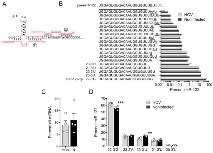
Figure 1. Natural variation in the 3 terminal bases of the HCV host factor, miR-122. (A) Base-pair interactions between miR-122 (red font) and the extreme 5end of the positive-strand HCV RNA genome (black font) established in previous mutational analyses. (S1) and (S2) represent seed-match sites, SL1indicates stem-loop 1 in the HCV 5UTR. Note the absence of base pairs involving the 3terminal 6 nucleotides of miR-122. (B) The 19 most abundant miR-122-5p (guide strand) variants identified in non-malignant liver tissue from HCV-infected and non-infected human subjects (n=4 each). Sequences are listed according to their abundance (percent of all small RNAs sharing the core 5miR-122 sequence ‘UGGAGUGUGACAAUGGUGUU’). Underlined bases represent non-templated additions and deletions. The pre-miR-122 sequence is shown at the top for comparison. Error bars represent the SEM. (C) Relative abundance of all miR-122 reads as the percentage of all miRNAs in HCV-infected (HCV) and non-infected (N) liver tissue. (D) Expanded view of the five most abundant miR-122 variants in HCV-infected versus non-infected human liver. Each symbol represents the result from an individual sample. Error bars represent SD,n=4. **P<0.01, ***P<0.001, by ANOVA with Holm-Sidak’s multiple comparisons test.
or deletions in H77S/GLucAAGS1p6m/dSL1 and
-S1p6m/Ins5U mutants were introduced by primer-directed
mutagenesis of the sequence spanning the NotI and AgeI
sites as described (8). Single base substitutions were created
within psiCHECK2/Luc-3HCV by inserting pre-annealed
oligonucleotides containing mutated sequences using the
XhoI and NotI sites. The 3UTR sequence of SLC7A1
containing natural miR-122 target sites (6) was amplified
using SuperScript® III One-Step RT-PCR System with
Platinum® Taq DNA Polymerase (Thermo Scientific)
and primers 5-AATTCTCGAGGACATAAGCTGT
CTGGCCTCTCTGT-3 and 5-TTATATGCGGCCGC
CTCAGTGGAACGCTCCACCCA-3 from total RNA
extracted from Huh-7.5 cells and cloned into psiCHECK2 using the XhoI and NotI sites. Base changes and the integrity of the surrounding sequences were confirmed by DNA sequencing.
RNA oligonucleotides
RNA oligonucleotides were synthesized by Dharmacon, and miRNAs were transfected as miRNA/miRNA*
du-plexes as described (19).
RNA transcription
RNA transcripts were synthesizedin vitroas described
pre-viously (29).
Transfections
HCV RNA (5 g) and duplex miRNA (250 pmol) were
mixed with 5×10⁶FT3-7 or PH5CH8 cells in a 4-mm
cu-vette and pulsed once at 250 V, 950F and 50in a Gene
Pulser Xcell Total System (Bio-Rad). MEFs were
Gaussia luciferase assay
Cell culture supernatant fluids were collected at intervals following transfection and cells then refed with fresh media. Secreted Gaussia luciferase (GLuc) activity was measured
as described (29).
Immunoblots
Immunoblotting was carried out using standard methods with the following antibodies: mouse monoclonal antibod-ies (mAbs) to human Ago2 (Clone 1B1-E2H5, MBL In-ternational) or mouse Ago2 (Clone 2D4, Wako Chemi-cals); and rabbit monoclonal antibody to Ago2 (Cell Sig-naling Technology). Protein bands were visualized with an Odyssey Infrared Imaging System (Li-Cor Biosciences).
Ago2-RNA co-immunoprecipitation
Five million MEFs or FT3-7 cells were electroporated with
5g HCV RNA and 250 pmol duplex miRNAs and seeded
onto a 10 cm dish. Five hours later, cells were harvested in lysis buffer [150 mM KCl, 25 mM Tris-HCl pH 7.4, 5 mM EDTA, 1% Triton X-100, 5 mM DTT, Complete protease
inhibitor cocktail (Roche) and 100 U/ml RNaseOUT
(In-vitrogen)]. Lysates were centrifuged, and filtered through a
0.22 m syringe filter. Filtrates were incubated with
anti-mouse Ago2 mAb (Wako Chemicals), anti-human Ago2
mAb (MBL International) or isotype control IgG at 4◦C
for 2 h, followed by the addition of 30 l of Protein
G Sepharose (50% Slurry, GE Healthcare) for 1 h. The Sepharose beads were washed three times in wash buffer [1
×TBS, 1.2% Triton X-100, 5 mM DTT, Complete protease
inhibitor cocktail (Roche), 80 U/ml RNaseOUT
(Invitro-gen)] and RNA extracted using the RNeasy Mini Kit (Qi-agen). HCV RNA associated with Ago2 protein was
quan-tified by cDNA synthesis using SuperScript® III
First-Strand Synthesis SuperMix for qRT-PCR (Invitrogen) fol-lowed by TaqMan qPCR analysis using iQ Supermix (Bio-Rad), or measured semi-quantitatively by RT-PCR as
de-scribed (19).
psiCHECK2 reporter assay
PH5CH8 cells or MEFs seeded on 96-well plates were co-transfected with 20 ng of psiCHECK2 DNA and 50 nM
du-plex miRNA using siLentFect™Lipid Reagent (Bio-Rad).
Twenty-four hours later, the cells were lysed in 30l of
Pas-sive Lysis Buffer (Promega). Firefly (FLuc) and Renilla lu-ciferase (RLuc) reporter activities were measured using a
Dual-Luciferase Reporter Assay System (Promega) (8).
CLEAR-CLIP data analysis
Previously described CLEAR (covalent ligation of endoge-nous Argonaute-bound RNAs)-CLIP sequence data from a
study of HCV RNA-associated miRNAs (15) was retrieved
from the NCBI Gene Expression Omnibus (GSE76967).
Reads containing miR122 sequence fused at its 3 end to
sequence near the 5 end of HCV RNA were extracted
by filtering for nts 2–21 of the miR-122 sequence and the
S1/S2 seed site sequences of HCV. miR-122 3 end
vari-ants were enumerated and percentages calculated. Data
were considered only from accessions in which>1000 such
miR-122 reads were identified (SRX1534493, SRX1534494, SRX1534496, SRX1534497 and SRX1534500).
Statistical analyses
Unless noted otherwise, all between-group comparisons were carried out by one-way or two-way ANOVA using Prism 6.0 software (GraphPad Software, Inc.).
RESULTS
Relative expression of miR-122 variants in liver from HCV-infected versus non-HCV-infected individuals
Mature miRNAs, including miR-122, are subject to a
va-riety of 3 modifications, including non-templated
adeny-lation, uridylation and trimming, that influence both their
stability and function (27,30). We previously reported the
use of high-throughput sequencing to profile in an unbi-ased fashion the expression of small non-coding RNAs in liver tissue collected from a series of Japanese adults with
chronic viral hepatitis (31). To determine the relative
abun-dance of different 3miR-122 variants in human liver, we
re-analyzed data from eight of these subjects, four with chronic HCV infection and four non-infected control individuals undergoing resection of metastatic tumors. We identified between 258 000 and 738 000 individual reads in each
sam-ple that shared a common 5 20 nt miR-122 sequence (5
-UGGAGUGUGACAAUGGUGUU-3) but had varied 3
ends. The most abundant of these, a 22 nt RNA with a 3
guanosine (the canonical miR-122-5p in miRBase, which
we term ‘22–3G’ in this report) accounted for no more than
63% of all miR-122 reads in any one sample (Figure1B).
Al-though the proportion of all miRNAs comprised by miR-122 reads varied considerably between individual study
sub-jects (Figure1C), the relative abundance of the five most
highly expressed variants of miR-122 (22–3G, 23–3A, 23–
3U, 21–3U and 20–3U) was remarkably constant (Figure
1D). Notable exceptions were 23–3U, which represented
a lower proportion of all miR-122 reads in HCV-infected
compared to non-infected subjects, and 22–3G, which was
reciprocally increased (Figure1D). These results are
consis-tent with reductions in the percentage of 23–3U isoforms
of miR-122 recently described by Kimet al. (32) in a small
number of HCV-infected individuals, although the
reduc-tion in 23–3U was lesser in magnitude and not associated
with an increase in 21–3U reported in that study.
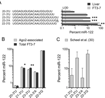
FT3-7 cells are derived from human hepatoma Huh-7
cells and are highly permissive for HCV infection (19).
Us-ing a similar high-throughput sequencUs-ing strategy, we found
the overall pattern of expression of the 3miR-122 variants
in these cells differed significantly from that in liver tissue.
22–3G had a greater relative abundance, representing about
80% of all miR-122 reads, whereas 23–3U and especially
23–3A were reduced (Figure2A). These differences were
highly significant statistically. To determine whether the 3
Figure 2. miR-122 3end variants expressed in a hepatoma cell line, FT3-7. (A) Relative abundance of the top five miR-122 variants expressed in FT3-7 cells compared to liver tissue. LOD=limit of detection (100 reads). ***P<0.001 by two-way ANOVA with Holm-Sidak multiple comparisons test. (B) The five most abundant miR-122 variants are loaded into Ago2 with minimally different efficiencies. Error bars represent the SEM,n= 3 independent cultures. *P<0.05, **P<0.01 by ANOVA with Holm-Sidak multiple comparisons test. (C) Relative abundance of the four major miR-122 3variants associated with HCV RNA in infected Huh-7 cells, as determined by a re-analysis of previously published CLEAR-CLIP data (15). A total of 19 129 miR-122 sequences with 3fusion to HCV 5UTR RNA were characterized. Data shown are means±SD;n=5 experiments.
Ago2 immunoprecipitate (31). Although statistically
signif-icant, differences between their relative abundance in the Ago2 extract versus total cell lysate were small in magni-tude and unlikely to be of biological importance (Figure
2B). Thus, the major 3 miR-122 variants are loaded into
Ago2 with only minor differences in efficiency.
To ascertain whether these 3 miR-122 variants are
re-cruited to replicating viral RNAs within infected human hepatoma cells, we analyzed CLEAR-CLIP sequencing
data generated in a previously published study (15) in which
Ago2-associated miRNAs were covalently ligated to their cellular and viral RNA targets in HCV-infected Huh-7.5 cells. We filtered these data to identify chimeric sequences
in which miR-122 was fused at its 3 end to HCV RNA
containing the S1 and/or S2 sites near the 5 end of the
genome, and enumerated the different 3variants present.
Although we found substantial variation in the distribution of the variants in different experiments, the overall
propor-tions of each of the four major 3miR-122 variants did not
differ significantly from what we had found in FT3-7 cells
(Figure2C). Thus, the minor 3variants are not only loaded
into Ago2 complexes, they are also recruited to the 5end
of HCV RNA in infected cells expressing an abundance of
the canonical 22–3G isoform.
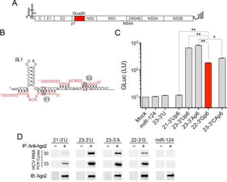
Differential capacities of miR-122 variants to support HCV genome amplification
We next assessed the capacities of the 3miR-122 variants
to support replication of an HCV RNA reporter genome
(H77S.3/GLuc-S1-S2p6m) in Huh-7.5 cells (Figure 3A).
This HCV reporter RNA contains A-to-U substitutions at each of the bases pairing with nucleotide 6 of miR-122
in the S1 and S2 seed match sites (Figure3B). This
pre-vents replication of the reporter RNA genome unless it is co-transfected with a complementary mutant duplex
miR-122 mimic (designated ‘miR-miR-122p6’) (7). Since replication
of the genome can be monitored non-invasively by mea-suring GLuc activity secreted by transfected cells, this pro-vides a simple system for assessing the capacity of miR-122p6 variants to support replication in cells expressing en-dogenous, wild-type miR-122. We thus co-transfected the reporter genome individually with duplex miR-122p6
mim-ics representing each of the four most abundant 3variants
(22–3G, 23–3A, 23–3U and 21–3U, Figure1A), and
mon-itored secreted GLuc activity over the ensuing 72 h. As neg-ative controls, we also co-transfected the reporter genome with miR-124, an unrelated brain-specific miRNA or
wild-type miR-122 (23–3U). In each case, we used duplex RNAs
as mimics because the two-nucleotide 3 overhang of
ma-ture miRNA is important for efficient loading of the guide strand into miRISC complexes.
Surprisingly, the most abundant 3 miR-122p6 variant
(22–3G) was not the most active in facilitating
amplifi-cation of the reporter virus genome (Figure3C). Both of
the 23 nt long miR-122p6 variants (23–3U and 23–3A)
were 3–4 fold more efficient than 22–3G variant in
pro-moting replication, whereas the 21–3U variant was
com-pletely inactive. Similar results were obtained with a second
HCV RNA which, in contrast to the genotype 1a 5UTR
in the H77S.3/GLuc-S1-S2p6m reporter virus, contains the
5UTR sequence of a genotype 2a virus, JFH1
(Supplemen-tary Figure S1). These results suggest that either the length
and/or the composition of the 3 terminal nucleotides of
miR-122 are important for its ability to support efficient HCV RNA replication. To distinguish between these pos-sibilities, we tested a 23 nt miR-122p6 mutant in which the
3terminal nucleotides (nts 22 and 23) were substituted with
complementary bases (23–3CAp6). This miR-122 mimic
did not function as well as either the 23–3U or 23–3A
(Fig-ure3C). We conclude from these results that both the length
and the composition of the 3terminal nucleotides of
miR-122 are important for its HCV host factor activities. We demonstrated previously that the ability of miR-122 to stabilize the HCV RNA genome and to promote its syn-thesis depends upon its recruitment of Ago2 to the viral
5UTR (4,8). Thus, we next ascertained whether the
dif-ferences we observed in the host factor activity of these
3 miR-122 variants correlate with differences in their
ca-pacity to recruit Ago2 to the viral genome. To accomplish this, we immunoprecipitated Ago2 from lysates of cells co-transfected with HCV RNA and the miR-122 variants and then assessed the amount of HCV RNA associated with Ago2 using semi-quantitative RT-PCR. These experiments revealed that there was less viral RNA associated with Ago2
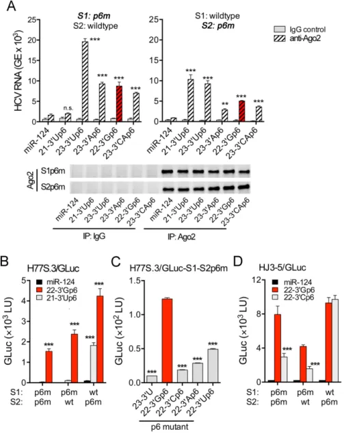
Figure 3. The capacity of miR-122 variants to promote HCV genome amplification correlates with their ability to bind HCV RNA as a complex with Ago2. (A) Schematic of the HCV GLuc reporter genome, showing insertion of GLuc2A sequence between the p7 and NS2 coding regions (see ‘Materials and Methods’ section). (B) Base-pair interactions between HCV RNA carrying p6m mutations in the miR-122 seed match sequences (black font, with red nucleotide substitution) and two copies of Ago2-associated miR-122p6 with complementary base substitutions (red font with black nucleotide substitution). (S1) and (S2) indicate seed-sequence interaction sites;SL1=stem-loop 1. (C) H77S.3/GLuc RNA containing double S1 and S2 p6m mutations were co-transfected into Huh-7.5 cells with the indicated wild-type or mutant duplex miR-122s or the control miRNA, miR-124. Data shown represent the GLuc activity secreted into media between 48 and 72 h post-transfection. 22–3Gp6 is representative of the most abundant miR-122 variant (22–3G, see Figure1), and is shaded in red here and in subsequent figures. Selected pair-wise comparisons are shown: *P<0.05, **P<0.01 by one-way ANOVA with Dunnett’s correction for multiple comparisons. Error bars represent SD;n=3 technical replicates. Results shown are representative of multiple independent experiments with similar results (see also Supplementary Figure S1). (D) Semi-quantitative RT-PCR results of HCV RNA associated with Ago2 precipitated from lysates of murine embryonic fibroblast (MEFs) that had been co-electroporated with the non-replicating H77S.3/AAG RNA and either a miR-122 variant or the control miR-124. Immunoblots of immunoprecipitated Ago2 are shown in lower panels.
or 23–3A variants, and very little HCV RNA associated
with Ago2 isolated from 21–3U-transfected cells (Figure
3D). These results correlate well with the HCV host factor
activities of the 3miR-122 variants (Figure3C and
Supple-mentary Figure S1). Since the variants demonstrate similar
efficiencies for loading into Ago2 (Figure2B), these results
suggest that the 3terminal nucleotides of miR-122 play an
important role in determining the affinity of the miR-122– Ago2 complex for the viral RNA. This conclusion stands in sharp contrast to the absence of any interactions between
the 3terminal miR-122 nucleotides and HCV RNA in the
model structures shown in Figures1A and3B.
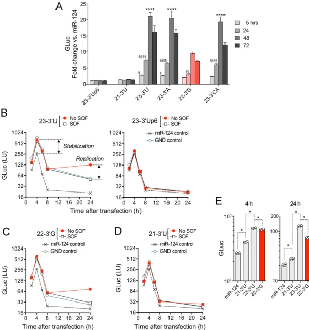
3miR-122 variants differ in their capacity to stabilize HCV RNA and promote its replication in hepatocyte-derived cells lacking endogenous miR-122 expression
To exclude the possibility that the results described above might reflect aberrant miR-122 target site specificities related to the p6m mutations present in the reporter viruses, we carried out similar experiments in a second human hepatocyte-derived cell line. PH5CH8 cells are T antigen-transformed, non-neoplastic adult human hepato-cytes. They have no detectable miR-122 expression, mak-ing HCV replication in these cells completely dependent
upon transfection of exogenous miR-122 (33). We
trans-fected PH5CH8 cells with a genotype 2a HCV reporter
virus RNA similar to that shown in Figure3A that
con-tains the wild-type miR-122 seed-binding and downstream
5UTR sequence (JFH1-QL/GLuc) together with duplex
mimics of the various wild-type 3miR-122 variants.
Nega-tive miRNA controls for these experiments included duplex
miR-122p6 or miR-124. As shown in Figure4A,
amplifica-tion of reporter virus RNA (marked by GLuc expression be-tween 24–72 h post-transfection) was maximal when it was
co-transfected with the 23–3U or 23–3A miR-122 variant.
Co-transfection with 22–3G resulted in significantly lower
levels of replication (P<0.0001), whereas co-transfection
with 21–3U completely failed to support replication
(Fig-ure4A). These results thus mirror closely those described
above from experiments in Huh-7.5 cells supplemented with
p6 mutant versions of these 3 miR-122 variants (Figure
3C), and thereby exclude any confounding effect of the p6
mutation. The 23–3CA mutant was also demonstrably less
active than 23–3A and 23–3U in promoting HCV
replica-tion in PH5CH8 cells (P<0.0001 at 72 h), similar to the
23–3CAp6 mutant in Huh-7.5 cells (Figure3C).
In an effort to discern whether differences in the capacity
of the 3miR-122 variants to support HCV genome
Figure 4.Capacity of natural miR-122 variants to promote stabilization and amplification of the HCV genome. (A) PH5CH8 cells were co-transfected with the replication competent, genome-length JFH1-QL/GLuc reporter virus RNA, which contains the wild-type HCV 5UTR sequence and the indicated wild-type 3miR-122 variants. Supernatant fluids were collected at 5 h, and then 24 h intervals thereafter and assayed for GLuc activity. Results are presented as the fold-increase in GLuc relative to that in cells co-transfected with the control miRNA, miR-124. ****P<0.0001 by two-way ANOVA for comparisons with the 22–3G variant at 24, 48 and 72 h;§P<0.05,§§P<0.01,§§§§P<0.0001 for comparisons with the miR-122 mutant 23–3Up6 at the indicated time point. (B) PH5CH8 cells were co-electroporated with HCV JFH1/GLuc RNA and the (left) 23–3U variant or (right) 23–3Up6 mutant, then cultured in the presence or absence of the NS5B inhibitor sofosbuvir (SOF). As negative controls for viral RNA amplification, parallel cultures were co-electroporated with miR-124 or a non-replicating HCV RNA containing a lethal GND mutation in the NS5B polymerase. Secreted GLuc activity was followed at intervals. Similar experiments are shown involving transfection of (C) 22–3G or (D) 21–3U. (E) Comparison of 4 h (stabilization) and 24 h results (stabilization + replication) from the experiments shown in panels A–C. Symbols represent GLuc values from individual cultures. In all panels, error bars represent the SD fromn=4 technical replicates. *P=0.03 by two-sided Mann–Whitney test. Note that GLuc activity is shown on a log scale in each panel. The data shown are representative of two independent experiments.
carried out additional experiments assessing GLuc expres-sion immediately following transfection of PH5CH8 cells. To distinguish GLuc expression resulting from genome replication versus translation of the transfected input RNA, we transfected cells with a reporter viral RNA contain-ing a lethal mutation in the NS5B RNA polymerase (JFH1/GLuc-GND) that prevents viral RNA synthesis. We also cultured the cells in the presence or absence of
sofosbu-vir (SOF), a potent antisofosbu-viral inhibitor of HCV replication. Secreted GLuc activity was measured at 2 h intervals up to 8 h post-transfection, and again at 24 h. Results from a
typ-ical experiment involving transfection of the 23–3U variant
are shown in Figure4B (left). GLuc secreted between 2 and
8 h after transfection reflects translation of the input RNA
(4,8,11,19), and was similar for both replication-competent
and those of others show that miR-122-mediated increases in secreted GLuc activity at these early time points reflect largely if not entirely increased stability of the transfected
RNA (8,11–12). Similar increases were observed in the
pres-ence of SOF, a potent inhibitor of replication (Figure4B,
left). Later miR-122-mediated increases in GLuc activity (24 h) reflect enhanced RNA replication in addition to sta-bilization, and were not observed with SOF treatment or in cells transfected with the GND mutant despite
stabiliza-tion of the RNA (Figure4B, left). These data thus reveal
that 23–3U both stabilizes the HCV RNA and enhances its
replication (Figure4B, left). In contrast, neither enhanced
stability nor increased replication followed transfection of
23–3Up6 or the irrelevant miRNA control, miR-124
(Fig-ure4B, right). Similar experiments confirmed that 22–3G
is active in both stabilizing the viral RNA as well as
stim-ulating RNA synthesis (Figure4C), albeit significantly less
efficiently than 23–3U (Figure4E). In contrast, whereas the
21–3U variant demonstrated a statistically significant
ca-pacity to stabilize the HCV RNA, this effect was much less
than that observed with 23–3U and 22–3G (Figure4D and
E). Its ability to stimulate viral RNA synthesis was
negligi-ble (Figure4D). These results are consistent with those
ob-tained with the p6 miR-122 mutants in Huh-7.5 cells
(Fig-ure 3C) and the wild-type miR-122 variants in PH5CH8
cells (Figure4A).
miRNA-induced miRISC activity of naturally occurring 3 miR-122 variants
To determine whether the 3miR-122 variants differ in their
capacity to form functionally active miRISC complexes, we co-transfected the variants into cells together with a re-porter plasmid psiCHECK2 expressing an RLuc transcript that has the natural miR-122 SLC7A1 (a.k.a. CAT1) tar-get sequence containing three miR-122 binding sites in its
3UTR (6). RLuc activities were normalized to FLuc
activ-ity produced from a second, independent ORF expressed by the reporter plasmid. Interestingly, the four major miR-122
variants, including 21–3U, demonstrated comparable
sup-pressive activity when co-transfected with the reporter
plas-mid into MEFs (Figure5A, left) or PH5CH8 cells (right),
both of which do not express endogenous miR-122. In
con-trast, in many (but not all) experiments the 21–3U variant
was modestly less active than the other variants in suppress-ing expression of a reporter containsuppress-ing nts 1–45 of the HCV
genome within its 3UTR (Figure5B). This trend was
evi-dent in both cell types, but statistically significant only in PH5CH8 cells. miR-122 suppression of the HCV reporter
was entirely dependent on Ago2, because the 23–3U
vari-ant had no effect on RLuc expressed from this reporter in
genetically deficientAgo2−/−MEFs (Figure5C). We
con-clude from these results that the 21–3U variant is less active
in targeting the HCV RNA sequence than the other major
3UTR variants, although the magnitude of this difference
is much less in reporter gene suppression assays (Figure5)
than in the HCV host factor assays described above
(Fig-ures3C and4A–D).
To ascertain whether the 21–3U variant is less active
in targeting one or the other of the two HCV seed match sites, we constructed additional reporter plasmids with a
p6m mutation (Figure 3B) in either S1 or S2. When
co-transfected with the S1-p6m reporter plasmid, wild-type
miR-122 is directed to the S2 site and vice versa.
Inter-estingly, the 21–3U variant was incapable of suppressing
RLuc expression from the HCV 3UTR reporter in MEFs
when directed to the S1 site, whereas it suppressed RLuc ex-pression as efficiently as the other miR-122 variants when
directed to the S2 site (Figure5D). The 22 and 23 nt long
variants had similar activities when directed to either S1 or
S2. These results indicate that the 3terminal nucleotides of
miR-122 are important for miR-122 to properly bind and recruit Ago2 to the S1 site, but that binding to the S2 site alone is largely sufficient for miRISC activity when the
tar-get sequence is placed in the 3UTR of a reporter construct.
The 3 terminal nucleotides of miR-122 are required for re-cruiting Ago2 to the S1 site and essential for efficient promo-tion of HCV RNA replicapromo-tion
To confirm that the 21–3U variant is deficient in targeting
the S1 site in the context of genome-length HCV RNA, we carried out a series of Ago2-pulldown experiments. HCV RNAs containing the p6m substitution in either S1 or S2 were co-transfected into MEFs together with duplex miR-122p6 mimics containing a complementary p6 mutation.
Since MEFs lack endogenous expression of miR-122 (8), we
were able to assess the capacity of each variant to bind and recruit Ago2 to the viral RNA by quantifying the amount of HCV RNA present in Ago2 immunoprecipitates.
Consis-tent with the results of the reporter assays, 21–3Up6 failed
to recruit Ago2 to the S1 site of the S1p6m RNA (Figure
6A, left), whereas it efficiently recruited Ago2 to the S2 site
in the S2p6m RNA (right). This difference was highly
sig-nificant statistically (P=0.710 for miR-124 versus 21–3U
in the S1p6m-transfected cells, versus P < 0.0001 in the
S2p6m-transfected cells, by two-way ANOVA).
We confirmed the inability of 21–3U to interact with
the S1 site by determining whether supplementing Huh-7.5
cells with 21–3Up6 or 22–3Gp6 promotes replication of
HCV RNAs containing the p6m substitution in S1 or S2. These cells express endogenous miR-122 capable of inter-acting with the non-mutated seed match site in these RNAs, making replication of the RNA dependent upon the ca-pacity of the transfected p6 mutant miR-122 to bind to
the mutated S1 or S2 site. As anticipated, 21–3Up6
pro-moted replication when directed to S2, but not S1, whereas
22–3Gp6 promoted replication when directed to either site
(Figure6B). 22–3Gp6 also stimulated replication of a
dou-ble S1+S2p6m mutant RNA, whereas 21–3Up6 did not.
We validated these results using a HCV target genome with a different mutation in the S1 site involving the third nu-cleotide in the seed sequence binding region, ‘p3m’. A
cog-nate 21–3Up3 miR-122 variant had much weaker activity
in promoting replication of this RNA than 23–3Up3,
con-firming that the inability of 21–3U to promote replication
is not an artifact caused by the p6m substitution in the HCV
genome (Supplementary Figure S2). Thus, the 21–3U
vari-ant lacks HCV host factor activity because it has a reduced capacity to bind the S1 site and recruit Ago2 to it.
It is puzzling that 21–3U should have less affinity for the
Figure 5.Capacity of miR-122 variants to suppress translation of RLuc expressed by a capped reporter mRNA containing (A) the natural SLC7A1 (CAT1) 3UTR miR-122 target or (B) nts 1–45 of the HCV RNA genome in its 3UTR. Experiments were carried out in MEFs (left panels) or PH5CH8 cells (right). Results are shown as percent suppression for each miR-122 variant relative to 23–3Up6 that was included as a negative control. Error bars represent SEM from three independent experiments (2 for SLC7A1 tested in MEFs), each with 3–4 technical replicates. *P<0.05 by one-way ANOVA with Dunnett’s correction for multiple comparisons. (C) Suppression of RLuc translation was measured under the condition of (B) in wild-type orAgo2−/− MEFs. Error is the SEM of duplicate cultures. **P<0.01 by two-sidedt-test. (D) MEFs were co-transfected with psiCHECK2 expressing RLuc reporter mRNA transcripts containing in their 3UTR the HCV miR-122 target sequence (HCV nts 1–45) with p6m mutations (Figure3A) in either the S1 or S2 site, and either a miR-122 variant or miR-124. miR-122 was directed to the wild-type binding site (red font) in these experiments. Results were normalized to FLuc activity expressed from a different open reading frame in the same DNA. Error bars represent the SEM of triplicate cultures. ***P=0.0002 for wild-type S1 versus S2 seed match site, by two-way ANOVA with Sidak’s correction for multiple comparisons.
whereas it is only 6 nts long in S2 (Figure 1A). However,
structural studies of Ago2 complexed with a 21 nt guide RNA have revealed that nts 13–16 of the RNA are exposed for target recognition as well as nts 2–8 (the seed region)
(34). Furthermore, base pairs involving nucleotides 3of the
seed contribute substantially to miR-122 binding at both
S1 and S2 (19). Although this region of the miR-122 guide
strand (nts 13–17) is constant in the 3variants we studied,
the upstream HCV nucleotides at the S2 site (‘. . . CCAU. . . ’) provide for four Watson–Crick accessory pairs, whereas se-quence upstream of S1 (‘GCCA. . . ’) provides for only three
with a possible additional wobble G-U pair (Figure1A). To
exclude a role for this difference in the lesser affinity of 21–
3U for the S1 site, we created a ‘. . . CCAU. . . ’ sequence
up-stream of the S1 seed match similar to that upup-stream of S2 by inserting a U at nt 5 of the HCV genome. This did not
result in Ago2 recruitment to S1 by 21–3U
(Supplemen-tary Figure S3A). We also considered the possibility that
the binding of 21–3U to the S1 might be compromised by
a stable stem-loop (SL1) in the HCV sequence that exists between the seed match sequence and upstream HCV
nu-cleotides that are known to form 3auxiliary base-pair
inter-actions with miR-122 (19) (Figure1A). However, removal
of the stem-loop did not rescue recruitment of Ago2 to the
S1 site by 21–3U (Supplementary Figure S3B).
Collectively, these results reveal that significant difference
exist in the targeting of the 21–3U variant to the S1 versus
the S2 site in the HCV RNA. Changes in the 3 terminal
nucleotide of 22 nt long miR-122 variants also conferred substantial differences in HCV host factor activity when
targeted to S1 but not the S2 site. Replacing the 3
termi-nal 22G nucleotide with adenosine (22–3Ap6), uridine (22–
3Up6) or especially cytidine (22–3Cp6) markedly reduced
the capacity of the 22–3Gp6 variant to promote
replica-tion of either H77S.3 or HJ3-5 RNAs containing p6m
mu-tations at both sites (Figure6C and Supplementary Figure
S4A). However, this difference was noted only when the 22
nt variant was targeted to the S1 site, as 22–3Cp6 promoted
RNA replication as well as 22–3Gp6 directed to S2 (Figure
6D). In contrast, when we replaced the 3terminal U of 23–
3Up6 with guanosine, there was no reduction in its capacity
to promote HCV RNA replication (Supplementary Figure S4B). This demonstrates that it is the guanosine residue at nt 22 of miR-122 that is important, not the presence of a
guanosine at the 3terminal position.
Figure 6.Length and composition of miR-122 3 terminal nucleotides are important for recruiting an Ago2 complex to the S1 site in the HCV 5UTR. (A) Ago2 complexed with the 21–3U miR-122 binds S2 but not S1. Lysates were prepared from wild-type MEFs co-electroporated with (left) H77S/S1p6m/AAG or (right) H77S/S2p6m/AAG RNA mixed with miR-122p6 variants, and immunoprecipitated with anti-Ago2 antibody. Ago2-associated HCV RNA was quantified with quantitative RT-PCR. Error is the SEM of triplicate experiments. Immunoblots of immunoprecipitated Ago2 are shown below. Results versus miR-124: **P<0.01, ***P<0.001 by one-way ANOVA with Dunnett’s test for multiple comparisons. n.s.=not-significant. (B) H77S.3/GLuc RNAs bearing either single or double S1 and S2 p6m mutations were co-transfected with the indicated duplex miRNAs. Error is the SD of triplicate cultures. ***P<0.001 versus miR-124 by two-way ANOVA with Holm-Sidek’s multiple comparisons test. (C) H77S.3/GLuc RNA bearing double S1 and S2 p6m mutations was co-transfected with 22–3Gp6 or similar 22 nt long RNA with either C, A or U at nt 22. Similar results were obtained with HJ3-5/GLuc RNA which contains genotype 2a sequence in the 5UTR (see Supplementary Figure S4A). ***P<0.001 by one-way ANOVA with Dunnett’s test for multiple comparisons. (D) HJ3-5/GLuc RNAs bearing either single or double S1 and S2 p6m mutations were co-transfected with the indicated duplex miRNAs. ***P<0.001 versus 22–3G by two-way ANOVA with Dunnett’s multiple comparison test. (B–D) GLuc activity secreted into media between 48–72 h post-transfection is shown. Error bars represent the SD of triplicate cultures.
DISCUSSION
Recent studies indicate that the 3-terminal nucleotides of
miRNAs can contribute to target site selection, either by direct base-pair formation with the target RNA or by mod-ulating key interactions of the miRNA with the PAZ
do-main of Ago (23–24,26). The 3terminal sequences of
miR-NAs are often modified by non-templated additions and deletions and, as we show here, miR-122 variants with
non-canonical 3end sequence are abundant in human liver
(Fig-ure 1).In vitro, 22–3G is the only product generated by
Dicer cleavage of pre-miR-122, with the 23–3A variant
gen-erated subsequently via engagement of the non-canonical
cytoplasmic poly(A) RNA polymerase GLD2 (27). 23–3A
is likely to be more stable than 22–3G (35), potentially
con-tributing to its abundance. 22–3G (and possibly 23–3A) is
also subject to trimming to shorter variants as well as 3
uridylation mediated by terminal uridyl transferases
dissociation of passenger strand RNA from newly formed miRISC complexes remains uncertain, although the GLD2 polymerase is more active on a single-strand substrate
sug-gesting it may act after passenger strand loss (27).
CLEAR-CLIP studies have suggested that the 3
termi-nal nucleotides of miR-122 influence its selection of mRNA
targets in Huh-7 cells (26), but whether differences in the 3
sequence also affect HCV host factor activity has not been studied. Our results suggest several important conclusions. The first is that there is a substantial, previously unrecog-nized difference in the structural and sequence requirements for miR-122 to function optimally as an HCV replication factor versus its canonical role in regulating host gene
ex-pression. Both the length and base composition of the 3end
of miR-122 is critically important in determining its ability to promote HCV replication, but not gene regulation. This
is evidenced by the fact that the 21–3U isoform loads into
Ago2 and functions in suppressing gene expression (Figures
2B,5A and B), but fails to promote HCV replication
(Fig-ures3C and4D and E; Supplementary Figure S1). A related
but equally important conclusion is that the current model
of the interaction of miR-122 with HCV RNA (Figure1A)
is incomplete, as it offers no explanation for the requirement that miR-122 be at least 22 nts in length with a guanosine
at position 22 (Figure6C and Supplementary Figure S4A).
While we do not fully understand the molecular basis for these requirements, we have shown that it centers on the in-ability of miR-122 isoforms lacking these criteria to func-tionally bind the S1 site that plays the major role in driving genome replication. A third major conclusion is that HCV replication is likely driven to a substantial extent by a
mi-nor 3miR-122 variant, 23–3U. A review of previously
de-scribed Ago2 CLEAR-CLIP data from HCV-infected cells
(15) suggests that 23–3U comprises about 26% of
endoge-nously expressed miR-122 isoforms binding the 5 end of
the viral RNA, whereas the major, canonical 22–3G
iso-form represents about 63% (Figure2C). Since the 23–3U
variant is∼3-fold as active as 22–3G in promoting HCV
replication (Figures3C,4E and Supplementary Figure S1),
both variants likely contribute equivalently to HCV repli-cation.
miR-122 promotes the replication of HCV by two distinct mechanisms. First, by binding to the S1 and S2 sites in asso-ciation with Ago2, miR-122 physically stabilizes the RNA, slowing its rate of decay by protecting it from the major
cy-toplasmic 5exoribonuclease, Xrn1 (8,11–12,36). A number
of studies have also suggested that miR-122 might act to
en-hance HCV IRES-directed translation (9,18,37–38).
How-ever, it is difficult to distinguish the impact of enhanced RNA template stability from greater IRES efficiency in driving increases in viral protein expression, particularly in biologically relevant cell-based studies using full-length, replication-competent viral RNAs. When protection from
Xrn1-mediated 5HCV RNA decay is carefully controlled
for, we and others have concluded that most if not all miR-122-driven increases in protein translation are due to
en-hanced RNA template stability (4,12–13). We have shown
recently that miR-122 directly stimulates the synthesis of HCV RNA within infected cells as measured by increases in the rate of incorporation of 5-ethynyl uridine into nascent
HCV RNA (13). Notably, this occurs in the absence of any
increase in [35S]-Met incorporation into nascent viral
pro-tein (13).
Although miR-122-mediated stabilization of HCV RNA and increased viral RNA synthesis are antithetical to the typical actions of miRNAs in downregulating host gene ex-pression, both of these actions are dependent upon Ago2
(8–9,13). Previous studies have suggested that the
recruit-ment of Ago2 to the viral RNA by miR-122 follows a canonical pattern of miRNA binding at both the S1 and
S2 sites (17–19), with targeting dependent upon both the
seed sequence (nt 2–8) of the guide strand as well as supple-mentary base pairs involving nts 13–16 that often stabilize miRNA target interactions to repress translation and
desta-bilize mRNAs (34,39–40). Current models for the
interac-tions of miR-122 with the 5end of the viral RNA, such as
that shown in Figure1A (17–19), thus show most of the 5
40 nucleotides of HCV RNA to be engaged in base pair-ing, either with other HCV bases in stem-loop 1 (SL1), or in interactions with one of two copies of miR-122. These
models predict no involvement of the 3terminal 5–7 nts of
miR-122 (Figure1A). Thus, it is surprising that only
miR-122 species equal to or longer than 22 nucleotides support
HCV replication (Figure3C), and that there is a strong
pref-erence for the canonical guanosine at nt 22 (Figures3C,6C
and Supplementary Figure S4A), as we show here.
Our data demonstrate that nts 22–23 of miR-122 play a key role in binding to and recruiting Ago2 to the S1 site
within the HCV genome (Figure6A), as well as to the S1 site
in mRNA reporter transcripts containing nts 1–45 of HCV
RNA within their 3UTR (Figure5D). In contrast,
Ago2-targeting to the S2 site was not sensitive to changes in
ei-ther the length or composition of the 3terminal nucleotides
of miR-122. While 3adenylation can increase the physical
stability of miR-122 (27), reduced stability cannot explain
the selective absence of HCV host factor activity associated
with the 21–3U variant as it was fully functional in
sup-pressing RLuc expression from reporter mRNAs
contain-ing its native SLC7A1 3UTR target sequence (Figure5A).
These data also exclude insufficient transfection as an
ex-planation for the failure of 21–3U to support HCV genome
replication. The S1-site specific nature of the 21–3U variant
also argues against the notion that variation in the length
and composition of the 3nts 22–23 is simply affecting the
stability of miR-122 (Figures5D and6A).
The binding of miR-122 to S1 is dominant over S2 in
fa-cilitating HCV RNA replication (7,12,19). Thus,
substitu-tions that ablate miR-122 binding to S1 have a more severe impact on replication than those that ablate binding to S2, which is in quantitative agreement with the two-fold greater abundance of RNA pulled down by antibody to Ago2 when miR-122 is directed to the S1 rather than the S2 site
(Fig-ure6A). This is consistent with the fact that S1 contains a
7 base seed-match site that base-pairs with nts 2–8 of miR-122 as well as an adenosine nucleotide opposite miR-miR-122 nt
1 that is likely to increase the affinity to Ago2 (34). In
con-trast, S2 has a shorter, 6 base seed-match target that pairs with nts 2–7 of miR-122. Thus, a greater affinity for Ago2– miR-122 complex formation at the S1 site is likely to be the basis for its dominance over S2. This may explain why the
(Fig-ure 6), despite its efficient loading into Ago2 (Figure2B), is so severely detrimental to its functions in stabilizing and promoting replication of HCV RNA. This dominance of complex formation at S1 over S2 is not evident in miR-122 suppression of reporter mRNA transcripts containing the
HCV target sequence within their 3UTR. Although the S1
site also fails to bind 21–3U when placed in the 3UTR of
such transcripts (Figure5D), 21–3U demonstrated
surpris-ingly little diminution in its miRISC activity in these assays
(Figure5A and B).
Our findings stand in contrast to a previous report de-scribing cell-free biophysical experiments that suggested miR-122 binds with greater affinity at the S2 site than
at S1 (20). These studies examined interactions between
RNA representing the 5UTR of HCV and miR-122 guide
molecules in the absence of Ago2 and other cellular RNA
binding proteins in a reconstituted in vitro system, and
yielded very different results from what we observed in our
cell-based system. Whereas we found that 21–3U failed to
bind and recruit Ago2 to the S1 site (Figure6A), a 19 nt
miR-122 mutant (20–23) appeared capable of
function-ally binding to both S1 and S2in vitro(20). This difference
highlights the potential importance of Ago2, which was
ab-sent in thein vitrostudies, in miR-122 target selection.
Although binding to the S1 site is dominant in the host factor activity of miR-122, the miRNA has a greater im-pact on replication when bound to both tandem sites than
when bound to either alone (10,12,19). Recent studies of the
binding of miRNAs to closely spaced target sites show that the shuttling of an Ago2–miRNA complex between neigh-boring sites on a single RNA molecule synergistically pro-motes Ago2 retention time during initial interactions with the target RNA, providing a mechanism by which RNAs containing adjacent miRNA binding sites may function as
potent miRNA sponges (16,23). Importantly, dynamic
in-teractions between the 3 end of the guide strand and the
Argonaute protein PAZ domain can have a substantial in-fluence on this process, potentially regulating Ago2
shut-tling between adjacent target sites (23,41). In an effort to
determine whether anchoring of the 3terminal nucleotides
in the PAZ domain plays a distinct role in miR-122 target
selection at S1 versus S2, we biotinylated 23–3Up6 at its
3end, a modification that should disrupt such anchoring.
This 3biotinylated miR-122 failed to promote HCV RNA
replication when directed to either the S1 or S2 site, and it had very little activity in suppressing translation of reporter RNA transcripts containing either the HCV or SLC7A1
target sequences in their 3UTR (data not shown). However,
the fact that the 21–3U-Ago2 complex selectively fails to be
recruited to S1 in the absence of a functional S2 site
(Fig-ures5D and6A) argues against this resulting from a defect
in lateral diffusion between the two sites.
In summary, the HCV host factor activity of miR-122 en-tails a strict requirement for a length of at least 22 nts and a preference for guanosine at nt 22 for functional target-ing of the S1 site in the RNA genome of HCV. Why this is so remains enigmatic. Although the preference for guano-sine at nt 22 suggests the possibility of specific base-pairing, this is not represented by any existing model of the com-plex formed by the viral genome and miR-122, nor is there any obvious nucleotide in the HCV sequence with which it
might pair. Unfortunately, past efforts to characterize the structure of the S1 site in the presence and absence of
miR-122 using SHAPE have not yielded clear results (20,21), and
thus shed no light on this possibility. Both the length of miR-122 and the presence or absence of G-22 could
influ-ence tethering of the 3miRNA tail within the PAZ domain
of Ago, which could be important for target site selection
(24), but an influence on interactions with a yet-to-be
dis-covered RNA-binding protein acting in concert with Ago2
cannot be excluded. The unique 3 base composition and
length required for miR-122 HCV host factor activity is likely to be explained only by a high resolution structural analysis of the HCV RNA–miR-122-Ago2 ternary com-plex.
SUPPLEMENTARY DATA
Supplementary Data are available at NAR Online.
ACKNOWLEDGMENT
The authors are grateful to Scott Hammond and Troels Scheel as well as members of the Lemon laboratory for help-ful discussions.
FUNDING
National Institutes of Health [AI095690, R01-CA164029 to S.M.L., in part]; University of North Carolina Cancer Research Fund (in part). Funding for open access charge: National Institutes of Health.
Conflict of interest statement.None declared.
REFERENCES
1. Westbrook,R.H. and Dusheiko,G. (2014) Natural history of hepatitis C.J. Hepatol.,61, S58–S68.
2. Yamane,D., McGivern,D.R., Masaki,T. and Lemon,S.M. (2013) Liver injury and disease pathogenesis in chronic hepatitis C.Curr. Top. Microbiol. Immunol.,369, 263–288.
3. Scheel,T.K. and Rice,C.M. (2013) Understanding the hepatitis C virus life cycle paves the way for highly effective therapies.Nat. Med., 19, 837–849.
4. Li,Y., Yamane,D., Masaki,T. and Lemon,S.M. (2015) The yin and yang of hepatitis C: synthesis and decay of hepatitis C virus RNA. Nat. Rev. Microbiol.,13, 544–558.
5. Jopling,C.L., Yi,M., Lancaster,A.M., Lemon,S.M. and Sarnow,P. (2005) Modulation of hepatitis C virus RNA abundance by a liver-specific microRNA.Science,309, 1577–1581.
6. Chang,J., Nicolas,E., Marks,D., Sander,C., Lerro,A., Buendia,M.A., Xu,C., Mason,W.S., Moloshok,T., Bort,R.et al.(2004) miR-122, a mammalian liver-specific microRNA, is processed from hcr mRNA and may downregulate the high affinity cationic amino acid transporter CAT-1.RNA Biol.,1, 106–113.
7. Jangra,R.K., Yi,M. and Lemon,S.M. (2010) Regulation of hepatitis C virus translation and infectious virus production by the microRNA miR-122.J. Virol.,84, 6615–6625.
8. Shimakami,T., Yamane,D., Jangra,R.K., Kempf,B.J., Spaniel,C., Barton,D.J. and Lemon,S.M. (2012) Stabilization of hepatitis C virus RNA by an Ago2-miR-122 complex.Proc. Natl. Acad. Sci. U.S.A., 109, 941–946.
9. Wilson,J.A., Zhang,C., Huys,A. and Richardson,C.D. (2011) Human Ago2 is required for efficient miR-122 regulation of HCV RNA accumulation and translation.J. Virol.,85, 2342–2350.
11. Li,Y., Masaki,T., Yamane,D., McGivern,D.R. and Lemon,S.M. (2013) Competing and noncompeting activities of miR-122 and the 5 exonuclease Xrn1 in regulation of hepatitis C virus replication.Proc. Natl. Acad. Sci. U.S.A.,110, 1881–1886.
12. Thibault,P.A., Huys,A., Amador-Canizares,Y., Gailius,J.E., Pinel,D.E. and Wilson,J.A. (2015) Regulation of hepatitis C virus genome replication by Xrn1 and microRNA-122 binding to individual sites in the 5untranslated region.J. Virol.,89, 6294–6311. 13. Masaki,T., Arend,K.C., Li,Y., Yamane,D., McGivern,D.R., Kato,T.,
Wakita,T., Moorman,N.J. and Lemon,S.M. (2015) miR-122 stimulates hepatitis C virus RNA synthesis by altering the balance of viral RNAs engaged in replication versus translation.Cell Host Microbe,17, 217–228.
14. Janssen,H.L., Reesink,H.W., Lawitz,E.J., Zeuzem,S.,
Rodriguez-Torres,M., Patel,K., van der Meer,A.J., Patick,A.K., Chen,A., Zhou,Y.et al.(2013) Treatment of HCV infection by targeting microRNA.N. Engl. J. Med.,368, 1685–1694.
15. Scheel,T.K., Luna,J.M., Liniger,M., Nishiuchi,E., Rozen-Gagnon,K., Shlomai,A., Auray,G., Gerber,M., Fak,J., Keller,I.et al.(2016) A broad RNA virus survey reveals both miRNA dependence and functional sequestration.Cell Host Microbe,19, 409–423. 16. Luna,J.M., Scheel,T.K., Danino,T., Shaw,K.S., Mele,A., Fak,J.J.,
Nishiuchi,E., Takacs,C.N., Catanese,M.T., de Jong,Y.P.et al.(2015) Hepatitis C virus RNA functionally sequesters miR-122.Cell,160, 1099–1110.
17. Machlin,E.S., Sarnow,P. and Sagan,S.M. (2011) Masking the 5 terminal nucleotides of the hepatitis C virus genome by an unconventional microRNA-target RNA complex.Proc. Natl. Acad. Sci. U.S.A.,108, 3193–3198.
18. Roberts,A.P., Lewis,A.P. and Jopling,C.L. (2011) miR-122 activates hepatitis C virus translation by a specialized mechanism requiring particular RNA components.Nucleic Acids Res.,39, 7716–7729. 19. Shimakami,T., Yamane,D., Welsch,C., Hensley,L., Jangra,R.K. and
Lemon,S.M. (2012) Base pairing between hepatitis C virus RNA and microRNA 122 3of its seed sequence is essential for genome stabilization and production of infectious virus.J. Virol.,86, 7372–7383.
20. Mortimer,S.A. and Doudna,J.A. (2013) Unconventional miR-122 binding stabilizes the HCV genome by forming a trimolecular RNA structure.Nucleic Acids Res.,41, 4230–4240.
21. Pang,P.S., Pham,E.A., Elazar,M., Patel,S.G., Eckart,M.R. and Glenn,J.S. (2012) Structural map of a microRNA-122: hepatitis C virus complex.J. Virol.,86, 1250–1254.
22. Li,Y., Masaki,T., Shimakami,T. and Lemon,S.M. (2014) hnRNP L and NF90 interact with hepatitis C virus 5-terminal untranslated RNA and promote efficient replication.J. Virol.,88, 7199–7209. 23. Chandradoss,S.D., Schirle,N.T., Szczepaniak,M., MacRae,I.J. and
Joo,C. (2015) A dynamic search process underlies microRNA targeting.Cell,162, 96–107.
24. Hur,J.K., Zinchenko,M.K., Djuranovic,S. and Green,R. (2013) Regulation of Argonaute slicer activity by guide RNA 3end interactions with the N-terminal lobe.J. Biol. Chem.,288, 7829–7840. 25. Wang,Y., Juranek,S., Li,H., Sheng,G., Wardle,G.S., Tuschl,T. and
Patel,D.J. (2009) Nucleation, propagation and cleavage of target RNAs in Ago silencing complexes.Nature,461, 754–761.
26. Moore,M.J., Scheel,T.K., Luna,J.M., Park,C.Y., Fak,J.J., Nishiuchi,E., Rice,C.M. and Darnell,R.B. (2015) miRNA-target chimeras reveal miRNA 3-end pairing as a major determinant of Argonaute target specificity.Nat. Commun.,6, 8864.
27. Katoh,T., Sakaguchi,Y., Miyauchi,K., Suzuki,T., Kashiwabara,S. and Baba,T. (2009) Selective stabilization of mammalian microRNAs by 3adenylation mediated by the cytoplasmic poly(A) polymerase GLD-2.Genes Dev.,23, 433–438.
28. Spaniel,C., Honda,M., Selitsky,S.R. and
Yamane,D. (2013) microRNA-122 abundance in hepatocellular carcinoma and non-tumor liver tissue from Japanese patients with persistent HCV versus HBV infection.PLoS One,8, e76867. 29. Yamane,D., McGivern,D.R., Wauthier,E., Yi,M., Madden,V.J.,
Welsch,C., Antes,I., Wen,Y., Chugh,P.E., McGee,C.E.et al.(2014) Regulation of the hepatitis C virus RNA replicase by endogenous lipid peroxidation.Nat. Med.,20, 927–935.
30. Ren,G., Chen,X. and Yu,B. (2014) Small RNAs meet their targets: when methylation defends miRNAs from uridylation.RNA Biol.,11, 1099–1104.
31. Selitsky,S.R., Baran-Gale,J., Honda,M., Yamane,D., Masaki,T., Fannin,E.E., Guerra,B., Shirasaki,T., Shimakami,T., Kaneko,S. et al.(2015) Small tRNA-derived RNAs are increased and more abundant than microRNAs in chronic hepatitis B and C.Sci. Rep.,5, 7675.
32. Kim,G.W., Lee,S.H., Cho,H., Kim,M., Shin,E.C. and Oh,J.W. (2016) Hepatitis C virus core protein promotes miR-122 destabilization by inhibiting GLD-2.PLoS Path.,12, e1005714.
33. Dansako,H., Yamane,D., Welsch,C., McGivern,D.R., Hu,F., Kato,N. and Lemon,S.M. (2013) Class A scavenger receptor 1 (MSR1) restricts hepatitis C virus replication by mediating toll-like receptor 3 recognition of viral RNAs produced in neighboring cells.PLoS Path., 9, e1003345.
34. Schirle,N.T., Sheu-Gruttadauria,J. and MacRae,I.J. (2014) Structural basis for microRNA targeting.Science,346, 608–613.
35. Burns,D.M., D’Ambrogio,A., Nottrott,S. and Richter,J.D. (2011) CPEB and two poly(A) polymerases control miR-122 stability and p53 mRNA translation.Nature,473, 105–108.
36. Li,Y., Yamane,D. and Lemon,S.M. (2015) Dissecting the roles of the 5exoribonucleases Xrn1 and Xrn2 in restricting hepatitis C virus replication.J. Virol.,89, 4857–4865.
37. Henke,J.I., Goergen,D., Zheng,J., Song,Y., Schuttler,C.G., Fehr,C., Junemann,C. and Niepmann,M. (2008) microRNA-122 stimulates translation of hepatitis C virus RNA.EMBO J.,27, 3300–3310. 38. Roberts,A.P., Doidge,R., Tarr,A.W. and Jopling,C.L. (2014) The P
body protein LSm1 contributes to stimulation of hepatitis C virus translation, but not replication, by microRNA-122.Nucleic Acids Res.,42, 1257–1269.
39. Bartel,D.P. (2009) MicroRNAs: target recognition and regulatory functions.Cell,136, 215–233.
40. Wee,L.M., Flores-Jasso,C.F., Salomon,W.E. and Zamore,P.D. (2012) Argonaute divides its RNA guide into domains with distinct functions and RNA-binding properties.Cell,151, 1055–1067. 41. Jung,S.R., Kim,E., Hwang,W., Shin,S., Song,J.J. and Hohng,S. (2013)