RESEARCH ARTICLE
Cryptosporidium
Priming Is More Effective
than Vaccine for Protection against
Cryptosporidiosis in a Murine Protein
Malnutrition Model
Luther A. Bartelt1*, David T. Bolick2, Glynis L. Kolling2, James K. Roche2†, Edna
I. Zaenker2, Ana M. Lara3, Francisco Jose Noronha2, Carrie A. Cowardin2, John H. Moore2, Jerrold R. Turner4,5, Cirle A. Warren2, Gregory A. Buck3, Richard L. Guerrant2
1Division of Infectious Diseases, University of North Carolina at Chapel Hill, Chapel Hill, North Carolina, United States of America,2Division of Infectious Diseases and Center for Global Health, University of Virginia, Charlottesville, Virginia, United States of America,3Molecular Biology and Genetics, Virginia Commonwealth University, Richmond, Virginia, United States of America,4Department of Pathology, The University of Chicago, Chicago, Illinois, United States of America,5Departments of Pathology and Medicine
—Gastroenterology, Brigham and Women’s Hospital, Boston, Massachusetts, United States of America
†Deceased.
Abstract
Cryptosporidiumis a major cause of severe diarrhea, especially in malnourished children. Using a murine model ofC.parvumoocyst challenge that recapitulates clinical features of severe cryptosporidiosis during malnutrition, we interrogated the effect of protein malnutri-tion (PM) on primary and secondary responses toC.parvumchallenge, and tested the dif-ferential ability of mucosal priming strategies to overcome the PM-induced susceptibility. We determined that while PM fundamentally alters systemic and mucosal primary immune responses toCryptosporidium, priming withC.parvum(106oocysts) provides robust pro-tective immunity against re-challenge despite ongoing PM.C.parvumpriming restores mucosal Th1-type effectors (CD3+CD8+CD103+T-cells) and cytokines (IFNγ, and IL12p40) that otherwise decrease with ongoing PM. Vaccination strategies withCryptosporidium
antigens expressed in theS. Typhi vector 908htr, however, do not enhance Th1-type responses toC.parvumchallenge during PM, even though vaccination strongly boosts immunity in challenged fully nourished hosts. Remote non-specific exposures to the attenu-atedS. Typhi vector alone or the TLR9 agonist CpG ODN-1668 can partially attenuateC.
parvumseverity during PM, but neither as effectively as viableC.parvumpriming. We con-clude that although PM interferes with basal and vaccine-boosted immune responses toC.
parvum, sustained reductions in disease severity are possible through mucosal activators of host defenses, and specificallyC.parvumpriming can elicit impressively robust Th1-type protective immunity despite ongoing protein malnutrition. These findings add insight into potential correlates ofCryptosporidiumimmunity and future vaccine strategies in malnour-ished children.
a11111
OPEN ACCESS
Citation:Bartelt LA, Bolick DT, Kolling GL, Roche JK, Zaenker EI, Lara AM, et al. (2016)Cryptosporidium
Priming Is More Effective than Vaccine for Protection against Cryptosporidiosis in a Murine Protein Malnutrition Model. PLoS Negl Trop Dis 10(7): e0004820. doi:10.1371/journal.pntd.0004820
Editor:Joseph M. Vinetz, University of California San Diego School of Medicine, UNITED STATES
Received:January 28, 2016
Accepted:June 11, 2016
Published:July 28, 2016
Copyright:© 2016 Bartelt et al. This is an open access article distributed under the terms of the Creative Commons Attribution License, which permits unrestricted use, distribution, and reproduction in any medium, provided the original author and source are credited.
Data Availability Statement:All relevant data are within the paper and its Supporting Information files.
Author Summary
Cryptosporidiumattributable morbidities in malnourished children are increasingly recog-nized. Exactly how malnutrition interferes with host mucosal immunity to diarrheal path-ogens and mucosal vaccine responses remains unclear. Dissecting these interactions in an experimental model of cryptosporidiosis can uncover new insights into novel therapeutic approaches against a pathogen for which effective therapies and vaccines are currently unavailable. We demonstrate that although malnutrition diminishes baseline (primary) Th1-type mucosal immunity these deficits can be partially overcome via non-specific mucosal strategies (S. Typhi and CpG) and completely restored after a sub-clinical (low-dose) exposure to viableC.parvum. These results add insight into preventive strategies to help alleviateCryptosporidium-specific diarrhea in children in low-resource settings and abrogate prolonged post-infection sequelae.
Introduction
Malnutrition affects an estimated 165 million children worldwide [1], contributes to an esti-mated 45% of early childhood mortality [2], and interferes with immune responses to enteric pathogens and mucosally delivered vaccines [3].Cryptosporidium sp., a ubiquitous waterborne apicomplexan intestinal protozoan, is a prototypic pathogen that is more severe in malnour-ished children. Independent of socioeconomic status, early childhoodCryptosporidium infec-tion associates with excess mortality in West Africa (hazard ratio 2.9; 1.7–4.9), sub-Saharan Africa, and South Asia (HR 2.3; 1.3–4.3) where malnutrition prevalence remains high. Crypto-sporidiuminfection associates with up to a 4-fold risk for persistent diarrhea (>14 days) [4–7] increases likelihood of recurrent diarrheal episodes, and associates with growth decrements [8,9]. Even non-diarrhealCryptosporidiuminfections can acutely impair growth [10], and sus-tained linear growth shortfalls may persist for months following infection [11,12].
While severe manifestations ofCryptosporidiuminfection in patients living with advanced HIV/AIDS [13] and studies in animal models confirm an undisputed role for Th1-effector mediated clearance ofCryptosporidium[14–16], whether and how malnutrition increases sus-ceptibility toCryptosporidiumin children is not well understood. Unlike the protective effect of IFN-γseen in jejunal tissues of sensitized healthy volunteers who rapidly clearCryptosporidium
[17], fecal IFN-γlevels are paradoxically lower in malnourished children infected with Crypto-sporidiumthan uninfected controls [18,19]. In contrast, stool cytokines in malnourished chil-dren with active cryptosporidiosis demonstrate increased TNF-α, IL-8, and IL-13, and serum IgE, but not IgG is elevated [19]. Also, whereas circulating CD4+and CD8+T-cells from infected individuals produce IFN-γupon re-stimulation withCryptosporidiumantigens [20], cell-mediated immune (CMI) responses are generally impaired during infection in malnour-ished children, but serum and fecal antibodies are increased [21]. Whether this apparent skew in the immune response is characteristic not only of active, but also responses to recurrent
Cryptosporidiuminfection in malnourished children has yet to be determined. Although reduced systemic IFN-γhas been documented in some malnourished children [2], CD4+T-cell quantity and activation is not consistently impaired, and one follow-up study in malnourished children demonstrated partial reconstitution of CMI through six weeks post-Cryptosporidium
infection [21].
We have previously reported that undernourished neonatal and weaned mice have enhanced susceptibility toCryptosporidium[22–24] infection concurrent with diminished baseline mucosal IFN-γsecretion [23]. Restoration of IFN-γlevels via systemic exposure to the study design, data collection and analysis, decision to
publish, or preparation of the manuscript.
TLR9 agonist CpG immediately prior to infection can partially attenuateCryptosporidium sus-ceptibility during malnutrition [24]. Despite apparently diminished basal IFN-γresponses dur-ing malnutrition, however, mice vaccinated with theSalmonella entericaserovar Typhi strain CVD 908-htr intranasal vector expressing theCryptosporidiumsporozoite antigen Cp15 [25] had unexpectedly preserved splenocyte CMI, including IFN-γrecall responses, through two weeks post vaccination [26]. This finding coupled with a rise in IFN-γat later timepoints post-infection [27] suggests that despite constitutively diminished IFN-γsecretion, these malnour-ished hosts could produce IFN-γin adaptive responses.
In the present study, we dissected how protein malnutrition influences both primary and secondary immune responses to naturalC.parvuminfection and tested whether mucosal deliv-ered strategies could overcome the resultant immunodeficiency. We first established that severe protein malnutrition (PM) differentially recapitulates short and long-term features of severe childhood cryptosporidiosis, and combines withC.parvumto worsen intestinal epithe-lial cell tight-junction disruption and mucosal architecture. These changes were co-incident with a fundamentally altered basal (or primary) immune response toCryptosporidiumantigens in both the systemic and mucosal compartment that resembled findings in malnourished chil-dren (i.e. increased IL13, decreased IFNγ). Surprisingly, however, Th1-type secondary responses were not only preserved, but enhanced during PM. Even at low (106) doses in this model, priming with viableC.parvumoocysts was sufficient to provide protective immunity. CD8+T-cells predominated the secondary mucosal immune response, and were accompanied by Th-1 type cytokines (IFNγ, IL12p40) along with the lymphocyte chemoattractant, CCL5. Vaccination with either of twoCryptosporidiumsporozoite antigens expressed in theS. Typhi vector, however, was unable to provide protective immunity in malnourished mice, despite robust boosted responses in nourished hosts. We also found that while remote non-specific mucosal exposures to eitherS. Typhi or CpG could partially attenuate the course of cryptospo-ridiosis during PM, neither alone nor in combination was as effective asC.parvumpriming.
Results
Protein malnutrition differentially enhances gut disruption and weight
loss during
C
.
parvum
infection in mice
The severity ofCryptosporidiuminfection in malnourished children directly correlates with quantitative fecal parasite burden [28], altered gut function [9], and growth impairments that persist beyond the period of active parasite shedding [11]. To optimize a malnutrition model that best recapitulated these features, we simultaneously performedCryptosporidiumchallenge in weaned mice maintained on three different isocaloric diets: a full nutrient control diet (CD); a 7% protein, 5% fat, reduced vitamin diet (Regional Basic Diet-RBD) [29,30]; or a 2% protein, 15% fat, vitamin sufficient diet (isolated protein deficient-PD) [23,24,31] (Fig 1A). We found that 5 x 10⁷C.parvumoocysts administered after 5 days of dietary acclimation led to weight loss and greater parasite shedding in PD-fed mice (Fig 1B). Severe zinc deficiency, which increases susceptibility to other enteric infections [32], did not further enhance theC.parvuminfection phenotype (S1 Fig). Intestinal damage as measured by villus:crypt ratios was most significantly altered inCryptosporidium-infected PD-fed mice (Fig 1B). Live oocysts were necessary to cause weight loss, confirming that the resultant pathology was due to activeC.parvuminfection and not a maladaptive response to parasite products in the malnourished host (Fig 1C).
Both the PD diet andC.parvumdisrupted epithelial tight-junction expression. Specific tight-junction protein expression was measured using immunofluorescence four days afterC.
parvumchallenge (Fig 1D). Neither expression nor localization of the tight junction scaffolding protein ZO-1 was altered under any of the conditions studied, providing a useful internal
Fig 1. Severe protein malnutrition in mice selectively enhances intestinal disruption and severity of cryptosporidiosis.(A) Experimental timeline. 3-week-old C57Bl/6 female mice were initiated on experimental malnutrition diets (RBD or PD) or control diet (CD) immediately upon receipt from supplier. Challenge with 106or 107Cryptosporidium parvumoocysts, heat-inactivatedC.parvum
oocysts (ΔC.parvum) or PBS occurred via oral gavage 5 days after initiating diet. Serial weights were collected daily post-challenge, and fecal parasite shedding was determined by RT-PCR. On day 3–4 post-challenge, tissue parasite burden and mucosal injury was assessed by measuring ileum villus:crypt ratios and alterations in epithelial tight-junction proteins. (B) Impact of diet on growth (**P<0.01,***P<0.001C.parvumPDvs.C.parvumorC.parvumRBD), fecal parasite shedding (**P<0.01 PD or RBD vs. CD day 2,
*P<0.05 PD vs. RBD or CD day 3), tissue parasite burden (*P<0.05 PD vs CD or RBD ileum,*P<0.05 PD vs CD colon), and ileum villus:crypt ratios (**P<0.01 PBSCDvs PBSRBD,***P<0.001 PBSCDvs PBSPD,****P<0.0001C.parvumCDorC.parvumRBDvsC.
control. In contrast, occludin expression was reduced by PD alone.C.parvuminfection also affected occludin, primarily by inducing redistribution to the intracellular vesicular pool.C.
parvuminfection with PD resulted in both reduced expression and enhanced internalization of occludin. As reported previously [33], little claudin-2 expression was present under basal con-ditions. This was not affected by PD alone.C.parvuminfection upregulated claudin-2 expres-sion, though only part of the claudin-2 expressed localized to the tight junction. In contrast,
C.parvuminfection with PD resulted in markedly increased claudin-2 expression, nearly all of which was concentrated at the tight junction. Finally, similar to the long-term growth impair-ments that are greater in symptomatic cryptosporidiosis than asymptomatic infections [10], challenge with 107C.parvumcompared with 106C.parvum, led to more sustained concentra-tion-dependent growth impairments through 21 days post-challenge (Fig 1E), despite a similar duration of shedding (Fig 1F).
Protein malnutrition alters basal immune responses to
Cryptosporidium
antigens, while enhancing secondary responses following natural
infection
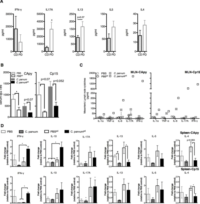
We hypothesized that increased severity of cryptosporidiosis during PD was due to impaired Th1-type immunity. To compare basal immune responses toCryptosporidiumantigens during malnutrition, we stimulated splenocytes from uninfected CD or PD-fed mice (S1 Fig) with two different immunogenic recombinantCryptosporidiumsporozoite antigens (Cp15 and CApy) [34]. Primary responses to eitherC.parvumantigen were fundamentally different between naïve CD and PD-fed mice. Rather than IFN-γ, IL17A predominated in PD-fed mice along with a relatively IFN-γand with tendency toward Th2-type cytokines (Fig 2A). Serum IgG titres were also constitutively lower in PD-fed mice (Fig 2B).
Secondary immune responses to eitherCryptosporidiumantigen at 13–15 days afterC. par-vumchallenge were also diet-dependent, but strikingly opposite of the primary response in PD-fed mice. Nourished infected mice cleared parasites with little evidence of a serological response to either antigen. Serum IgG geometric mean titre (GMT) was attenuated both at baseline and after infection in all PD-fed mice (Fig 2C). Only the more heavily infected PD-fed
C.parvumchallenged mice, however, demonstrated robust secondary cytokine responses in mesenteric lymph nodes (MLNs), including IFN-γ(Fig 2C). Splenocytes of infected CD-fed mice mirrored the minimal cytokine responses in MLNs (Fig 2D).C.parvumchallenged PD-fed animals unexpectedly demonstrated a 14- (CApy) and 41-fold (Cp15) increase in post-stimulation IFN-γsecretion, whereas the increases in IL-13, IL-5, and IL-4 seen in primary responses were reversed (Fig 2D).
10
6C
.
parvum
protects against subsequent severe cryptosporidiosis
and restores effector Th1-type immunity despite ongoing protein
malnutrition
Since we previously observed decreased IFN-γin ileal tissues of PD-fed mice during peak infec-tion [23], we hypothesized that despite robust systemic IFN-γrecall followingC.parvum chal-lenge, PD would interfere with effective mucosal immune responses to serialC.parvum fecal parasite shedding (n = 5-10/group,*P<0.05,**P<0.01). (D) Immunofluorescence staining of epithelial cell tight-junction proteins (ZO-1, occludin, claudin-2) in ileum of CD and PD-fed infected mice and uninfected controls (n = 4/group). (E) Dose dependent persistent growth faltering (n = 10/group,*P<0.05,**P<0.01,***P<0.001,****P<0.0001C.parvum107vsC.parvum106; and
####P<0.0001C.parvumvs PBS) and (F) fecal parasite shedding (**P<0.01,****P<0.0001) through 21 days post-challenge. Data is
representative of 2 replicate experiments.
doi:10.1371/journal.pntd.0004820.g001
Fig 2. Protein malnutrition alters basal immune responses to primaryC.parvumexposure, but secondary responses are intact.Immunologic responses to two different recombinantCryptosporidiumsporozoite antigens (CApy and Cp15) were performed at 13–15 days postC.parvumchallenge in mice fed either control-diet (C.parvumCD) or protein-deficient diet (C.parvumpd) and results
were compared with naïve age and diet-matched uninfected controls (PBSCDand PBSpd). Mice began respective diets 12 days prior to
C.parvumchallenge and remained on the same diets post-challenge. (A) Cytokine secretion in splenocytes of naïve (uninfected) CD or PD-fed mic after stimulation withCryptosporidiumantigens. (B) Serum antibody production as anti-CApy or anti-Cp15 IgG titer (*P<0.05). (C) Cytokines secreted after CApy or Cp15 antigen stimulation in (C) mesenteric lymph nodes. (D) Cytokine secretion in splenocytes expressed as fold change relative to CD-fed uninfected controls. (*P<0.05 as indicated). Data is representative of pooled individual responses from two separate tissue harvests (n = 4-5/group).
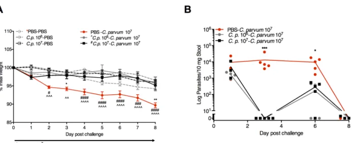
exposures. PD-fed mice were challenged with either a priming dose of 106C.parvumoocysts or the standard challenge dose of 107oocysts similar to re-challenge models in gnotobiotic pig-lets [35]. We confirmed clearance of parasite shedding within 11 days and through day 22 post-challenge in both groups prior to re-challenge withC.parvum107oocysts on day 22 (Fig 1F). Despite ongoing PD, priorC.parvumexposure, unexpectedly, completely protected against weight loss (Fig 3A) and diminished parasite burden (Fig 3B).
Increases in mucosal CD8+CD103+T-cells dominate the protective immune response to
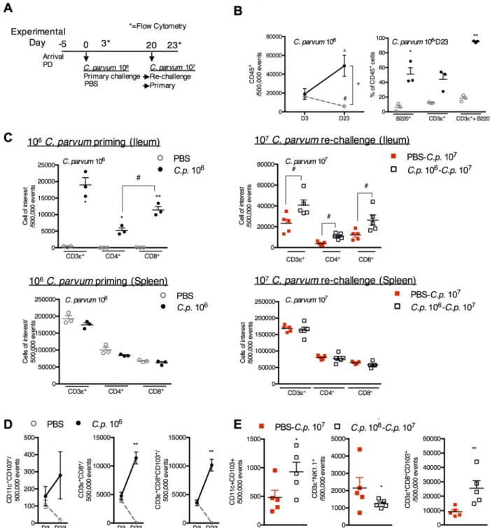
C.parvumre-challenge. To determine whetherC.parvumpriming had restored Th1-type immunity despite ongoing PD, flow cytometry was performed on ileal tissues of PD-fed mice three days after 106C.parvumpriming and again 23 days later (Fig 4A). At peak parasite shed-ding (day 3), total numbers of CD45+cells (Fig 4B) and proportions of B-cells (B220+), T-cells (CD3ε+) and dendritic cells (CD11c+) were similar between both primed and naïve groups (S2 Fig). Although the CD45+population progressively declined with ongoing PD,C.parvum
priming dramatically expanded all CD45+cells including both B-and T-cell populations (Fig 4B). CD8+T-cells became predominant in the mucosal compartment at 23 days post-priming and corresponded with a modest but non-significant reciprocal decrease in splenic T-cells (Fig 4C, left). CD8+T-cells also further increased in the ileum upon re-challenge, and a lack of reciprocal reductions in splenic T-cells suggested local expansion of this cell type (Fig 4C, right).
Further interrogation revealed that several cell-types associated with IFN-γproduction (CD11c+CD103+dendritic cells, NKT (CD3ε+NK1.1+) cells, and CD8+T cells) were depleted in the mucosa during prolonged PD, whereas, these cells progressively increased inC.parvum
primed mice (Fig 4D). The majority of the CD3ε+CD8+cells co-expressed theαE:β7 integrin, CD103+, confirming a mucosal imprinted phenotype (Fig 4D). Compared with 107C.parvum
challenge in naïve mice, CD8+CD103+cells in previously primed re-challenged mice increased more than 3-fold (Fig 4E).
Fig 3.C.parvumpriming protects against re-challenge despite continuous protein malnutrition.Growth (A) and parasite fecal shedding (B) following challenge with 107C.parvumoocysts in either naïve (PBS-C.parvum107) PD-fed mice, or mice previously exposed to either 106(Cp 106) or 107(Cp 107)C.parvumoocysts 21 days prior as indicated (n = 5/group). (A)*P<0.05,**P<0.01 for PBS-PBS vs PBS-C.parvum107,#P<0.05,###P<0.001####P<0.0001 for Cp 106-C.parvum107vs. PBS-C.parvum107,^P<0.05, and
^^P<0.01, ^^^^P<0.0001 for Cp 107-C.parvum107vs. PBS-C.parvum107. (B)*P<0.05,**P<0.01 for Cp 106-C.parvum107or Cp 107
-C.parvum107vs. PBS-C.parvum107.
doi:10.1371/journal.pntd.0004820.g003
106C.parvumpriming restores mucosal Th-1 type immune mediators during ongoing
protein malnutrition. Ileal chemokine and cytokine secretion at peak infection (day 3) and
23 days after exposure toC.parvumpriming confirmed a restructured mucosal immune envi-ronment (Fig 5A). Although cell populations were identical between primed and naïve mice at day 3, CXCL9 and CXCL10 increased inC.parvumprimed mice consistent with epithelial cell monolayer responses toC.parvumexposure [36,37] (Fig 5A). CCL-3, CCL-5 (aka RANTES-Regulated on Activation, Normal T-cell Expressed and Secreted), and CCL-11 were also ele-vated at day 3 in primed mice (Fig 5A). Only CCL5, however, remained significantly elevated through 23 days post-priming (Fig 5A). Similar to systemic primary splenocyte responses, mucosal IL-13, but not IFNγ, was elevated 3 days after priming (Fig 5B) [18]. Through 23 days post-priming, however,C.parvumexposure had remodeled mucosal inflammatory mediators. Otherwise progressive increases in TNFα, IL1β, IL-8 (CXCL1), CCL11 and Th2-type cytokines (IL-4, IL-5, and IL-13) (P<0.05 for IL-8 and IL-5) seen during prolonged PD in naïve controls were reversed inC.parvumprimed mice (Fig 5A and 5B).
Compared with primary 107C.parvumchallenge during prolonged PD (25 days),C. par-vumprimed PD-fed mice (Fig 4A) demonstrated robust elevations in CCL-3 and CCL-5 upon re-challenge (Fig 6A). IFN-γ, IL12p40, and IL-10 were all greater in secondary compared with primary challenge (Fig 6B). IL-13 was increased 100-fold during primary infection (Fig 6B), but was undetectable in secondary infection (P<0.05). Thus, coincident with the robust expan-sion of Th1-type effector cells (Fig 4D and 4E),C.parvumpriming restored mucosal Th1-type cytokine responses despite ongoing PD.
Vaccination strategies with
Cryptosporidium
antigens expressed in
S
.
Typhi do not overcome protein malnutrition-induced susceptibility to
Cryptosporidium
, however
S
. Typhi alone partially reduces disease
severity
Protein malnutrition interferes with vaccine-boosted immune responses to
Cryptospo-ridiumchallenge. We next determined whether the PM-associated immune deficiency could be overcome with other mucosal vaccine strategies. We previously established that humoral and cell-mediated recall responses to vaccine were preserved during PD, but vaccination was ineffective againstC.parvumchallenge [26]. We hypothesized, but had not previously tested, that PD therefore affected effector phases of vaccine responses at the time ofC.parvum chal-lenge, rather than development of memory during vaccination. To test this, we independently expressed either of two sporozoite antigens, the previously tested Cp15 and a novel calcium apyrase (CApy) vaccinogen, in the attenuatedS. Typhi908htrvector system (S. TyphiCp15orS. TyphiCApy) as previously described [25,36]. Vaccine was delivered according to our optimized intransal prime-prime intramuscular boost strategy (Fig 7A). Immune responses and challenge studies were compared with the identicalS. Typhi vector devoid ofCryptosporidiumantigens as well as a‘double-sham’group of animals exposed only to PBS.
Fig 4.C.parvumpriming promotes a progressive increase in CD3+CD8+CD103+cells that further expand upon re-challenge.
(A) Experimental timeline of 3 week-old C57Bl/6 mice initiated on the maintained on PD diet beginning 5 days prior to priming withC.
parvum106(●) or PBS control (grey◦). Day 0 is designated as the day ofC.parvumpriming. Mice were challenged withC.parvum107 20 days later indicated as PBS-C.p. 107(red◻) orC.p. 106-C.p. 107(◻). Flow cytometry was obtained on day 3 (D3) and day 23 (D23)
afterC.parvumpriming. (n = 3/group D3; n = 4-5/group D23). Day 23 also corresponds to 3 days after challenge withC.p. 107. (B)
Intestinal leukocytes in the ileum decrease between D3 and D23 in uninfected mice, but both B-cell (B220+) and T-cells (CD3ε+) increase inC.parvumprimed mice. ^P<0.05 forCp106D3 vsCp106D23, #P<0.05 for PBS D3 vs PBS D23,*P<0.05 for PBS vs.Cp
106. C) Expansion of T-cells inC.parvum-primed mice through D23 afterC.parvumpriming mice is specific to the ileum and
accompanied by a predominance of CD8+T-cells (left) that further increase 3 days afterC.parvumre-challenge (right).*P<0.05 for PBS
vs.Cp106,#P<0.05 between groups as indicated. (D) CD11c+CD103+, CD3ε+CD8+and CD3ε+CD8+CD103+cells expand in the ileum through D23 afterC.parvumpriming.**P<0.01 for PBS vs.Cp106. (E)C.parvumpriming leads to expansion of CD8+CD103+cells and CD11c+CD103+but not NK1.1+ cells compared with initialC.parvum107challenge.*P<0.05,**P<0.01.
doi:10.1371/journal.pntd.0004820.g004
[26], transition to PDaftervaccination also led to diminished IFNγrelease in MLNs ofS. TyphiCp15group. This post-vaccine PD effect was antigen-specific, however, and did not inter-fere with IFNγrelease in theS. TyphiCApygroup (S3 Fig).
Despite waning of serum IgG GMT to either antigen at 11 weeks post-vaccination relative to our observations at earlier timepoints [26,34],C.parvumchallenge unmasked strongly boosted immunity in vaccinated mice (Fig 7B). PD, however, blunted the vaccine-boosted serum IgG effect (Fig 3B). Similarly, post-challenge splenocyte IFN-γresponses in vaccinated CD-fed were markedly enhanced compared with natural infection, but not in the PD-fed vacci-nated mice (Fig 7C). In contrast, IL-17A responses dominated in the vaccinated PD-fed mice (Fig 7D). Irrespective of diet, IL17A responses were driven by exposure to theS. Typhi vector rather than eitherCryptosporidiumantigen. Real-time PCR forS. Typhi at 24 hours after each intranasal exposure and again 24 hours prior to the next prime/boost challenge confirmed exposure to the vector was transient (S4 Fig).
RemoteS. Typhi vector exposure non-specifically facilitates post-infection recovery.
Although disease was mild in the CD-fed groups,S. TyphiCp15orS. TyphiCApyvaccinated mice demonstrated a relative growth improvement, compared with challenge in
‘double-sham’(PBS)-exposed animals. This 5.8–7.7% relative growth advantage reached significance on days 8, 12, and 13 post-infection in theS. TyphiCp15group (Fig 4E, left), but appeared to be partially driven by anyS. Typhi exposure. Similarly, in PD-fed mice, anyS. Typhi exposure, independent ofCryptosporidiumantigen expression, partially mitigated weight loss (nadir 8.7% weight loss on day 3 for aggregatedS. Typhi groups vs 13.1% on day 10 in the sham-exposed group,S4 Fig), and promoted growth recovery (Fig 3E, right).
Fig 5.C.parvumpriming leads to sustained changes in ileal tissue chemokine and cytokine profiles during protein malnutrition.Mice were conditioned on PD for 5 days prior to infection with 106C.parvum(Cp106). Luminex was performed for
measurement of chemokines and cytokines in ileal tissues at. day 3 (D3) and day 23 (D23) post challenge compared to uninfected controls (PBS) (n = 3-4/group). (A) PrimaryC.parvuminfection led to increases in CXCL9, CXCL10, CCL-3, CCL-5, and CCL11 on D3. On D23, TNFα, IL1β, and IL-8 were diminished in infected mice relative to uninfected controls, however, CCL-5 continued to be elevated and other chemokines had returned to baseline. (B) Only IL12p40 and IL-13 were modestly elevated three days after primaryC.parvum
challenge. There was a relative decrease in all Th2-type cytokines through 23 days post-C.parvumcompared with uninfected controls. (n = 3-4/group).*P<0.05 for PBS vs Cp106as indicated; #P<0.05 for Cp106D3 vs Cp106D23 as indicated.
Within each diet cohort,C.parvumshedding was similar across all experimental groups (Fig 7F). Intestinal damage as measured by decreased villus:crypt (V:C) ratios was worse in all PD-fedC.parvumchallenged mice compared with diet-matched uninfected controls. Whereas in CD-fed mice, eitherS. TyphiCApyorS. TyphiCp15reduced intestinal damage compared with
S. Typhi alone (Fig 7G), in PD-fed mice onlyS. TyphiCApyreduced intestinal damage. Thus, vaccination with eitherS. TyphiCp15orS. TyphiCApyin the CD-fed mice was effective at boost-ing post-infection immune responses and diminishboost-ing disease severity without an apparent reduction inC.parvumshedding. PD attenuated the vaccine-boosted IgG and IFN-γresponses to infection, but amplified the over-exuberant IL17A response driven by anyS. Typhi exposure. In PD-fed mice any aggregated remoteS. Typhi exposure facilitated post-infection recovery with a modest decrease inC.parvumshedding (S4 Fig).
Priming with viable
C
.
parvum
oocysts is more effective than remote
intranasal exposures to the non-specific immune activators CpG and
S
.
Typhi
In subsequent experiments, even in the absence ofCryptosporidiumantigen expression, intra-nasalS. Typhi vector exposures at 6 and 4 weeks prior to 107C.parvumchallenge partially reduced disease severity (S4 Fig). The effect was likely not due toS. Typhi-enhanced IL17A responses. WhileIL17expression in the ileal mucosa remained elevated several weeks afterS. Typhi exposure in some mice, IL17RA mice, which lack IL17 signaling, were relatively less sus-ceptible toC.parvumchallenge than wild-type controls (S4 Fig). Rather, the benefit ofS. Typhi more resembled the partial benefit seen in PD-fed mice receiving an intraperitoneal TLR9 ago-nist CpG-ODN 1668 at the time of infection (S5 Fig). Intriguingly, remote intranasal CpG,
Fig 6.C.parvumpriming enhances Th1-type cytokine responses to re-challenge in protein malnourished mice.(A) Ileal inflammatory mediators and chemokines and (B) cytokines measured three days after 107C.parvumchallenge in previously uninfected (PBS) compared with mice primed with 106C.parvum(Cp106) 20 days prior to re-challenge.*P<0.05,**P<0.01,***P<0.001. doi:10.1371/journal.pntd.0004820.g006
Fig 7. Protein malnutrition interferes with vaccine-boosted immunity, but theS. Typhi vector improves recovery afterC.parvumchallenge.(A) Timeline for immunization, growth monitoring, infection, and analysis of immune responses. 21 day-old mice acclimated for 4 days prior to weight-matched randomization (n = 8-19/group). Intranasal immunization withS.entericaTyphi 908htrvector expressing either of two recombinant sporozoite antigens, ClyA-Cp15 (S. TyphiCp15(aqua)) or ClyA-CApy (S. TyphiCApy(blue)) was administered at two-week intervals. TheS. Typhi vector
though incompletely protective, was more effective than intraperitoneal CpG delivered at the time of infection. There was no apparent additional benefit toS. Typhi combined with CpG (S5 Fig).
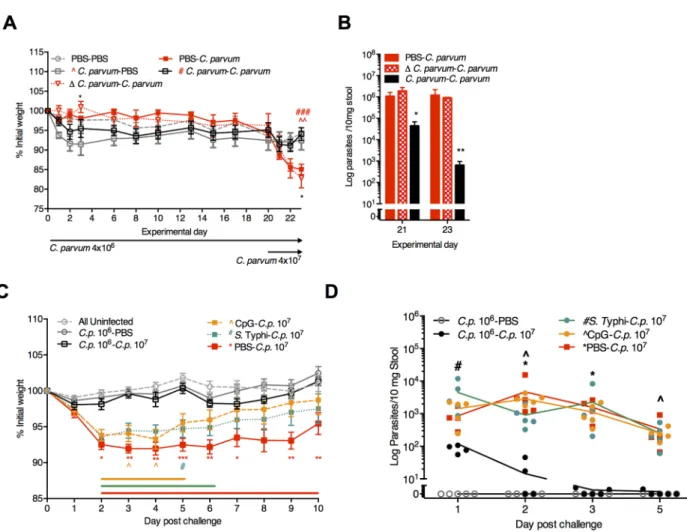
Since either CpG-ODN in PD-fed weanling mice [24] or oral CpG in neonates [38] restores mucosal IFN-γ, we hypothesized that remote intranasal CpG might be as efficacious asC. par-vumpriming. To test this hypothesis we first confirmed that only viableC.parvumpriming, but not heat-inactivatedC.parvum, established protective immunity (Fig 8A and 8B) and increased Th1-type cytokines (S6 Fig). While even a single mucosal exposure to intranasal CpG-ODN 1668 (CpG-C.parvum) and evenS. Typhi (S. Typhi-C.parvum) 21 days prior toC.
parvumchallenge facilitated post-infection recovery, only the single 106C.parvumpriming afforded complete protection against weight loss and rapid parasite clearance (Fig 8C and 8D;
S5 Fig). Thus, while enhancing host defenses through transient exposures during PD has a last-ing impact on cryptosporidiosis severity, onlyC.parvumpriming, which activated secondary Th1-type effector responses, resulted in protective immunity.
Discussion
The epidemiology of malnutrition andCryptosporidiumare intimately associated, but mecha-nisms whereby malnutrition increases severity of cryptosporidiosis are poorly understood. In order to improve understanding of how malnutrition-induced mucosal immune deficits influ-enceCryptosporidiuminfection and immune responses, we isolated which nutritional deficien-cies have the greatest impact on mucosal host defenses againstCryptosporidium. We then determined which of several anti-cryptosporidial preventive strategies would most effectively overcome the resultant immunodeficiency. Protein malnutrition (PM) in mice selectively repli-cated clinical, histological, and immunological features of activeCryptosporidiuminfection in malnourished children. Using remote exposures to non-specific mucosal defense enhancers (S. Typhi vaccine vector and CpG), vaccination withCryptosporidiumantigens expressed in anS. Typhi vector, and viableC.parvumpriming we demonstrated that while PM fundamentally alters Th1-type basal and vaccine-boosted immune responses at the time of infection, the resul-tant immunodeficiency is not insurmountable. Rather, robust and protective adaptive
Th1-type effector responses can occur when priming with viableC.parvum. These findings, summarized inFig 9, provide new insights into nutrient-dependent mucosal immune deficien-cies relevant toCryptosporidiumoutcomes and a viable model in mice for future comparisons ofCryptosporidiumprevention strategies.
Cryptosporidiumis a challenging parasite to study and there is a lack of established models that confidently mimic human disease. Thus, we first wanted to establish which model would
obtained throughout the vaccination protocol (S1 Fig). On day 107 (9 weeks afterS. Typhi exposure), mice were transitioned to either PD or CD diets (n = 4-10/group) and continued on respective diets throughout the remainder of the experiment. Mice were challenged with 5x10⁷C.parvum(Cp) on day 119 and followed for 13–15 days post-challenge. (B) Serum geometric mean IgG titers (GMT) inC.parvumchallenged groups. (C) IFN-γand (D) IL-17A cytokine secretion recall responses to homologous vaccinogen as indicated. For (B-D),*P<0.05,**P<0.01,***P<0.001, One-way ANOVA, Tukey post-test analysis,#P<0.05 by Student’st-test. (E) Growth as percentage of weight change on the day ofC.
parvuminfection beginning on the day of transition to either CD (left) or PD (right) diets. (left:*P<0.05,S. TyphiCp15vs PBS-Cp; right: colored bars indicateP<0.05 for PBS-Cp(red),S. Typhi-Cp(green),S. TyphiCp15(aqua), andS. TyphiCApy
(blue) vs. uninfected controls,*P<0.05 for individual vaccine groups [S. Typhi-Cp(green),S. TyphiCp15(aqua), andS.
TyphiCApy(blue)] vs. PBS-Cp. (F) Parasite shedding for infected groups: PBS (red),S. Typhi (green,S. TyphiCp15(aqua), andS. TyphiCApy(blue). (G) Ileal villus:crypt for CD-fed (left) and PD-fed (right) mice in eachC.parvuminfected group or combined uninfected controls as indicated. Left:***P<0.001 PBS-Cpvs uninfected controls,###P<0.001S. TyphiCp15or
S. TyphiCApyvsS. Typhi; Right:***P<0.001 for PBS-Cp,S. Typhi, orS. TyphiCp15vs uninfected controls,###P<0.001S.
TyphiCApyvs PBS-Cp. ^P<0.05 for PD-PBS and PD-S. TyphiCp15vs DD-PBS and DD-S. TyphiCp15. ns = not significant vs. uninfected controls.
doi:10.1371/journal.pntd.0004820.g007
best recapitulate known clinical features of activeCryptosporidiuminfection in malnourished children. While we and others have used various malnutrition protocols to intensify enteric infections [23,24,31,29,39] it was not known which nutrients were most essential for Crypto-sporidiumsusceptibility. Only isolated protein malnutrition, but not multinutrient deficiency, sufficiently replicated dose-dependent disease severity evidenced by weight loss and persistent growth impairment, intestinal villus injury and epithelial tight junction disruption, and the ele-vated IL13 but paradoxically diminished IFNγresponse seen in malnourished children. Amino
Fig 8. ViableC.parvumpriming provides greater protection against re-challenge than either CpG-ODN orS. Typhi.(A, B) Comparison of protective immunity following priming with either viable and heat-inactivated (Δ)C.parvum106. (A) Growth of PD-fed
mice through 23 days post-priming with either 4x10⁶viable (C.parvum) or 4x10⁶heat-inactivated (ΔC.parvum). Mice were challenged
with viable 4x10⁷
C.parvumoocysts on day 20 post-priming.*P<0.05 forΔC.parvum-C.parvumvs.C.parvum-C.parvum(d3 and d23); ^^P<0.01 forC.parvum-PBS vs PBS-C.parvum(d23);###P<0.001 forC.parvum-C.parvumvs. PBS-C.parvum(d23). (B) RT-PCR of
Cryptosporidiumstool shedding on experimental days 21 and 23 (day 1 and day 3 afterC.parvum107challenge, respectively).
*P<0.05 and*P<0.01 forC.parvum-C.parvumvs either PBS-C.parvumorΔC.parvum-C.parvum. (C,D) 3-week-old C57Bl/6 mice were conditioned on PD for 7 days prior to orogastric inoculation with 106
C.parvum, intranasal (i.n.) 109
S. Typhi 908htr, i.n. CpG-ODN 1668 (100 mcg), or PBS (100 mcl) as indicated (n = 10/group). On day 21, mice were re-challenged with either PBS orC.parvum107. (C) Growth as percentage of initial weight, normalized to the day of 107C.parvumchallenge (Day 0). The group labeled“All uninfected”
includes animals that received either PBS during both inoculations, CpG followed by PBS, orS. Typhi followed by PBS (n = 5/group x 3 = 15) given all three groups grew similarly and were never exposed toC.parvum(S4 Fig).*P<0.05,**P<0.01,***P<0.001 for PBS–
C.p.107(red) vsC.p.106-C.p.107, ^P<0.05 for CpG-C.p.107(yellow) vsC.p.106-C.p.107, and#P<0.05 forS. Typhi-C.p.107(green) vsC.
p.106-C.p.107). Horizontal lines designate significant differences atP<0.05 between CpG-C.p.107(yellow),S. Typhi-C.p.107(green),
and PBS-C.p.107(red) vs. All uninfected controls, respectively. (D) Parasite fecal shedding in serial fecal pellets collected on indicated
experimental days postC.parvum107challenge.*P<0.05 for PBS–C.p.107vs.C.p.106-C.p.107, ^P<0.05 for CpG-C.p.107vsC.
p.106-C.p.107, and#P<0.05 forS. Typhi-C.p.107vsC.p.106-C.p.107. Data is representative of two replicate experiments.
acid dependent immune pathways therefore may be most relevant forCryptosporidium out-comes. In contrast, isolated zinc deficiency less markedly enhanced cryptosporidiosis even though zinc can shorten duration of diarrhea and lessens severity and virulence of enteroaggre-gativeEscherichia coliinfection in our models [32]. Indeed, current standard WHO-recom-mended oral rehydration therapy and zinc treatment appears to be less effective for
cryptosporidiosis than other diarrheal pathogens in children [6]. Rather, targeted amino acid therapies such as alanyl-glutamine [24,40] or arginine [41] may more selectively diminish severity ofCryptosporidiuminfection.
Prolonged PM in our model, like total caloric restriction [42], leads to progressive depletion in multiple IFNγ-producing effector cell populations (CD8+ T-cells, CD11c+CD103+dendritic cells, and NKT cells) that should otherwise be steadily increasing in maturing young mice [43]. Consequently, like in malnourished children with active cryptosporidiosis [18], either peak pri-maryC.parvumchallenge during PM in mice or stimulation of their naïve lymphocytes with
C.parvumantigens elicits a basal IL13 response rather than IFNγ. Consistent with this obser-vation, a systematic review concluded that malnutrition most significantly impairs effector T-cell responses and Th1-type cytokines but not total T- and CD4+T-cell numbers. Malnutrition also associated with a tendency toward Th2-type cytokines [2]. We find that PM alone is bio-logically sufficient to recapitulate a similar immunological alteration. These data contribute to increasing recognition that immune effector function is differentially altered, rather than glob-ally suppressed, by select nutrient deficiencies. Vitamin A deficiency, for example,
Fig 9. Schematic of non-specific (S. Typhi and CpG) and specific (C.parvumpriming) mucosal exposures that modulate host immunity and protect against cryptosporidiosis during malnutrition.Strategies to enhance immune defenses against
Cryptosporidiuminfection during malnutrition were investigated in a protein deficient murine model that replicates clinical features of childhood cryptosporidiosis. Whereas the well-nourished host (black) clearsCryptosporidiumwith little evidence of a secondary immune response (dark blue), mucosal vaccination withCryptosporidiumantigens expressed in anS. Typhi vector can elicit strongly boosted IFNγ-predominant immune responses to subsequent challenge (light blue). Vaccine attenuates the mild disease caused by
Cryptosporidiumin well-nourished hosts. In protein malnourished hosts (light grey) there is ongoing depletion of mucosal lymphocytes including Th1-type effectors. This results in enhanced disease after primaryC.parvumchallenge with a response characterized by decreased IFNγbut increased IL13 and tendency toward Th2-type cytokines (red). Unlike in nourished hosts, vaccine does not further enhance IFNγto primaryC.parvumchallenge, but rather theS. Typhi vector alone drives increased IL17A and partially attenuates disease severity similar to the TLR9 agonist CpG (yellow).C.parvumpriming, however, leverages a robust secondary Th1-type response toC.parvumduring protein malnutrition, and even at low-doses in this model establishes a mucosal imprinted population of CD8+T-cells along with protective immunity to subsequent re-challenge (dark grey).
doi:10.1371/journal.pntd.0004820.g009
disproportionately alters ratios but not absolute numbers of mucosal innate lymphoid subsets, increasing susceptibility toCitrobacter rodentiumwhile paradoxically enhancing immunity against intestinal nematodes [44].
In the case ofCryptosporidiuminfection, a skewed response during PM may not only increase susceptibility to primary infection, but also contribute to the pathogenesis. Our studies confirm for the first timein vivotight-junction disruption seen inin vitro Cryptosporidium
models [45,46]. Of the studied epithelial cell proteins, the most marked disruptions were seen in the cytokine-inducible tight-junction proteins occludin and claudin-2. Both internalization of occludin and IL-13 induced claudin-2 upregulation have also been reported in human and experimental inflammatory bowel disease models [33,47]. Internalization of occludin occurs during acute TNF-αinduced barrier loss and diarrhea [48].In vitroandin vivostudies have shown that removal of occludin from the tight junction, either by endocytosis or reduced expression, facilitates paracellular flux of small and large molecules via the‘leak pathway’. This complements the increased‘pore pathway’permeability to water and small cations following increases in claudin-2 expression [49]. In addition to potentially promoting diarrhea, the inflammatory response toCryptosporidiumduring PM corresponds with persistent growth impairment, and may resembles the chronic T-cell activation seen in the environmental enter-opathy associated with malnutrition [50]. Thus, not only heavierCryptosporidiuminfection, but potentially aberrant host responses during malnutrition contribute to the greater severity of diarrhea and subsequent post-infection sequelae.
While PM-induced depletion of several cell types may be independently important for anti-cryptosporidial defenses, CD11c+CD103+cells that co-express the CXCR3 receptor were recently shown to be more consequential for earlyCryptosporidiumclearance in primary infec-tion in neonatal mice than loss of either NK cells or T-cells [43]. Although CD11c+CD103+ cells did increase followingC.parvumpriming in the current study, the influx of CD3+CD8+ T-cells was even more striking. Sensitized CD8+T-cells have a direct role in clearing Cryptospo-ridiumfrom human epithelial cells [51] as well as in experimental models ofC.murisinfection [52,53] and have been shown to accumulate after infection in neonatal mice [54,55]. Preced-ing the influx CD3+CD8+T-cells in this model was an increase in Th1-type chemoattractants CXCL9, CXCL10, CCL3 and CCL5 at peak primary infection. Epithelial cells are a major source of these chemokines in response to exposure toCryptosporidium[43,56,57], and appear to be functional despite PM. Interestingly, however, CCL5 but not CXCL9 or CXCL10, dominated re-challenge responses during PM. This is in contrast with the predominance of CXCL10 rather than CCL5 in patients with HIV/AIDS duringCryptosporidiuminfection, and is consistent with the likelihood that the influx of T-cells afterC.parvumpriming in this model contributed to elevated CCL5 [56]. Ours is the first study to identify that the majority of CD3 +-CD8+also co-express theαE:β7 marker CD103+indicative of intestinal honing [58] in
response toCryptosporidium. A subpopulation of these cells has recently been classified as tis-sue resident effector memory (Trm) cells that produce higher amounts of IFNγthan CD3 +-CD8+CD103-cells, accumulate locally following intracellular infections at mucosal sites [59,
60] and thus represent an emerging target for mucosal vaccine development [61,62]. Further elucidation of the role of Trms inCryptosporidiuminfections may therefore further inform vaccine development [13].
an oral route may more optimally target vaccine responses to the small intestine than the intra-nasal route used in our studies, includingCryptosporidiumantigens expressed in an attenuated
S. Typhimirium vector [61,64]. Thus, oral rather than intranasalCryptosporidiumvaccine delivery might better mimicC.parvumpriming. Intranasal vaccination with either Cryptospo-ridiumantigen in fully nourished hosts leads to a robust enhancement in the immune response, similar to human volunteers that demonstrated serum IgG antibodies only after secondary, but not primaryC.parvuminfection [65]. As fully nourished mice appear to clearC.parvum infec-tion via innate defenses, there was no apparent decrease in parasite clearance following vacci-nation. These findings raise important implications in the limitations of inferring correlates of immunity in healthy volunteers and desired effective responses in malnourished target popula-tions. Similarly, Maier et al. demonstrated that malnourished mice infected with rotavirus demonstrated diminished seroconversion, but were unexpectedly protected against viral re-challenge [66].
We present a series of experiments designed to compare the effect of both specific (sporozo-ite antigen vaccine and homologous re-challenge) and non-specific (the attenuatedS. Typhi vector and the TLR9 agonist CpG) antigen exposures. In this model, we demonstrate that while less effective thanC.parvumpriming, even remote non-specific exposures can diminish disease severity, even if not reducing parasite shedding. Others have reported partial efficacy of vector-based anti-Cryptosporidiumvaccines in animal models [67], including apparent unex-plained phenomena of the vector alone [68,69]. In the present studyS. Typhi enhanced IL-17A production, most strikingly during PM. IL-IL-17A responses are also increased in naturally acquiredS. Typhi [70] and Ty21a vaccination [71]. While the loss of IL-17A producing lym-phocytes promotes disease severity in simian immunodeficiency virus andCryptosporidium
co-infection [72] a protective role of IL-17A againstCryptosporidiumremains undefined. Alternatively, we speculate thatS. Typhi provided broad activation of innate immunity and thus modulated mucosal responses to microbial products such lipopolysaccharide [26], or influenced claudin protein expression and barrier function [33,47,73,74]. Oral immunization withSalmonellavaccine strains has also been shown to protect against non-Salmonella bacte-rial infections via sustained changes in TLR expression and non-specific macrophage activation [75]. Signaling TLR3 in combination with TLR5 [76,77], TLR4 [78] and TLR9 [24,38] inde-pendently influenceCryptosporidiuminfection outcomes. While prior studies have focused on TLR activation immediately prior to infection, we found that even remote selective TLR activa-tion with CpG mimicked theS. Typhi effect and was more effective than CpG administration immediately prior to infection. Such observations are aligned with the concept of“trained innate immunity”with potential for enhanced responses to repeated exposures to non-specific microbial products [79]. These concepts may be highly relevant in malnourished children given potentially divergent microbiota and frequent exposures to enteropathogens associated with childhood malnutrition [80].
Important questions remain regarding primary and secondary immune responses to Cryp-tosporidiumin malnourished children. First, although many features of our model overlap with childhood cryptosporidiosis during malnutrition, murine immunology is not a surrogate for children, and further investigations are needed to delineate whether malnourished children similarly demonstrate such contrasting immune responses to primary and secondary infection. Indeed, despite our finding robust protective immunity after even low-inoculumC.parvum
exposure, recurrent and multipleCryptosporidiuminfections are well documented in malnour-ished children [8,81]. The prevalence of asymptomatic infections is only beginning to be defined as detection of these exposures may require highly sensitive molecular diagnostics [28], and when applied outnumber diarrheal infections ~10:1 [12]. If asymptomatic exposure does induce a protective response, the duration of immunity in children may be short lived. While
recurrence was unlikely within one month of infection in one cohort [82], by nine weeks post-infection only 54% of children in a different study demonstrated antibody to an immunodomi-nantCryptosporidiumantigen gp15 through nine weeks [83]. Although humoral and cell-mediated responses to specificCryptosporidiumantigens are not universally concordant [20,
84], these findings suggest that immunity in children may rapidly wane. Also, our findings are restricted to homologousC.parvumre-challenge and may as such be strain-specific. We did not have access toC.hominisand important anthroponotic strains ofCryptosporidium parvum
[5,85] that when given to gnotobiotic piglets reveal incomplete heterologous protection [35]. This is important since sequential infections with differentCryptosporidium spp. subtype are documented [82,86]. Finally, the role of passive maternal immunity in infants [87] and the role of intestinal microbiota remain important considerations not addressed in the present study.
In conclusion, in a PM model that replicates several clinical and immunologic features of
Cryptosporidiuminfection in malnourished children, our findings raise important future direc-tions for understandingCryptosporidiumpathogenesis and immunity during malnutrition. First, the stark contrasts in immune responses in naturalC.parvuminfection and post vaccine-boosted immunity during PM compared with fully nourished animals reinforces the need for further investigation to distinguish primary from secondary responses toCryptosporidium sp. in malnourished children. Second, elucidating the independent roles of direct consequences of
Cryptosporidium-induced damage from a potentially deleterious host inflammatory response on tight-junction alterations may advance understanding of cryptosporidiosis pathogenesis and raise novel candidate therapeutics. Finally, since alterations in basal immune responses during malnutrition appear to have the greatest impact at the time of infection, a successful anti-Cryptosporidiumvaccine for malnourished children may need to consider not only sys-temic correlates of immunogenicity, but a careful examination of which mucosal effector responses are most indicative of future protection. Thus, overcoming defective mucosal T-cell effector responses, such as enriching intestinal CD3+CD8+CD103+cells population, may be a component of successful strategies to eliciting protective anti-cryptosporidial immunity during malnutrition.
Methods
Ethics statement
This study included the use of mice. This study was conducted in strict accordance with recom-mendations in the Guide for the Care and Use of Laboratory Animals of the National Institutes of Health. The protocol was approved by the the International Animal Care and Use Commit-tee at the University of Virginia (Animal Care and Use CommitCommit-tee Protocol number: 3315). Tissue procurement was performed under anesthesia that was induced and maintained with ketamine hydrochloride and xylazine, and all efforts were made to minimize suffering.
Animals and malnutrition
vaccine experiments at protocolized timepoints prior to infection. All mice remained on their respective diets diets throughout the remainder of the experiment. Mice in experiments designed for testing boosted vaccine responses were originally on in-house chow (Harlan) prior to transitioning to respective diets between 5–12 days prior toCryptosporidium parvum
infection, a range that shows a consistent phenotype in this model [23,24]. IL17RA-/-mice on a C57Bl/6 background were obtained through a material transfer agreement with Amgen.
Cryptosporidium parvum
infection
Oocysts ofC.parvum(Iowa isolate) were purchased from Waterborne, Inc. (New Orleans, LA) at a concentration of 1 x 10⁹/50 mL PBS. Oocysts were rinsed in PBS using centrifugation at 650gprior to infection [24]. The final concentration of the challenge inoculum was determined using a hemacytometer and the pellet was re-suspended in PBS to yield a final concentration of 1–5 x 10⁷oocysts in 100μl or as a 1:10 dilution (1–5 x 10⁶oocysts/100μl) for low-dose
inocu-lum experiments. For experiments using heat-inactivatedCryptosporidium, lot-matched oocysts were placed in a dry block shaker (Thermomixed, Eppendorf) at 90°C for 10 minutes with 300 rpm shaking per prior protocols [88].
Histology and immunofluorescence staining
Ileal tissue histology was performed at 14 days post-C.parvuminfection. Three-cm segments of ileum were cut in cross section and fixed in 10% zinc-formalin for 48 hours prior to transfer into 70% ethanol. Ileal villus length and crypt depth (10 villus:crypt pairs/mouse) were mea-sured in a blinded manner (HH) as previously described using Image J software [23].
Ileal tissues from 4-week-old mice fed either 20% control diet or 2% protein deficient diet for 7 days prior toC.parvum107challenge were harvested on day 4 post-infection. Ileal tissue was cut into 0.5 cm segments and embedded in an optimum cutting temperature (OCT) media-filled cryomold on dry ice. Embedded tissues were stored at -80°C. Frozen sections (5μm) were fixed in 1% paraformaldehyde in phosphate-buffered saline and immunostained with mouse anti–ZO-1 (Invitrogen), rabbit anti–claudin-2 (Abcam), or mouse anti–occludin (Invitrogen) followed by Alexa Fluor 488–or Alexa Fluor 594–conjugated secondary antibod-ies (Invitrogen), along with Hoechst 33342 (Invitrogen). Stained sections were mounted in Pro-Long Gold (Invitrogen) and images were captured using a Coolsnap HQ camera (Roper Scientific) mounted on an Axioplan 2 epifluorescence microscope equipped with a Plan-Neo-fluar 63× NA 1.3 objective (Zeiss) and ET-sputtered single band filter sets (Chroma Technol-ogy) [89]. The microscope was controlled using MetaMorph 7 (Molecular Devices). Exposure times were matched between conditions for each antigen, and all post-acquisition processing was standardized for each antigen. Overlays were created using MetaMorph 7 and subse-quently rotated using Adobe Photoshop CS6.
Salmonella enterica
Typhi CVD 908-htr vaccination
each inoculation at VCU and transported to UVa for immediate use. Mice remained on the house vivarium chow throughout the duration of vaccine protocol and for another 51 days prior to transitioning to customized diets. Each mouse was weighed periodically throughout the vaccination and malnutrition protocol. In a separate experiment interval stools were col-lected forSalmonelladetection using TaqMan (AgPath-ID OneStep RT-PCR kit (Life Technol-ogies, Cat#4387391) for theinvAtarget (5’-3’sequences: F- TCGGGCAATTCGTTATTGG; R-GATAAACTGGACCACGGTGACA; Probe-FAM-AAGACAACAAAACCCACCGC-MGB) [28] to confirm absence of shedding 24 hours after the first intranasal dose and immediately before the second intranasal dose.
Re-challenge experiments
Infection with 106or 107Cryptosporidiumoocysts, intranasal 5x10⁹liveS. Typhi, or 100μg intranasal CpG-ODNs (1668 5’TCCATGAGCTTCCTGATGCT’3; Sigma-Aldrich) occurred after 5–7 days of conditioning on the 2% protein diet at 26–28 days of life. The dose of CpG-ODN was determined from prior experience [24] and pilot experiments.S. Typhi for these experiments was regrown from frozen aliquots from an initial shipment from VCU using previously published methods [26]. Mice were weighed serially following initial challenge for 20–22 days. On post-challenge day 20–22, mice were re-challenged with 107C.parvumoocysts
and weighed daily thereafter for 3–10 days. Stools were collected every other day following pri-mary or secondary challenge.
DNA extraction and real-time PCR for parasite detection. DNA was extracted from
thawed stool stamples using QIAmp DNA stool extraction protocol on the QIAcube (Qiagen) with minor modifications. DNA from tissue samples was extracted from frozen tissue samples using QIAmp DNA tissue extraction protocol on the QIAcube (Qiagen). Qiagen reagents were used for all extractions. The master mix solution and primer sequence targeting the 18S ribo-somal subunit utilized are described in detail elsewhere (5’-3’primers: forward:
CTGCGAATGGCTCATTATAACA, reverse: AGGCCAATACCCTACCGTCT) [91]. Quanti-fication of the infection was performed in a Bio-Rad iCycler iQ PCR Detection System by inter-polating Ct values of each run with a standard curve of known amounts ofC.parvumranging from 106to 101and transformed into number of organisms per 10 milligrams of stool or tissue sample. The optimized protocol consisted of amplification for 5 minutes at 95°C, followed by 40 cycles of 10 seconds at 95°C and 30 seconds at 60°C, followed by melt curve analysis starting at 60°C with 0.5°C increments. The limit of detection of was set at 101/10 mg stool given that at this Ct value (~36) the specificity of the assay diminished and samples with a Ct value>37 were assigned a‘zero’value designating undetectable.
Immunology assays
Antigen-specific serum antibody responses. Antigen-specific serum IgG antibody titers
were determined using a customized ELISA in microtiter plates (96 wells) coated with recombi-nant protein (2μg/ml rCp15 or rCApy) as previously described [34]. Geometric mean titers were calculated using GraphPad Prism 6. Controls were defined as uninfected, unvaccinated mice on CD-diet and fold-changes were derived relative to this group.
Recall assays. Post-stimulation cytokine profiles from splenocytes and mesenteric lymph
nodes were determined as previously described [26]. Briefly, cells were harvested from mice at the UVa and transported in RPMI 1640 with 10% FBS and antibiotic-antimycotic to VCU. Specimens from individual mice or pooled were plated and cultured in medium (RPMI 1640 with10% FBS) containing recombinant Cp15 protein (10 mg/mL), rCApy (10 mg/mL), crude
(ConA. 10μg/mL) was used as a positive control for proliferation. Optimal concentrations of rCp15, rCApy,C.parvumlysate, and ConA were established in previous studies. After incuba-tion for 3 days at 37°C in an atmosphere containing 5% CO2, multiplex cytokine quantificaincuba-tion was performed on 60μl aliquots of post-stimulation supernatants using the Luminex 100 IS System at the UVa Biomolecular Core facility. Controls were defined as uninfected, unvacci-nated mice on CD-diet and fold-changes were derived relative to this group.
Flow cytometry. Flow cytometry of lamina propria cells was performed according to our
previously published protocols [31,92]. For isolation of cells from ileum segments, suspensions of small intestinal lamina propria cells were prepared from 4 cm segments of distal small intes-tine beginning 1 cm from the ileocecal valve. After segments were PBS-flushed and cleaned of gross debris and mucus, they were incubated at 37°C in HBSS buffer containing 50mM EDTA and 1mM DTT for 30 minutes in a shaking incubator at 250 rpm in order to remove epithelial-layer cells. The digested tissue was passed through a 100-μm filter and the filtrate centrifuged as previously described [92]. For lamina propria cell isolations, the tissue pieces were minced and suspended in 10 ml RPMI media with 4% FBS containing 1.2 mg/ml collagenase Type IV, 1.0 mg/ml dispase, and 25–40 U/ml DNase I enzyme solution for 30 minutes at 37°C in a shaking incubator and strained through a 40-μm filter. The resulting pellets were resuspended in 1% BSA-PBS buffer. Fluorophore-conjugated purified mAbs used in flow cytometry were pur-chased from BD Biosciences (CD8-FITC, CD4-PE-Cy7, NK1.1-PE, CD3ε-BV421, CD45-V500, CD11c-APC-Cy7, and CD103-APC) and Biolegend (B220 [CD45R]-PerCP), and cell surface staining was performed according to the manufacturer’s instructions. All samples were acquired on a CyAn ADP LX analyzer (BD Biosciences and Cytek Development). The leukocyte popula-tion was gated based on forward/side scatter and equal events within the gate collected (i.e., 500,000). Cell analysis was performed using FlowJo version 9.3.3 software (Tree Star).
Intestinal cytokine secretion. For mucosal cytokine and chemokine responses, 0.5–1.0
cm of ileum were immediately placed in liquid nitrogen at the time of euthanasia and stored at -80°C until use. Protein was collected from ileum lysates, which were made using a lysis buffer containing 50 mM HEPES, 1% Triton X-100, and Halt protease inhibitor on ice and homoge-nized in Zirconia beads (Biospec) using a Mini-Beadbeater (Biospec) for 60 seconds. Clarified supernatants were stored at -80°C. Multiplex protein quantification was performed using Luminex 100 IS System at the University of Virginia Biomolecular Core facility. Cytokine and chemokine levels were normalized to total lysate protein as determined by bicinchoninic assay (BCA) (Thermoscientific) at 562nm absorbance (Biotek ELISA plate reader) after 30 minutes incubation of sample with reagent at room temperature.
IL17 gene expression. Total cellular RNA was obtained from each intestinal tissue using
an RNeasy kit, and cDNA was synthesized from 1μg RNA using iScript. For quantitative PCR analyses of cytokine mRNA abundance, the cDNA was diluted 1: 8 and 4μl of this dilution was used for each PCR. Reagents from the real-time PCR kit containing SYBR Green were used for quantitative PCR assays. The primer sequences used were as follows:β-actin, sense 50-CCACC ATGTACCCAGGCATT-30and antisense 50-CGGACTCATCGTACTCCTGC-30; IL-17, sense 50-AGGGAGCCTGAGAGCTGCCC-30and antisense 50-AATCGAGGCCACGCAGGTGC-30; The PCR conditions were: 95°C for 13 min, followed by 40 cycles of 95°C for 30 s, 58°C for 30 s and 72°C for 30 s, followed by melt-curve analysis. Data were analysed and are presented based on the relative expression method as previously described [30].
Statistical analysis
Data analyses were performed with GraphPad Prism 6 software (GraphPad Software). All sta-tistical analyses were done with the use of analysis of variance, Studentttests, and Bonferroni
or Tukey post hoc analysis where applicable. Differences were considered significant at
P<0.05. Data are represented as means ± standard errors of the mean unless otherwise
specified.
Supporting Information
S1 Fig. Effect of select nutrient deficiency (zinc or protein) on cryptosporidiosis.(A)
Growth of mice infected withC.parvumon protein deficient (PD) compared with isolated zinc deficient diet (ZD) (n = 4/group).P<0.00 andP<0.0001 forC.parvumpd vs. PBSpd; ##P<0.00 and ####P<0.0001 forC.parvumzdvs. PBSpd; ^^^^P<0.0001 forC.parvumzdvs.C.
parvumpd. (B) Fecal parasite shedding per 10 mgfecesasdetermined by RT-PCR. (C) Growth of control diet (CD) and protein deficient (PD) diet-fed mice beginning 12 days prior toC. par-vuminfection. Mice were challenged on experimental day 0.P<0.05 andP<0.0001 forC.
parvumpd vs PBSpd, ^P<0.05 forC.parvumpdvsC.parvum, 2-way ANOVA, Bonferroni post-test anlaysis. (D) Fecal parasite shedding per 10 mg feces as determined by RT-PCR. The break in the y-axis of the graph represents the assay limit of detection, and‘0’indicates no detection.
P<0.05, Student’st-test. (E) Ileal intestinal morphometry measured as villus length, crypt
depth, andvillus:cryptratios. Data are representative of tissues at 13–15 days post-infection (n = 10 villus/crypt pairs per animal).P<0.05,P<0.01,P<0.001 One-Way ANOVA, Tukey post-test analysis.
(PDF)
S2 Fig. Effect ofC.parvum106priming on ileum CD45+ cell populations at three days
post-challenge.Numbers of (A) dendritic cells (CD11c+), (B) B-cells (B220+), (C) T-cells
(CD3ε+), (D) CD4+and (E) CD8+cells as % relative to CD45+ events. (PDF)
S3 Fig. Mouse growth during vaccination withCryptosporidiumsporozoite protein
expressingS. Typhi in nourished and protein malnourished mice.Growth during the
vacci-nation and malnutrition protocol, prior to Cryptosporidium challenge. Mice were randomized into intervention groups (PBS- sham, S. Typhi, S. TyphiCp15, or S. TyphiCApy), and then to be challenged with either C. parvum or PBS control. (A) Depicts growth of each group of mice during the vaccination period. Inset bar graph is weights on day 107 when mice were transi-tioned to either control or protein-deficient diet (pd). (B) Absolute weight of mice after 12 days on either the control or protein-deficient (pd) diets. Top: Mice are grouped according to whether they were allocated to be challenged with eitherC.parvumor PBS. Bottom: Distribu-tion of weights for all mice according to intervenDistribu-tion arm. (C,D) Effects of 12 days of control (C) or protein-deficient diet (pd) (D) in each individual group shown as % of initial weight. For all figures,P<0.001 andP<0.05. (E) Serum IgG anti-Cp15 or anti-CApy to homologous vaccinogen for uninfected S. TyphiCp15or S. TyphiCApyvaccinated mice compared with unvac-cinated controls (pooled eitherS. Typhi or PBS) at 75–77 days after vaccination. (left)P<0.05 for S. TyphiCp15vs unvaccinated in CD-fed mice; #P<0.05 S. TyphiCp15vs unvaccinated in PD-fed mice. (right) mice grouped by vaccine group only, regardless of diet. (F) Cytokine secretion after stimulation with homologous vaccinogens in pooled mesenteric lymph nodes from uninfected vaccinated mice compared with uninfected unvaccinated PBS controls. Con-trol diet (CD) (left) and PD-fed (right) mice as indicated.
(PDF)
S4 Fig. Effect of any remoteS. Typhi exposure onC.parvumchallenge, ileal IL17A
expres-sion, andC.parvumchallenge outcomes in IL17RA-/-mice.(A) Control-diet fed growth
P<0.05 (red = all uninfected vs. PBS infected; green = AllS. Typhi infected vs. PBS infected; black = all uninfected vs. AllS. Typhi infected);forP<0.01forP<0.001for
P<0.0001, respectively. (C) PostC.parvuminfection shedding by RT-PCR in serial fecal pellets in pd-fed animals aggregated as AllS. Typhi infection or PBS-only. Significant values are indi-cated asforP<0.05. (D)S. Typhi RT-PCR in feces of mice prior to exposure, 24 hours after intranasal exposure, and at 2 weeks post-exposure (n = 8/group). (E) IL17 mRNA expression normalized to house keeping gene measured in the ileum at seven weeks after intranasal expo-sure to theS. Typhi vector expressed as fold change relative to intranasal PBS exposure (n = 4–7 per group). (F) Growth (left) andC.parvumshedding (right) in wild type C57Bl/6 and IL17RA KO mice through three days after 107C.parvumchallenge (n = 3-4/group). (PDF)
S5 Fig. Impact of various routes and combinations of non-specific innate immune
adju-vants (CpG-ODN 1668 andS. Typhi vector) on experimental cryptosporidiosis.8 week old
CD-fed C57Bl/6 females were administered either CpG-ODN 1668 (intranasal = IN), theS. Typhi vector, or PBS-sham intranasally, and again at 10 weeks of life. All mice also received either intramuscular alum or intramuscular alum+NUS at 12 weeks of life. Mice were transi-tioned to PD diet at 13 weeks of life and challenged withC.parvum1072 weeks later. CpG-ODN intraperitoneal (IP) or PBS IP was given to each of two previously sham-only treated groups beginning 3 days prior, the day ofC.parvum, and 3 days afterC.parvum chal-lenge. A) Growth as percentage of initial weight on the day ofC.parvumchallenge.P<0.05 for
CpG-ODN IN vs PBS-any sham. B) The“PBS-any sham group, n = 12”represents 3 separate groups (n = 4 each) consisting of either PBS+alum, PBS+alum+NUS, or a sham intraperitoneal CpG-ODN controls. Growth was similar in all sham groups. C) Post-challenge shedding by 18S RT-PCR.P<0.05 for indicated groups. D) Comparison of CpG withS. Typhi or each alone for growth and E) parasite shedding. For D,E) There were no significant differences between any of the groups.
(PDF)
S6 Fig. Growth after inoculation with either PBS,S. Typhi908htrintranasal, CpG-ODN
1668 intranasal, orC.parvum106orogastric gavage through 22 days as absolute weight in
grams (A) and percentage of initial weight (B).(C) Parasite burden as Log10 per 10 mg fecal
pellet after primary challenge withC.parvum106. (D) Growth as percentage of initial weight beginning on experimental day 23 (post re-challenge day 0) for indicated groups. The groups labeled PBS-PBS,S. Typhi-PBS, and CpG-PBS are aggregated as“All uninfected”inFig 8A and 8B. (E) Ileal CCL, IL12p40, and IFNγat 3 days afterC.parvum107challenge in mice previously primed with eitherC.parvumorΔC.parvumboth at 106inoculum compared with PBS con-trols.P<0.05,P<0.001 as indicated (n = 5/group).
(PDF)
S7 Fig. Gating strategy for mucosal T-cell subsets. (PDF)
Acknowledgments
We thank Jim Galen at the University of Maryland for generously providing theSalmonella
vector system. We thank the directors and staff of the University of Virginia Biomolecular Research Facility for tissue Luminex assays and Flow Cytometry Equipment, the Biorepository and Tissue Research Facility for histology preparation and staining and Heather Herman for morphometric analysis, IL17 mRNA expression assays andS. Typhi TaqMan detection. We