i
THE HUMAN ANTIBODY RESPONSE TO DENV2 INFECTION AND VACCINATION
Emily Newman Gallichotte
A dissertation submitted to the faculty at the University of North Carolina at Chapel Hill in particial fulfillment of the requirements for the degree of Doctor of Philosophy in the Department
of Microbiology and Immunology.
Chapel Hill 2018
Approved by:
ii
© 2018iii
ABSTRACT
Emily Newman Gallichotte: The Human Antibody Response to DENV2 Infection and Vaccination
(Under the direction of Aravinda de Silva and Ralph Baric)
Dengue viruses (DENVs) are mosquito-borne flaviviruses that are estimated to infect 390 million people each year. Dengue is a major global public health concern because people infected with the virus can develop dengue fever or severe dengue hemorrhagic fever and shock syndrome. Vaccines offer the best hope for controlling the current global DENV pandemic. The major goal of my thesis project was to define the properties of neutralizing and protective human antibodies stimulated by natural DENV infections and the leading live attenuated DENV vaccines.
iv
I sought to determine if DENV vaccination is able to elicit the same types of DENV2 antibodies implicated in protective immunity following natural infections. I observed that two different live DENV vaccines were able to elicit antibodies targeting epitopes similar to those targeted by antibodies following natural DENV2 infections, suggesting that these vaccines might be protective against DENV2 challenge.
v
ACKNOWLEDGMENTS
The de Silva and Baric laboratories have been an excellent environment for me to both conduct my research, and also grow and develop as a scientist over the last five years. When I first came to UNC, I wanted to find a lab that would allow me to ask fundamental research questions that could be applied to issues of global health. I found not one, but two labs that fit this perfectly. I want to thank Aravinda for giving me so much independence when I first rotated in his lab and throughout my entire graduate career. He pushed me to take ownership of my project, and ask the questions that I’m interested. Aravinda has provided incredible scientific and personal support throughout my time at UNC. Ralph gave me freedom and independence, pushed me to reach my potential, and provided me with encouragement and resources to achieve my goals.
vi
I have many individuals to thank in the Baric lab. I thank Doug and Boyd for my introduction to flaviviruses when I first rotated, and now Ellen who has been my dengue partner here for the last few years. Tim, Sarah, Kenny, Ethan and everyone else in Little Switzerland, for being wonderful Swiss labmates and eating all the baked treats I bring in. Lisa G for our nearly weekly Al’s lunches and all the talking that goes with them. Vineet, for being a mentor, supporting me scientifically, challenging me daily, and being an amazing role model and friend. My time as a graduate student would have been incredibly different if it weren’t for him.
I thank Dixie Flannery, and Toni Baric and everyone in the Microbiology & Immunology and Epidemiology offices for their administrative help. I have worked with many talented and generous collaborators including Anna Durbin and Steve Whitehead, Carlos Sariol, Sheemei Lok, Sanofi Pasteur and VaxDesign, Eva Harris, and many others who have all supported my research. I owe many thanks to my friends and classmates, Sarah, Kara, Laura and Zach, who have been with me from the beginning, and have made these last few years so much fun.
vii
TABLE OF CONTENTS
LIST OF FIGURES ... xii
LIST OF TABLES ... xiv
LIST OF ABBREVIATIONS ... xvi
CHAPTER 1 – The Molecular Specificity of the Human Antibody Response to Dengue Virus Infections ... 1
1.1 Summary ... 1
1.2 DENV Structure ... 1
1.3 Antibod Response to DENV Infection ... 2
1.4 Methods to Study the Molecular Specificity of Human Antibodies to DENVs ... 3
1.5 Molecular Specificity of Neutralizing MAbs from Primary Cases ... 4
1.6 Differences in Neutralizing MAb Epitopes Across Serotypes ... 5
1.7 Cryptic Epitopes ... 6
1.8 Other Flaviviruses - Zika Virus MAbs ... 6
1.9 Mapping the Molecular Specificity of Polyclonal Serum Antibody Response ... 7
1.10 Molecular Specificity of Neutralizing Antibodies Following Secondary DENV Infection ... 7
1.11 NS1 and prM MAbs ... 9
1.12 Mechanisms of Neutralization ... 9
1.13 Implications for Evaluating Antibodies to DENV Live Attenuated Vaccines (LAVs) ... 10
1.14 Objectives of this Dissertation ... 11
CHAPTER 2 – A new quaternary structure epitope on dengue virus serotype 2 is the target of durable type-specific neutralizing antibodies ... 24
viii
2.2 Importance ... 24
2.3 Introduction ... 25
2.4 Results ... 26
Design of recombinant DENV4/2 chimeric virus ... 26
Monoclonal antibody binding and neutralization of rDENV4/2 ... 27
Polyclonal sera neutralization of rDENV4/2 ... 28
DENV2 type-specific antibodies require complex epitope ... 29
2.5 Discussion ... 30
2.6 Materials and Methods ... 32
Virus construction ... 32
Cells ... 33
DENV Type-Specific PCR and RFLP Analysis ... 33
Binding ELISA ... 33
DENV Immune Sera ... 34
Virus Titration and Focus Reduction Neutralization Test (FRNT) ... 34
Growth Curves ... 35
Immunoblotting ... 35
Depletion of DENV2-Specific Antibodies from Immune Sera ... 36
Depletion of rEDIII-Specific Antibodies from Immune Sera ... 36
ELISA Confirmation of DENV2 or rEDIII Depleted Sera ... 36
CHAPTER 3 – Human dengue virus serotype 2 neutralizing antibodies target two distinct quaternary epitopes ... 48
3.1 Summary ... 48
3.2 Author Summary ... 49
3.3 Introduction ... 49
ix
Human monoclonal antibodies target quaternary epitopes on DENV2 ... 52
HMAbs 2D22 and 1L12 bind to proximal but distinct epitopes ... 52
3F9 targets a complex EDI epitope ... 53
DENV2 polyclonal neutralizing antibodies target epitopes defined by hMAbs ... 54
3.5 Discussion ... 56
3.6 Methods ... 59
Virus Construction ... 59
Cells ... 59
Ethics Statement ... 59
Virus Titration and Immunostaining ... 60
Binding Enyzme-Linked Immunosorbent Assay (ELISA) ... 60
Blockade of Binding Assay ... 61
Focus Reduction Neutralization Test ... 61
CHAPTER 4 - Human DENV2-specific antibody response following tetravalent dengue vaccination ... 71
4.1 Introduction ... 71
Dengue Vaccines ... 71
Antibody Response to Dengvaxia Vaccination ... 72
Results from Dengvaxia Phase II/III Trials ... 73
Outstanding Research Questions ... 76
4.2 Approach and Results ... 77
Determine quality of Dengvaxia induced DENV2 neutralizing antibodies ... 77
Determine epitopes targeted by DENV2 serotype-specific antibodies ... 78
Impact of antigenic diversity on vaccine response ... 79
4.3 Conclusions ... 82
x
Depletion assays to remove cross-reactive antibodies ... 83
Focus reduction neutralization assay (FRNT) ... 83
CHAPTER 5 - Epitope addition and ablation via manipulation of a DENV1 infectious clone ... 89
5.1 Summary ... 89
5.2 Importance ... 90
5.3 Introduction ... 90
5.4 Results ... 92
Design and construction of dengue 1 full-length infectious clone ... 92
Replication kinetics of rDENV1ic ... 93
The display of antibody epitopes on rDENV1ic ... 93
Antibody neutralization of rDENV1ic ... 94
Gain of DENV4 monoclonal binding and neutralization ... 95
Ablation of DENV1 monoclonal binding and neutralization ... 95
5.5 Discussion ... 96
5.6 Materials and Methods ... 99
Virus Construction ... 99
rDENV1ic-EDI Mutant Construction ... 100
Cells ... 100
Virus Titration and Immunostaining ... 100
Infectious Center Assay ... 101
Growth Curves ... 101
Binding Enzyme-Linked Immunosorbent Assay (ELISA) ... 102
Neutralization Assays ... 102
CHAPTER 6 - Discussion ... 113
xi
6.2 My Contributes to the DENV Antibody Field ... 114
6.3 Implications of DENV Antibody Immunity, Vaccines and Enhanced Disease ... 116
6.4 Overview of Progress and Pitfalls of DENV Vaccines ... 117
6.5 Comparison to other Viruses ... 118
6.6 Future Directions for the Flavivirus Field ... 119
Appendix ... 121
xii
LIST OF FIGURES
Figure 1.1. Structure of DENV ... 15
Figure 1.2. Antibody response following DENV infection ... 16
Figure 1.3. Methods to dissect DENV antibody response ... 17
Figure 1.4. Epitopes recognized by DENV serotype-specific human neutralizing antibodies ... 19
Figure 1.5. From MAbs to polyclonal serum Abs ... 20
Figure 1.6. Model of B-cell maturation following sequential DENV infections ... 21
Figure 1.7. EDE and other cross-reactive epitopes ... 23
Figure 2.1. Design and characterization of rDENV4/2 ... 37
Figure 2.2. DENV4 infectious clone ... 38
Figure 2.3. Recognition and neutralization of rDENV4/2 by DENV-specific monoclonal antibodies ... 39
Figure 2.4. rDENV4/2 neutralization by human and macaque DENV immune sera ... 40
Figure 2.5. Depletion of DENV2- and rEDIII-binding antibodies ... 41
Figure 2.6. rDENV4/2 neutralization by DENV2- and rEDIII-depleted sera ... 42
Figure 2.7. Validation of recombinant virus purity ... 43
Figure 3.1. Sequence of rDENVs ... 62
Figure 3.2. DENV2 serotype-specific hMAbs use multiple quaternary epitopes ... 63
Figure 3.3. HMAbs 2D22 and 1L12 use EDIII in their epitopes ... 64
Figure 3.4. HMAbs 2D22 and 1L12 use different critical residues in their epitopes ... 65
Figure 3.5. HMAb 3F9 uses an epitope contained within EDI ... 66
Figure 3.6. DENV2 polyclonal antibodies target EDIII and EDI epitopes ... 67
Figure 3.7. DENV2 polyclonal neutralizing antibodies target two distinct epitopes ... 68
xiii
Figure 4.2. Dengvaxia immune sera contains carying amounts of DENV2
serotype-specific antibodies ... 85
Figure 4.3. DENV2 neutralization titer does not correlate with percentage of serotype-specific antibodies ... 86
Figure 4.4. Dengvaxia-elicited DENV2 serotype-specific antibodies target an EDIII epitope ... 87
Figure 4.5. DENV genotypic variation can impact polyclonal neutralization ... 88
Figure 5.1. DENV1 infectious clone design, characterization, and growth ... 104
Figure 5.2. rDENV1ic is bound only by DENV1 serotype-specific antibodies ... 105
Figure 5.3. rDENV1 maintains fidelity to DENV1 serotype-specific antibodies in a focus reduction neutralization test ... 106
Figure 5.4. rDENV1ic is neutralized only by DENV1 serotype-specific antibodies in a flow cyotometry-based neutralization test ... 107
Figure 5.5. Virus amino acid sequences and MAb epitopes ... 108
Figure 5.6. rDENV1ic-EDI grains binding and neutralization to DENV4 serotype-specific MAb 5H2 ... 109
Figure 5.7. rDENV1ic-EDI loses binding and neutralization to EDI DENV1 serotype-specific MAbs ... 110
xiv
LIST OF TABLES
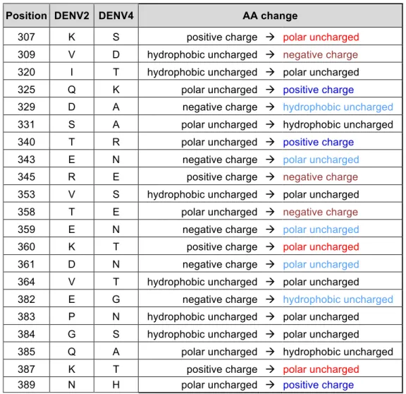
Table 2.1. Summary of amino acid changes in rDENV4/2 introducing a change
of charge or polarity ... 44
Table 2.2. Summary of MAbs used in this study ... 45
Table 2.3. Summary of DENV immune sera used in this study ... 46
Table 2.4. Neutralization of rDENV4/2 by human immune sera depleted of DENV2- or rEDIII-binding antibodies ... 47
Table 3.1. DENV2 monoclonal antibodies ... 69
Table 3.2. Percent polyclonal neutralization tracking with each epitope ... 70
xv
LIST OF ABBREVIATIONS
AA – amino acid Ab – antibody
ADE – antibody dependent enhancement cDNA – complementary deoxyribonucleic acid CR – cross-reactive
cryo-EM – cryo-electron microscopy CYD – chimeric yellow fever dengue
CYD-TDV – chimeric yellow fever dengue-tetravalent dengue vaccine
DC-SIGN – dendritic cell-specific intercellular adhesion molecule-3-grabbing non-integrin DENV – dengue virus
DNA – deoxyribonucleic acid E – envelope
E. coli - Escherichia coli ED – envelope domain
EDE – envelope dimer epitope EDI – envelope domain I
EDI/II – envelope domain I and II EDII – envelope domain II
EDIII – envelope domain III
ELISA – enzyme-linked immunosorbent assay FC – fragment crystallizable
FFU – focus forming unit
xvi
HIV – human immunodeficiency virusHMAb – human monoclonal antibody IgG – immunoglobulin G
IgM – immunoglobulin M LAV – live attenuated vaccine LLPC – long lived plasma cell MAb – monoclonal antibody MBC – memory B-cell NAb – neutralizing antibody Neut – neutralization assay Neut50 – 50% neutralization titer
NHP – non-human primate NHS – normal human sera
NIH – National Institutes of Health NS – non-structural protein
OD – optical density PAb – polyclonal antibody
PCR – polymerase chain reaction pr – precursor protein
prM – precursor membrane protein rDENV – recombinant dengue virus rE – recombinant envelope protein
rEDIII – recombinant envelope domain III protein RFLP - restriction fragment length polymorphism RNA – ribonucleic acid
xvii
TS – type-specificWNV – West Nile virus WT – wildtype
1
CHAPTER 1 – The Molecular Specificity of the Human Antibody
Response to Dengue Virus Infections1
1.1 Summary
Dengue viruses (DENV) are mosquito-borne positive sense RNA viruses in the family Flaviviridae. The four serotypes of DENV (DENV1, DENV2, DENV3, DENV4) are widely distributed and it is estimated over a third of the world’s population is at risk of infection (1). While the majority of infections are asymptomatic, DENV infection can cause a spectrum of disease, from mild flu-like symptoms, to the more severe DENV hemorrhagic fever and shock syndrome (2). Over the past 20 years, there have been intense efforts to develop a tetravalent live-attenuated DENV vaccine (3). The process of vaccine development has been largely empirical, because effective live attenuated vaccines have been developed for other flaviviruses like yellow fever and Japanese encephalitis viruses. However, recent results from Phase III live attenuated DENV vaccine efficacy trials are mixed with evidence for efficacy in some populations but not others (4). In light of unexpected results from DENV vaccine trials, in this chapter we will review recent discoveries about the human antibody response to natural DENV infection and discuss the relevance of this work to understanding vaccine performance.
1.2 DENV Structure
The DENV genome encodes a single open reading frame that is translated into a polyprotein. Viral and host proteases cleave the polyprotein into three structural and seven
2
structural viral proteins. The structural envelope protein (E) contains three domains, domain I (EDI), domain II (EDII) and domain III (EDIII) (5). Two envelope monomers come together in a head-to-tail orientation, forming the E dimer (Figure 1.1). Three E dimers form the dimer raft, and 30 dimer rafts cover the surface of the DENV virion in icosahedral orientation with both 3-fold and 5-3-fold axes of symmetry. Domain II contains the hydrophobic fusion peptide, which mediates fusion between the virus and host cell membrane. To prevent fusion with the host-membrane during egress from infected cells, the pre-host-membrane (prM) protein covers the fusion loop. As the virus moves through the endosome, pH changes triggers the host protease furin to cleave the prM protein (6). As the virus is released from cells, cleaved prM dissociates from the virion. This process is inefficient however, leaving a heterogeneous population of fully mature (no prM present), fully immature (containing prM), and partially mature virions (7). While cell culture grown virus shows a spectrum of maturation states, it is now clear that the overall maturation state of virions can vary between strains and even between different preparations of the same strain (8). As we discuss later, maturation state can influence the ability of some antibodies to bind and neutralize DENV and other flaviviruses.
1.3 Antibody Response to DENV Infection
3
neutralizing antibodies and some cross reactive poorly neutralizing antibodies are maintained for decades following infection and appear to protect against subsequent re-infection with the same serotype, but do not protect against the other serotypes (Figure 1.2). Conversely, cross-reactive antibodies not only are non-protective, but can enhance subsequent infection via a mechanism known as antibody dependent enhancement (ADE) whereby non-neutralizing antibodies bind the virus and the antibody-virus complex is taken up by cells via FC-receptor mediated endocytosis (13). Although ADE is poorly understood, the response is important in natural infection and vaccine development but will not be discussed in this review. Readers are recommended to refer to these earlier reviews for additional information on ADE and DENV (13-15).
1.4 Methods to Study the Molecular Specificity of Human Antibodies to DENVs
4
has shown that the discontinuous residues that comprise these complex epitopes can be transplanted to a different serotype to generate chimeric DENVs that encode neutralizing epitopes from multiple DENV serotypes, and which can be used to map and confirm the binding and neutralization of individuals MAbs (20, 21).
Polyclonal sera contains a complex mixture of DENV-specific IgG antibodies, those that are neutralizing or non-neutralizing, and those that are specific to a serotype or cross-reactive to multiple serotypes (Figure 1.2). Depletion assays can be used to determine the percentage of neutralizing serotype-specific antibodies to neutralizing cross-reactive antibodies (Figure 1.3A). To remove cross-reactive Abs, primary infection sera can be incubated with beads coated with a heterologous serotype (e.g. a primary DENV2 sera can be incubated with DENV1/DENV3/DENV4-coated beads). Cross-reactive Abs will bind to the virus on the beads and be pelleted out, leaving only DENV2 serotype-specific Abs. Neutralization assays using depleted sera allow one to calculate the fraction of neutralization due to serotype specific Abs, relative to the total neutralization coming from both serotype-specific and cross-reactive Abs (22-24). These depletion techniques, in addition to use of epitope transplant chimeric rDENVs described above, has allowed us to study the amount of polyclonal antibodies targeting epitopes represented by individual MAbs (21, 23).
1.5 Molecular Specificity of Neutralizing MAbs from Primary Cases
5
their ability to bind recombinant envelope monomeric protein (rE). This has biased our study of MAbs to those that recognize simple epitopes; their epitopes are contained within a single E protein. Recent work by many groups has expanded this study to screen antibodies based on their ability to bind the entire DENV virion (25, 26). This work has identified antibodies from each serotype that recognize complex, quaternary epitopes, which are assembled from higher-order structures that span across monomers, dimers, rafts, or require specific angles or presentation of E, that is only present on the intact virus (Figure 1.4). Additionally, it has been found that while antibodies using simple epitopes can be neutralizing, it is the antibodies recognizing complex epitopes that are ultimately responsible for polyclonal neutralization (21, 27, 28). Antibodies recognizing quaternary epitopes are not unique to DENV; West Nile Virus (WNV) and Zika Virus (ZIKV) have also been shown to generate human MAbs recognizing similar complex epitopes (29-32).
1.6 Differences in Neutralizing MAb Epitopes Across Serotypes
6
antibodies from a few immune individuals. To fully define the boundaries of the polyclonal neutralizing epitopes against each serotype, additional antibodies from more individuals will need to be studied.
1.7 Cryptic Epitopes
The majority of human epitopes studied are present on the surface of the intact virion. Some studies have identified mouse MAbs that target cryptic epitopes not readily accessible on the surface of the virus. However, at elevated temperature these cryptic epitopes are transiently displayed, allowing antibody binding and neutralization (39). Recent studies suggest that there are antibodies present in human immune sera that also target these cryptic epitopes, potentially allowing the virus to be neutralized when it is under specific conditions exposing these epitopes (39). However, we need to identify human MAbs that target these cryptic epitopes and determine their role and importance in neutralization and protective immunity.
1.8 Other Flaviviruses – Zika Virus MAbs
7
1.9 Mapping the Molecular Specificity of the Polyclonal Serum Neutralizing Antibody
Response
While MAbs are isolated or generated from memory B-cells, circulating polyclonal antibodies come from plasma cells (40). The memory B-cell derived human MAbs can be used as tools to interrogate the properties and specificity of the more complex polyclonal serum antibody response (Figure 1.5). Work by multiple groups have shown that individual MAbs can be representative of the anti-DENV B-cell repertoire, polyclonal Abs from the same individual, and polyclonal Abs across other naturally infected and vaccinated individuals, confirming the importance of studying individual monoclonals (21, 23, 41). Importantly, depletion assays have revealed that after primary DENV infections, the majority of polyclonal neutralization comes from serotype-specific antibodies, not cross-reactive ones (22-24). Additionally, we have found that epitopes defined by individual MAbs that are complex and quaternary, are representative of the polyclonal epitopes targeted by neutralizing serotype-specific antibodies (27, 28). With the rapid emergence of ZIKV, similar techniques as described above were applied to dissecting the antibody response to ZIKV infection. Multiple groups have found that strongly neutralizing ZIKV MAbs target complex quaternary epitopes (29-31). Additional work using depletion assays, has identified that primary ZIKV infections can results in ZIKV specific Abs, despite populations of Abs that cross-neutralize DENV (42).
1.10 Molecular Specificity of Neutralizing Antibodies Following Secondary DENV
Infection
8
encountered by the person (43). Human cohort studies in dengue-endemic countries have also established that tertiary infections are nearly always mild or inapparent, implicating a protective role for these broadly cross-neutralizing antibodies that develop after a second DENV infection (44). Figure 1.6 presents a model to explain the evolving antibody response following sequential DENV infections with different serotypes. The model is based on the premise that low affinity DENV cross reactive memory B-cells derived from primary infections undergo antibody somatic hyper mutation and each subsequent DENV exposure selects and expands rare affinity matured clones with greater neutralization breadth and potency (24). The model is supported by recent studies demonstrating that serotype cross-reactive antibodies derived from secondary infections had stronger neutralization potencies and higher binding avidities than those derived from patients with primary infections (45-49).
9
molecular mechanisms leading to the evolution of cross-neutralizing antibodies from the memory B-cell pool from a primary infection are also unclear.
1.11 NS1 and prM MAbs
While E is the major antigenic protein of DENV, antibodies are also generated targeting the viral proteins NS1 and prM. The host sees prM protein in several configurations. As mature viruses are released from infected cells, prM protein dissociates from the virus and is released as an antigen. Additionally, immature viruses have prM present on their surface, allowing the immune system to recognize them as part of the virus. prM antibodies are predominantly non-neutralizing enhancing antibodies; they allow non-infectious immature viruses to be taken up into cells via FC-receptor-mediated endocytosis (52).
DENV NS1 protein has many roles depending on its interactions and location (53, 54). NS1 can exist as a monomer, dimer or hexamer, and is important in viral RNA replication, viral assembly and release, and immune evasion. NS1 is secreted from infected cells primarily as a hexamer, which can bind to endothelial cells, triggering hyperpermeability, leading to vascular leakage seen in severe DENV disease (55). Clinically, levels of circulating NS1 are correlated with disease severity (56, 57). People infected with DENV make antibodies directed against NS1, but is unclear if these are an important part of the protective immune response, or are merely a consequence of high levels of circulating viral antigen (54).
1.12 Mechanisms of Neutralization
10
mechanisms (37, 38, 51, 58). DENV maturation state (amount of prM present) and virus breathing are important factors for virus neutralization. A fully immature virus (i.e. 180 copies of prM present) is non-infectious, and therefore cannot be neutralized, but a partially mature virus, can still be infectious (59). Alternatively, under certain temperature conditions, some DENV strains can undergo reversible conformational changes where the E proteins expand and contract analogous to “breathing”. These expansion and contraction changes can reveal or hide epitopes, limiting neutralization by antibodies recognizing these epitopes to specific conditions (39, 60-62). While DENV maturation and “breathing” have been studied in cell culture systems, the importance of these phenomenon in natural infection, and therefore the potential impact on antibody neutralization, is not well understood.
1.13 Implications for Evaluating Antibodies to DENV Live Attenuated Vaccines (LAVs)
Recently we have learned important lessons from DENV tetravalent vaccine clinical trials. The leading tetravalent vaccine had variable efficacy depending on DENV serotype and vaccinated population (4). The vaccine had higher efficacy in DENV-primed individuals compared to DENV naïve individuals who received the vaccine, establishing the impact of immunological memory on vaccine performance (63). The population with the greatest need for a DENV vaccine is young children, the majority of whom will be DENV-naïve at vaccination. As discussed above, in people exposed to primary natural DENV infections, the neutralizing and protective antibody response is dominated by type-specific antibodies to quaternary epitopes. Therefore, in this population the success of tetravalent vaccination is likely to require balanced replication of the four vaccine viruses leading to type-specific antibodies that target quaternary epitopes in each serotype.
11
DENV serotypes, driven by the sequential infection and robust replication of two different serotypes of DENV (24). A similar mechanism is likely to be responsible for the superior performance of tetravalent LAVs in DENV-primed individuals. In a subject with pre-existing DENV-specific MBCs, even unbalanced replication of one or two vaccine components is likely to activate MBCs and expand somatically mutated higher-affinity cross-reactive clones with capacity to broadly neutralize multiple serotypes.
Immune correlates of protection and vaccine efficacy are urgently needed. For the leading DENV vaccine, the mere presence of in vitro neutralizing antibodies was not sufficient for protection because many individuals experienced breakthrough infections despite having neutralizing antibodies to the breakthrough serotype (63). The lessons we have learned from natural infections studies about the molecular specificity of human antibodies to DENV infection may also lead to more robust correlates of vaccine efficacy than mere levels of total neutralizing antibodies (3). Certainly, the reagents and tools are now available to interrogate vaccine responses in a manner similar to that we have described here for natural DENV infections.
1.14 Objectives of this Dissertation
12
Early research on immunity to DENVs relied heavily on monoclonal antibodies (MAbs) generated in mice. Extensive studies with mouse MAbs identified a primary epitope in EDIII of the envelope glycoprotein recognized by strongly neutralizing antibodies (64). Subsequent studies revealed that, while this epitope is immuno-dominant in mice, only a small portion of the human serological response targets this EDIII epitope (28, 64, 65). This research revealed that the human antibody response to DENV infection is distinct from that in a mouse, and therefore to fully characterize it, we must isolate and charactize the activies of human DENV antibodies (64). Unlike mouse EDIII epitopes, it was found that human strongly neutralizing MAbs for DENV1, DENV3 and DENV4 target an epitope centered near the EDI/II hinge region (23, 27). Importantly, these are not simple epitopes present on recombinant proteins, but quaternary; they are only present on the fully assembled infectious virion (23, 27). Recent advances have shown that polyclonal serum neutralizing antibodies are also directed to quaternary structure epitopes defined using MAbs (23, 27).
13
DENV polyclonal immune sera are comprised of multiple populations of antibodies, including type-specific antibodies specific to the infecting serotype, and weakly neutralizing cross-reactive antibodies (12). It is thought that cell-culture based assays can over-represent the neutralization potency by cross-reactive antibodies. Using depletion techniques we can study the contribution of each of these populations of antibodies on total neutralization (27, 70). Recent work identified that Dengvaxia vaccine sera contains large populations of DENV4 serotype-specific neutralizing antibodies, however DENV1-3 neutralization is driven primarily by cross-reactive antibodies (22). Analysis of vaccine sera from individuals who experienced breakthrough infections, versus those who did not, we can potentially determine an improved antibody and epitope based correlate of protection, by specifically measuring the quality of antibodies elicited by vaccination.
Identifying the dominant epitopes targeted by human DENV2 serotype-specific monoclonal and polyclonal antibodies is critical for refining our understanding of response to infection and vaccination. Detailed characterization of these DENV2 antibodies and the epitopes they target, will improve our understanding of the complex adaptive immune response to DENV infection, and can be harnessed to improve design and evaluation of DENV vaccines.
We hypothesize that 1) DENV2 serotype-specific neutralizing antibodies target multiple
quaternary epitopes on the envelope glycoprotein, 2) that generating a robust DENV2-specific
antibody response directed to these epitopes is critical for vaccine-elicited protection, and that
3) DENV1-specific neutralizing epitopes are distinct from those of DENV2, but can be mapped
14
AIM 1: Map the epitopes of human DENV2 serotype-specific monoclonal and polyclonal
antibodies using recombinant DENVs, chimeric epitope transplant viruses, monoclonal blockade/competition assays, binding and neutralization assays.
AIM 2: Evaluate the DENV2 serotype-specific neutralizing antibody response following
tetravalent DENV vaccination, by determining the quality of DENV2 neutralizing antibodies, the epitopes they target, and whether these responses differ in individuals who experienced DENV2 breakthrough infections, versus those who did not.
AIM 3: Map the epitopes of DENV1 serotype-specific monoclonal antibodies by characterizing a
15
Figure 1.1. Structure of DENV. A) Linear schematic of DENV envelope (E) protein. DENV E
protein dimer composed of two monomers with domains I, II and III colored in red, yellow and blue respectively. B) DENV virion structure composed of 30 rafts, each containing three E dimers.
A
B
II II
I I I III
pr M TM
16
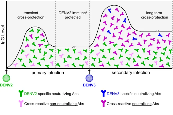
Figure 1.2. Antibody response following DENV infection. Following primary DENV2
infection, there is an IgG response composed of neutralizing DENV2 serotype-specific antibodies, a transient population of reactive neutralizing antibodies, and long-lived cross-reactive non-neutralizing antibodies. After a secondary infection, in this case with DENV3, the cross-reactive non-neutralizing antibodies become strongly neutralizing. It is also possible to generate a new population of neutralizing serotype-specific antibodies to the second infecting serotype.
secondary infection primary infection
DENV2
Ig
G
L
eve
l
DENV3
DENV2-specific neutralizing Abs DENV3-specific neutralizing Abs
Cross-reactive non-neutralizing Abs Cross-reactive neutralizing Abs transient
17
Figure 1.3. Methods to dissect DENV antibody response. A) Human DENV antibodies can
be studied using a variety of approaches. PBMCs from a DENV immune donor can be EBV-transformed to generate MAb producing hybridomas, or antibody DNA sequences can be single-cell sequenced, cloned and recombinant expressed to generate MAbs. DENV polyclonal immune sera can be depleted of different populations of antibodies using beads coated with
DENV2
immune blood
PBMCs
DENV MAbs
Control
Deple+on Heterotypic Deple+on Homotypic Deple+on
Polyclonal DENV2 immune sera
Binding Assays
rEDIII rE Whole DENV Chimeric rDENV
DENV1
DENV2
DENV3
DENV4
rDENV1/3
rDENV2/4
Neutralization Assays
Whole
DENV Chimeric rDENV
DENV1
DENV2
DENV3
DENV4
rDENV1/3
rDENV2/4
A
18
19
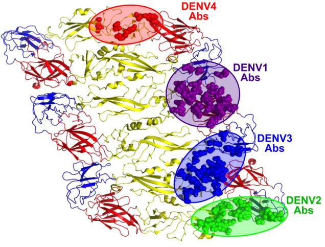
Figure 1.4. Epitopes recognized by DENV serotype-specific human neutralizing MAbs.
Serotype-specific neutralizing human MAbs isolated from primary infections recognize different quaternary structure epitopes displayed on the viral envelope.
DENV1
Abs
DENV2
Abs
DENV3
Abs
20
Figure 1.5. From MAbs to polyclonal serum Abs. Complex host generic diversity, exposure
history, and immune differences can make it challenging to study DENV polyclonal antibody responses across a population. Studying DENV antibody immunity in a single individual can simplify these analyses, however there is still the polyclonal nature of the adaptive immune response. Conversely, we can characterize the properties of individual MAbs from DENV immune donors. Information learned from MAbs can then be used to inform study of the B-cell repertoire from that, and other donors. Additionally, it can be determined whether the individual MAbs represent the polyclonal antibodies in that donor, and in a larger DENV immune population.
DENV immune/vaccinated population
Polyclonal DENV immunity
PBMCs
21
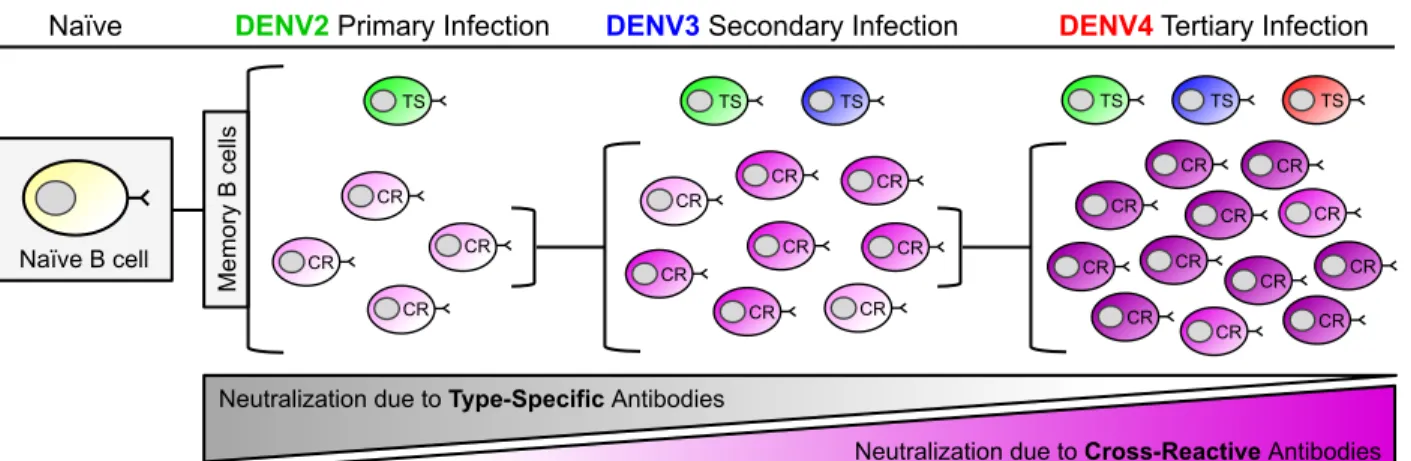
Figure 1.6. Model of B-cell maturation following sequential DENV infections. With each
successive DENV infection, the ratio of serotype-specific (TS) and cross-reactive (CR) antibodies that contribute to DENV neutralization changes. During a primary infection (DENV2 in this example), dengue-specific naïve B-cells are activated and these cells give rise to both memory B-cells (MBCs) and antibody secreting long lived plasma cells (LLPCs). This primary response is dominated MBC and LLPCs clones producing low affinity, weakly neutralizing serotype CR antibodies. The primary response also contains rare MBC and LLPCs producing TS antibodies that strongly neutralize DENV2. Following a secondary infection with a new serotype (DENV3 in this example), the overall DENV-specific B-cell response will be dominated by the activation and expansion of DENV2 and 3 cross-reactive MBCs induced by the primary infection. MBCs producing CR antibodies that bind to the second infecting serotype with high affinity will be preferentially activated. These activated cells will reenter germinal centers and undergo further rounds of somatic hyper mutation. CR B-cells with high affinity for the second serotype will be selectively expanded to give rise to cross-reactive MBC and LLPCs that strongly cross-neutralize multiple serotypes. In the figure this increase in affinity and neutralization is depicted by an increase in the color gradient (light pink to bright pink) of CR B-cells. Following a tertiary infection (DENV4 in this example), this process is repeated again and results in a population of CR MBCs and LLPCs that dominate the neutralizing antibody
Naïve B cell
TS
Naïve
CR CR
CR
CR
CR CR
CR CR CR CR CR CR TS TS TS
DENV4Tertiary Infection
CR CR
CR CR CR CR
CR CR
CR CR
CR CR Me mo ry B ce lls
Neutralization due to Type-Specific Antibodies
Neutralization due to Cross-Reactive Antibodies
DENV2 Primary Infection
TS TS
22
23
Figure 1.7. EDE and other cross-reactive epitopes. Envelope dimer epitope 1 (EDE1) targets
EDIII of one monomer and spans over the fusion loop region of EDII of the neighboring monomer. EDE2 uses a similar epitope, but is shifted to also expand into EDI of the first monomer. Another class of cross-reactive antibodies targets the highly conserved bc-loop region of EDII.
EDE1
Abs
EDE2
Abs
24
CHAPTER 2 – A new quaternary structure epitope on dengue virus serotype 2
is the target of durable type-specific neutralizing antibodies2
2.1 Summary
Dengue virus serotype 2 (DENV2) is widespread and responsible for severe epidemics. While primary DENV2 infections stimulate serotype-specific protective responses, a leading vaccine failed to induce a similar protective response. Using human monoclonal antibodies (hMAbs) isolated from dengue cases and structure-guided design of a chimeric DENV, here we describe the major site on the DENV2 envelope (E) protein targeted by neutralizing antibodies. DENV2-specific neutralizing hMAb 2D22 binds to a quaternary structure epitope. We engineered and recovered a recombinant DENV4 that displayed the 2D22 epitope. DENV2 neutralizing antibodies in people exposed to infection or a live vaccine tracked with the 2D22 epitope on the DENV4/2 chimera. The chimera remained sensitive to DENV4 antibodies, indicating that the major neutralizing epitopes on DENV2 and -4 are at different sites. The ability to transplant a complex epitope between DENV serotypes demonstrates a hitherto underappreciated structural flexibility in flaviviruses, which could be harnessed to develop new vaccines and diagnostics.
2.2 Importance
Dengue virus causes fever and dengue hemorrhagic fever. Dengue serotype 2 (DENV2) is widespread and frequently responsible for severe epidemics. Natural DENV2 infections stimulate serotype-specific neutralizing antibodies, but a leading DENV vaccine did not induce a
25
similar protective response. While groups have identified epitopes of single monoclonal antibodies (MAbs), the molecular basis of DENV2 neutralization by polyclonal human immune sera is unknown. Using a recombinant DENV displaying serotype 2 epitopes, here we map the main target of DENV2 polyclonal neutralizing antibodies induced by natural infection and a live DENV2 vaccine candidate. Proper display of the epitope required the assembly of viral envelope proteins into higher-order structures present on intact virions. Despite the complexity of the epitope, it was possible to transplant the epitope between DENV serotypes. Our findings have immediate implications for evaluating dengue vaccines in the pipeline as well as designing next-generation vaccines.
2.3 Introduction
26
The DENV envelope glycoprotein (E) is the main target of protective antibodies (5). The E protein is composed of three domains: I, II and III (designated EDI, EDII and EDIII). Each DENV particle has 180 monomers of E that are organized into 90 dimers that cover the entire surface of the virus (76). The arrays of E proteins are arranged with icosahedral symmetry, with each asymmetric unit containing three E proteins. Some human monoclonal antibodies (hMAbs) that neutralize DENVs bind to quaternary structure epitopes that require assembly of E protein into homodimers or higer order structures (27, 36, 38, 51, 77). Followng infection or vaccination, it is a DENV-specific serum polyclonal antibody response that is responsible for protection. The principle targets of the human polyclonal antibody responses that neutralize DENVs have remained elusive.
We recently described hMAb 2D22, which is a DENV2-specific strongly neutralizing antibody isolated from a person exposed to a primary DENV2 infection (27). A point mutation at amino acid position 323 in EDIII (residue highlighted in magenta in Figure 2.1A and B) led to complete escape from 2D22 neutralization, indicating that the epitope includes EDIII residues (27). Recently Fibriansah et al solved the structure of 2D22 bound to DENV2 and demonstrates that the antibody bound to a quaternary epitope that was formed by EDIII and EDII on two different monomers within a single dimer (38). While the structure of hMAb 2D22 was a major advance, it is not known if the structurally intriguing epitope defined by 2D22 is the main target of neutralizing and protective antibodies in people exposed to DENV2 infections or a vaccine.
2.3 Results
Design of recombinant DENV4/2 chimeric virus
27
clone to create a recombinant virus, designated rDENV4/2 (Figure 2.2). The recombinant virus, which had 40 amino acid changes in EDIII compared to the parental wild-type (wt)DENV4 strain (Figure 2.1A and Table 2.1), grew to similar levels as the wt viruses in C6/36 insect cells and in a human monocytic cell line (U937) expressing dendritic cell-specific intercellular adhesion molecule-3-grabbing non-integrin (DC-SIGN), a know dengue receptor, but was partially growth impaired in Vero cells (Figure 2.1C). DENVs are assembled inside cells as immature virions containing premembrane proteins (prM), which are cleaved in the Golgi resulting in the formation of mature virions. Proteolytic cleavage of prM is inefficient and the population of virions released from infected cells is an admixture displaying different stages of maturation. As the maturation state of DENVs may influence the display of some epitopes and sensitivity to antibody neutralization (59, 62, 78), immunoblots were performed to compare the maturation state of the rDENV4/2 chimera and the wt parental strains. From C6/36 cells, DENV2 particles had high levels of prM and DENV4 particles had low levels of prM relative to E protein, indicating that DENV4 virions were more mature (Figure 2.1D). The rDENV4/2 chimera had a maturation state similar to DENV4 indicating that insertion of EDIII from serotype 2 minimally altered the maturation state of the backbone serotype 4 virus (Figure 2.1D).
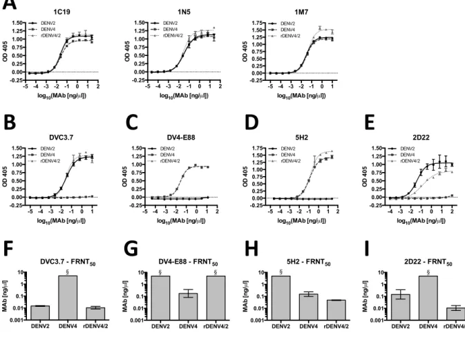
Monoclonal antibody binding and neutralization of rDENV4/2
28
MAb with an EDI epitope, is not disrupted in rDENV4/2, showing we have not affected epitopes present on other domains (Figure 3.2D, H and Table 2.2) (80). Overall, these results demonstrate that the rDENV4/2 chimera displays epitopes in a manner consistent with display on a properly folded and functional chimeric E protein. Transplantation of DENV2 EDIII into DENV4 also restored binding and neutralization by MAb 2D22, even though this antibody did not bind to DENV2 recombinant EDIII alone (Figure 2.3E, I and Table 2.2). We predict that the full 2D22 epitope required for antibody binding and neutralization includes EDIII as well as some conserved residues on adjacent domains, but that the residues on EDIII alone determine DENV2 specificity. Indeed the cryo-electron microscopy structure of 2D22 bound to DENV2 demonstrates that the epitope consists of residues on EDIII and EDII of different monomers within a single dimer (38).
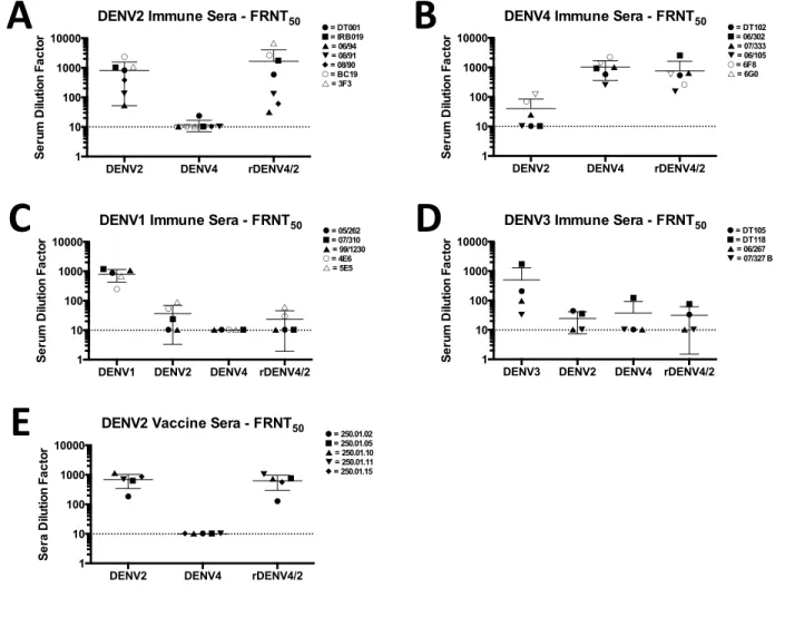
Polyclonal sera neutralization of rDENV4/2
29
reflecting a global increase in sensitivity to neutralization by any dengue immune serum, a panel of primary DENV1 and DENV3 immune sera were tested against the same three viruses. The rDENV4/2 virus did not display increased sensitivity to neutralization by DENV1 or 3 immune sera (p>0.05; p>0.05), demonstrating that the chimeric virus was not globally sensitive to antibody neutralization (Figure 2.4C and D).
DENV2 type-specific antibodies require complex epitope
30
To determine if dengue vaccines can induce “2D22-like” quaternary epitope targeted neutralizing antibodies, we tested sera from 5 subjects who had developed DENV2 neutralizing antibodies after receiving a monovalent live attenuated DENV2 vaccine developed by the NIH (82). The vaccine sera neutralized DENV2 and the rDENV4/2 chimera but not DENV4 demonstrating that the vaccine induced neutralizing antibodies that tracked with the transplanted EDIII (Figure 2.4) (p<0.01). To determine if the vaccine induced antibodies also recognized a quaternary epitope that extended beyond EDIII, three vaccine sera were depleted of antibodies binding intact DENV2 virions or recombinant DENV2 EDIII and then tested for ability to neutralize the chimeric virus. In all three samples depletion with whole virus led to a nearly complete loss of neutralizing antibodies (Table 2.4). Removal of EDIII specific antibodies resulted in a loss of neutralizing antibodies in one vaccine sample, while the other two samples retained the majority of neutralizing antibodies after EDIII depletion (Table 2.4). Thus, the vaccine induced neutralizing antibodies that bind to epitopes contained within EDIII or more complex epitopes that extend beyond EDIII.
2.5 Discussion
We have described an approach using whole domain replacement to identify principal antigenic sites targeted by polyclonal antibodies following natural DENV infection or experimental live attenuated DENV vaccination. With the DENV4/2 chimera, we observed a clear gain of DENV2 neutralization and no loss of sensitivity to neutralization by DENV4 sera, suggesting that the principal DENV4 neutralizing epitopes are distinct from DENV2 epitopes. Importantly, these data demonstrate that a single recombinant DENV can be designed that encodes major neutralizing epitopes from two virus serotypes.
31
recognize distinct quaternary structure epitopes centered at the EDI/II hinge. However, only a small fraction (<3%) DENV-specific memory B-cell clones produce strongly neutralizing antibodies (83). It has not been clear if epitopes defined using human MAbs are the main targets of the polyclonal serum neutralizing antibody response as well. Our studies here demonstrate that the DENV2 serotype-specific epitopes targeted by a human MAb, and polyclonal immune sera are closely related if not identical. The epitope is a complex, quaternary epitope and includes critical residues in EDIII that determine serotype specificity.
The results reveal the fundamental importance of complex quaternary structures on the surface of DENV particles for driving potent antibody immune responses. Our results are entirely consistent with the 2D22 epitope structure reported by Fibriansah et al (84) demonstrating that antibody footprint contains critical contact residues on EDIII of one monomer, as well as the fusion loop and BC-loop of EDII on the adjacent monomer, bridging across the dimer. The structure also demonstrates that the 2D22 antibody contact sites on EDIII are not conserved between serotypes but the contact sites on EDII are highly conserved between DENV2 and 4 (84). Thus, the serotype specificity of the 2D22 is determined by EDIII and transplantation of this domain into DENV4 was sufficient to create the complete epitope, functional epitope.
32
The rDENV4/2 chimera is a powerful tool for evaluating antibody site-specific responses following infection and vaccine trials. Recombinant DENVs expressing quaternary neutralizing antibody epitopes from 2 or more serotypes may lead to simpler and more effective vaccines than current tetravalent formulations.
2.6 Materials and Methods
Virus construction
Recombinant viruses were constructed using a four-cDNA cloning strategy, the same strategy used to create wt DENV infectious clones (Figure 2.2). Patterned after coronavirus cDNA clones (85, 86), the DENV-4 genome was subcloned into four separate cDNA plasmids. A T7 promoter was introduced into the 5ʹ end of the A fragment, and unique type IIS restriction
endonuclease cleavage sites are introduced into the 5ʹ and 3ʹ end of each fragment to allow for
systematic assembly into a genome-length cDNA from which full-length transcripts can be derived (85-87).
33
C6/36 cells, centrifuged to removed cellular debris, and stored at -80°C. Passage 3 represents our working stock.
Cells
Mosquito Ae. albopictus C6/36 cells were grown in MEM (Gibco) media at 32°C. Vero-81 cells were maintained in DMEM and U937+DC-SIGN were maintained in RPMI at 37°C. Medium was supplemented with FBS (10% for Vero-81 and 5% for C6/36 and U937+DC-SIGN), which was lowered to 2% after infection. C6/36 and U937+DC-SIGN media was supplemented with non-essential amino acids, and U937+DC-SIGN was also supplemented with L-glutamine and 2-mercaptoethanol. All media were additionally supplemented with 100 U/mL penicillin and 100 µg/mL streptomycin. All cells were incubated in 5% CO2 as previously described by our
group (87).
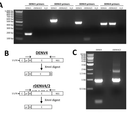
DENV Type-Specific PCR and RFLP Analysis
Total RNA was isolated from viral supernatants and used as template for cDNA synthesis using standard molecular techniques. Serotype-specific PCR and restriction endonuclease analyses were performed on cDNA samples in order to validate purity of the recombinant viral preparations (Figure 2.7).
Binding ELISA
34 DENV Immune Sera
De-identified human DENV immune sera were collected from individuals with confirmed previous natural DENV infections (Table 2.3). All donations were collected in compliance with the Institutional Review Board of the University of North Carolina at Chapel Hill (Protocol #08-0895). De-identified human immune sera previously collected from adults given the NIH monovalent DENV2 vaccine (ClinicalTrials.gov identifier: NCT00920517) was provided by Anna Durbin and Stephen Whitehead. All sera was collected following informed consent and approval by the Western Institutional Review Board. Non-human primate immune sera were collected following experimental DENV infection, and kindly provided by Carlos Sariol (Supplemental Table 2). All procedures were reviewed and approved by the Institute’s Animal Care and Use Committee at Medical Sciences Campus, University of Puerto Rico (IACUC-UPR-MSC), and performed in a facility accredited by the Association for Assessment and Accreditation of Laboratory Animal Care (AAALAC) (Animal Welfare Assurance number A3421; protocol numbers, 7890108, 7890208, 7890209, and 7890210).
Virus Titration and Focus Reduction Neutralization Test (FRNT)
35
(Sigma), diluted 1:2,500 in blocking buffer. Plates were washed, foci were developed with TrueBlue HRP substrate (KPL), and then foci were counted.
For the FRNT assay, either MAbs or sera were diluted four-fold and mixed with ~40 focus forming units (FFUs) virus, then incubated for 1 hr at 37°C. After incubation, virus and MAb or serum dilutions were added to cells for 1 hr at 37°C, then overlay was added and processed as above.
Growth Curves
Either Vero or C6/36 cells were inoculated at a multiplicity of infection (MOI) of 0.01. Every 24 hrs, culture supernatant was harvested and centrifuged to remove cellular debris. Samples were frozen at -80°C until use. Fresh medium was replaced each day. Viruses were titered on their propagating cell type, as described above. U937+DC-SIGN cells were infected at an initial infection of 1%, and every 12 hours a sample of cells was harvested, fixed, permeabilized, and probed with 2H2 (anti-prM antibody) conjugated to 488. Infected cells were quantified using a Guava flow-cytometer (Milipore).
Immunoblotting
36
Depletion of DENV2-Specific Antibodies from Immune Sera
Polyclonal immune sera were depleted of DENV2-binding antibodies as previously described (27). Briefly, polystyrene microspheres (Polysciences, 17135) were coated with purified DENV2 antigen (Microbix, EL-22-02-001) or BSA control. Immune sera were depleted of antibodies by incubating with coated beads for 45 minutes at 37°C for at least three rounds, until maximum depletion of antibodies was measured. Depletions of antibodies were confirmed by ELISA.
Depletion of rEDIII-Specific Antibodies from Immune Sera
Polyclonal immune sera were depleted of rEDIII-binding antibodies as previously described for rE-binding antibodies (27). Briefly, Dynabeads® (Life Technologies, 14302D) were covalently conjugated to DENV2 rEDIII protein following manufacturers protocol or BSA control. Immune sera were depleted of antibodies by incubating with conjugated beads for 45 minutes at 37°C for at least three rounds, until maximum depletion of antibodies was measured. Depletions of antibodies were confirmed by ELISA.
ELISA Confirmation of DENV2 or rEDIII Depleted Sera
37
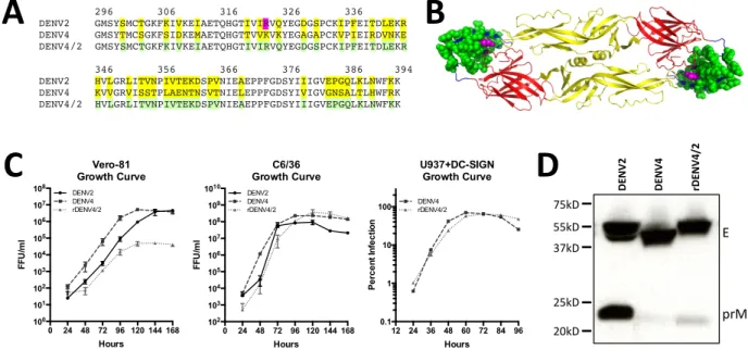
Figure 2.1. Design and characterization of rDENV4/2. (A) Amino acid alignment of DENV2
and DENV4 linear envelope domain III (EDIII) sequence, residues 296-395 of entire E sequence (99 aa total). Residues differing between DENV2 and DENV4 are highlighted in yellow. Recombinant DENV4 virus containing EDIII from DENV2, designated rDENV4/2, replaces differing residues from DENV4 with those from DENV2, highlighted in green (40 aa total). Residue generated from escape mutant highlighted in magenta. (B) Crystal structure model of DENV2 E protein dimer, with swapped residues colored in green, and DENV2 type-specific MAb 2D22 escape mutant residue highlighted in magenta. (C) Vero-81 or C6/36 cells were inoculated and viral supernatants were collected every 24 hrs and subsequently titered on the respective cell type, or 1% of U937+DC-SIGN were infected and total percent infection was measured every 12 hours (mean ± s.d.) (D) Immunoblotting of C3/36 grown viruses with anti-E and anti-PrM antibodies. E = 55 kD, prM = 21 kD.
0 24 48 72 96 120 144 168 100 101 102 103 104 105 106 107 108 Hours FFU /m l Vero-81 Growth Curve DENV2 DENV4 rDENV4/2
0 24 48 72 96 120 144 168 102 103 104 105 106 107 108 109 1010 Hours FFU /m l C6/36 Growth Curve DENV2 DENV4 rDENV4/2
12 24 36 48 60 72 84 96 0.1 1 10 100 Hours Pe rc e n t I n fe c tio n U937+DC-SIGN Growth Curve DENV4 rDENV4/2
296 306 316 326 336 DENV2 GMSYSMCTGKFKIVKEIAETQHGTIVIRVQYEGDGSPCKIPFEITDLEKR DENV4 GMSYTMCSGKFSIDKEMAETQHGTTVVKVKYEGAGAPCKVPIEIRDVNKE DENV4/2 GMSYSMCTGKFKIVKEIAETQHGTIVIRVQYEGDGSPCKIPFEITDLEKR
346 356 366 376 386 394 DENV2 HVLGRLITVNPIVTEKDSPVNIEAEPPFGDSYIIIGVEPGQLKLNWFKK DENV4 KVVGRVISSTPLAENTNSVTNIELEPPFGDSYIVIGVGNSALTLHWFRK DENV4/2 HVLGRLITVNPIVTEKDSPVNIEAEPPFGDSYIIIGVEPGQLKLNWFKK
38
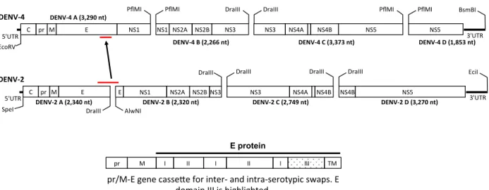
Figure 2.2. DENV4 infectious clone. Reverse genetics system for manipulating DENV4
genome. DENV genome was divided into four plasmid cassettes that can be mutated individually, ligated together, and electroporated into cells to generate recombinant virus. DENV4-A cassette contains the envelope gene, EDIII is highlighted grey. Replacing EDIII residues with those from DENV2, in DENV4 backbone, creates rDENV4/2 recombinant virus. A total of 58 nucleotide changes were introduced into DENV4-A cassette (highlighted in red).
EcoRV&
E& NS1&
C& pr& M& 5'UTR&
DENV%4''
NS1&NS2A& NS2B& NS3&
PflMI& PflMI&
NS3& NS4A& NS4B& NS5&
DraIII& DraIII&
NS5&
PflMI& PflMI& BsmBI&
3'UTR&
DENV%4'A'(3,290'nt)'
DENV%4'B'(2,266'nt)' DENV%4'C'(3,373'nt)' DENV%4'D'(1,853'nt)'
I& II&
pr& M& I& II& I& III& TM&
E protein
pr/M?E&gene&casseCe&for&inter?&and&intra?serotypic&swaps.&E& domain&III&is&highlighted.&
DraIII&
5'UTR& 3'UTR&
DraIII& AlwNI&
DraIII& DraIII& DraIII& EciI&
DENV%2''
E& NS2A& NS2B& NS3& NS4A& NS4B& NS4B& NS5&
DENV%2'A'(2,340'nt)' DENV%2'B'(2,320'nt)' DENV%2'C'(2,749'nt)' DENV%2'D'(3,270'nt)'
E& C& pr& M&
SpeI&
39
Figure 2.3. Recognition and neutralization of rDENV4/2 virus by DENV-specific
monoclonal antibodies. ELISA capture assay with (A) cross-reactive MAbs, (B)
DENV2-specific EDIII MAb DVC3.7, (C) DENV4-DENV2-specific EDIII MAb DV4-E88, (D) DENV4-DENV2-specific EDI MAb 5H2, and (E) DENV2-specific MAb 2D22 (mean ± s.d.). Vero-81 cell based Focus Reduction Neutralization Test (FRNT) was performed using (F) DENV2-specific EDIII MAb DVC3.7, (G) DENV4-specific EDIII MAb DV4-E88, (H) DENV4-specific EDI MAb 5H2, or (I) DENV2-specific MAb 2D22and FRNT50 (concentration of antibody required to neutralize 50% of
infection) values were calculated (mean ± 95% CI), § = FRNT50 >5 ng/µl
-5 -4 -3 -2 -1 0 1 -0.25 0.00 0.25 0.50 0.75 1.00 1.25 1.50
log10(MAb [ng/µl])
OD 4 0 5 DVC3.7 DENV2 DENV4 rDENV4/2
-5 -4 -3 -2 -1 0 1 2 -0.25 0.00 0.25 0.50 0.75 1.00 1.25 1.50
log10(MAb [ng/µl])
OD 4 0 5 DV4-E88 DENV2 DENV4 rDENV4/2
-4 -3 -2 -1 0 1 2
-0.25 0.00 0.25 0.50 0.75 1.00 1.25 1.50
log10(MAb [ng/µl])
OD 4 0 5 2D22 DENV2 DENV4 rDENV4/2
-5 -4 -3 -2 -1 0 1 2 -0.25 0.00 0.25 0.50 0.75 1.00 1.25 1.50 1.75
log10(MAb [ng/µl])
OD 4 0 5 5H2 DENV2 DENV4 rDENV4/2
-5 -4 -3 -2 -1 0 1 2
-0.25 0.00 0.25 0.50 0.75 1.00 1.25 1.50
log10(MAb [ng/µl])
OD 4 0 5 1C19 DENV2 DENV4 rDENV4/2
-5 -4 -3 -2 -1 0 1 2 -0.25 0.00 0.25 0.50 0.75 1.00 1.25 1.50
log10(MAb [ng/µl])
OD 4 0 5 1N5 DENV2 DENV4 rDENV4/2
-5 -4 -3 -2 -1 0 1
-0.25 0.00 0.25 0.50 0.75 1.00 1.25 1.50 1.75
log10(MAb [ng/µl])
OD 4 0 5 1M7 DENV2 DENV4 rDENV4/2
DENV2 DENV4 rDENV4/2 0.001 0.01 0.1 1 10 MA b [n g / µ l]
2D22 - FRNT50 §
DENV2 DENV4 rDENV4/2 0.001 0.01 0.1 1 10 MA b [n g / µ l]
5H2 - FRNT50
§
DENV2 DENV4 rDENV4/2
0.001 0.01 0.1 1 10 MA b [n g / µ l]
DVC3.7 - FRNT50
§
DENV2 DENV4 rDENV4/2 0.001 0.01 0.1 1 10 MA b [n g / µ l]
DV4-E88 - FRNT50
§ §
B"
C"
D"
G"
F"
E"
H"
I"
40
Figure 2.4. rDENV4/2 neutralization by human and macaque DENV immune sera. Vero-81
cell-based Focus Reduction Neutralization Test (FRNT) was performed using (A) primary DENV2, (B) primary DENV4, (C) primary DENV1, (D) primary DENV3, and (E) monovalent DENV2 vaccine immune sera, and FRNT50 (sera dilution factor required to neutralize 50% of
infection) values were calculated (mean ± 95% CI). Solid symbols = human sera, empty symbols = rhesus macaque sera. Sera that did not block 50% of infection at lowest sera dilution factor were assigned a value of 10 (½ the lower limit of detection) for graphing and statistical analysis.
DENV2 DENV4 rDENV4/2 1 10 100 1000 10000 ! " ! " # ! " ! " # ! " ! " # Se ru m D ilu tio n F a c to r
DENV2 Vaccine Sera - FRNT50
! = 250.01.02 " = 250.01.05 ! = 250.01.10 " = 250.01.11 # = 250.01.15
DENV2 DENV4 rDENV4/2
1 10 100 1000 10000 ! " # ! " # $ ! " #! " $#
! " # ! " # $ Se ru m D ilu tio n F a c to r
DENV2 Immune Sera - FRNT50
! = DT001
" = IRB019 " = 06/94 # = 08/91
$ = 08/90
# = BC19 ! = 3F3
A"
DENV2 DENV4 rDENV4/2
1 10 100 1000 10000 ! "! # " # ! " ! # "
# !" ! # " # Se ru m D ilu tio n F a c to r
DENV4 Immune Sera - FRNT50
! = DT102
# = 06/302 " = 07/333 # = 06/105
" = 6F8 $ = 6G0
DENV1 DENV2 DENV4 rDENV4/2
1 10 100 1000 10000 ! ! " # " !! " #
" ""!!# ! ! " # " Se ru m D ilu tio n F a c to r
DENV1 Immune Sera - FRNT50
" = 05/262 # = 07/310 " = 99/1230 ! = 4E6 ! = 5E5
DENV3 DENV2 DENV4 rDENV4/2 1 10 100 1000 10000 ! " !
" ! " !" ! " ! " ! " !" Se ru m D ilu tio n F a c to r
DENV3 Immune Sera - FRNT50
! = DT105
" = DT118
! = 06/267 " = 07/327 B
DENV2 DENV4 rDENV4/2
1 10 100 1000 10000 ! " ! " # ! " ! " # ! " ! " # Se ra D ilu tio n F a c to r
DENV2 Vaccine Sera - FRNT50
! = 250.01.02 " = 250.01.05 ! = 250.01.10 " = 250.01.11 # = 250.01.15
B"
C"
D"
41
Figure 2.5. Depletion of DENV2- and rEDIII-binding antibodies. (A) DENV2 immune sera
were depleted using beads coated with virus (DV2-depleted) or BSA (BSA-depleted) and removal of DENV2 binding antibodies was confirmed by ELISA, with wells directly coated with DENV2 antigen. (B) DENV2 immune sera were depleted using Dynabeads with DENV2 rEDIII (rEDIII-depleted) or BSA (BSA-depleted) and removal of DENV2 rEDIII-binding antibodies was confirmed by ELISA, with wells directly coated with rEDIII protein. DT001, IRB019, D031 are sera from people exposed to natural DENV2 infections. 250.01.02, 250.01.05 and 250.01.19 are sera from people who received the NIH DENV2 vaccine. NHS = normal human serum.
DT001 IRB019 DT031 DT110 DT134 ss08/90 250.01.02 250.01.05 250.01.19 NHS 0.0
0.2 0.4 0.6 0.8 1.0
Sera
OD
4
0
5
DENV2-depleted sera Undepleted
BSA-depleted DENV2-depleted
DT001 IRB019 DT031 DT110 DT134 ss08/90 250.01.02 250.01.05 250.01.19 NHS
0.0 0.5 1.0 1.5 2.0 2.5
Sera
OD
4
0
5
rEDIII-depleted sera Undepleted
BSA-depleted rEDIII-depleted