Mabika Barthelemy et al JMSCR Volume 07 Issue 05 May 2019 Page 1167
Quantitative Morphological Analysis of Infectious Endocarditis by the
SAMBA System
Authors
Mabika Barthelemy
1, Ngoua Meye Misso Rick-Léonid
2, Lepidi Hubert
3, Didier Raoult
31
Laboratory of Anatomical Pathology, University of Health Sciences, Libreville, Gabon
2
Laboratory of Biochemistry Research, University of Sciences and Technology of Masuku, Franceville, Gabon
3
Unité Des Rickettsies, CNRS UMR 6020, Faculte de Medecine, Universite de la Mediterranee *Corresponding Author
MABIKA Barthélemy
Laboratory of Anatomical Pathology, University of Health Sciences, Libreville, Gabon Email: [email protected]
Abstract
The aim of this work was to determine the criteria for histologically differentiating infectious endocarditis from degenerative valve processes on native valves, bioprostheses and mechanical valves using the SAMBA technique.
The Cardiac Surgery Service of the Center hospital-universities of Timone provided 630 valves. Histological sections stained with hematoxylin eosin saffron (HES) were reviewed. The histological lesions analyzed on slides stained with HES were vegetation, fibrosis and calcifications. Those analyzed after immunostaining were the cells of the inflammatory response (neutrophils labeled with the CD15 antibody, the CD68 macrophages and the CD3 lymphocytes). For endothelial cells, Factor VIII antibody was used. White slides from each case were used to practice immunohistochemistry techniques to mark the different cells of the inflammatory response and the endothelial cells of the neovascular valve.
Of the 630 valve devices examined, 350 were native valves, 200 were bio-prostheses and 80 were mechanical prostheses. Quantitative analysis with the SAMBA technique has shown that infectious endocarditis always has vegetation associated with a multicellular inflammatory infiltrate. In addition, the composition of this inflammatory infiltrate was practically superimposable on the three types of valves. In contrast, the inflammatory response accompanying valvular pathologies other than infectious endocarditis (aortic valve stenosis, chronic rheumatic valvulopathy), was almost composed of mononuclear cell elements. The vegetation was absent. While the phenomena of repair accompanied the two varieties of inflammatory infiltrates.
These results open up new perspectives in the management of patients with infectious endocarditis with negative blood cultures, where the pathologist can, by the technique of quantitative analysis of the valvular histological lesions, minimize the risk of diagnosing an infectious endocarditis because of the evidence of an inflammatory infiltrate.
Keywords: SAMBA Quantitative Analysis, Valvular Devices, Inflammation and Infectious Endocarditis.
www.jmscr.igmpublication.org Index Copernicus Value: 79.54
Mabika Barthelemy et al JMSCR Volume 07 Issue 05 May 2019 Page 1168
Introduction
Infectious endocarditis is currently one of the most serious and rapidly life threatening diseases in the absence of appropriate antibiotic therapy for the causative pathogen. It is defined by the invasion of the heart valves by bacterial or fungal microorganisms[1]. Infectious endocarditis can pose diagnostic problems and in about 10% of cases it is endocarditis with negative blood cultures, that is to say without microbial identification[2]. The histological study of cardiac valves represents an excellent diagnostic approach to infectious endocarditis[3]. The definition of an infective endocarditis, which is the infection of the endocardium, implies for the pathologist the in situ
visualization of microorganisms within the valvular tissue through the use of histochemical staining or immunohistochemical revelation[4,5]. This last condition is not always fulfilled and histologically two major lesions can guide the diagnosis or strongly suspect it: it is the presence of valvular inflammation and valvular vegetation (inflammatory thrombus). Other valvular lesions contributing to the diagnosis of infective endocarditis are foci of necrosis, fibrosis, calcification and neovascularization[3].
One of the main histological difficulties of the pathologist is to differentiate the inflammatory infiltrates accompanying an infectious process from those, which are of a reactive nature. These are very frequently found in many lesional processes: degenerative valve pathology, prosthetic valve material. Degenerative valve pathologies are by far the most common causes of valve dysfunction. They are characterized histologically by a fibrous and often calcified degeneration of the heart valves. It is not uncommon for these degenerative lesions to be accompanied by inflammatory changes that should not be confused with an infectious process.
In order to obtain precise histological criteria necessary to diagnose infective endocarditis during an anatomopathological examination of the heart valves, we performed a quantitative morphological analysis of several histological parameters on a large number of cardiac valves. The purpose of this
work is to determine criteria for histologically differentiating infectious endocarditis from degenerative valvular processes. For this study, a computerized image analysis system called the automatic scanning microphotometric analysis system (SAMBA) was used.
Material and Methods Biological material
630 heart valves from La Timone's cardiac surgery department were studied.
Treatment of valve devices
As soon as the valve was surgically removed, specimens were taken from the Rickettsia Unit of the Faculty of Medicine. The remainder of the specimen was fixed in 10% formaldehyde and then embedded in paraffin. Serial sections 5 μm thick were made from the paraffin blocks and then stained with hematoxylin eosin saffron (HES) and white slides reserved for other special stains.
Macroscopic study
The management was different depending on whether it was a native valve, a bio-prosthesis or a mechanical prosthesis. For the native valves, the size of the piece has been specified, its various components such as valvular tissue, ropes, pillars and papillary muscles. Subsequently, samples were taken along the long axis of the cusps and according to the identified lesions.
For bio-prostheses, the same procedure as for the native valves was carried out with in addition a careful examination for the peri-annular area.
For the mechanical valves, only the annular ring area was sampled in search of foci of infection. When calcification foci existed, a decalcification step was performed to facilitate microtome tissue cutting.
Histological study
Mabika Barthelemy et al JMSCR Volume 07 Issue 05 May 2019 Page 1169
Table 1: Diagnosis of Infective Endocarditis
Major criteria Minor criteria
Vegetation Neovascularization Active endocarditis: cellular
polymorphism of the inflammatoryinfiltrate
Mononuclear inflammatory infiltrate (lymphocytes or macrophages)
Necrosis Demonstration of
micro-organisms
Fibrosis Calcification
Diagnosis of infectious endocarditis in the native valves
Infectious endocarditis was diagnosed by two main alterations including vegetation and inflammation.
a. Etiology of infectious endocarditis Etiology of infectious endocarditis was determined by the size and appearance of the vegetation. Infectious endocarditis of fungal etiologies has a large vegetation compared to those produced by bacterial etiologies[6]. Q fever has been identified by very small or absent vegetation[1].
b. Evolution of the infectious process The evolution of the infectious process was determined by the presence of different types of inflammatory cells (neutrophils, macrophages and lymphocytes). Indeed, the presence of neutrophils corresponds to an active lesion whereas their absence associated with the identification of macrophages or lymphocytes on a fibrous background orients towards cicatricial lesion. The characterization of the germs was achieved through the use of special histochemical stains. Acute infectious endocarditis has been associated with high virulence germs such as Staphylococcus aureus. Valvular destruction is rapid and important, associated with many polynuclear and few fibroblastic elements. The vegetation is often large and friable. Subacute infectious endocarditis has been associated with less virulent germs such as hemolytic α streptococci, which causes slowly progressive lesions. Mononuclear cells associated with fibrosis of variable importance dominate the inflammatory elements.
Beside these two main lesions, others occurring in a variable way can be found. These are tissue necrosis, neovascularization, fibrosis and calcification. The normal valve is avascular. Neovascularization
occurs during the inflammatory process. Fibrosis and calcification may be responsible for the diffuse thickening of the valve and its retraction.
Diagnosis of infectious endocarditis in valve prostheses
The diagnosis of infective endocarditis in valve prostheses required a different approach depending on whether it was a bio-prosthesis or a mechanical prosthesis.
For mechanical prostheses, the diagnosis of infectious endocarditis was made by the inflammation of the peri-prosthetic tissue more precisely to the insertion areas of the device with the possibility of development of ring abscess. The infection did not destroy the mechanical valves but could cause their dysfunction and even their disinsertion.
For bioprostheses, the diagnosis of infectious endocarditis was made by the presence of organized thrombus, fibrin, calcification or perforation, by the presence of the infectious process at the sutures in the annular zone, the presence of large vegetations were and foreign body granulomas frequent especially at the insertion areas of the prosthesis were independent of the infectious process of the endocardium.
Quantitative analysis
Mabika Barthelemy et al JMSCR Volume 07 Issue 05 May 2019 Page 1170
The histological parameters studied with HES staining were fibrosis, calcification and vegetations. With immunohistochemistry techniques, the studied parameters are: neo-vascularization and inflammatory response.
For the evaluation of the neovascularization we apply on deparaffinized tissue sections a monoclonal anti-factor VIII antibody (Dako, diluted to 1/200).
To analyze the inflammatory response, we quantify the different cellular components. For macrophages we used CD68 Dako monoclonal antibody at a concentration of 1:50. For T cells the polyclonal CD3 antibody at 1/25. Finally, for polynuclear CD15 Dako monoclonal antibody (kit ready for use). With these last two cell types a step of antigenic restoration by heat was used (microwave waves, 20 minutes at 750W in a citrate buffer at pH 6).
For neovascularization and inflammatory response, the revelation was carried out using the Dako LSAD kit, including a peroxidase neutralization step (3% oxygenated water for 10 min) and endogenous biotin (Dako biotin blocking system). The chromogen used was diaminobenzidine with counterstaining with Mayer's hematoxylin for 3 min.
Etiological diagnosis of infectious endocarditis Histochemical staining
The etiological diagnosis of infectious endocarditis is rarely performed using HES staining. In case of inflammatory infiltrate a Gram stain was systematically performed. The Gram method is based on the ability of certain bacteria to retain a crystal-iodine violet complex when subjected to basic differentiation and counterstaining. Gram-positive bacteria appear blue and Gram-negative bacteria appear red. However, if the wall of the bacterium is damaged, this coloring will be of no help. In the case of negativity, other complementary stains were performed: Schiff periodic acid staining (SPA) and Gomori-Grocott silver staining for a fungal agent.
Stainings from Warthin Starry and Giemsa helped identify Bartonella and Rickettsia bacteria. For chlamydia, the use of Machiavello staining was
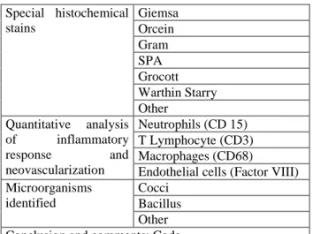
more appropriate. While Gimenez staining was used to identify C.burnetii, bartonella and legionellasp. The rarely used Ziehl-Neelson stain was used to detect acid-fast bacilli, especially mycobacteria. But these are rarely involved in infectious endocarditis. All histochemical stains usually employed are shown in Table 2.
Immunohistochemical methods
With immunohistochemistry techniques, the parameters studied were neovascularization and inflammatory response.
For evaluation of neovascularization, a monoclonal anti-factor VIII antibody (Dako diluted 1/200) was applied to deparaffinized tissue sections.
To analyze the inflammatory response, different cellular components were quantified: For macrophages, the CD68 Dako monoclonal antibody at the concentration of 1/50 was used. For T cells, polyclonal CD3 antibody at 1: 25 was used and for polynuclear CD15 Dako monoclonal antibody (kit ready for use). With these last two cell types, a step of antigenic restoration by heat was used (microwaves, 20 min at 750W in a citrate buffer at pH 6).
For neovascularization and inflammatory response, the revelation was carried out using the Dako LSAD kit, including a peroxidase neutralization step (3% oxygenated water for 10 min) and endogenous biotin (Dako biotin blocking system). The chromogen used was diaminobenzidine with counterstaining with Mayer's hematoxylin for 3 min. The histopathological account of a heart valve operated for an infectious endocarditis included the response items recorded in Table 2
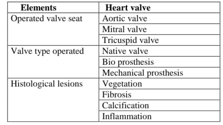
Table 2: Elements of a histological report of heart valve
Elements Heart valve
Operated valve seat Aortic valve Mitral valve Tricuspid valve Valve type operated Native valve
Bio prosthesis Mechanical prosthesis Histological lesions Vegetation
Mabika Barthelemy et al JMSCR Volume 07 Issue 05 May 2019 Page 1171
Special histochemical stains
Giemsa Orcein Gram SPA Grocott Warthin Starry Other
Quantitative analysis of inflammatory response and neovascularization
Neutrophils (CD 15) T Lymphocyte (CD3) Macrophages (CD68)
Endothelial cells (Factor VIII) Microorganisms
identified
Cocci Bacillus Other Conclusion and comments: Code
Statistical analysis
The Mann and Whitney U test were used to perform a comparison of inflammatory infiltrates accompanying infectious endocarditis and other valvular pathologies, respectively. With this test, the difference is significant when P <0.005.
Results
Of the 630 valve devices analyzed, 350 were of native valves, 200 of bio prostheses and 80 of mechanical prostheses. Their seat was exclusively aortic and mitral.
For all types of valve devices we analyzed, a code was assigned to each conclusion of the histological report. Table 3 summarizes the different conclusions reached where each corresponds to an encrypted code.
Table 3: Different conclusions asked where each corresponds to an encrypted code.
Type of anatomo-pathological finding Code
Certain infectious endocarditis with visible germ or positive immunohistochemistry
1
Certain infectious endocarditis without visible germ or negative immunohistochemistry
2
Infectious endocarditis possible 3 Degenerative changes with reactive inflammatory infiltrates
4
Non-inflammatory degenerative changes 5 Marathon endocarditis or non-bacterial thrombotic endocarditis
6
Other (myxoid degeneration) 7
The type of valve examined, the various histological parameters analyzed, the code, the valve seat, the mean age of the patients and the sex ratio are shown
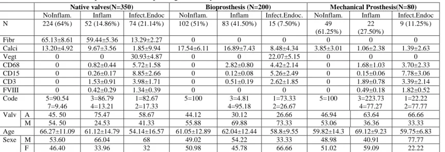
in Table 4. We identified for the native valves, the bioprostheses and the mechanical valves three subsets corresponding to degenerative, inflammatory lesions without identified microorganisms and infectious endocarditis with evidence of the pathogen.
The histological parameters studied during the morphological analysis with the SAMBA technique are: fibrosis, calcifications and vegetations on the one hand, whose analysis is done on slides stained with hematoxylin eosin saffron. The analysis of the inflammatory response and neo-vascularization is done on slides treated with immunolabeling techniques. We used anti mouse monoclonal antibodies directed respectively against macrophages (CD68), neutrophils (CD15), lymphocytes (CD3) and endothelial cells (Factor VIII) for the inflammatory response, respectively. For the distribution of the average age of patients according to the type of valve device analyzed, for the native prostheses, the average age of the patients was 60.51 ± 14.13 years, the extremes varying from 52 to 78 years and a sex ratio H / F of 0.7.
Mabika Barthelemy et al JMSCR Volume 07 Issue 05 May 2019 Page 1172
Table 4: Results of quantitative morphological analysis of valve devices
Native valves(N=350) Bioprosthesis (N=200) Mechanical Prosthesis(N=80)
NoInflam. Inflam Infect.Endoc NoInflam. Inflam Infect.Endoc. NoInflam. Inflam Infect.Endoc
N 224 (64%) 52 (14.86%) 74 (21.14%) 102 (51%) 83 (41.50%) 15 (7.50%) 49
(61.25%)
22 (27.50%)
9 (11.25%)
Fibr 65.13±8.61 59.44±5.36 13.29±2.27 0 0 0 0 0 0
Calci 13.20±4.92 9.67±3.56 1.85±9.94 17.54±6.11 16.89±7.43 8.48±4.34 3.85±3.01 1.06±2.38 1.39±2.63
Vegt 0 0 30.93±4.87 0 0 22.07±5.15 0 0 0
CD68 0 0.82±0.44 5.72±1.58 0 2.82±0.80 4.42±2.14 0 1.68±1.03 3.70±2.33
CD15 0 0.26±0.17 8.85±2.66 0 0.12±0.08 5.26±2.49 0 0.15±0.06 7.78±3.06
CD3 0 1.53±0.91 3.98±1.71 0 0.51±0.19 2.62±1.85 0 1.89±0.78 3.39±2.14
FVIII 0 0.42±0.29 1.34±0.39 0 0 0 0 0.49±0.18 1.82±0.52
Code 5=90.54
7=9.46
3=86.79 4=13.21
1=82.67 2=17.33
5=100 3=4.81
4=95.18
1=73.33 2=26.67
5=100 3=223.73
4=77.27
1=22.22 2=77.77
Valv A 45. 50 75.47 58.67 44.12 30.12 26.66 46.94 63.64 66.66
M 54. 50 24.53 41.33 55.88 69.88 73.33 53.06 36.36 33.33
Age 66.27±11.09 61.12±14.79 54.14±16.57 61.05±12.89 62.04±12.44 58.8±9.55 59.82±14.3 69.12±9.23 59.75±6.83
Sexe M 53.60 66.04 68 49.02 54.22 33.33 48.98 40.91 77.77
F 46.40 33.96 32 50.98 45.78 66.66 51.02 59.09 22.22
N=nombre, Fibr= fibrosis, Calci= calcifications, Vegt= vegetations, Valv A, M= Aortic, Mitral valve, No Inflam= No inflammatory, Inflam=inflammatory, Infect. Endoc= Infectious endocarditis
Discussion
During this work we proceeded to the morphological quantitative analysis of the histological parameters identified on routine staining (hematoxylin eosin saffron) or on slides treated with the immunolabeling techniques. This analysis system comprises various programs allowing the analysis of each of the parameters[2,7]. The histological signs analyzed on HES stained section are: vegetation, fibrosis and calcifications. The parameters analyzed after immunostaining are the inflammatory response and the neovascularization. Vegetation is an almost constant sign of infectious endocarditis. It consists of a platelet fibrin aggregate infiltrated with inflammatory elements and sometimes microorganisms. In a recent work, Lepidi et al[3] noted that during Q endocarditis the vegetation was small. For infectious endocarditis on mechanical valves, the absence of vegetation could be explained by the fact that the infectious agents do not attack the inert constituents of these prostheses.
With respect to the inflammatory infiltrate, quantitative analysis allowed us to distinguish on the three types of valves an inflammatory infiltrate with endocarditis and an inflammatory infiltrate without endocarditis. The respective composition of these two types of infiltrates was almost superimposable regardless of the type of valve being examined. With the Mann and Whitney U test,
the comparison of these two types of inflammatory infiltrates showed a significant difference (P<0.005). Neovascularization is a histological sign that accompanies any valvular inflammatory process[8]. It is not specific for infectious endocarditis. Although it is sometimes absent on mechanical prostheses and bioprostheses, this is due to their intrinsic constitution. Infectious involvement is possible on the collar of the fibrous ring and on the suture son.
Mabika Barthelemy et al JMSCR Volume 07 Issue 05 May 2019 Page 1173
monoclonal antibody directed against a specific germ to characterize it.
Among the histological signs encountered with infectious endocarditis is inflammatory infiltrate, which from time to time leads to misdiagnosis[3]. This is why the quantitative analysis of this histological parameter should be generalized because, as we saw in the results of our work, the inflammatory infiltrate that accompanied the infective endocarditis in each type of valvular device presented a composition stackable.
Conclusions
The results provided by this technique are objective and reproducible, which could make it possible to carry out multicenter studies and to be able to make comparisons between different working groups.
Conflict of Interest
The authors declare that there are no competing interests. All the authors read and approved the final version.
References
1. MAURIN M, RAOULT D FEVER. Q fever. Clin
Microbiol Rev 1999; 12: 518–553.
2. LEPIDI H, FOURNIER P-E, RAOULT D.
Quantitative analysis of valvular lesions during Bartonella endocarditis. Am J Clin Pathol 2000; 114: 880–889.
3. LEPIDI H, HOUPIKIAN P, LIANG Z, RAOULT
D. Cardiac valves in patients with Q fever endocarditis: microbiological, molecular, and histologic studies. J Infect Dis 2003; 187: 1097–1106.
4. MYLONAKIS E, CALDERWOOD SB. Infective
endocarditis in adults. N Engl J Med 2001; 345: 1318–1330.
5. BROUQUI P, DUMLER JS, RAOULT D. Immunohistologic demonstration of Coxiella burnetii in the valves of patients with Q fever endocarditis. Am J Med 1994; 97: 451–458.
6. PE F. Marrie tJ, raoult d. diagnosis of Q fever. J clin Microbiol 1998; 36: 1823–1834.
7. CHARPIN C,MARTIN PM,DEVICTOR Bet al. Multiparametric study (SAMBA 200) of estrogen receptor immunocytochemical assay in 400 human breast carcinomas: analysis of estrogen receptor distribution heterogeneity in tissues and correlations with dextran coated charcoal assays and morphological data. Cancer Res 1988; 48: 1578–1586.
8. LEPIDI H, DURACK DT, RAOULT D.
Diagnostic methods current best practices and guidelines for histologic evaluation in infective endocarditis. Infect Dis Clin North Am 2002; 16: 339–61.
9. BROUQUI P, RAOULT D. Endocarditis due to