Background - Red blood cell (RBC) exchange (RCE) transfusion therapy is indicated for certain patients with sickle cell disease (SCD). Although
beneficial, this therapy is costly and inconvenient to patients, who may require it monthly or more often. Identification of blood and plasma
biomarkers that could improve or help individualise RCE therapy is of interest. Here we examined relevant blood and plasma metabolites and biomarkers of vasoactivity and RBC fragility in a pilot study of SCD patients undergoing RCE using either standard RBC units or RBC units treated with a US Food and Drug Administration (FDA)-approved additive solution containing phosphate, inosine, pyruvate, and adenine (“PIPA”).
Materials and methods -In this prospective, single-blind, cross-over pilot clinical trial, patients were randomised to receive either standard RBC exchange or PIPA-treated RBC exchange transfusion with each RCE session over a 6-month treatment period. Pre- and post-transfusion blood samples were obtained and analysed for RBC O2 affinity, ATP, purine metabolites, RBC
microparticles, and cell free haemoglobin.
Results -Red blood cell O2 affinity was maintained after PIPA-RCE in contrast to standard RCE, after which P50 fell (net O2 affinity rose). Plasma ATP did not change significantly after RCE using either of the RBC unit types. Exchange
transfusion with PIPA-treated RBC units led to modest increases in plasma
inosine and hypoxanthine. Plasma cell free haemoglobin fell after either standard or PIPA-treated RBC exchange transfusion (novel findings), and to a similar extent. RBC-derived microparticles in the plasma fell significantly and similarly after both standard and PIPA-treated RCE transfusion.
Discussion -In summary, treatment of RBCs with PIPA prior to RCE elicited favourable or neutral changes in key metabolic and vascular biomarkers.
Further study of its efficacy and safety is recommended and could ultimately
serve to improve outcomes in chronically transfused SCD patients.
Keywords: sickle cell disease, exchange transfusion. Arrived: 1 October 2019
Revision accepted: 20 December 2019 Correspondence: Tim J. McMahon e-mail: [email protected]
1Department of Pediatrics, Duke University Medical Center, Durham, NC; 2Department of Anesthesiology, Duke University Medical Center, Durham, NC; 3Department of Pediatrics, University of North Carolina at Chapel Hill, Chapel Hill, NC; 4Department of Medicine, Duke
University Medical Center, Durham, NC; 5Department of Pathology, Duke
University Medical Center, Durham, NC; 6Durham Vetern Affairs Health Care System, Durham, NC; United States of America
Effects of repleting organic phosphates
in banked erythrocytes on plasma
metabolites and vasoactive mediators
after red cell exchange transfusion in
sickle cell disease
adhere to the vascular endothelium4,6. Additionally,
they are depleted of 2,3-diphosphoglycerate (2,3-DPG) and adenosine triphosphate (ATP), and have lower P50 (ie, higher O2 affinity)7. As RBCs are cleared from circulation, they produce additional burdens of RBC breakdown byproducts, including cell free haemoglobin (CFH) and RBC microparticles (RMPs)8-10. RMPs are already elevated in SCD due to
sickling and haemolysis and can impair vasodilation, decrease microcirculatory oxygenation, and damage vascular endothelium11-13. Thus, these burdens of
cleared RBCs exacerbate problems already present in this high-risk patient population. Additionally, SCD patients who routinely undergo RCE face major health care burdens arising from the need for central vascular access, transfusion complications, work and school absences, and high costs14.
RBC “rejuvenation” (incubation with “PIPA” additive solution, containing phosphate, inosine, pyruvate, and adenine) is a US Food and Drug Administration (FDA)-approved, standardised blood bank process that restores the organic phosphates 2,3-DPG and ATP, and raises the P50, all of which are reduced during storage15,16. RBC transfusate incubation with
PIPA increases RBC membrane stress tolerance, and microcirculatory oxygenation, while decreasing RMP generation and vascular endothelial adhesion17-19.
In this pilot study, we studied a cohort of clinically stable, crisis-free, chronically RCE-transfused SCD patients and compared the effects of standard RBC exchange transfusion (STD-RCE) vs PIPA-treated RBC exchange transfusion (PIPA-RCE) on plasma vasoactive biomarkers/mediators. Specifically, we examined purine metabolites including and related to the vasoactive mediator ATP (which is augmented in PIPA-treated RBCs), and the RBC haemolysis biomarkers and vasoactive mediators plasma CFH and red cell-derived RMPs. Plasma ATP originating in RBCs or other cells may act as a vasodilator and antiadhesive agent20,21. ATP can also elicit
pro-inflammatory effects22. CFH and RMPs reflect
the balance between RBC fragility and related clearance mechanisms, and may also contribute to vascular pathophysiology in SCD or after RBC transfusion10. We hypothesise that changes in these
INTRODUCTION
Sickle cell disease (SCD), the most common inherited blood disorder in the United States, is characterised by abnormal sickle haemoglobin (HbS) production. As a result, the capacity of sickle red blood cells (RBCs) to transport and deliver oxygen is impaired. In low-oxygen conditions, HbS polymerises, altering RBC morphology and susceptibility to lysis and triggering recurrent vaso-occlusive episodes and pain crises1. Chronic periodic RBC exchange transfusion is indicated for patients with SCD for primary and secondary stroke prophylaxis and for patients with recurrent vaso-occlusive crises2,3.
Red blood cell exchange (RCE) transfusion is a therapeutic intervention in SCD involving the removal of native sickle (SS) RBCs in exchange for the administration of multiple units of packed healthy RBCs. In RCE transfusion, RBCs containing normal adult haemoglobin (HbA) are given to largely replace the recipient’s diseased HbS-containing RBCs and to reach a target clinically determined % HbA (e.g. >50%) level. The interval between RBC exchanges (typically every 3-6 weeks) is determined based in part on the projected rate of HbA clearance. This partial exchange of HbS for HbA reduces the number of SS RBCs present, lowering viscosity, and protecting against sickling and vaso-occlusion2. This
can in turn reduce the rate of serious complications, including recurrent pain crisis, acute chest syndrome, stroke, and renal and other end organ damage. This intervention is critical in patients with SCD who require chronic transfusion for a number of indications, including secondary stroke prevention. However, the duration of benefit from transfusion is limited due, in part, to the clearance over time of transfused RBCs, which have already aged during storage4. Additionally, large-volume RBC transfusion
is associated with other complications including iron overload, iron-mediated hypercoagulability, and progressive development of alloimmunisation, motivating the study of determinants of optimal RBC unit processing5.
to Rejuvesol® RBC Processing Solution (Zimmer Biomet Labs, LLC, Braintree, MA, USA), per standard protocol24. Briefly, RBC units are infused with PIPA
solution for 1 hour at 37 ˚C, washed with saline in a Cobe 2991 Cell Washer (TerumoBCT, Lakewood, CO, USA), and suspended in 0.9% saline/0.2% dextrose for transfusion. The 4-unit treatment was based on practical considerations while projecting that this would equate to PIPA treatment of a high fraction of the patient’s final circulating RBC volume.
P₅₀ analysis
Whole-blood P50 was analysed before and after either standard or PIPA-treated RCE using a TCS Hemox analyser (TCS Scientific Corporation, New Hope, PA, USA), according to the manufacturer’s instructions. ATP, purine metabolites, and cell-free haemoglobin assays
Plasma from patient samples was analysed for ATP concentration, purine metabolites, and CFH. ATP was measured using a commercially available luciferase ATP assay (Sigma). Purine metabolites were measured using a previously described mass spectrometric method25. CFH was measured using
multi-wavelength absorbance spectroscopy for haemoglobin, as we described previously26.
Red blood cell microparticle analysis
Plasma from patient samples was incubated at room temperature with RBC-specific CD235a antibody (Invitrogen; 1:30 concentration, 50 µL samples) for 30 min as previously described to distinguish MPs derived from RBCs (RMPs) from other microparticles27. The number of RMPs in the
plasma samples was determined by volumetric flow cytometry (Cytek, Aurora, CO, USA). A SCD blood sample was used as a positive control to set appropriate gates to identify RMPs based on size and CD235a staining; these gates were applied to all study plasma samples.
Statistical analysis
All values are reported as means±Standard Error of Mean (SE) unless otherwise noted. Outliers were defined as ±3 Standard Deviation (SD) from the mean in any sample set. No data points fit that criterion, and therefore no outliers were excluded.
biomarkers might therefore provide information on the risk-benefit balance of metabolic modification of RBCs and ultimately be of benefit in frequently transfused SCD patients.
MATERIAL AND METHODS
Selection and description of research participants This prospective, single-blind, cross-over pilot clinical trial allowed for randomisation of patients to receive either PIPA-treated or standard RBC exchange transfusion with each treatment session over a 6-month treatment period. Other results from this study have been published previously23. Eligible
patients were ≥18 years of age, in stable clinical condition, therapy-compliant, chronically transfused SCD patients (HbSS) who were maintained with repeated RCE therapy and who had been crisis-free for a period covering at least the previous three consecutive sessions. No patient had tolerated chronic hydroxyurea therapy and none received it during the study period. Exclusion criteria included a baseline need for washed RBCs and pre-treatment SaO2 <92%. After written, informed consent under a protocol approved by the Duke University Medical Center Institutional Review Board, blood samples were collected from these established SCD patients. Demographic data including age, sex, race, height and weight were obtained for each patient. Pre- and post-transfusion session vital signs were recorded and blood was collected for analysis just before and within 60 minutes after RCE.
Red blood cell units
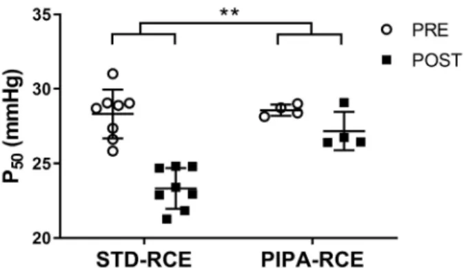
P50 after PIPA-treated RCE transfusion (p=0.0024). The interaction between pre vs post P50 effects and STD vs PIPA effects was significant by two-way ANOVA (p=0.0064).
A mixed error-component statistical model would be the ideal approach for the analysis of such data, but is not recommended when N <528. Thus, ANOVA
and t-test analyses were performed using GraphPad software version 8.2 (San Diego, CA, USA). p<0.05 was considered statistically significant. For each biomarker, at least one pair of values (pre and post) was available for each patient from one STD-RCE session and one PIPA-RCE session (n=4 throughout). In some cases, data from multiple STD-RCE or PIPA-RCE sessions was used for a given patient in the principal statistical analysis (n >4).
RESULTS Demographics
The 4 patients were clinically stable, African-American males with HbSS disease, aged 19-32 years; none of the patients was currently receiving hydroxyurea. They were maintained free from vaso-occlusive crisis on a monthly RCE transfusion programme. Based on established %HbS targets and patient size, the RBC requirements at each RCE session were 7 units for 3 patients (post-RCE HbA target 75-80%) and 10 units for the remaining patient (post-RCE HbA target 80-85%). Patients underwent up to 3 standard and 3 PIPA-treated RCE sessions in varying sequence over the 6-month study period. The average storage age for blood units was 16±5 days for standard units and 22±13.5 days for the PIPA-treated units (p=0.002 vs STD units). Vital signs were stable throughout all RCEs, and no transfusion reactions were reported. All participants remained crisis-free with stable renal function during the study period (data not shown). No adverse effects (AEs) of any kind (including serious AEs and treatment-emergent AEs) occurred during the 4 weeks following the RCE transfusions of either standard or PIPA-exposed (rejuvenated) type.
Whole-blood P₅₀
We compared patients' whole-blood P50 before and after either a standard RCE transfusion or RCE transfusion using PIPA-treated RBCs (Figure 1). The P50 decreased after STD transfusion (pre, 28.31±1.6 vs post, 23.33±1.4) in contrast to no change after PIPA-treated RCE transfusion (pre, 28.57±0.4 vs post, 27.17±1.3). There was improved maintenance of the
Plasma ATP
Plasma ATP levels were measured before and after either standard or PIPA-treated RCE transfusion. There was no greater preservation of ATP in the PIPA-RCE group post transfusion (Figure 2). There was also no overall difference in plasma ATP in pre- vs post-RCE transfusion when analysing both transfusion conditions (STD-RCE vs PIPA-RCE; p=0.23). There was no significant interaction between the pre vs post and the STD vs PIPA effects (two-way ANOVA).
Plasma purine metabolites
To further evaluate the impact of rejuvenation on RBC metabolism, we used a mass spectrometric method to measure ATP pathway metabolites in plasma from a subset of samples pre- and post-RCE with PIPA-treated RBCs. The primary ATP metabolic products, AMP and adenosine, were unchanged post transfusion (Figure 3). We observed significant increases in plasma inosine (p=0.003), a component of Rejuvesol, and in the inosine metabolite hypoxanthine (p=0.007) post-PIPA-RCE. However, further downstream metabolites including adenosine, xanthine and uric acid, were unchanged post-PIPA-RCE.
Figure 1 - P50 before and after standard or phosphate, inosine, pyruvate, and adenine (PIPA)-treated red blood cell exchange (RCE) transfusion showing improved maintenance of P50 with PIPA-treated RCE transfusion
Figure 2 - Plasma ATP before and after standard (STD) or phosphate, inosine, pyruvate, and adenine (PIPA)-treated RCE Two-way ANOVA was non-significant.In addition, there was no significant effect of PIPA and no overall post-red blood cell exchange (RCE) effect on plasma ATP.
Figure 3 - Mass-spectrometric analysis of plasma obtained pre- and post-exchange transfusion of phosphate, inosine, pyruvate, and adenine (PIPA)-exposed red blood cells (RBCs)
Post-transfusion concentrations of inosine (p=0.003) and hypoxanthine
(p=0.007) increased significantly without changes in other purine metabolites. Concentrations are in μM except for uric acid, which is in mM.
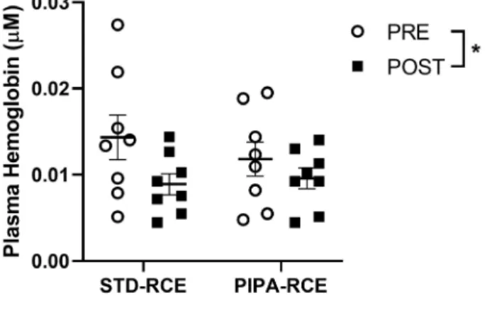
Figure 4 - Plasma cell-free haemoglobin (CFH) before and after standard (STD) or phosphate, inosine, pyruvate, and adenine (PIPA)-treated red blood cell exchange (RCE) There is a significant decrease in CFH after both STD and PIPA-treated RCE
(p=0.047).
Figure 5 - Plasma red blood cell microparticles (RMPs) before and after standard (STD) or phosphate, inosine, pyruvate, and adenine (PIPA)-treated red blood cell exchange (RCE) There is a significant decrease in RMPs after both STD and PIPA-treated RCE
(p=0.01). Plasma cell-free haemoglobin and red blood cell
microparticles
Plasma haemoglobin levels were measured pre and post either standard or PIPA-treated RCE transfusion. Figure 4 demonstrates the nanomolar CFH concentration. There is a statistically significant decrease in CFH after both STD or PIPA-treated RCE (p=0.047). There was no significant difference based on the RCE transfusion type (STD-RCE vs PIPA-RCE) in plasma RMPs quantified
pre and post either standard or PIPA-treated RCE transfusion (Figure 5).
DISCUSSION
This pilot study examined the effects of PIPA treatment of RBC transfusates on changes in plasma metabolic and vasoactive biomarkers in patients with SCD requiring chronic RCE transfusion therapy. Previous studies have examined metabolic changes that develop secondary to RBC storage, but few have investigated the metabolic and functional changes in the plasma and RBCs of SCD patients transfused with metabolically modified RBC units. In theory, RBC transfusate modification such as PIPA treatment could extend the interval between RCE visits and thereby positively influence quality of life in SCD patients by reducing costs and the incidence of complications. We reasoned that a carefully controlled demonstration of favourable and safe changes in blood biomarkers of RBC metabolism and vasoactivity after PIPA-RCE in SCD was warranted before such an outcome study.
We found a statistically significant decrease in patient blood P50 (increase in RBC Hb O2 affinity) after standard RCE transfusion. This finding was expected, given that RBCs transfused after storage have depressed 2,3-DPG, and thus elevated O2 affinity (lower P50) relative to fresh (or native HbA) RBCs. The P50-lowering effect of RCE was essentially eliminated with PIPA-treated RCE transfusion. The relative preservation of patient blood P50 using stored-RBC exposed to PIPA is not novel, and is consistent with the well-established effect of PIPA in augmenting stored-RBC organic phosphates (ATP and 2,3-DPG)29,30. This corresponds to an increased O2
off-loading capacity of the PIPA-treated (vs standard) transfused RBCs, which may be of clinical benefit in this patient population who have physiologically compensated for their anaemia by right-shifting their O2 dissociation curve at baseline. Specifically, SCD patients compensate for their anaemia by increasing the production of 2,3-DPG in RBCs, in turn lowering RBC O2 affinity (raising the P50). Additionally, although RCE transfusion is generally beneficial, the overall decrease in P50 (leftward shift in the O2-binding curve) resulting from transfusion of stored HbA RBCs could limit the potential O2-delivery benefits of transfusion, especially in
SCD patients. In addition, it is possible that O2 exchange may take place between the transfused HbA RBCs (with high O2 affinity) and the native HbS RBCs (having low O2 affinity). The net effect could be the movement of O2 from low-affinity HbS RBCs to high affinity HbA RBCs and a net decrease in oxygen delivery from RBCs to the tissue. This could promote the sickling of SS RBCs and/or lead to local tissue hypoxia. Pre-transfusion PIPA exposure of HbA RBCs would tend to attenuate this exchange by restoring organic phosphates in the stored RBCs, thus moving P50 nearer to that of the native HbS RBCs.
Red blood cell exchange transfusion (both STD-RCE and PIPA-RCE) did not significantly alter plasma ATP concentration. The plasma ATP may reflect the net balance resulting from the steady-state rates of RBC export of ATP and intravascular haemolysis when SS RBCs are exchanged for AA RBCs, on one hand, and the hydrolysis of ATP on the other20,21. Thus, the
RBC ATP-repleting effect of PIPA treatment of the transfused units may not result in increased ATP into the plasma20,21. PIPA treatment of stored RBCs
does augment ATP export in vitro29, but the net effect
of transfusion of PIPA-treated RBCs on plasma ATP levels in SCD patients may also be driven by a slowing of intravascular haemolysis rates (tending to lead to lower plasma ATP). Notably, the effects of PIPA on post-RCE plasma ATP may have been blunted by the fact that the storage age of the PIPA-treated RBC units was (by chance) greater than that of the STD RBC units used in RCE.
In our purine analysis, we did not observe any changes in plasma adenosine monophosphate (AMP) or adenosine (ADO) post-RCE with PIPA-treated RBCs, suggesting that ATP metabolism was not altered by this process. Plasma ADO is of particular interest in terms of safety because it can promote pathophysiology in SCD when ADO promotes production of 2,3-DPG in erythrocytes, which shifts the O2 affinity curve to the right (lower P50). This lowered affinity for O2 thus favours HbS deoxygenation, RBC sickling, and organ dysfunction31. The increased plasma inosine post
in PIPA, as was the increase in the inosine metabolite hypoxanthine, consistent with our prior findings32.
Increased plasma hypoxanthine is also a biomarker of disease and can contribute to oxidative stress through its metabolism by xanthine oxidase32. The
impact of RCE with PIPA-treated RBCs on oxidative stress will need to be determined in future studies. In SCD, RMP generation is elevated at baseline as a result of accelerated RBC vulnerability and turnover33. We found that exchange transfusion
(standard or PIPA-treated) decreased the overall steady-state RMP concentration in SCD patient plasma after transfusion, consistent with prior reports34,35. We report for the first time that RBC
exchange transfusion also reduced plasma levels of CFH (resulting from intravascular haemolysis) regardless of RCE type. Because CFH and RMPs can promote intercellular adhesion, blood flow dysregulation, and coagulation, all contributing to vaso-occlusion and organ damage in SCD, their net reduction after RCE is expected to be favourable. However, transfused RBC units also contain (supernatant) CFH and RMPs, and the amount of clearance with RCE would depend in part on the RMP content of the transfused units, which may vary with factors including RBC storage time (which happened to be significantly longer in the PIPA group). Finally, the washing that necessarily follows PIPA treatment might, on the one hand, “clear” CFH and RMPs from the aging RBC unit, but can also contribute to generation of CFH and RMPs through the added mechanical shear stress of centrifugation and osmotic perturbations with washing. It is, therefore, reassuring that we observed a net decrease in plasma CFH and RMPs, and this favourable effect was quantitatively similar in both transfusion types (STD-RCE vs PIPA-RCE).
Collectively, these findings suggest some benefit of transfusing PIPA-treated RBCs with a smaller post transfusion drop in P50 after rejuvenated transfusion. Plasma ATP, exported from RBCs or other cells or sources, can act as a regional vasodilator and antiadhesive20,21, and so the maintenance of
ATP concentration after RCE (STD or PIPA) may also be reassuring in SCD. Additionally, PIPA-RCE
demonstrates a similar favourable safety profile to STD-RCE with respect to decreased CFH and RMPs post transfusion. It also does not appear to contribute harmful purine metabolites in the plasma. This study is limited, however, in that it is a pilot study with a small patient sample. It would be logical in future studies to determine whether pre-transfusion rejuvenation of stored RBCs, or other strategies designed to preserve or replete organic phosphates, lead to longer post-transfusion circulating survival time after RCE, and to investigate patient-reported and clinical outcomes.
CONCLUSIONS
Chronic RBC transfusion including RCE is a required maintenance therapy for many patients with SCD and chronic complications. Metabolic optimisation of RBC units could extend the interval between RCE sessions, would reduce costs and risks, and could improve quality of life for SCD patients. As such, our findings support further investigation of the outcomes of measures to improve the metabolic and vasoactive sequelae of RBC transfusions in order to improve the risk-benefit balance in chronic transfusion therapy for SCD patients.
ACKNOWLEDGEMENTS
IW received funding for this study from the NC TraCS Institute supported by the CTSA grant (UL1TR001111) and Investigator Initiated Grant Funding to study transfusion in volunteers; Rejuvesol® was supplied by Zimmer Biomet (Warsaw, IN, USA) free of charge to support this study. Grants NIH R01 GM-113838 and VA Merit BX-003478 to TJM also supported the study.
DISCLOSURE OF CONFLICTS OF INTEREST
REFERENCES
1. Rees DC, Williams TN, Gladwin MT. Sickle-cell disease. The Lancet 2010; 376: 2018-31.
2. Biller E, Zhao Y, Berg M, et al. Red blood cell exchange in patients with sickle cell disease-indications and management: a review and consensus report by the therapeutic apheresis subsection of the AABB. Transfusion 2018; 58: 1965-72.
3. Padmanabhan A, Connelly-Smith L, Aqui N, et al. Guidelines on the use of therapeutic apheresis in clinical practice – Evidence-based approach from the Writing Committee of the American Society for Apheresis: the eighth special issue. J Clin Apheresis 2019; 34: 171-354.
4. Barshtein G, Manny N, Yedgar S. Circulatory Risk in the transfusion
of red blood cells with impaired flow properties induced by
storage. Transfus Med Rev 2011; 25:24-35.
5. Shah N, Welsby IJ, Fielder MA, et al. Sickle cell disease is associated with iron mediated hypercoagulability. J Thromb Thrombolysis 2015; 40: 182-5.
6. Raval JS, Waters JH, Seltsam A, et al. The use of the mechanical fragility test in evaluating sublethal RBC injury during storage. Vox Sang 2010; 99: 325-31.
7. Brecher ME, Zylstra-Halling VW, Pineda AA. Rejuvenation of erythrocytes preserved with AS-1 and AS-3. Am J Clin Pathol 1991; 96: 767-9.
8. Hod EA, Brittenham GM, Billote GB, et al. Transfusion of human volunteers with older, stored red blood cells produces extravascular hemolysis and circulating non-transferrin-bound iron. Blood 2011; 118: 6675-82.
9. Tissot J-D, Rubin O, Canellini G. Analysis and clinical relevance of microparticles from red blood cells. Curr Opin Hematol 2010; 17: 571-7.
10. Donadee C, Raat NJH, Kanias T, et al. Nitric oxide scavenging by red blood cell microparticles and cell-free hemoglobin as a mechanism for the red cell storage lesion. Circulation 2011; 124: 465-76.
11. Tantawy AAG, Adly AAM, Ismail EAR, et al. Circulating platelet and erythrocyte microparticles in young children and adolescents with sickle cell disease: Relation to cardiovascular complications. Platelets 2013; 24:605-614.
12. Camus SM, De Moraes JA, Bonnin P, et al. Circulating cell membrane microparticles transfer heme to endothelial cells and trigger vasoocclusions in sickle cell disease. Blood 2015; 125: 3805-3814.
13. Jy W, Johansen ME, Bidot C, et al. Red cell-derived microparticles (RMP) as haemostatic agent. Thromb Haemost 2013; 110: 51-760. 14. Tsitsikas DA, Ekong A, Berg L, et al. A 5-year cost analysis of
automated red cell exchange transfusion for the management of recurrent painful crises in adult patients with sickle cell disease. Transfus Apher Sci 2017; 56: 466-9.
15. Lockwood WB, Hudgens RW, Szymanski IO, et al. Effects of
rejuvenation and frozen storage on 42-day-old AS-3 RBCs. Transfusion 2003; 43: 1527-32.
16. Meyer EK, Dumont DF, Baker S, Dumont LJ. Rejuvenation capacity of red blood cells in additive solutions over long-term storage. Transfusion 2011; 51: 1574-9.
17. Raat NJH, Hilarius PM, Johannes T, et al. Rejuvenation of stored human red blood cells reverses the renal microvascular
oxygenation deficit in an isovolemic transfusion model in rats.
Transfusion 2009; 49: 427-4.
18. Koshkaryev A, Zelig O, Manny N, et al. Rejuvenation treatment of stored red blood cells reverses storage-induced adhesion to vascular endothelial cells. Transfusion 2009; 49: 2136-43. 19. Qureshi S, Patel N, Wozniak M, et al. Red cell rejuvenation targets
inflammatory microparticles, free haemoglobin and redox active
iron: key mediators of transfusion related acute lung injury in cardiac surgery. Heart 2015; 101 (Suppl 4): A110.
20. Ellsworth ML, Forrester T, Ellis CG, Dietrich HH. The erythrocyte as a regulator of vascular tone. Am J Physiol-Heart Circ Physiol 1995; 269: H2155-61.
21. Zhu H, Zennadi R, Xu BX, et al. Impaired adenosine-5′-triphosphate
release from red blood cells promotes their adhesion to
endothelial cells: A mechanism of hypoxemia after transfusion*.
Crit Care Med 2011; 39: 2478-86.
22. Cauwels A, Rogge E, Vandendriessche B, et al. Extracellular ATP
drives systemic inflammation, tissue damage and mortality. Cell
Death Dis 2014; 5: e1102.
23. Gehrke S, Shah N, Gamboni F, et al. Metabolic impact of red blood cell exchange with rejuvenated red blood cells in sickle cell patients. Transfusion 2019; 59: 3102-12.
24. Citra Lab [Internet]. Directions for rejuvenation of CPD, CPDA-1, or CDP/AS-1 RBC prior to cryopreservation. Available at: http://
www.citra-labs.com/fileLibrary/FL7000-rejuvesol.pdf. Accessed
on 29/03/2019.
25. Esther CR, Coakley RD, Henderson AG, et al. Metabolomic
evaluation of neutrophilic airway inflammation in cystic fibrosis.
Chest 2015; 148: 507-15.
26. Rogers S, B. Dosier L, Mcmahon T, et al. Red blood cell phenotype
fidelity following glycerol cryopreservation optimized for research
purposes. PLOS ONE 2018; 13: e0209201.
27. Bennett-Guerrero E, Kirby BS, Zhu H, et al. Randomized study of washing 40- to 42-day-stored red blood cells. Transfusion 2014; 54: 2544-52.
28. Moen EL, Fricano-Kugler CJ, Luikart BW, O’Malley AJ. Analyzing clustered data: why and how to account for multiple observations nested within a study participant? PLOS ONE 2016; 11: e0146721. 29. Kirby BS, Hanna G, Hendargo HC, McMahon TJ. Restoration of intracellular ATP production in banked red blood cells improves inducible ATP export and suppresses RBC-endothelial adhesion. Am J Physiol-Heart Circ Physiol 2014; 307: H1737-44.
30. Valeri CR, Hirsch NM. Restoration in vivo of erythrocyte adenosine triphosphate, 2,3-diphosphoglycerate, potassium ion, and sodium ion concentrations following the transfusion of acid-citrate-dextrose-stored human red blood cells. J Lab Clin Med 1969; 73: 722-33.
31. Zhang Y, Dai Y, Wen J, et al. Detrimental effects of adenosine
signaling in sickle cell disease. Nat Med 2011; 17: 79-86.
32. Quinlan GJ, Lamb NJ, Tilley R, et al. Plasma hypoxanthine levels in ARDS: implications for oxidative stress, morbidity, and mortality. Am J Respir Crit Care Med 1997; 155: 479-84.
33. Hebbel RP, Key NS. Microparticles in sickle cell anaemia: promise and pitfalls. Br J Haematol 2016; 174: 16-29.
34. Mahfoudhi E, Lecluse Y, Driss F, et al. Red cells exchanges in sickle cells disease lead to a selective reduction of erythrocytes-derived blood microparticles. Br J Haematol 2012; 156: 545-7.