399
© 2017 by the Serbian Biological Society How to cite this article: Zhao JF, Wang Q, Ge YM, Tan PL, Chen YM, Yan Jie. Distribution of β-lactamase genes of Klebsiella pneumoniae isolates in Zhejiang province, China, and regulation of gene expression. Arch Biol Sci. 2017;69(3):399-407.
Distribution of β-lactamase genes of
Klebsiella pneumoniae
isolates in Zhejiang province,
China, and regulation of gene expression
Jin-Fang Zhao1, Qiang Wang2, Yu-Mei Ge2, Pan-Li Tan1, Yi-Min Chen1 and Jie Yan2,*
1Department of Clinical Laboratory, the First Affiliated Hospital of Zhejiang Chinese Medical University, Hangzhou,
Zhejiang 310006, P.R. China
2Department of Medical Microbiology and Parasitology, Zhejiang University School of Medicine, Hangzhou, Zhejiang
310058, P.R. China
*Corresponding author: [email protected]
Received: May 12, 2016; Revised: July 21, 2016; Accepted: August 15, 2016; Published online: October 31, 2016
Abstract: Klebsiella pneumoniae is a common causative agent of nosocomial infections with a high level of resistance toward β-lactam antibiotics. Our previous study showed that TEM-1 and SHV-11 are the predominant β-lactamase-encoding genes of K. pneumoniae isolates in the Zhejiang area, China. In this study, more clinical K. pneumoniae isolates were collected for detecting their β-lactamase-encoding gene profiles by PCR and sequencing. qRT-PCR was then performed to determine the
role of cefotaxime or penicillin in low concentrations to induce the β-lactamase gene expression of K. pneumoniae isolates.
Moreover, the K. pneumoniae isolates were pretreated with closantel (CLO), a histidine kinase inhibitor, before antibiotic
treatment, and qRT-PCR and the β-lactamase phenotype confirmatory test were then applied to determine the effect of CLO on the expression of the β-lactamase genes. The results showed that, except for KPC-2, the 1/4 MIC cefotaxime or penicillin induced significant mRNA elevation of the TEM-1, CTX-M-14, SHV-11 and OXA-1 β-lactamase genes, but this induction could be inhibited by CLO. After pretreatment with CLO, 78.4~81.4% of the β-lactam-resistant isolates became sensitive and the positive rate of the β-lactamase production phenotype in the isolates was decreased from 100% to 27.1%. The data indicate that TEM-1 (70.7%), SHV-11 (64.2%) and CTX-M-14 (40.5%) are the predominant β-lactamase genes
of the K. pneumoniae isolates in Zhejiang and sublethal dosage of β-lactam antibiotics can induce the β-lactamase gene
expression of K. pneumoniae through histidine kinase-mediatedtwo-component signaling systems.
Keywords: Klebsiella pneumonia; resistance; β-lactamases; gene expression; closantel
INTRODUCTION
Klebsiella pneumoniae, an important member of the
Enterobacteriaceae family, is present as a saprophyte
in the human gastrointestinal tract, nasopharynx and skin [1]. However, as an opportunistic pathogen, K.
pneumoniae is frequently involved in nosocomial
infections [2], causing many different diseases, such as pneumonia, urinary and biliary tract infections, wound and soft tissue infection, osteomyelitis and septicemia [3]. Cephalosporins, a group of β-lactam antibiotics, are the most valuable and frequently used drugs for treatment of Klebsiella infection. However,
K. pneumoniae isolates commonly show high
resist-ance rates toward β-lactam antibiotics, leading to inefficient antibiotic therapy and prolonged hospital stay and greater hospital charges for patients [4-6].
The most important resistance mechanism against β-lactam antibiotics is the production of β-lactamases [7-9]. Many different β-lactamases such as extended-spectrum β-lactamases (ESBLs) and carbapenemases
from K. pneumoniae isolates have been identified,
among which TEM, CTX-M, SHV and OXA are the most common ESBLs, and KPC is also frequently de-tected [10-12]. However, there is diversity between the predominant ESBLs and carbapenemase genes in
K. pneumonia isolates from different areas [13-15].
upregulate the expression of enzymes to inactivate dif-ferent antibiotics in S. aureus, E. coli and A. baumannii
[19], and two-component signaling systems (TCSS) are involved in the regulation of the gene expression [20]. TCSS, usually composed of a transmembrane histidine kinase (HK) and an intracellular response regulator (RR), are frequently administered as the drug targets of antimicrobials [21]. Therefore, inhibi-tors of TCSS, such as closantel (CLO), have a potential for developing novel antibacterial drugs [22].
In our previous study, we detected the β-lactamase gene profile of 118 β-lactam antibiotic-resistant K.
pneumoniae isolates in Zhejiang province, and TEM-1
and SHV-11 were the predominant β-lactamase genes in these isolates [23]. In the present study, another
118 K. pneumoniae isolates were collected for
detect-ing their β-lactamase gene profiles. To avoid possible interference among different β-lactamase genes, the isolates that only carry a single β-lactamase gene were subjected to 1/4 MIC β-lactam antibiotic treatment to determine the induction of sublethal dosage of β-lactam antibiotics on the expression of β-lactamase genes. Moreover, CLO, an HK inhibitor, was applied to understand the probable inhibitory effect of HK-based TCSS-mediated β-lactamase gene expression.
MATERIALS AND METHODS
Source and identification of K. pneumoniae isolates
In our previous study, we obtained 118 β-lactam antibiotic-resistant K. pneumoniae isolates [23]. In the present study, 272 K. pneumoniae isolates were collected. All the isolates were isolated by fractional cultivation on Columbia blood plates from sputum, pleural effusion, urine, peripheral blood of patients with pneumonia, upper respiratory tract infection, pleurisy, urethritis, bacteremia or septicemia from eight hospitals in Zhejiang province, and subsequently identified using the VITEK 2 Compact Automatic Microbial identification system plus GNI bacterial verification card (BioMérieux, France). Duplicates of each of the specimens were collected at the same time from the same sampling sites.
Drug sensitive tests
Susceptibility of the 272 K. pneumoniae isolates to penicillin (CPN), ampicillin (AMP), cefoxitin (CFX), cefotaxime (CTX) and ceftazidime (CAZ) was deter-mined using the microdilution method as recom-mended by the Clinical and Laboratory Standards Institute (CLSI) of USA [24]. To obtain the accurate minimal inhibitory concentration (MIC) values of CTX and CPN against the β-lactam antibiotic-resist-ant isolates, E-tests were performed using CTX and CPN strips (BioMérieux). In these tests, Escherichia
coli ATCC25922 and K. pneumoniae ATCC700603
were used as control strains.
Phenotypic detection of β-lactamases
Activities of ESBLs of the β-lactam antibiotic-resistant
K. pneumoniae isolates were detected by double paper
disk synergy screening test as recommended by the CLSI [24]. Briefly, the isolates were spread on Mueller Hinton (MH) agar plates (BioMérieux) and then CAZ and CAZ-clavulanic acid disks, and CTX and CTX-clavulanic acid disks (Oxoid, England) were placed on the seeded agar plates and the plates were incubated at 37°C for 24 h. Diameters exceeding 5 mm between the inhibition zones of the inhibitor compound anti-biotic disks and single antianti-biotic disks were consid-ered to be positive for β-lactamase. In the test, E. coli
ATCC25922 and the K. pneumoniae ATCC700603 were used as the control strains. Furthermore, the Hodge test was applied to detect carbapenemase activity of the β-lactam antibiotic-resistant K.
pneu-moniae isolates from this study and the 118 β-lactam
antibiotic-resistant K. pneumoniae isolates from our previous study using meropenem paper disks (Oxoid) according to the CLSI protocol [23,24].
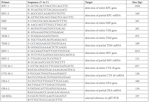
Detection of β-lactamase genes
According to the reported consensus primer sequenc-es [25-28], the PCR primers for amplification of the ESBL TEM, CTX-M, SHV and OXA genes and car-bapenemase KPC, IMP and VIM genes were synthe-sized by Invitrogen Co. in Shanghai, China (Table 1). Several PCRs were performed to detect these genes in the β-lactam antibiotic-resistant K. pneumoniae
by Invitrogen Co. [23]. The obtained sequencing data were compared to the corresponding sequences of β-lactamase genes of K. pneumoniae in GenBank using BLAST software.
Measurement of β-lactamase gene-mRNA levels
To avoid possible interference among different β-lactamase genes, 68 K. pneumoniae isolates carry-ing a scarry-ingle β-lactamase gene (of which 32 isolates are from our previous study and 36 isolates are from the present study) were selected for treatment with 1/4 MIC of CTX or CPN for 0.5, 1, 2 or 4 h at 37ºC, and then the TEM, CTX-M, SHV, OXA and KPC mRNAs were detected by real-time fluorescent quantitative RT-PCR (qRT-PCR). Briefly, total bacterial RNAs were extracted with TRIzol (Sigma, USA), and then the cDNAs were synthesized from the total RNAs us-ing a PrimeScript™ RT reagent kit (TaKaRa).Using the cDNAs as templates, the target gene mRNA levels
were assessed by qRT-PCR using a SYBR® Premix Ex-Taq™ II Kit (TaKaRa) on a LightCycler 480 Real-Time PCR System (Roche, Germany). The primers used in the qRT-PCR were designed by Primer Premier 6.0 Software and synthesized by Invitrogen Co. (Table 1). In the qRT-PCR, 16S rRNA of K. pneumoniae was used as the internal control. The qRT-PCR data were analyzed using the ΔΔCT model and randomization test in REST2005 software [29].
Detection of CLO toxicity against K. pneumoniae
One hundred isolates were randomly selected from
the K. pneumoniae isolates for treatment with 25, 50,
100, 250 or 500 μg/mL CLO (Sigma) at 37°C for 30 min [30], and then the MICs of CLO against the iso-lates were detected using the microdilution method as described above. Subsequently, 0.1 mL of each of the CLO-treated bacterial suspensions was inoculated on MH agar plates (BioMérieux) for a 24-h incubation
Table 1. Sequences of the primers used in PCR or qRT-PCR.
Primer Sequence (5ʹ to 3ʹ) Target Size (bp)
KPC-1 F: GCTACACCTAGCTCCACCTTC detection of entire KPC gene 1050
R: TCAGTGCTCTACAGAAAACC
KPC-2 F: AGCCGCCAAAGTCCTGTTC detection of partial KPC-mRNA 169
R: ATTGCTACACCTAGCTCCACCTTC
IMP F: CTACCGCAGCAGAGTCTTTG detection of entire IMP gene 587
R: AACCAGTTTTGCCTTACCAT
VIM F: AGTGGTGAGTATCCGACAG detection of entire VIM gene 261
R: ATGAAAGTGCGTGGAGAC
TEM-1 F: TCGGGGAAATGTGCG detection of entire TEM gene 972
R: TGCTTAATCAGTGAGGCACC
TEM-2 F: CCCAGAAACGCTGGTGAAA detection of partial TEM-mRNA 109
R: GGGGCGAAAACTCTCAAGG
SHV-1 F: GCCGGGTTATTTTATTTGTCGC
detection of entire SHV gene 1015
R: TCTTTCCGATGCCGCCGCCAGTCA
SHV-2 F: TTGATCCGCTCCGTGCT detection of partial SHV-mRNA 121
R: CCACAATCCGCTCTGCTTT
CTX-M-1 F: GTTACAGCCCTTCGGCGATGATTC detection of entire CTX-M gene 877
R:GCGCATGGTGACAAAGAGAGTGCA
CTX-M-2 F:TGCGGCTGGGTAAAATAGGT
detection of partial CTX-M-mRNA 120
R:GTCGTGGACTGTGGGTGATAAG
OXA-1 F:CTGTTGTTTGGGTTTCGCAAG detection of entire OXA gene 440
R:CTTGGCTTTTATGCTTGATG
OXA-2 F:TATGGCATTTGATGCGGAAA detection of partial OXA-mRNA 134
R:GCGAAACCCAAACAACAGAAA
16S RNA F:CGGTCTGTCAAGTCGGATGT internal reference in qRT-PCR 197
R:TTTGCTCCCCACGCTTTC
at 37°C. The bacterial growth was observed to deter-mine the minimal bactericidal concentration (MBC) of CLO against the isolates [24]. In the detections, the CLO-untreated isolates were used as controls.
Detection of β-lactamase gene-mRNA levels after CLO treatment
The 68 K. pneumoniae isolates carrying a single β-lactamase gene were treated with 25, 50, 100, 250 or 500 μg/mL CLO (Sigma) at 37°C for 30 min, and then CTX or CPN with 1/4 MIC was added for a 60-min incubation period at 37°C. The TEM, CTX-M, SHV or OXA mRNA were detected by qRT-PCR as described above. In the assay, the CLO-untreated isolates were used as the controls.
Detection of β-lactamase activity after CLO treatment
One hundred µg/mL CLO was used to pretreat the 68
K. pneumoniae isolates carrying a single β-lactamase
gene at 37C for 30 min. The change in β-lactamase ac-tivity in the isolates or the MICs of CPN, AMP, CFX, CTX and CAZ against the isolates were detected by the double paper disk synergy screening test or micro-dilution method as described above. In the assay, the CLO-untreated isolates were used as the controls.
Data analysis
Data from a minimum of three experiments were av-eraged and presented as means±standard deviation (SD). The χ2 test and t-test were used to determine significant differences. Statistical significance was defined as p<0.05.
RESULTS
Resistance rate and β-lactamase phenotypes of the
K. pneumoniae isolates
In this study, 118 of the 272 K. pneumoniae isolates were resistant to CPN, AMP, CFX, CTX and CAZ, with a resistance rate of 43.4% (118/272). The E-test result showed that the MICs of CTX and CPN against theβ-lactam antibiotic-resistant K. pneumoniae
iso-lates were 4~64μg/mL. The phenotype confirmatory tests showed that all the β-lactam antibiotic-resistant isolates were phenotypically positive for ESBL produc-tion, but only 23 of the isolates were phenotypically positive for carbapenemase production.
Predominant β-lactamase genes of the K.
pneumoniae isolates
Consistent with our previous study [23], the PCR and sequencing data showed that 91.5% of the 118 β-lactam antibiotic-resistant K. pneumoniae isolates in this study were positive for TEM-1, CTX-M-14, SHV-11, OXA-1 and/or KPC-2 genes according to a com-parison with the reported β-lactamase gene sequences in GenBank (Accession No.: TEM-1/NC_009651.1, CTX-M-14/NC_016839.1, SHV-11/NC_016845.1, OXA-1/JQ235810.1, KPC-2/NC_016846.1). A com-bination of this result with the β-lactamase gene detection data from our previous study (107 in the 118 isolates) revealed that 68.4% of the β-lactam antibiotic-resistant K. pneumoniae isolates (147/215) possess more than two β-lactamase genes (Table 2), with TEM-1 (70.7%, 152/215) and SHV-11 (64.2%, 138/215) and CTX-M-14 (40.5%, 87/215) as the pre-dominant genes of ESBLs. TEM-1 plus SHV-11 was the common combination of β-lactamase encoding genes carried by the isolates (30.7%, 66/215) (Table 2).
Table 2. β-lactamase gene profiles of the 215 K. pneumoniae iso-lates.
β-lactamase genes and combination Strains (n) Percentage (%)
KPC-2 2 0.9
TEM-1 30 14.0
CTX-M-14 19 8.8
SHV-11 16 7.4
OXA-1 1 0.5
KPC-2+CTX-M-14 2 0.9
KPC-2+SHV-11 2 0.9
TEM-1+CTX-M-14 17 7.9
TEM-1+SHV-11 66 30.7
CTX-M-14+SHV-11 14 6.5
OXA-1+SHV-11 4 1.9
KPC-2+TEM-1+CTX-M-14 6 2.8
KPC-2+TEM-1+SHV-11 7 3.3
KPC-2+CTX-M-14+SHV-11 3 1.4
TEM-1+CTX-M-14+SHV-11 25 11.6
KPC-2+CTX-M-14+TEM-1+SHV-11 1 0.5
Increase of β-lactamase gene-mRNA levels after antibiotic treatment
In the total of 68 β-lactam antibiotic-resistant K.
pneu-moniae isolates carrying a single β-lactamase gene,
the mRNA levels of TEM-1 (30 isolates ), CTX-M-14 (19 isolates), SHV-11 (16 isolates) and OXA-1 (one isolate) genes, but not that of KPC-2 genes (2 isolates), were rapidly increased after treatment with 1/4 MIC of CTX or CPN at different time intervals (Fig. 1).
Inhibition of CLO on antibiotic-induced β-lactamase gene-mRNA level elevation
The toxicity test showed that 25~500 µg/mL CLO nei-ther inhibited nor killed the K. pneumoniae isolates. When the 68 β-lactam antibiotic-resistant K. pneumo-niae isolates carrying a single β-lactamase gene were pretreated with 50~500 µg/mL CLO, the 1/4 MIC of or CPN-induced elevation of the TEM-1, CTX-M-14, SHV-11 or OXA-1 mRNA levels was signifi-cantly inhibited in a dose-dependent manner (Fig. 2).
Fig. 1. Effect of CTX and CPN on upregulation of β-lactamase-mRNA levels. A total of 68 K. pneumoniae isolates carrying a single β-lactamase gene were subjected to 1/4 MIC of CTX or CPN treatment for the indicated times and the mRNA levels of KPC (2 isolates), TEM (30 isolates), CTX-M (19 isolates), SHV (16 isolates) and OXA (1 isolate) were detected by qRT-PCR. Bars show the means±SD of three independent experiments. *p<0.01vs. the β-lactamase-mRNA levels in the CTX- or CPN-untreated isolates.
Fig. 2. Effect of CLO on inhibition of CTX- or CPN-induced β-lactamase-mRNA level elevation. The 68 K. pneumoniae isolates car-rying a single β-lactamase gene were pretreated with different concentrations of CLO for 30 min, followed by treatment of 1/4 MIC of CTX or CPN for 1 h, and the TEM, CTX-M, SHV and OXA mRNA levels were detected by qRT-PCR. Bars show the means±SD of three independent experiments. *p<0.01vs. the β-lactamase-mRNA levels in the CTX- or CPN-untreated isolates; #p<0.01vs. the β-lactamase-mRNA levels in the CLO-untreated but CTX- or CPN-treatedisolates.
Table 3. Changes of β-lactam antibiotic-resistance rate in CLO-treated K. pneumoniae isolates.
100 µg/mL CLO CPN Resistant isolates / sensitive isolates / sensitive rate (%)AMP CFX CTX CAZ
Before treatment 236/0/0 236/0/0 236/0/0 236/0/0 236/0/0
After treatment 51/185/78.4* 46/190/80.5* 49/187/79.2* 45/191/80.9* 44/192/81.4*
Changes of antibiotic resistance and ESBL phenotype after CLO treatment
When the 236 β-lactam antibiotic-resistant K.
pneu-moniae isolates from our previous study and the
present study were pretreated with 100 µg/mL CLO as above, a great portion of the resistant isolates became sensitive to CPN, AMP, CFX, CTX or CAZ, resulting in the following percentages of sensitized isolates of 78.4% (185/236), 80.5% (190/236), 79.2% (187/236), 80.9% (191/236) and 81.4% (192/236) (Table 3). In addition, the rate of phenotypically positive ESBLs in the CLO-pretreated isolates was decreased from 100% (236/236) to 27.1% (64/236).
DISCUSSION
Drug resistance of bacteria is a global major public health challenge, which has resulted in the inefficacy of antibiotic treatments for infectious diseases caused by bacteria, including K. pneumonia [31]. Recent data announced by the CHINET bacterial resistance surveillance network showed that more than 70% of bacterial infectious diseases were caused by infec-tion of Gram-negative bacteria in China, of which K.
pneumonia infection accounted for 16.1% [32].The
CHINET resistance data revealed that the positive rate of different β-lactamase genes in K. pneumoniae iso-lates was 43.6%, while 31.8% of the isoiso-lates contained detectable ESBL genes [32]. Therefore, K. pneumoniae
is a prominent causative agent of infectious diseases with a high rate of antibiotic resistance in China.
β-lactam antibiotics are commonly used for the treatment of bacterial infectious diseases. However, many bacteria, including K. pneumoniae, can resist β-lactam antibiotics through the production of dif-ferent β-lactamases [33]. The most frequently found β-lactamases in clinical isolates of β-lactam antibiotic-resistant bacteria includes three major genetic groups: TEM, SHV and CTX-M types [10-12]. Recently, KPC- or OXA-positive K. pneumoniae isolates were also frequently reported [12,34]. However, distribu-tion of the bacteria with different β-lactamase genes presents a geographical or local diversity. In North America and Western Europe, TEM and SHV types still dominate [12], while in Eastern Europe, Asia and South America, the CTX-M type has replaced TEM
and SHV as the predominant ESBL [13,35-37]. In par-ticular, CTX-M-9, CTX-M-14 and CTX-M-15 are the predominant CTX-M subtypes in Japan, China and India, respectively [38-40]. The diversity of ESBLs in bacteria was also found in different areas of China. For example, CTX-M is predominant in bacteria from Guangdong province but TEM is the most common ESBL in bacteria from Shanxi province [41,42]. Our previous study revealed the prevalence of TEM-1 and SHV-11 in K. pneumoniae isolates from Zhejiang province [23]. In this study, more K. pneumoniae iso-lates were collected and the high prevalence of both TEM-1 and SHV-11 in the isolates from Zhejiang province was further confirmed, which differed from the epidemiology of β-lactamase genes in Guangdong or Shanxi province [41,42]. Moreover, our results also showed that the multiple β-lactamase gene carrying rate in the isolates was significantly higher than the single gene carrying rate (p<0.05), where TEM-1 plus SHV-11 was the most common carrying mode.
TEM-1, CTX-M-14, SHV-11 or OXA-1 mRNA level, but it was inhibited by the administration of CLO. The data indicated that sublethal dosage of CTX or CPN acts as an inducer for the expression of the four ESBL genes in the K. pneumonia isolates probably through HK-related TCSS.
To eliminate the possible toxicity of CLO to the K.
pneumoniae isolates that also can inhibit the elevation
of antibiotic-induced ESBL-mRNAs, both MIC and MBC of CLO against the isolateswere determined. The results showed that 25~500 μg/mL CLO could not present any effects to inhibit or kill the isolates tested, indicating that inhibition of HK but not its toxicity is involved in the role of CLO on the inhibition of the CTX- or CPN-induced ESBL-mRNA elevation. In particular, the addition of 100 µg/mL CLO caused the majority of the β-lactam antibiotic-resistant K.
pneu-moniae isolates to become sensitive to the five β-lactam
antibiotics tested, and the phenotype of ESBLs in most of the CLO-pretreated isolates to disappear. Since HK-related TCSS is absent in eukaryotes [46,49], CLO may have a potential as a candidate for developing a novel drug against ESBL-producing bacteria.
CONCLUSIONS
TEM-1, SHV-11 and CTX-M-14 are the predominant β-lactamase genes in the β-lactam antibiotic-resistant
K. pneumoniae isolates from Zhejiang province of
China, and TEM-1 plus SHV-11 is the predominant combination of β-lactamase genes carried by the iso-lates. Sublethal concentrations of CTX or CPN act as an extrinsic inducer to upregulate the expression of TEM-1, SHV-11, CTX-M-14 and OXA-1 genes in the isolates, but it can be inhibited by CLO, a bacte-rial histidine kinase inhibitor. CLO was also capable of changing the sensitivity to β-lactam antibiotics and to limit the phenotype of ESBLs in the β-lactam antibiotic-resistantisolates.
Acknowledgments: This study was supported by a grant from the National Natural Science Foundation of China (No.: 81271893) and a grant from Zhejiang Provincial Program for the Cultivation of High-level Innovative Health talents (No.: 2012-241). We thank the Department of Clinical Laboratory of the First Affiliated Hos-pital of Zhejiang Chinese Medical University, the Second Affili-ated Hospital of Zhejiang Chinese Medical University, the Second Affiliated Hospital of Zhejiang University, Zhejiang Provincial
People’s Hospital, Hangzhou Second People’s Hospital, Zhejiang Hospital, Ningbo First Hospital and Jinhua People’s Hospital for providing of the K. pneumoniae isolates.
Conflict of interest disclosure: No conflict of interests is declared.
REFERENCES
1. Struve C, Krogfelt KA. Pathogenic potential of environ-mental Klebsiella pneumoniae isolates. Environ Microbiol. 2004;6(6):584-90.
2. Podschun R, Ullmann U. Klebsiella spp. as nosocomial pathogens: epidemiology, taxonomy, typing methods, and pathogenicity factors. Clin Microbiol Rev. 1998;11(4):589-603.
3. Vuotto C, Longo F, Balice MP, Donelli G, Varaldo PE. Anti-biotic resistance related to biofilm formation in Klebsiella pneumoniae. Pathogens. 2014;3(3):743-58.
4. Bouza E, Cercenado E. Klebsiella and enterobacter: anti-biotic resistance and treatment implications. Semin Respir Infect. 2002;17(3):215-30.
5. Paterson DL. Resistance in gram-negative bacteria: entero-bacteriaceae. Am J Med. 2006;119(6 Suppl 1):S20-8;S62-70. 6. Lautenbach E, Patel JB, Bilker WB, Edelstein PH, Fishman
NO. Extended-spectrum beta-lactamase-producing Esch-erichia coli and Klebsiella pneumoniae: risk factors for infec-tion and impact of resistance on outcomes. Clin Infect Dis. 2001;32(8):1162-71.
7. Livermore DM. Beta-lactamases in laboratory and clinical resistance. Clin Microbiol Rev. 1995;8(4):557-84.
8. Thomson JM, Bonomo RA. The threat of antibiotic resis-tance in Gram-negative pathogenic bacteria: beta-lactams in peril! Curr Opin Microbiol. 2005;8(5):518-24.
9. Bush K. Bench-to-bedside review: The role of beta-lac-tamases in antibiotic-resistant Gram-negative infections. Crit Care. 2010;14(3):224.
10. Paterson DL, Bonomo RA. Extended-spectrum beta-lactamases: a clinical update. Clin Microbiol Rev. 2005;18(4):657-86.
11. Chong Y, Ito Y, Kamimura T. Genetic evolution and clinical impact in extended-spectrum β-lactamase-producing Esch-erichia coli and Klebsiella pneumoniae. Infect Genet Evol. 2011;11(7):1499-504.
12. Lynch JP 3rd, Clark NM, Zhanel GG. Evolution of anti-microbial resistance among Enterobacteriaceae (focus on extended spectrum β-lactamases and carbapenemases). Expert Opin Pharmacother. 2013;14(2):199-210.
13. Kiratisin P, Apisarnthanarak A, Laesripa C, Saifon P. Molecular characterization and epidemiology of extended-spectrum-beta-lactamase-producing Escherichia coli and Klebsiella pneumoniae isolates causing health care-associated infection in Thailand, where the CTX-M family is endemic. Antimicrob Agents Chemother. 2008;52(8):2818-24. 14. Molton JS, Tambyah PA, Ang BS, Ling ML, Fisher DA.
15. Feizabadi MM, Delfani S, Raji N, Majnooni A, Aligholi M, Shahcheraghi F, Parvin M, Yadegarinia D. Distribution of bla(TEM), bla(SHV), bla(CTX-M) genes among clinical isolates of Klebsiella pneumoniae at Labbafinejad Hospital, Tehran, Iran. Microb Drug Resist. 2010;16(1):49-53. 16. Yim G, Wang HH, Davies J. Antibiotics as signalling
molecules. Philos Trans R Soc Lond B Biol Sci. 2007; 362(1483):1195-200.
17. Fajardo A, Martínez JL. Antibiotics as signals that trigger specific bacterial responses. Curr Opin Microbiol. 2008; 11(2):161-7.
18. Bruchmann J, Kirchen S, Schwartz T. Sub-inhibitory con-centrations of antibiotics and wastewater influencing biofilm formation and gene expression of multi-resistant Pseudomo-nas aeruginosa wastewater isolates. Environ Sci Pollut Res Int. 2013;20(6):3539-49.
19. Wu Y, Sun A, Zhao J, Ge Y, Yan J. [Distribution of drug inac-tive enzyme genes in bacterial isolates and mechanism of its induction and inhibition]. Zhejiang Da Xue Xue Bao Yi Xue Ban. 2013;42(2):131-40. Chinese.
20. Depardieu F, Podglajen I, Leclercq R, Collatz E, Courvalin P. Modes and modulations of antibiotic resistance gene expres-sion. Clin Microbiol Rev. 2007;20(1):79-114.
21. Gotoh Y, Eguchi Y, Watanabe T, Okamoto S, Doi A, Utsumi R. Two-component signal transduction as potential drug targets in pathogenic bacteria. Curr Opin Microbiol. 2010;13(2):232-9.
22. Schreiber M, Res I, Matter A. Protein kinases as antibacterial targets. Curr Opin Cell Biol. 2009;21(2):325-30.
23. Wang Q, Ge YM, Sun AH, Liu JF, Wang Y, Yan J. [Genotypes of β-lactamase in Klebsiella pneumoniae isolates and induc-tion and inhibiinduc-tion of the β-lactamase gene expression]. Zhonghua Min Guo Wei Sheng Wu Ji Mian Yi Xue Za Zhi. 2013;33(12):916-21. Chinese.
24. Clinical and Laboratory Standards Institute. Performance Standards for Antimicrobial Susceptibility Testing, Twenty-fifth Informational Supplement. CLSI document M100-S25. Wayne, PA: Clinical and Laboratory Standards Institute, USA. 2015.
25. Bradford PA. Extended-spectrum β-lactamases in the 21st century: Characterization, epidemiology, and detection of this important resistance threat. Clin Microbiol Rev. 2001;14(4):933-51.
26. Turner MS, Andersson P, Bell JM, Turnidge JD, Harris T, Giffard PM. Plasmid-borne blaSHV genes in Klebsiella pneu-moniae are associated with strong promoters. J Antimicrob Chemother. 2009;64(5):960-4.
27. Shi W, Qin J, Mi ZA. Klebsiella pneumoniae sputum culture isolate from China carrying blaOXA-1, blaCTX-M-55 and aac(6’)-Ib-cr. J Med Microbiol. 2008;57(Pt 12):1588-99. 28. Holstein A, Grillon A, Yzon L, Morange V, Baty G,
Lar-tigue MF, Mereghetti L, Goudeau, A, Lanotte P. Prevalence of extended-spectrum beta-lactamases of the CTX-M type producing Escherichia coli and Klebsiella pneumoniae in Bretonneau hospitals (CHRU Tours). Pathol Biol (Paris). 2010;58(1):67-9.
29. Pfaffl MW, Horgan, GW, Dempfle L. Relative expression software tool (REST) for group-wise comparison and
statis-tical analysis of relative expression results in real-time PCR. Nucl Acids Res. 2002;30(9):e36.
30. Kumagai Y, Cheng Z, Lin M, Rikihisa Y. Biochemical activi-ties of three pairs of Ehrlichia chaffeensis two-component regulatory system proteins involved in inhibition of lyso-somal fusion. Infect Immun. 2006;74(9):5014-22.
31. Gootz TD. The global problem of antibiotic resistance. Crit Rev Immunol. 2010; 30(1):79-93.
32. Hu FP, Zhu DM, Wang F, Jiang XF, Sun ZY, Chen ZJ, Hu ZD, Li J, Xie Y, Kang M, Xu YC, Zhang XJ, Zhang ZX, Ji P, Wang CQ, Wang AM, Ni YX, Sun JY, Yu YS, Lin J, Chu YZ, Tian SF, Xu YH, Shen JL, Shan B, Du Y, Zhuo C, Su DH, Zhang H, Kong J, Wei LH, Wu L, Hu YJ, Ai XM. CHINET 2013 surveillance of bacterial resistance in China. Clin J Infect Chemother. 2014;14(5):365-74. Chinese.
33. Brolund A. Overview of ESBL-producing Enterobacte-riaceae from a Nordic perspective. Infect Ecol Epidemiol. 2014;4:24555.
34. Tzouvelekis LS, Markogiannakis A, Psichogiou M, Tassios PT, Daikos GL. Carbapenemases in Klebsiella pneumoniae and other Enterobacteriaceae: an evolving crisis of globaldi-mensions. Clin Microbiol Rev. 2012;25(4):682-707. 35. Livermore DM, Canton R, Gniadkowski M, Nordmann P,
Rossolini GM, Arlet G, Ayala J, Coque TM, Kern-Zdano-wicz I, Luzzaro F, Poirel L, Woodford N. CTX-M: chang-ing the face of ESBLs in Europe. J Antimicrob Chemother. 2007;59(2):165-174.
36. Ling TK, Xiong J, Yu Y, Lee CC, Ye H, Hawkey PM. Multi-center antimicrobial susceptibility survey of gram-negative bacteria isolated from patients with community-acquired infections in the People’s Republic of China. Antimicrob Agents Chemother. 2006;50(1):374-8.
37. Rossi F. The challenges of antimicrobial resistance in Brazil. Clin Infect Dis. 2011;52(9):1138-43.
38. Chong Y, Shimoda S, Yakushiji H, Ito Y, Miyamoto T, Kamimura T, Shimono N, Akashi K. Community spread of extended-spectrum β-lactamase-producing Escherichia coli, Klebsiella pneumoniae and Proteus mirabilis: a long-term study in Japan. J Med Microbiol. 2013;62(Pt 7):1038-43. 39. Xia S, Fan X, Huang Z, Xia L, Xiao M, Chen R, Xu Y,
Zhuo C. Dominance of CTX-M-type extended-spectrum β-lactamase (ESBL)-producing Escherichia coli isolated from patients with community-onset and hospital-onset infection in China. PLoS One. 2014;9(7):e100707.
40. Muzaheed, Doi Y, Adams-Haduch JM, Endimiani A, Sidja-bat HE, Gaddad SM, Paterson DL. High prevalence of CTX-M-15-producing Klebsiella pneumoniae among inpatients and outpatients with urinary tract infection in Southern India. J Antimicrob Chemother. 2008;61(6):1393-4. 41. Zhuo C, Su DH, Li HY, Wang LX, Liao K, Wang M, Zhi ZQ,
Guo ZH, Wei YC, Geng SN, Jin GY, Zhong NS. Study on CTX-M type ESBLs-producing Escherichia coli and Kleb-siella pneumoniae in Guangzhou. Chin J Lab Med. 2009; 32(10):1114-9. Chinese.
43. Lee N, Yuen KY, Kumana CR. Clinical role of beta-lactam/beta-lactamase inhibitor combinations. Drugs. 2003;63(14):1511-24.
44. Chen J, Shang X, Hu F, Lao X, Gao X, Zheng H, Yao W. β-Lactamase inhibitors: an update. Mini Rev Med Chem. 2013; 13(13):1846-61.
45. Mitrophanov AY, Groisman EA. Signal integration in bacte-rial two-component regulatory systems. Genes Dev. 2008; 22(19):2601-11.
46. Hansen J, Mailand E, Swaminathan KK, Schreiber J, Angelici B, Benenson Y. Transplantation of prokaryotic two-compo-nent signaling pathways into mammalian cells. Proc Natl Acad Sci U S A. 2014;111(44):15705-10.
47. Worthington RJ, Melander C. Combination approaches to combat multidrug resistant bacteria. Trends Biotechnol. 2013;31(3):177-84.
48. Bacon JA, Ulrich RG, Davis JP, Thomas EM, Johnson SS, Conder GA, Sangster NC, Rothwell JT, McCracken RO, Lee BH, Clothier MF, Geary TG, Thompson DP. Comparative in vitro effects of closantel and selected beta-ketoamide anthel-mintics on a gastrointestinal nematode and vertebrate liver cells. J Vet Pharmacol Ther. 1998; 21(3):190-8.