Research Article
July
2017
Computer Science and Software Engineering
ISSN: 2277-128X (Volume-7, Issue-7)
Survey on Ocular Blood Vessel Segmentation
R. Hannah Roseline, R. Jemina Priyadarsini
Department of Computer Science, Bishop Heber College, Bharathidasan University, Tiruchirappalli, Tamil Nadu, India
DOI: 10.23956/ijarcsse/V7I7/0114
Abstract: The eye is sometimes said to provide a window into the health of a person for it is only with the eye that one can actually see the exposed flesh of the subject without using invasive procedures. There are a number of diseases, particularly vascular disease that leave telltale markers in the retina. The retina can be photographed relatively straightforwardly with a funds camera and now with retinal image processing there is much interest in computer analysis of retinal images for identifying and quantifying the effects of diseases such as cardio vascular diseases. A retinal image provides a snapshot of what is happening inside the human body. In particular, the ceremonial of the retinal blood vessels has been shown to imitate the cardiovascular condition of the body. Retinal images provide considerable information on pathological changes caused by local ocular disease which reveals diabetes, hypertension, arteriosclerosis, cardiovascular disease and stroke. Computer-aided study of retinal image plays a central role in diagnostic procedures. However, automatic retinal segmentation is complicated by the fact that retinal images are often noisy, poorly contrasted, and the vessel widths can vary from very small to very large. So in this survey we can review various segmentation techniques to improve the accuracy in blood vessel extraction.
Keywords: Retinal image processing, Blood vessels, Cardio vascular, Segmentation, Computer analysis
I. INTRODUCTION
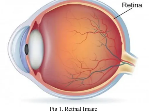
Eye is the one of the critical organ in our body, called as organs of vision. Blood vessel extraction could be very essential as many eye sicknesses are identified by inspecting the blood vessel. These blood vessel convey sparkling oxygenated blood from heart to feed vitamins‟s to tissues and cells present in retina after which deliver the deoxygenated blood from eye to heart. There are especially styles of blood vessels arteries those deliver sparkling blood and vein the ones bring oxygenated blood. The eye is a window to the retinal vascular gadget that is uniquely accessible for the non-invasive, in vivo study of a continuous vascular mattress in human beings. Retinal blood vessels have been shown to exchange in diameter, branching angles or tortuosity, due to a disorder. The retina is the most effective place in which blood vessels can be directly visualized non-invasively in vivo. Increasing technology main to the development of digital imaging structures over the last two decades has revolutionized fundal imaging. Whilst digital imaging does not nonetheless have the resolution of traditional pictures, modern digital imaging systems offer very high-decision pix which might be enough for maximum medical eventualities. In addition, virtual imaging has the benefit of simpler storage on media that don't become worse in best with time, may be transmitted over quick distances throughout a sanatorium or over large distances through digital transfer (allowing professional „„at-distance‟‟ opinion in big rural communities), may be processed to enhance image exceptional, and subjected to photograph evaluation to carry out objective quantitative evaluation of fundal photos and the capacity for automatic prognosis. In the studies or screening setting, massive databases of fundal pictures can be mechanically categorized and managed greater comfortably than exertions-in depth observer-driven techniques. Automated analysis can also be a useful resource choice-making for optometrists. The primary format of retinal photograph is shown in fig 1.
Fig 1. Retinal Image
ISSN(E): 2277-128X, ISSN(P): 2277-6451, DOI: 10.23956/ijarcsse/V7I7/0114, pp. 318-324
retina and the choroid. In this condition, a system of small blood vessels, known as choroidal neovascularization (CNV), arises in the choroid and taking a portion of the blood offering the retina. As the amount of blood offering the retina is reduced, the sight may be degraded and inside the extreme cases, blindness may additionally arise. The physicians attempt to deal with this risky disease by means of applying optical electricity to photocoagulate the neovascularization. Argon laser is utilized in photocoagulation functions to cauterize the small vessels which increases the quantity of blood supplying the retina and as a result maintaining the sight. This treatment modality is carried out in lots of periods. The doctor asks the affected person to fixate his/her eye so as to direct the laser beam to the affected region. The current achievement charge of this system is beneath 50% for eradication of CNV following one treatment session with a recurrence and/or staying power charge of about 50%. The latter situation requires repeating the treatment.
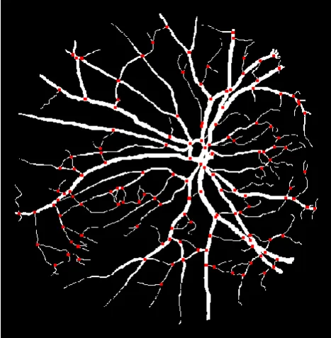
In this survey, outline the principles upon which retinal virtual photo analysis is primarily based on modern strategies used to routinely detect landmark features of the fundus, including blood vessels. And can also provide evaluation of retina inside the computerized prognosis of pathology (with particular connection with diabetic retinopathy). Additionally evaluate its function in defining and appearing quantitative measurements of vascular topography, how those entities are based totally on „optimization‟ concepts and how they've helped to explain the relationship among systemic cardiovascular sickness and retinal vascular adjustments. We also review the capacity destiny use of fundal picture evaluation in telemedicine. The vascular shape can be proven in fig 2.
Fig 2: Retinal image processing
II. RELATED WORK
A. Krizhevsky, et al. [1] proposed a multi-scale response to stumble on linear systems in 2D pics. The provided detection algorithm is divided into two steps. First, we gift a flux based anisotropic diffusion method and apply it to denoise images corrupted by way of an additive Gaussian noise. In order to extract handiest the pixels belonging to a vessel area, we use a Gaussian version of the vessels for decoding the eigen values and the eigenvectors of the Hessian matrix. Then, we compute the multi-scale reaction from responses computed at a discrete set of scales. First, we gift a flux primarily based anisotropic diffusion approach and apply it to denoise snap shots corrupted through an additive Gaussian noise. The unique pathological situations (illnesses) of retina are: diabetic retinopathy (DR), macular degeneration and glaucoma. One of the serious complication in present day era is the Diabetic Retinopathy (DR) takes place because of development of diabetes. It reasons damage to the one-of-a-kind elements of the retina and ends in vision loss. DR is a current sickness which procedures from non-proliferative diabetic retinopathy (NPDR) to the proliferative diabetic retinopathy (PDR). Micro aneurysms, the small purple dots in the coloration photos are the first clinical signal for the presence of DR. During the PDR degree the tiny blood vessels are blocked.
ISSN(E): 2277-128X, ISSN(P): 2277-6451, DOI: 10.23956/ijarcsse/V7I7/0114, pp. 318-324
Fabiola M,et.Al. [3] applied reliable vessel extraction that's a prerequisite for subsequent retinal picture evaluation and processing because vessels are the foremost and maximum stable structures performing in those pictures. Accurate segmentation of retinal snap shots influences immediately the performance of trivia extraction. If extra background regions are included within the segmented retinal image, greater fake capabilities are introduced; if a few elements of the foreground are excluded, useful function factors can be overlooked. Advances in vascular imaging generation have furnished radiologists with non-invasive imaging modalities which could deliver correct vascular facts, which enables the doctor to outline the character and quantity of a vascular ailment, assisting analysis and prognosis. As stated previously, accurate vascular extraction is the number one task in automatic ophthalmic photo evaluation.
George Azzopardi,et.Al [4] introduced a unique technique for the automated segmentation of blood vessels in retinal fundus pictures. It is primarily based at the Combination of Receptive Fields (CORF) computational model of a easy cell in visual cortex and its implementation referred to as Combination of Shifted Filter Responses (COSFIRE). We endorse a bar-selective COSFIRE filter, or B-COSFIRE for brevity, that may be effectively used to discover bar-shaped structures together with blood vessels. The B-COSFIRE filter out that we advise is non-linear because it achieves orientation selectivity with the aid of multiplying the output of a collection of Difference-of-Gaussians (DoG) filters, whose helps are aligned in a collinear manner. It approach that the selectivity of the filter out isn't always predefined inside the implementation but it is determined from a person-distinct prototype sample (e.G. A straight vessel, a bifurcation or a crossover point) in an automatic configuration process. The reaction of a B-COSFIRE clear out is computed as the weighted geometric imply, basically the product, of the responses of the involved DoG filters inside the facilities of the corresponding circles. The positions at which we take their responses are decided via an automated analysis method of the reaction of a DoG filter to a prototype bar structure; the positions alongside these circles at which these responses attain vast nearby maxima are the positions of the points that represent the dominant intensity variations round the point of interest.
Yitian Zhao,et.Al [5] proposed vascular illnesses are often existence-crucial for individuals, and gift a hard public fitness problem for society. The drive for better understanding and control of these situations clearly motivates the want for stepped forward imaging techniques. The detection and evaluation of the vessels in scientific pics is a fundamental assignment in many scientific programs to assist early detection, diagnosis and most excellent remedy. In line with the proliferation of imaging modalities, there's an ever-growing call for for computerized vessel evaluation systems for which where blood vessel segmentation is the first and most essential step.
Srivastava,et.Al,..[6] implemented to robotically finding the optic nerve and blood vessel in retinal pictures the usage of graph reduce approach. To phase and decide the blood vessels and optic disc from retinal photos, the input retinal pix are read which then beneath is going preprocessing, then to discover the optical disc segmentation and blood vessel segmentation. The coloration based totally segmentation and gradient approach used for blood vessel segmentation for accomplishing exquisite performance in segmenting the blood vessel and Hough circular rework is used for optical disc segmentation. For rule based totally validation is implemented to validate proper optical disc segmented or not similarly validation of blood vessel segmentation.
Y. Bengio,et.Al,… [7] provided an inpainting technique has a special recognition. Indeed, neither texture synthesis nor a visually doable picture is needed. Our purpose is to fill structures along with exudates in retinal snap shots so that, when vessel enhancement is implemented, the wide variety of nearby fake positives is substantially decreased. This purpose is simplest performed if exudates are crammed in a clean manner that reduces or removes viable edges. A multiple-scale Hessian-based enhancement is applied to discover retinal vessels. This approach is fast and has established to be powerful whilst detecting vessels of ordinary eyes. The key idea of the proposed approach is to use Hessian-based enhancement after exudate inpainting. Moreover, it yields the nice performance on pathological images, the goal of most automated retinal photograph evaluation equipment. Indeed, a vessel segmentation set of rules is usually step one for the automated detection of eye diseases.
M. Fraz,et.Al,.. [8] carried out the work to extract the blood vessels with the aid of the usage of color fundus photo. Fundus image is an RGB shade picture, in widespread RGB photographs encompass three channels (purple, inexperienced and blue) This can be achieved via separation the retina picture to a few channels and using simplest certainly one of them (Green channel), the blue channel is characterised by using low comparison and does now not incorporate a good deal data. The vessels are visible inside the pink channel. In this technique, the retinal picture is taken because the input photo. Then the input retinal photo is pre-processed. In pre-processing stage, the enter photo is resized and the Red or Green channel picture is separated as the blood vessel seems brighter within the Red or green channel photograph. Then morphological operation is carried out at the Red or green channel photo. The number one morphological operations are dilation and erosion. The more complex morphological operations are beginning and last. Dilation is an operation that grows or thickens objects in a binary picture. The particular manner and volume of this thickening is controlled by means of shape referred to a structuring element. Dilation is defined in terms of set operation. Erosion shrinks or thins objects in a binary photo.
III. BLOOD VESSEL SEGMENTATION
ISSN(E): 2277-128X, ISSN(P): 2277-6451, DOI: 10.23956/ijarcsse/V7I7/0114, pp. 318-324 A. GRAPH TRACER ALGORITHM:
This Graph based set of rules truly identifies the true vessel photograph from thinned image where the vessels are sincerely marked with its suitable bifurcation and crossovers. Finally the width measurement is conducted on those true vessels. The vessel quality annotation tool is applied here for the reason of width measurement. The width values are as compared with Gold widespread width values. A new line-monitoring process is beginning from a small institution of pixels, derived from a brightness selection rule and extracting the vessel community. The multi-scale photograph map is derived after combining the individual picture maps alongside scales, containing the pixels self-assurance to belong in a vessel. The preliminary vessel community is derived after map quantization of the multi-scale confidence matrix. Median filtering is carried out in the initial vessel network, restoring disconnected vessel traces and disposing of noisy strains. Finally, filtering tactics gets rid of erroneous regions using directional attributes of vessels and morphological reconstruction. To become aware of genuine blood vessels Graph Tracer Algorithm” is implemented on this painting. This algorithm is a post processing approach to segmentation that utilizes the worldwide information of the segmented vascular structure to correctly pick out proper vessels in a retinal image. The segmented vascular shape is modeled as a vessel section graph and transforms the trouble of identifying real vessels to that of locating an most advantageous woodland inside the graph.
Let P be the set of all white pixels in a line photograph. Two pixels pi , pj ∈ P are adjacent, i.E., adj (pi , pj), if and best if pj ∈ neigh8 (pi), wherein neigh8 (p) = p1, p2 to p8 is the eight-neighborhood of p.
Pixel Crossing Number
Let p1 to p8 be a clockwise sequence of the eight neighbor pixels of pixel p. Then, xnum (p) is the number of black to nonblack transitions in this sequence of neighbor pixels of p. Junction Let white8 (p) ⊆ neigh8 (p) be the set of white pixels that are neighbors of p. The set of junction pixels in P is YP = {p ∈ P| xnum (p) | > 2 ∨ |white8 (p)| >3}. A junction is a set of connected junction Pixels, i.e., J ⊆ YP such that ∀pi , pj = i ∈ J, conn (pi , pj), where conn is restricted to the set YP. Then, the set of all junctions in P is JP.
Segment
A segment s is a sequence of unique white pixels p1 to pn in P such that all of the following conditions are true: a. n > 0 and ∀i ∈ [1, n], pi /∈ JP
b. ∀i ∈ {1, n}, |white8(pi)| = 1∨∃pj ∈ JP s.t. adj(pi , pj) c. n > 2 ⇒∀i ∈ [2, n − 1], xnum(pi) = 2.
Let SP be the set of all segments in P and NP = P −YP, i.e., NP contains non junction pixels that are part of segments. Then, s ∈ SP is adjacent to a junction J adj (s, J), if ∃pj ∈ J s.t. adj(pj , p1) ∨ adj(pj , pn).
B. FUZZY C MEANS CLUSTERING:
Fuzzy C-Means (FCM) is a method of clustering which lets in one piece of records to belong to two or more clusters. This method is regularly used in sample recognition. Basically this algorithm works via assigning membership to every statistics factor corresponding to each cluster center on the idea of distance among the cluster and the facts factor. More the records is close to to the cluster middle greater is its club toward the unique cluster middle. The iterative unsupervised Fuzzy C Means (FCM) algorithm is the maximum widely used clustering set of rules for photo segmentation.
Algorithm for FCM:
Step 1. The Color Retinal Fundus Image in Gray scale is transformed from inexperienced channel. Step 2. Adaptive histogram equalization is completed on the grey image.
Step 3. The background is subtracted from the foreground of the picture using median filter out. Step 4. FCM is implemented on the image accompanied by binarization and filtering.
Step five: The ground reality image is compared with the corresponding disorder
C. CNN BASED CLASSIFICATION:
ISSN(E): 2277-128X, ISSN(P): 2277-6451, DOI: 10.23956/ijarcsse/V7I7/0114, pp. 318-324
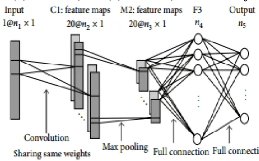
Fig 3: Convolutional Neural network for blood vessel segmentation
As illustrated in fig 3 the net contains five layers with weights, including the input layer, the convolutional layer C1, the max pooling layer M2, the full connection layer F3, and the output layer. Assuming 𝜃 represents all the trainable parameters (weight values), 𝜃 = {𝜃𝑖} and 𝑖 = 1, 2, 3, 4, where 𝜃𝑖 is the parameter set between the (𝑖−1)th and the 𝑖th layer. In HSI, each HSI pixel sample can be regarded as a 2D image whose height is equal to 1 (as 1D audio inputs in speech recognition). Therefore, the size of the input layer is just (𝑛1, 1), and 𝑛1 is the number of bands. The first hidden convolutional layer C1 filters the 𝑛1 × 1 input data with 20 kernels of size 𝑘1 × 1. Layer C1 contains 20 × 𝑛2 × 1 nodes, and 𝑛2 = 𝑛1 − 𝑘1 + 1. There are 20 × (𝑘1 + 1) trainable parameters between layer C1 and the input layer. The max pooling layer M2 is the second hidden layer, and the kernel size is (𝑘2, 1). Layer M2 contains 20 × 𝑛3 × 1 nodes, and 𝑛3 = 𝑛2/𝑘2. There is no parameter in this layer. The fully connected layer F3 has 𝑛4 nodes and there are (20 × 𝑛3 + 1) × 𝑛4 trainable parameters between this layer and layer M2. The output layer has 𝑛5 nodes, and there are (𝑛4 + 1) × 𝑛5 trainable parameters between this layer and layer F3. Consequently, the architecture of our proposed CNN classifier totally has 20 × (𝑘1 + 1) + (20 × 𝑛3 + 1) × 𝑛4 + (𝑛4 + 1) × 𝑛5 trainable parameters. Classifying a specified HSI pixel wants the corresponding CNN with the aforementioned parameters, where 𝑛1 and 𝑛5 are the spectral channel size and the number of output classes of the data set, respectively. In our experiments, 𝑘1 is better to be ⌈𝑛1/9⌉, and 𝑛2 = 𝑛1−𝑘1+1.
𝑛3 can be any number between 30 and 40, and 𝑘2 = ⌈𝑛2/𝑛3⌉. 𝑛4 is set to be 100.These choices might not be the best but are in effect for general HSI data. In our architecture, layer C1 and M2 can be analyzed as a trainable attribute extractor to the input HSI data, and layer F3 is a trainable classifier to the feature extractor. The output of subsampling is the real feature of the original data. In our proposed CNN structure, 20 features can be extracted from each original hyper-spectral, and each feature has 𝑛3 dimensions.
Algorithm – Convolutional neural network Constructing the CNN Model
function INITCNNMODEL (𝜃, [𝑛1–5])
layerType = [convolution, max-pooling, fully-connected, fully-connected]; layerActivation = [tanh(2), max(),softmax()]
model = new Model(); for𝑖=1 to 4 do
layer = new Layer(); layer.type = layerType[𝑖]; layer.inputSize = 𝑛𝑖
layer.neurons = new Neuron [𝑛𝑖+1]; layer.params = 𝜃𝑖;
model.addLayer(layer); end for
return model; end function
Training the CNN Model
Initialize learning rate 𝛼, number of maximum iteration ITERmax, minimum error ERRmin, training batchesBATCHEStraining, batch size SIZEbatch, and so on;
Compute 𝑛2, 𝑛3, 𝑛4, 𝑘1, 𝑘2, according to 𝑛1 and 𝑛5; Generate random weights 𝜃 of the CNN;
ISSN(E): 2277-128X, ISSN(P): 2277-6451, DOI: 10.23956/ijarcsse/V7I7/0114, pp. 318-324
while err >ERRmin and iter<ITERmax do err = 0;
forbach = 1 to BATCHEStraining do
[∇𝜃𝐽(𝜃), 𝐽(𝜃)] = cnnModel.train (TrainingDatas, TrainingLabels), as (4) and (8); Update 𝜃 using (7); err = err + mean(𝐽(𝜃));
end for err = err/BATCHEStraining; iter++;
end while
Save parameters 𝜃 of the CNN
This network varies affording to the spectral channel size and the number of output classes of input HSI data. So our proposed work overcomes irregular boundaries separation in hyper-spectral image classification with spectral and spatial features extraction.
IV. COMPARATIVE RESULTS
Quality assessment in segmentation of pix is a completely critical issue. Metrics are used for the contrast of automated segmented image acquired with the aid of the proposed technique with trendy-reference photo (also called floor fact photo). Both DRIVE and STARE databases provide preferred reference photos which are manually labelled by means of two observers. Various metrics used for assessment are sensitivity, specificity, accuracy. Sensitivity (additionally known as True Positive Rate or Recall Rate) is defined because the ratio of truly classified vessel pixels. Specificity, SP (also referred to as True Negative Rate) is the ratio of actually categorized non-vessel pixels.
Sensitivity = 𝑻𝑷 𝑻𝑷+𝑭𝑵
Specificity = 𝑻𝑵 𝑻𝑵+𝑭𝑷
Where TP is the variety of pixels which might be categorized as vessel pixels within the segmented photograph, which are successfully classified. TN is the wide variety of pixels which are categorized as non-vessel pixels, which can be efficaciously labeled. FP is the number of pixels classified as vessel pixels which might be incorrectly categorized. FN is the quantity of pixels labeled as non-vessel pixels which can be incorrectly categorized.
Accuracy = 𝑻𝑷+𝑻𝑵 𝑻𝑷+𝑻𝑵+𝑭𝑷+𝑭𝑵
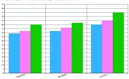
Based on these metrics, we can evaluate graph tracer algorithm, fuzzy c manner clustering and Convolutional neural community set of rules. The assessment outcomes as proven in fig 4.
Fig 4. Comparison Results
ISSN(E): 2277-128X, ISSN(P): 2277-6451, DOI: 10.23956/ijarcsse/V7I7/0114, pp. 318-324
V. CONCLUSION
In this paper, different types of segmentation algorithms are described. The main purpose of this paper to analyze the performance of segmentation analysis in retinal image. Here, Deep neural network, Graph Tracer and Fuzzy C means clustering applied on retinal blood vessel tracking. We find that deep neural network produced the better result in comparison with other segmentation algorithms.
REFERENCES
[1] A. Krizhevsky, I. Sutskever, and G. E. Hinton, “Imagenet classification with deep convolutional neural networks,” in Advances in neural information processing systems, 2012, pp. 1097–1105.
[2] X. Glorot, A. Bordes, and Y. Bengio, “Deep sparse rectifier neural networks,” in International Conference on Artificial Intelligence and Statistics, 2011, pp. 315–323.
[3] F. M. Villalobos-Castaldi, E. M. Felipe-Riverón, and L. P. Sánchez-Fernández, “A fast, efficient and automated method to extract vessels from fundus images,” J. Vis., vol. 13, no. 3, pp. 263–270, Aug. 2010.
[4] G. Azzopardi, N. Strisciuglio, M. Vento, and N. Petkov, “Trainable cosfire filters for vessel delineation with application to retinal images,” Medical image analysis, vol. 19, no. 1, pp. 46–57, 2015.
[5] Y. Zhao, L. Rada, K. Chen, S. P. Harding, and Y. Zheng, “Automated vessel segmentation using infinite perimeter active contour model with hybrid region information with application to retinal images,” Medical Imaging, IEEE Transactions on, vol. 34, no. 9, pp. 1797–1807, 2015.
[6] N. Srivastava, G. Hinton, A. Krizhevsky, I. Sutskever, and R. Salakhutdinov, “Dropout: A simple way to prevent neural networks from overfitting,” The Journal of Machine Learning Research, vol. 15, no. 1, pp. 1929– 1958, 2014.
[7] Y. Bengio, P. Lamblin, D. Popovici, H. Larochelle et al., “Greedy layer-wise training of deep networks,” Advances in neural information processing systems, vol. 19, p. 153, 2007.
[8] M. Fraz, P. Remagnino, A. Hoppe, B. Uyyanonvara, A. Rudnicka, C. Owen, and S. Barman, “Blood vessel segmentation methodologies in retinal images - a survey,” Comput. Methods Prog. Biomed. vol. 108, no. 1, pp. 407–433, Oct. 2012.
[9] J. Staal, M. D. Abràmoff, M. Niemeijer, M. A. Viergever, and B. van Ginneken, “Ridge-based vessel segmentation in color images of the retina,” Medical Imaging, IEEE Transactions on, vol. 23, no. 4, pp. 501– 509, 2004.
[10] A. Hoover, V. Kouznetsova, and M. Goldbaum, “Locating blood vessels in retinal images by piecewise threshold probing of a matched filter response,” Medical Imaging, IEEE Transactions on, vol. 19, no. 3, pp. 203–210, 2000.
[11] Y. Bengio, “Learning deep architectures for ai,” Foundations andtrendsR in Machine Learning, vol. 2, no. 1, pp. 1–127, 2009.
[12] J. Schmidhuber, “Deep learning in neural networks: An overview,” Neural Networks, vol. 61, pp. 85–117, 2015. [13] A. Krizhevsky and G. Hinton, “Learning multiple layers of features from tiny images,” Computer Science
Department, University of Toronto,Tech. Rep, vol. 1, no. 4, p. 7, 2009.
[14] A. Hyvärinen and E. Oja, “Independent component analysis: algorithms and applications,” Neural networks, vol. 13, no. 4, pp. 411–430, 2000.
[15] A. J. Bell and T. J. Sejnowski, “Edges are the" independent components" of natural scenes,” in NIPS, 1996, pp. 831–837.
[16] A. Dosovitskiy, J. T. Springenberg, M. Riedmiller, and T. Brox, “Discriminative unsupervised feature learning with convolutional neural networks,” in Advances in Neural Information Processing Systems, 2014, pp. 766– 774.
[17] X. Jiang and D. Mojon, “Adaptive local thresholding by verification based multithreshold probing with application to vessel detection in retinal images,” Pattern Analysis and Machine Intelligence, IEEE Transactions on, vol. 25, no. 1, pp. 131–137, 2003.
[18] A. Osareh and B. Shadgar, “Automatic blood vessel segmentation in color images of retina,” Iran. J. Sci. Technol. Trans. B: Engineering, vol. 33, no. B2, pp. 191–206, 2009.
[19] K. Simonyan and A. Zisserman, “Very deep convolutional networks for large-scale image recognition,” arXiv preprint arXiv:1409.1556, 2014.