Research Article
a
May
2018
Computer Science and Software Engineering
ISSN: 2277-128X (Volume-8, Issue-5)
Review: Dermatological Skin Disease Detection and
Classification Based on Wavelet Transform and Artificial
Neural Network
Revati Kadu*
PG Student, HVPM COET, Amravati, Maharashtra, India
Dr. U. A. Belorkar
Professor, & Head, Dept. of EXTC, HVPM COET, Amravati, Maharashtra, India
Abstract— One of the most common and augmenting health problems in the world are related to skin. The most unpredictable and one of the most difficult entities to automatically detect and evaluate is the human skin disease because of complexities of texture, tone, presence of hair and other distinctive features. Many cases of skin diseases in the world have triggered a need to develop an effective automated screening method for detection and diagnosis of the area of disease. Therefore the objective of this work is to develop a new technique for automated detection and analysis of the skin disease images based on color and texture information for skin disease screening. In this paper, system is proposed which detects the skin diseases using Wavelet Techniques and Artificial Neural Network. This paper presents a wavelet-based texture analysis method for classification of five types of skin diseases. The method applies tree-structured wavelet transform on different color channels of red, green and blue dermoscopy images, and employs various statistical measures and ratios on wavelet coefficients. In all 99 unique features are extracted from the image. By using Artificial Neural Network, the system successfully detects different types of dermatological skin diseases. It consists of mainly three phases image processing, training phase, detection and classification phase. Keywords— Skin disease, Wavelet Techniques, dermatological, Melanoma color, Artificial Neural Network
I. INTRODUCTION
Dermatology is the branch of medicine that deals with skin, hair and nails in the widest sense. A dermatologist detects dermatological and cosmetic diseases of the skin. Detection of diseases is very important in today’s world scenario because the epidemics of skin diseases cause severe losses to people all over the world. Especially in developing countries there is a need for automated diagnostic system that would reduce manual efforts and time consumption of dermatologists and patients. Melanoma is the most serious form of skin cancer. The major diagnostic and prognostic parameters of melanoma are the vertical thickness, three dimensional size and shape and color of the lesion. The other characteristic features of early melanoma are irregularities in the boundary of the lesion and the appearance of non uniform pigmentation with a variety of color. Melanocytic (pigmented) lesions are formed by groups of specialized cells called melanocytes. Under normal conditions melanocytes live in isolation under the epidermis, the top layer of skin, producing a brown pigment called melanin. Melanin is passed on to keratinocytes that migrate to upper epidermal layers, playing an important role in protecting the skin by absorbing ultraviolet radiation, reducing the incidence of DNA damage. Increasing UV radiation results in an increase in melanin production, darkening the skin tone. Whe n melanocytes group together, brown pigmented lesions may become visible on the skin. Such lesions may be benign or malignant Benign pigmented lesions, known as benign nevi, are typically symmetrical with clean, abrupt edges and may or may not be raised from the surrounding skin. Malignant melanoma is a cancerous condition where melanocytes grow unchecked, producing growths with variations in size, color, density and contour. The incidence of malignant melanoma has increased dramatically over the past few decades. Although a potentially fatal disease, malignant melanoma has a near 100%cure rate if detected and excised early. Early diagnosis is obviously dependent upon patient attention but also accurate assessment by a medical practitioner. Dermatologist will see numerous melanoma patients, knowing their capacity to make accurate diagnoses but general practitioners and other medical professional may not develop such an accurate eye. A computer aided diagnosis system is not meant to replace a trained dermatologist but to assist less trained practitioners. Such a system would be useful for remote medical clinics that do not have the luxury of a trained dermatologist on hand.
Due to the enhancements in skin imaging technology and image processing techniques in the recent years, there
ISSN(E): 2277-128X, ISSN(P): 2277-6451, pp. 1-6
subjectivity and uncertainty from the diagnostic process and provide a reliable second-hand opinion to dermatologists. However, it is widely acknowledged that much higher accuracy is required for computer-based algorithms to be adopted routinely in the diagnostic process [4, 5]. A computer aided diagnosis of melanoma generally comprises four components; image acquisition, border detection, feature extraction, and classification; the latter two are the main focus of this paper.
Feature extraction is used to extract the most important features that accurately characterize a lesion. These features are mainly similar to those visually detected by dermatologists. In computerized melanoma detection systems, feature extraction has been generally based on the conventional clinical algorithm of ABCD-rule of dermoscopy, where asymmetry, border irregularity, color and diameter (or differential structures) of the lesion are examined. Numerous feature extraction methods have been proposed and different image processing techniques have been employed [3] to obtain color, border and texture information of the lesion. It has been shown that using wavelet coefficient along with ABCD features augment the classification accuracy [6]. Texture-based features have been explored in the literature, however, in this paper we propose a complementary feature extraction method which extracts the textural information of the lesion using a 4-level wavelet decomposition on red, green, blue and luminance color channels, and applies a variety of measures and ratios.
Classification is the final step of the diagnosis process, where the extracted features are utilized to ascertain whether the lesion is malignant, benign or suspicious. The most popular classification methods that have been applied to computer-based melanoma recognition include discriminate analysis, artificial neural network, K-nearest neighbourhood, support vector machine and decision trees [3]. In this study we have used and compared various classifiers; namely, support vector machine, random forest, logistic model tree, and hidden naive bayes.
II. LITERATURE SURVEY
The primary purpose of our research is to detect a considerable number of dermatological diseases and indicate the percentage of infection to the fed image. Due to the lack of advances in this field in terms of medicine, there has been a need to conduct a research on the same. Initially, Shamsul et al[7] presented an automated dermatological diagnostic system for treatment of skin anomalies which uses k-means clustering and color gradient technique which was successful in merely detecting six diseases.
D.S. Zingade[2], One of the most common and augmenting health problems in the world are related to skin. The most
unpredictable and one of the most difficult entities to automatically detect and evaluate is the human skin disease because of complexities of texture, tone, presence of hair and other distinctive features.In this paper, we proposed a system which detects the skin diseases using Artificial Neural Network. The system successfully detects different types of dermatological skin diseases. It consists of mainly three phases image processing, training phase, detection phase. In image processing phase we apply algorithms like grey scale conversion, RGB to HSV conversion to the input image. After getting HSV values disease corresponding to the input image gets detected by artificial neural network algorithm. Also, as an addition to the detection the percentage of infection is identified.
Kabari and Bakpo [8] , constructed artificial neural network using feed forward technique and was capable of diagnosing selected skin diseases with a considerable accuracy.
Wasan Kadhim Saa'd [9], a study of the role of color information in detecting the edges of images was conducted. Therefore another color space (HIS) is implemented. Several edge detection techniques are applied such as Laplace and Prewitt, the results shows that the Laplace operator is more efficient than Prewitt operator in edge detection. Wavelet Transform plays an important role in the image processing analysis, especially in texture recognition of data. For its fine result when using Multiresolution modeling. The texture image will be entered to Wavelet Mother Function; this will segment the texture into sub bands. These sub bands contain information about the texture, then this information will be entered to feature extraction, the output from them represent the input to the Artificial Neural Network (ANN) which represents powerful tool for handling problems of large dimension. The idea of combining wavelets and neural networks is proposed to classify images. The output of ANN represents the type of texture
D.N.V.S.L.S. Indira [1],a different method to develop a Texture Analysis based Classification Module to improve the decision strategy and overall accuracy of the system. Asymmetry, Border Irregularity, Color variation, Diameter is the major symptoms which we will use in our processing algorithm. By applying multi-level Wavelet Transformation to the input image and then choosing a group of sub-bands to be restored for best defect detection, the procedure for Skin cancer detection and analysis was developed.
III. PROPOSED WORK
ISSN(E): 2277-128X, ISSN(P): 2277-6451, pp. 1-6
relying on support vector machines or fuzzy logic mechanisms, our detection system uses features extracted from input image of skin through image processing algorithms along with feed forward back propagation neural network for classification and detection purpose. Basic features of image are extracted from the region of interest of skin image. These features are then applied to Back-propagation Neural Network algorithm for training in database or detecting disease from database. The Skin disease detection system consists of two parts where the first part is processing the image of infected region using image processing algorithms. The next part is detecting the disease using back propagation artificial neural network.
The main objective of the system is to classify the images into three categories. The classifier used here is the ANN along with special feature extraction procedure. In the overall procedure, there are two phases of the system. Off-line phase and On-Off-line phase i.e. the database creation phase and the actual classification of image using the above mentioned classifiers.
A. Off line process
All the images in the dataset are of random size, hence they are resize to 256*256.This resized image is then decomposed into R, G and B channel. Wavelet Transform is used to extract the describing features of the image. These features in the form of vectors are stored in the database. For the proposed method, three category each containing 150 images were considered and there feature vectors were stored in the training database. The features of all the images constitute the database for our system.
Fig.1 Flowchart to Create database
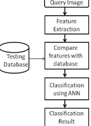
B. Online process
Fig. 2 shows the flowchart for disease classification. The Query image is resized and decomposed into R, G and B channel. The features are extracted using db-4 wavelet transform. The extracted feature of query image is compared with the feature in training database. The K Nearest Neighbor technique classifies the query image into one of the predefined categories by comparing the extracted feature with the feature in the database.
ISSN(E): 2277-128X, ISSN(P): 2277-6451, pp. 1-6
IV. TECHNIQUES USED 1. Wavelet-based texture analysis in dermoscopy image
Wavelets are an extension Fourier analysis. The mathematics of Fourier analysis dates back to the nineteen century but it wasn’t until the mid twentieth century, with the advent of fast algorithms and computers that Fourier analysis began to make an impact on the world. Widely used in signal analysis, hardly a scientific field hasn’t been impacted by this technique. The Fourier transform essentially converts a signal from the time domain to the frequency domain, giving a frequency representation of the signal . Fourier transformation relies on the fact that a signal can be represented by a combination o sine and cosine functions. Coefficients are calculated which determine how to combine sinusoid to produce the signal. Since sinusoids are not of limited duration, they extend from minus to plus infinity, coefficients calculated from these base waves loose all time information. Windowing techniques are often used to focus the transformation on a specific section of time but the accuracy is limited. There exists two wavelet structure;
1. Pyramid-structured wavelet transform which decomposes a signal into a set of frequency channels with narrower bandwidths in lower frequency channels, useful for signals which their important information lies in low frequency Components.
2. Tree-structured wavelet analysis which provides low, middle and high frequency decomposition which is done by decomposing both approximate and detail coefficients. In dermoscopy image analysis, the lower frequency components reveal information about the general properties (shape) of the lesion, which is clinically important, and the higher frequency decomposition provides information about the textural detail and internal patterns of the lesion which is also significant in the diagnosis. Thus the decomposition of all frequency channels are useful in this application. Therefore, the tree-structured wavelet analysis can be more informative for classification of skin lesions.
Wavelet analysis uses the terms approximations and details. Approximations are the high scale low frequency
components while details are the low scale high frequency components. Approximations are what give a signal its identity. The language content of a person’s voice is mostly distinguished by its approximations. Details provide distinct characteristics, such as unique tonal qualities in each individual’s voice Wavelet decomposition can be an iterative process occurring over several levels. The original signal is decomposed into approximations and details with the approximations feeding the next level of decomposition, creating a decomposition tree.
2. Artificial Neural Network Algorithm
An artificial neuron network (ANN) is a computational model. It is based on the structure and functions of biological neural networks. Neural Network facilitates in estimating the most cost-effective and ideal methods for reaching at solutions while defining computing functions or distributions. ANNs have three layers that are interconnected. The first layer consists of input neurons. Those neurons send data on to the second layer, which in turn sends the output to neurons of the third layer. There are different types of Neural Networks such as Feedback, Feed-forward, Back propagation, Classification-Prediction, etc. In our system,Neural Networks are used in the automatic detection of skin diseases by using Back propagation algorithm. Neural network is chosen as a detection tool due to its well known technique as a successful classifier.
Artificial Neural Network Algorithm contains two phases– training and testing phase and detection phase. In training phase these features are trained to the database using back propagation algorithm.The training and testing processes are among the crucial steps in developing an accurate process model using ANNs. The dataset for training and testing processes consists of two parts; the training features set which are used to train the nueral network model. While a testing features sets are used to verify the accuracy of the trained using the BP network. In the training part, connection weights were always updated until they reached the defined iteration number/suitable error. In detection phase the features of input image whose disease is to be recognized are compared with the features in database using Artificial Neural Network.
3. Image Feature Extraction
The accuracy of the system behaviour is majorly depends on the features used for training of classifier and the classifier structure itself. In all, 99 features are extracted from the image. The complete procedure for feature extraction is as shown in Fig.3
V. CONCLUSION
ISSN(E): 2277-128X, ISSN(P): 2277-6451, pp. 1-6
color information color moments are used. The three RGB bands of a color image in RGB model are used to extract the describing features. . The method applies tree-structured wavelet transform on different color channels of red, green, blue and luminance of dermoscopy images, and employs various statistical measures and ratios on wavelet coefficients. Feature extraction and a two-stage feature selection method, based on entropy and correlation. By using Artificial Neural Network, the system successfully detects different types of dermatological skin diseases which consists of mainly three phases image processing, training phase, detection phase. In image processing phase we apply algorithms like grey scale conversion, RGB to HSV conversion to the input image. After getting HSV values disease corresponding to the input image gets detected by artificial neural network algorithm.
Fig.3 Proposed technique for Feature extraction
REFERENCES
[1] D.N.V.S.L.S. Indira # , JYOTSNA SUPRIYA P , Detection & Analysis of Skin Cancer using Wavelet
Techniques, International Journal of Computer Science and Information Technologies, Vol. 2 (5) , 2011, 1927-1932
[2] D.S. Zingade, Manali Joshi,,Skin Disease Detection using Artificial Neural Network ,International Journal of Advance Engineering and Research Development Special Issue on Recent Trends in Data Engineering Volume 4, Special Issue 5, Dec.-2017 @IJAERD-2017,
[3] D.S. Zingade1, Manali Joshi2, Viraj Sapre3, Rohan Giri4 Maglogiannis and C. Doukas. Overview of advanced
computer vision systems for skin lesions characterization.IEEE Trans. on Information Technology in Biomedicine,
[4] 13(5):721–733, 2009American Academy of Dermatology, 56(3):417–421, 2007R. E. Sorace, V. S. Reinhardt, and S. A. Vaughn, “High-speed digital-to-RF converter,” U.S. Patent 5 668 842, Sept. 16, 1997.
[5] A. Perrinaud, O. Gaide, L. French, J. Saurat, A. Marghoob,and R. Braun. Can automated dermoscopy image analysisinstruments provide added benefit for the dermatologist? Astudy comparing the results of three systems.
British Journalof Dermatology, 157:926–933, 2007
[6] M. Elbaum, A. Kopf, H. Rabinovitz, R. Langley, andH. Kamino. Automatic differentiation of melanoma from
melanocytic nevi with multispectral digital dermoscopy: Afeasibility study. Journal of the American Academy of Dermatology.
[7] M. Shamsul Ari n, M. Golam Kibria, Adnan Firoze, M. Ashraful Amin, Hong
ISSN(E): 2277-128X, ISSN(P): 2277-6451, pp. 1-6
[8] L. G. Kabari and F. S. Bakpo, Member, IEEE, "Diagnosing Skin Diseases Using an Artificial Neural Network", 2016IEEE:
[9] Wasan Kadhim Saa'd, Method For Detection And Diagnosis Of The Area Of Skin Disease Based On Color
By Wavelet Transform And Artificial Neural Network, Al-Qadisiya Journal For Engineering Sciences [10] Girish Patil , Karan Belsare, An Approach to Natural Image Classification based on Wavelet Transform and
KNN, International Journal of Emerging Research in Management &Technology ISSN: 2278-9359 (Volume-4,