1
n
m
Hydrated metal ion Si tetrahedra Al octahedra Si tetrahedra
d
o
o
1
s
p
a
ci
n
g
H O
H H
O H
H O H
H O
H H
O H
H O H
SYNTHESIS AND CHARACTERIZATION OF
BIODEGRADABLE CLAY- POLYMER NANOCOMPOSITES
FOR ORAL SUSTAINED RELEASE OF
ANTI-INFLAMMATORY DRUG
Manpreet Kaur
[a]and Monika Datta
[b]*Keywords: diclofenac sodium, montmorillonite, PLGA nanocomposite, sustained release
In this paper, a clay based drug delivery system comprising of [poly(D,L-lactide-co-glycolide)] (PLGA)/montmorillonite (MMT) nanocomposite has been explored for the oral sustained release of diclofenac sodium (DS) using double emulsion solvent evaporation technique. Encapsulation of diclofenac sodium in PLGA matrix and clay matrix was also undertaken to assess the role of clay and PLGA in drug encapsulation and its subsequent release from the respective formulations. A drug encapsulation efficiency of 98 % was obtained for the synthesized PLGA/MMT nanocomposite system. The drug was found to be intercalated in the PLGA/MMT nanocomposite as confirmed by the X-Ray diffraction studies. The thermal analysis shows the crystalline nature of the encapsulated drug in the nanocomposite. The particle size of the drug loaded PLGA/MMT nanocomposite was found to be in the range of 10-20 nm as analysed by high resolution transmission electron microscopic (HRTEM) technique. The in vitro drug release studies of the drug encapsulated PLGA/MMT nanocomposite under simulated gastric fluid (phosphate buffer saline, PBS 1.2) shows no drug release while a sustained release was seen under simulated intestinal condition (phosphate buffer saline, PBS 7.4) releasing 51% drug in 8 hours.
*Corresponding Author
E-Mail: [email protected]
[a] Department of Chemistry, University of Delhi, Delhi-110007, India. E-mail:[email protected]
[b] Department of Chemistry, University of Delhi, Delhi-110007, India
INTRODUCTION
Polymer/clay nanocomposites are a class of hybrid systems in which clay/organo-clay nanoparticles (often montmorillonite) are dispersed in a polymer matrix. With small amount of the clay, they exhibit high thermal stability, enhanced mechanical1-6 and rheological properties etc.7
These benefits along with the good intercalation capacity offered by the clay mineral have been used to develop new sustained release systems, as documented by a number of patents.8-10 A number of polymers have been investigated
for designing the nanoparticulate delivery systems, but PLGA [copolymer of poly (lactide), (PLA) and poly (glycolide), (PGA)] has been most extensively used because of its biocompatibility, biodegradability, and versatile degradation kinetics. It is an FDA approved biodegradable and biocompatible polymer which has been in use for years.11-13 Also, its final degradation products (lactic and
glycolic acids) are completely safe, as they are either excreted by the kidneys or enter the Krebs’ cycle to be eventually eliminated as carbon dioxide and water.
Of all the clays, montmorillonite (MMT) has been extensively used in the pharmaceutical formulations. It is a FDA approved excipient and belongs to the 2:1 smectite group (general formula M+
x+y(Al2-x)(OH)2(Si4-yAly)O10) of
clay minerals. It is a naturally occurring layered silicate
having a unit thickness of 1 nm which makes it suitable for the synthesis of nano structured materials. It has rich interlayer chemistry, possesses high potential for ion exchange, is stable under acidic conditions and has high
chemical resistance. It is associated with good hydration and swelling properties and acts as a potent detoxifier.14
Structure of montmorillonite
Besides it has large specific surface area and exhibits standout mucoadhesive ability to cross the GI barrier. As a result, MMT is a common ingredient both as an excipient and active substance in pharmaceutical products.15,16
Diclofenac is currently the eighth largest-selling drug and the most frequently used nonsteroidal anti-inflammatory drug (NSAID) in the world, since its introduction in Japan in 1974. Diclofenac is among the better tolerated NSAIDs and is widely used in the long term treatment of degenerative diseases such as rheumatoid arthritis, osteoarthritis. Diclofenac sodium (DS) has analgesic and antipyretic actions and is one of the few NSAIDs used to treat ocular inflammatory conditions. But it has short biological half life of 1-2 hours17 therefore requires frequent dosing leads to
adverse gastrointestinal disturbances, peptic ulceration and gastrointestinal bleeding. The most common route of administration of this drug is oral.18,19 Thus in order to
O C
O
CH2 O CH
CH3
C O
m n
H OH
compliance, development of oral sustained-release formulations of this drug is highly desirable. To minimize the side effects, particularly to avoid gastric ulcers, diclofenac sodium is marketed as enteric coated and sustained release tablets. But even these formulations have shown GI toxicity in clinical studies.20
In this work, we propose novel formulation- diclofenac sodium encapsulated biodegradable PLGA/MMT nanocomposites, synthesized for the oral sustained delivery of diclofenac sodium.
Chemical structure of diclofenac sodium
The double emulsion solvent evaporation technique was used to synthesize these nanocomposites. Poloxamer 188 [polyoxyethylene–polyoxy-propylene–polyoxyethylene (PEO–PPO–PEO) block polymer], a non-ionic stabilizing surfactant, suitable for oral administration and widely used as wetting and solubilising agent was employed in the synthesis.
Chemical structure of poly(lactic-co-glycolic acid) (PLGA),
m=poly(glycolic acid) (PGA), n=poly(lactic acid) (PLA)
For comparative study, the drug loaded samples were synthesized in the absence of MMT and PLGA to investigate the effect of the two on the encapsulation efficiency and subsequent release kinetics of the drug under simulated intestinal conditions (PBS 7.4).
MATERIALS AND METHODS
Materials
The PLGA used in the present study was obtained from Sigma Aldrich, St. Louis MO USA and was used without any further purification. Poloxamer 188 (F68) having more than 98% purity was obtained from Fluka, Switzerland. The clay used in the present study, montmorillonite was obtained from Sigma Aldrich, St. Louis USA. The drugs - diclofenac sodium (DS) and diclofenac acid (DH) were obtained from Sigma Aldrich, St. Louis MO USA and were used without any further purification.
HPLC grade ethyl acetate having 99.8% essay was obtained from Research laboratories Ltd., Mumbai. HPLC grade dichloromethane (DCM) and methanol having 99.5% and 99.7% assay, respectively, were obtained from Merck,
Mumbai. NaOH and KH2PO4 used for making PBS 7.4
buffer were obtained from Merck, Mumbai.
Encapsulation of diclofenac sodium in PLGA/MMT composite
The double emulsion (w/o/w) solvent evaporation method was employed for the encapsulation of diclofenac sodium in PLGA/MMT nanocomposite. The multiple emulsions were prepared by using two step emulsification procedures. In the first step, the primary (w/o) emulsion was prepared by incorporating the aqueous drug (20 mg) phase into the oily phase containing PLGA (40 mg) dissolved in ethyl acetate and the resulting emulsion was sonicated for a minute at 25 °C using ultrasonics sonicator.
In the second step, the primary emulsion was added slowly into the aqueous phase containing a given amount of MMT and F-68 (at critical micelle concentration) to encapsulate the (w/o) emulsion. The resulting double emulsion, (w/o/w) was stirred at 700 rpm and the organic solvent was evaporated at 37 °C leaving behind diclofenac sodium encapsulated PLGA-MMT composite. The contents were then centrifuged at 20,000 rpm for 30 minutes at 20 °C using Sartorius 3K30 centrifuge. The filtrate was preserved and the residue was lyopholized for further characterizations (Scheme 1). A number of formulations were tried by varying different parameters before arriving at the optimized formulation designated as 002.
Scheme1. Schematic representation of synthesis of drug encapsulated clay polymer nanocomposite by w/o/w double emulsion solvent evaporation method
To investigate the effect of MMT on the drug encapsulation and its subsequent release from the synthesised formulation, diclofenac sodium was encapsulated in PLGA matrix using w/o/w double emulsion solvent evaporation technique. All the parameters and processing conditions were kept the same as in optimized formulation - 002 except for MMT, which was not added. The sample thus synthesised was designated as 010.
To see the effect of PLGA on the encapsulation of diclofenac sodium in the clay, the aqueous drug was added slowly under the same conditions to the aqueous suspension of MMT containing F-68. All the parameters and processing conditions were kept the same as in optimized formulation - 002 except that PLGA was not used. The sample thus synthesised was designated as 012.
N H
Na
O
O Cl
Quantitative estimation of diclofenac sodium in the synthesized formulations using high performance liquid chromatographic (HPLC) method
Method development and analysis of formulations
The amount of diclofenac sodium encapsulated in the synthesized formulations was determined by HPLC technique using a reversed phase C18 column (250 × 460 mm, Phenomex, USA) and UV-VIS photo-diode array. The samples were separated using an isocratic mobile phase consisting of CH3OH: PBS 6.8 (13:7 v/v) at a flow rate of
1mL min-1.
The drug was extracted from the PLGA-MMT composite system and PLGA matrix using a mixture of dichloromethane (DCM) and PBS 7.4 in 1:1 ratio. The aqueous phase containing the drug was quantitatively analysed by HPLC using a calibration curve and the % drug encapsulation efficiency (% EE) and % drug content (% DC) were determined using equations (1) and (2).
where
m1 – mass of the drug loaded in sample
m2 – mass of the total drug added
m3 – mass of the drug loaded sample
Characterizations of the optimal formulations
X-Ray diffraction (XRD) studies
The X-Ray diffraction (XRD) patterns of MMT, diclofenac sodium, F-68 and the synthesized formulations- 002, 010 and 012 were recorded using Cu K radiation (n
1 Ǻ) on a Philips X′ Pert-PRO MRD system operating at 50 kV and 100 mA in continuous scan mode with a scanning speed of 0.008°sec-1.
Differential scanning calorimetric (DSC) analysis
The differential scanning calorimetric (DSC) analyses of MMT, F-68, pristine diclofenac sodium, diclofenac acid and synthesized formulations- 002, 010 and 012 was performed using Perkin Elmer Q200 (V23.10 build 79) system operating at a heating rate of 20 °C min-1. The samples were
purged with nitrogen at a flow rate of 50.0 ml min-1.
Fourier Transformed Infrared (FT-IR) studies
The IR transmission spectra (4000400 cm1) were
recorded in a KBr matrix at 25 °C using a Perkin-Elmer FT-IR spectrum BX.
Scanning electron microscopic studies (SEM) with EDAX (energy dispersive X-ray analysis)
All the samples were sputter coated with gold (used as a conductive material) and the surface morphology of the samples was then examined using Zeiss EVO MA15 (Oxford instruments).
High resolution transmission electron microscopic (HRTEM) studies
The samples were prepared by depositing the aqueous suspensions of the samples on carbon film attached to a 400 mesh Cu grid. The images were recorded using TECNAI G2T30 FEI Instrument operated at 120 kV.
In vitro drug release kinetics
The study of in vitro drug release behaviour of pure diclofenac sodium and the synthesized formulations was carried out in simulated gastric (PBS 1.2) and intestinal fluid (PBS 7.4) using dialysis bag method for a period of 8 hours. A known amount of the formulation was dispersed in the dialysis bag and was immersed in 100 ml of dissolution media maintained at 37° ± 0.5°C with constant stirring at 300 rpm. After every one hour interval, 5.0 ml of the dissolution medium was withdrawn for the estimation of the drug content and at the same time 5 mL of the fresh solution was added to maintain the constant volume of the dissolution medium.21 The obtained 5 mL of the solution
was filtered through a membrane with a pore diameter of 0.45µm.The concentration of the drug released was determined by UV spectrophotometer at 277 nm, and then the cumulative percentage of DS released was calculated.
RESULTS AND DISCUSSION
Quantitative estimation of drug in the synthesized formulations by HPLC method
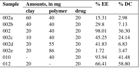
The % encapsulation efficiency and drug content of synthesised formulations as estimated using HPLC are tabulated in Table 1.
Table 1. Encapsulation efficiency and drug content in synthesised formulations
Sample Amounts, in mg % EE % DC
clay polymer drug
002a 60 40 20 15.31 2.98
002b 40 40 20 29.8 7.13
002 20 40 20 98.01 36.30
002c 10 40 20 45.25 24.14
002d 20 55 20 41.83 6.83
002e 20 86 20 1.72 3.47
010 - 40 20 93.94 41.48
012 20 - 20 66.41 58.80
%EE= % encapsulation efficiency; %DC= % drug content
Keeping the amount of clay and the drug content constant, the effect of the polymer concentration on encapsulation efficiency was investigated. As can be seen (table1), the %
1
2
1
3
100
100 m % EE
m
m % DC
m
(1)
10 20 30 40
In
te
n
s
it
y
c
o
u
n
ts
2 Theta
DS MMT F-68 012
10 20 30 40
In
te
n
s
it
y
c
o
u
n
ts
2 Theta MMT
DS F-68 002
encapsulation efficiency decreases from 98.01% to 1.72% with increase in the concentration of the polymer. To study the effect of the clay amount on % encapsulation efficiency, the amount of the clay was decreased from 60 mg to 10 mg, keeping the amount of the drug and the polymer constant. The encapsulation efficiency increased from 15.31% to 98.01% with decrease in the clay content upto 20mg, a further decrease in the clay content to 10 mg resulted in a decrease in the % encapsulation efficiency and drug content.
In case of 010 formulation (synthesized under the same set of experimental conditions and with same amount of the excipients as in 002 but minus MMT), an encapsulation efficiency of 93.94% was obtained. An encapsulation efficiency of 66.41% was achieved in case of 012 formulation (synthesized under the same set of experimental conditions and with same amount of the excipients as in 002 but minus PLGA).
Altogether, these results suggest that the highest encapsulation efficiency was achievable under the present experimental conditions with 002 formulation.
Characterizations of the optimal formulations
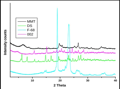
X-Ray diffraction (XRD) studies
The XRD pattern of 002 and 012 formulations show a shift in the peak in the lower angle region in the 001 plane from 2θ= 6.8° in the pristine MMT to 4.4° and 4.7° in 002 and 012 respectively, resulting in an increase in the corresponding d spacing from 13.4 Å to 20 Å and 18.6 Å respectively (Fig.1a and b). This increase in d spacing suggests that the drug has been intercalated within the clay interlayers21. Moreover, the diffractogram of 002 and 012
shows characteristic peaks of the drug22 which indicates that
the drug is present in crystalline state in the synthesized formulations.
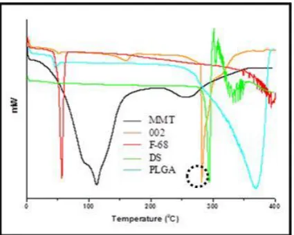
Differential scanning calorimetric (DSC) analysis
To investigate the physical state of diclofenac sodium in the synthesized formulations (002, 010 and 012) and to confirm the interaction of the drug with the excipients, DSC studies were performed. In the DSC curve of pure drug, the melting endothermic peak appears at 292 °C. This is followed by complex endothermic – exothermic phenomenon indicating decomposition of the drug.23
The small broad endotherm at 65 °C in the DSC trace of 012 formulation (Fig.2a) corresponds to the loss of surfaced adsorbed water followed by another sharp endothermic peak at 176 °C corresponding to the melting endothermic peak of diclofenac acid which indicates the conversion of diclofenac sodium into diclofenac acid in the clay matrix.24 The broad
endothermic peak at 303 °C (encircled) which is absent in pristine MMT and F-68 corresponds to the melting of the drug thus suggesting its presence in the crystalline state in the synthesized formulation-012.25 However, the
temperature at which this peak appears (303 °C) is lower than the melting endothermic peak of pure diclofenac acid (323 °C). This suggests that there may certain interaction between the drug and the excipients.
Figure 1a. XRD pattern of formulation 012, MMT, DS and F-68
Figure 1b. XRD pattern of formulation 002, MMT, DS and F-68
In case of 010 formulation (Fig.2b), the small endotherm at 50 °C corresponds to the glass transition temperature of PLGA.26 The decomposition temperature of PLGA and the
melting endotherm of the pure drug fall in the similar temperature range, therefore it is difficult to observe a clear well defined melting endothermic peak corresponding to the drug in this formulation. However a small endotherm (encircled) can be seen in the DSC curve of 010 formulation at 308 °C which is absent in the DSC curve of pure PLGA and F-68 which could be due to the melting endotherm peak of the diclofenac sodium. A big endotherm peak at 361 °C corresponds to the decomposition of PLGA.27
In the DSC curve of 002 formulation (Fig. 2c), the very small endotherm at 50 °C correspond to the glass transition temperature of PLGA. This is followed by another small endotherm c.a 158 °C which could be due to the loss of interlayer water in MMT.28 The sharp endothermic peak at
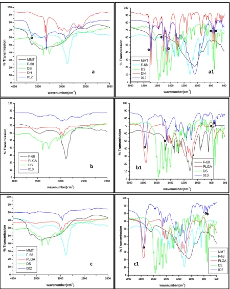
Fourier-transformed infrared (FT-IR) spectroscopic studies
The FT-IR spectral studies were carried out to confirm the presence of drug and to investigate the possible interaction between the drug and the excipients. In the 012 synthesized formulation (Fig.3a and 3a1), the band at 3622 cm-1
corresponds to the O-H stretching vibrations from the
structural water in the clay. The presence of characteristic vibrational band at 1044 cm-1 corresponding to Si-O
stretching indicates the presence of clay matrix.29 The
vibrational band at 3324 cm-1 corresponds to the N-H
stretching while the intense band at 1694 cm-1 has been
assigned to C=O stretching from the –COOH group in the drug.24 The presence of these two bands (at 3324 cm-1 and
1694 cm-1 ) suggests the conversion of diclofenac sodium
into diclofenac acid30,31 as is evident from the DSC results of
this formulation. The vibrational bands at 1508 cm-1
corresponds C-N-H in plane bending respectively from the drug molecule. The vibrational band at 1454 cm-1 has been
assigned to -CH2 bending from disubstituted benzene ring
from the drug molecule. The bands at 766 cm-1 and 742 cm-1
correspond to out of plane C-H ring bending and in plane ring deformation respectively in the drug.32,33
In the 010 formulation (Fig.3b and 3b1), the broad vibrational band at 3449 cm-1 corresponds to the O-H
stretching vibration from F-68.34 The weak vibrational band
at 2995 cm-1 corresponds to –CH, -CH
2 stretching while the
one at 1761 cm-1 corresponds to –C=O stretching from
PLGA.35 The vibrational band at 1454 cm-1 corresponds to
-CH2 bending from disubstituted benzene ring from the drug
molecule while the bands at 766 cm-1 and 742 cm-1
correspond to out of plane C-H ring bending and in plane ring deformation respectively from the drug molecule.
In the 002 formulation (Fig.3c and c1), the vibrational band at 1760 cm-1 corresponds to the –C=O stretching in
PLGA. The bands at 766 cm-1 and 742 cm-1 correspond to
out of plane C-H ring bending and in plane ring deformation respectively from the drug molecule. The shifting of the band from 3622 cm-1 in pristine MMT corresponding to the
O-H stretching vibrations from the structural water [(Al, Mg-) OH] in the clay to 3651 cm-1 in 002 sample suggests
the interaction of the structural hydroxyl groups in MMT with the polar groups of F-68.
The presence of characteristic absorption bands of the drug in the synthesized formulations confirms presence of the drug in these synthesized formulations. No significant shifting of the absorption bands of drug was observed in the synthesized formulations suggesting that there is no strong chemical interaction between the drug and the excipients. The characteristic absorption band at 1760 cm-1 in PLGA
corresponding to the C=O vibrational stretching was not shifted in 002 and 010 formulations suggesting that there is no strong chemical interaction of PLGA either with the encapsulated drug, F-68 or MMT.
Scanning electron microscopic studies (SEM) with EDAX (energy dispersive X-ray analysis)
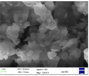
The morphology of 012 formulation seems to have become agglomerated as compared to pristine MMT (Fig. 4). The agglomeration may have taken place because of the presence of small amount of non- intercalated drug (free drug) and /or F-68on the clay surface. The EDAX data (S1, Supplementary material) shows the presence of drug as indicated by chlorine content.
The 010 formulation has rod like structures as clearly seen in the SEM images. The surface morphology of 002 formulation is different from that of pristine montmorillonite.
Figure 2a. DSC curves of formulation 012, MMT, DS, DH and F-68
Figure 2b. DSC curves of formulation 010, PLGA, DS and F-68
Figure 3. FTIR spectra a) 012 in 4000-2000 cm-1range, a1) 012 in 2000-600 cm-1range; b) 010 in 4000-2000 cm-1range, b1) 010 in
2000-600 cm-1range; c) 002 in 4000-2000 cm-1range, c1) 002 in 2000-600 cm-1 range along with MMT, DS, DH, F-68
The structure seems to have become more porous. The EDAX data of 002 formulation shows the presence of drug as suggested by the presence of chlorine. It is clear from the EDAX data that the % composition by weight of this formulation shows highest chlorine content as compared to 012 and 010 formulations. The EDAX figures can be seen in the supplementary material (S1). This is supported by
the fact that this formulation shows highest encapsulation efficiency of all the three formulations. This shows that it is clay which plays an important role in the entrapment of the drug in the PLGA/MMT composite as the composition of the 010 formulation is just the same but without clay and this formulation shows comparatively lower encapsulation efficiency.
2000 1800 1600 1400 1200 1000 800 600 0 10 20 30 40 50 60 70 80 90 100 % T ra n s m is s io n
wavenumber(cm-1)
MMT F-68 PLGA DS 002
4000 3500 3000 2500 2000
0 10 20 30 40 50 60 70 80 90 100 % T ra n s m is s io n
wavenumber(cm-1)
MMT F-68 PLGA DS 002
4000 3500 3000 2500 2000
0 10 20 30 40 50 60 70 80 90 100 % T ra n s m is s io n
wavenumber(cm-1) F-68
PLGA DS 010
a
a1
11
b
b1
11
c
c1
11
4000 3500 3000 2500 2000
0 10 20 30 40 50 60 70 80 90 100 % T ra n s m is s io n
wavenumber(cm-1) MMT
F-68 DS DH 012
2000 1800 1600 1400 1200 1000 800 600
0 10 20 30 40 50 60 70 80 90 100 % T ra n s m is s io n
wavenumber(cm-1) MMT
F-68 DS DH 012
2000 1800 1600 1400 1200 1000 800 600 0 10 20 30 40 50 60 70 80 90 100 % T ra n s m is s io n
wavenumber(cm-1)
Figure 4. SEM images of MMT and drug encapsulated formulations-012, 010 and 002
Figure 5. TEM images of drug encapsulated formulations-012, 010 and 002
High resolution transmission electron microscopic (HRTEM) studies
The particle size of the encapsulated drug in the 012 formulation was found to be in the range of 5 nm to 10 nm from the HRTEM image (Fig.5. image 012a). The well defined layered structure of clay observed in this case (image 012b) excludes the possibility of exfoliation and supports the XRD results which suggest intercalation. The HRTEM images of 010 formulation show rod like structures (also seen in the scanning electron micrographs) with a thickness of around 15 nm. In the HRTEM image of 002
formulation, the drug encapsulated PLGA particles can be seen entrapped in the clay layers (small circled) with a particle size in the range 10 nm to 20 nm. The characteristic Moiré fringes can be seen in the 002 micrograph (big circled) indicating misaligned stacking of the nanoplatelets in MMT. The misaligned stacking of the nanoplatelets for 002 formulation was believed to associated with their lamellar structure.36 The interplanar distance of 25 Å in the
Moiré fringes confirms the intercalation of the PLGA encapsulated drug in the clay in accordance with the XRD results, however the d spacing value obtained is slightly on the higher side as compared to the corresponding XRD results. Taking into account the d spacing of MMT (1.34 nm), PLGA/MMT nanocomposite (2.0 nm) and average particle size of the PLGA encapsulated drug particles (15 nm) in PLGA/MMT nanocomposite, it can be concluded that the entrapped drug particles are arranged at an angle and not in an absolute anti conformation in the clay interlayer in the synthesized PLGA/MMT nanocomposite.
Figure 6a. In vitro cumulative release profiles of pure DS and drug encapsulated formulations- 012, 010 and 002 in PBS 1.2.
Figure 6b. In vitro cumulative release profiles of pure DS and drug encapsulated formulations- 012, 010 and 002 in PBS 7.4.
In vitro drug release kinetics
From (Fig.6a), it is evident that no drug release takes place from any of the three formulations synthesized, in simulated gastric fluid (PBS 1.2). However, 4 % release was seen in the second hour and a total of 7 % release was observed in 8 hours in case of pure drug. It was observed that 60% of the pure diclofenac sodium was released in the first hour in simulated intestinal fluid (PBS 7.4) which rose
012 a
012 b
002 010
0 1 2 3 4 5 6 7 8
0 20 40 60 80 100
%
C
u
m
u
la
ti
v
e
r
e
le
a
s
e
Time(hours)
DS 012 010 002
0 1 2 3 4 5 6 7 8
0 20 40 60 80 100
%
C
u
m
u
la
ti
v
e
r
e
le
a
s
e
Time(hours)
to 80% in the second hour after which no drug release was seen (Fig. 6b). Whereas in the case of 012 formulation, 25 % of the drug was released in the first hour after which slow release was found to occur releasing up to 51 % of the drug by 5th hour after which no drug release was found to
occur. In the case of 010 formulation, 24.9 % of the drug was released in the first hour releasing up to 59.42 % of the drug up to 6th hour. After 6th hour no drug release was found
to take place. In the 002 formulation, 7 % of the drug was released in the first hour exhibiting sustained release afterwards, releasing 51 % of the drug by the 8th hour.
A sustained release was seen in all the formulations synthesized as compared to the pure drug. The 010 formulation exhibited more sustained release than 012 formulation and also a higher amount of the drug release was seen as compared to rest of the formulations synthesized. However, out of all, the most sustained release behaviour was seen in 002 formulation, with no burst release implying that the outer covering of the clay plays an important role in the sustained release of the drug encapsulated in the polymer.
Infact the drug encapsulated PLGA/MMT nanocomposite shows more sustained release as compared to the commercial sustained release Voltaren tablet which is known to release ~85 % of the drug by the 8th hour.37 The
almost no release of DS in pH 1.2 PBS and a sustained release in pH 7.4 PBS makes the PLGA/MMT nanocomposite an excellent pH-sensitive matrix for sustained drug release, which could be used for the fabrication of pharmaceutical formulation for oral intake.
Surface morphology of 002 formulation after drug release
There is a clear change in the morphology of 002 formulation after drug release (Fig.7). The SEM image of the 002 formulation show swelled clay particles with perforations (encircled region in the image) indicating the release of the encapsulated drug from the clay interlayers.
Figure 7. SEM image of 002 formulation after drug release
Mechanism of drug encapsulation in PLGA/ montmorillonite nanocomposite
The poloxamer 188 is supposed to form micelles in the clay interlayers (as critical micellar concentration of it has been used) (Fig. 8a) and intercalates through polar interactions with the structural hydroxyl groups in the clay.
Figure 8. a) Micellization of poloxamer 188, b) Encapsulation of drug encapsulated PLGA moeity in clay interlayers
This is evident from the FTIR spectral results of drug encapsulated PLGA/montmorillonite nanocomposite sample (002 formulation) wherein a shift in the vibrational band corresponding to the structural hydroxyl group in MMT was seen towards higher wavenumber from 3622 cm-1 in pristine
MMT to 3651 cm-1in the drug encapsulated
PLGA/montmorillonite nanocomposite. The PLGA encapsulate the drug through interaction of its polar groups with the polar groups of the drug. This drug encapsulated PLGA moiety then intercalates into the clay layers by encapsulation into the hydrophobic cavity of poloxamer 188 micelles through hydrphobic interactions with non polar groups of poloxamer 188 (Fig.8b).
CONCLUSION
Diclofenac sodium loaded PLGA/MMT nanocomposite was prepared by double emulsion solvent evaporation process. The XRD results confirmed the intercalation of the drug encapsulated PLGA moiety in the clay interlayers. The DSC results indicate the presence of the crystalline form of drug in PLGA/MMT nanocomposite. The FTIR results suggest that there is no strong chemical interaction between the drug and the excipients. The particle size of the encapsulated drug in the nanocomposite was found to be in the range 10 nm to 20 nm. The presence of MMT resulted in high drug encapsulation efficiency (98.01 %) as confirmed by the comparative studies performed to evaluate the role of clay. However, the presence of PLGA is also necessary as in its absence the drug encapsulation efficiency drastically decreases (66.41 %, formulation 012). No drug release was found to occur in simulated gastric fluid (PBS 1.2) in any of the formulations synthesized, indicating the stability of the formulations under strong acidic conditions prevailing in the stomach. The drug loaded PLGA/MMT nanocomposite (002 formulation) demonstrated the most sustained release behaviour in simulated intestinal conditions (PBS 7.4) as compared to the other formulations, releasing 51 % of the drug in the 8th hour. No burst release was seen during the
initial hour implying that the clay plays an important role in the sustained release of the drug encapsulated in the polymer as evident from the comparative in vitro release studies results (formulation 010).
Hydrophobic block
Hydrophilic block
(a)
(b)
Clay layer
Infact the drug loaded PLGA/MMT nanocomposite is found to show more sustained release in comparison to the commercial Voltaren tablet in simulated intestinal fluid (PBS 7.4). Thus on the basis of the present results, it can be concluded that PLGA/MMT nanocomposite may function as a suitable drug delivery vehicle for oral sustained release of diclofenac sodium.
ACKNOWLEDGEMENT
The authors are grateful to the Head, Department of Chemistry, Department of Geology and the Director of the University Science Instrumentation Centre of the University of Delhi for providing the Instrumentation facilities. We are also thankful for the financial assistance received from the University of Delhi in the form of annual research grant for carrying out the research work.
REFERENCES
1Chang, J. H., Ana, Y. U., Choa, D., Giannelis, E. P., Polymer.,
2003,44, 3715–3720.
2Cypes, S. H. W., Saltzman, M., Giannelis, E. P., J. Control.
Release.,2003, 90, 163–169.
3Lee, W. F., Fu, Y. T., J. Appl. Polym. Sci.,2003,89, 3652–3660.
4Kiersnowski, A., Pigłowski, J., Eur. Polym. J.,2004, 40, 1199–
1207.
5Lee, W. F., Chen, Y. C., J. Appl. Polym. Sci.,2004, 91, 2934–
2941.
6Puttipipatkhachorn, S., Pongjanyakul, T., Priprem, A., Int. J.
Pharm.,2005, 293, 51–62.
7Pongjanyakul, T., Priprem, A., Puttipipatkhachorn, S., J. Pharm.
Pharmacol., 2005a, 57, 429–434.
8Greenblatt, G. D., Hughes, L. D., Whitman, W., Patent number
EP1470823, 2004.
9Nagasaki, Y., Takahashi, T., Kataoka, K., Yamada, Y., PCT Int.
Appl., WO 2005005548, 2005.
10Zhong, S., U.S. Pat. Appl. Publ. US 2005181015, 2005.
11Jiang, W. L., Gupta, R. K., Deshpande, M. C., Schwendeman, S.
P., Adv. Drug Deliv. Rev., 2005, 57, 391–410.
12Zweers Miechel, L. T., Engbers Gerard, H. M., Grijpma Dirk, W.,
Jan, F. J., J. Control. Release,2004, 100, 347–356.
13Jain, R.A., Biomaterials,2000, 21, 2475–2490.
14Aguzzi, P.C., Viseras, C., Caramella, C., Appl. Clay Sci., 2007,
36, 22.
15Dong, Y., Feng, S. S., Biomaterials, 2005, 26, 6068.
16Takahashi, T., Yamada, Y., Kataoka, K., Nagasaki, Y., J. Control.
Release,2005, 107, 408.
17Saravanan, M., Bhaskar, K., Maharajan, G., Pillai, K. S., Int. J.
Pharm.,2004, 283, 71–82.
18Arias, J. L., López-Viota, M., Sáez-Fernández, E., Ruiz, M. A.,
Colloids Surf.,2010, 75, 204–208.
19Bertocchi, P., Antoniella, E., Valvo, L., Alimontia, S., Memoli,
A., J. Pharm. Biomed. Anal., 2005, 37, 679–685.
20Davies, N. M., J. Pharm. Pharm. Sci.,1999, 2, 5–14.
21Joshi , G. V., Kevadiya, B. D., Patel , H. A., Bajaj, H. C., Jasra,
R. V., Int. J. Pharm.,2009, 374, 53–57.
22Kenawi, I. M., Barsoum, B. N., Youssef, M. A., J. Pharm.
Biomed. Anal.,2005, 37, 655-661.
23Tudja, P., Khan, M. Z. I., Mestrovic, E., Horvat, M.,Golja, P.,
Chem. Pharm. Bull., 2001, 49, 1245-1250.
24Durairaj, C., Kim, S. J., Edelhauser, H. F., Shah, J. C., Kompella,
U. B., Invest. Ophthalmol. Vis. Sci.,2009, 50, 4887-4889.
25Dixit, M., Kulkarni, P. K., Panner, S., Int. Res. J. Pharm.,2011, 2,
207-210.
26Mukerjee, A., Vishwanatha, J. K., Anticancer Res., 2009, 29,
3867-3876.
27Mainardes, R. M., Gremião, M. P. D., Evangelista, R. C., Brazil.
J. Pharm. Sci.,2006, 42, 528.
28Del Hoyo, C., Rives, V., Vicente, M. A., Thermochim. Acta,
1996, 286, 89-103.
29Davarcioglu, B., Ciftci, E., Int. J. Nat. Eng. Sci., 2009, 3, 154-161.
30Jubert, A., Massa, N. E., Tevez, L. L., Okulik, N. B., Vibrat.
Spectros.,2005, 37, 161–178.
31Beck, R. C. R., Lionzo, M. I. Z., Costa, T. M. H., Benvenutti, E.
V., Ré, M. I., Gallas, M. R., Pohlmann, A. R., Guterres, S. S.,
Brazil. J. Chem. Eng.,2008, 25, 389–398.
32Kenawi, I. M., Barsoum, B. N., Youssef, M. A., J. Pharm.
Biomed. Anal.,2005, 37, 655–661.
33Iliescu, T., Baia, M., Kiefer, W., Chem. Phys.,2004, 298, 167–
174.
34Patil, S. B., Shete, D. K., Narade, S. B., Surve, S. S., Khan, Z. K.,
Bhise, S. B., Pore, Y. V., Drug Discov. Therap., 2010, 4,
435-441.
35Dey, S. K., Mandal, B., Bhowmik, M., Ghosh, L. K., Brazil. J.
Pharm. Sci.,2009, 45, 589.
36Lin, K-J., Jeng, U-S., Lin, K-F., Mater. Chem. Phys.,2011, 131,
120–126.
37Korkiatithaweechai, S., Umsarika, P., Praphairaksit, N.,
Muangsin, N., Mar. Drugs, 2011, 9, 1660.