Examination the Expression Pattern of HSP70 Heat Shock
Protein in Chicken PGCs and Developing Genital Ridge
Mahek Anand
1,2, Roland Tóth
2, Alayu Kidane
3, Alexandra Nagy
4, Bence Lázár
1, Eszter
Patakiné Várkonyi
5, Krisztina Liptói
5, Elen Gócza
*21SZIU, Doctoral School of Animal Husbandry Science – 2100 Gödöllő, Práter K. str. 1, Hungary
2
NARIC, ABC, Animal Biotechnology Department – 2100 Gödöllő, Szent-Györgyi A. str. 4, Hungary 3SZIU, Faculty of Agricultural and Environmental Sciences – 2100 Gödöllő, Práter K. str. 1, Hungary
4SZIU, Faculty of Veterinary Science – 2100 Gödöllő, Práter K. str. 1, Hungary
5Research Centre for Farm Animal Gene Conservation – 2100 Gödöllő, Isaszegi str. 200, Hungary
Abstract
Chicken Primordial Germ cells (PGCs) are emerging pioneers in the field of applied embryology and stem cell technology. Now-a-days transgenic chickens are promising models to study human disease pathophysiology and drug designing. However, most of the molecular mechanism, which govern the stemness and pluripotency of chicken PGCs, not known in details.
Recent studies have indicated the role of HSP70 in early embryonic development in many vertebrate species. Exposure of chicken to heat stress result in activation of heat shock factors which activate the transcription of HSP70. Exposure chicken eggs to acute heat stress effects HSP70 expression in PGCs and gonads. HSP70 helps in maintaining the integrity of chicken PGCs. A new emerging role of HSP70 in apoptosis has emerged.
In our lab, we aim to characterize the expression of cHsp70 in chicken PGCs and gonads during embryonic development by subjecting the parents to acute levels of heat stress. Chickens whose parents subjected to heat stress showed varied expression of cHsp70 and also improved thermo tolerance.
In the future we plan to study other factors and miRNAs, which is characterized as an emerging player in regulating heat shock protein response in chicken and also plays an important role in apoptosis.
Keywords: apoptosis, heat stress, HSP70, genital ridges, PGCs
1. Introduction
HSP70 are a family of conserved molecular chaperones. They aid in the folding of proteins, thus preventing unwanted accumulation of proteins in the cell. HSP70 expression has been studied widely in chicken embryos and PGC cells. HSPs in chicken embryo and PGC help in improving thermo-tolerance and also help in maintaining the integrity of the chicken genome [1]. Chicken eggs subjected to acute heat stress tend to show activation of group of transcription
* Corresponding author: Elen Gócza, Tel.: +36 28 526 162, Fax: +36 28 526 151, Email: [email protected]
factors that activate HSP70 expression. This group of activation factors is called Heat Shock Factors (HSF) [2].
A large number of different animal model studies have been done to characterize expression of HSP70 in early embryonic development and during periods of acute heat stress. These studies infer the role of HSP70 in both pre-implantation and post-implantation stages as well as the role of HSP70 in apoptotic pathways; by a recent study conducted on frog and zebra fish model [3]. Our lab primarily objective is study the expression
of cHsp70 in early genital ridges in chicken
embryo and PGCs as well as to conduct in vitro lab assays to study the role of cHsp70 in apoptotic pathway in chicken. We are looking for apoptotic markers in chicken PGCs that may indicate
cHsp70 expression in PGCs during heat stress
induced cell death. In the future we also want to characterize the expression of gga-miR-191 in PGCs during the periods of extreme cell death, as both HSP70 and miR-191 are observed to be players in apoptotic signal pathways.
2. Materials and methods
Treatment of Naked Neck Chicken
All of the procedures used in the present study were reviewed and approved by the Animal Care and Use Committee of NARIC Agricultural Biotechnology Institute and were performed in accordance with the Guiding Principles for the Care and Use of Laboratory Animals.
Fertilized eggs from Transylvanian Naked Neck Chicken were collected from Research Center for Farm Animals Gene Conservation in Gödöllő, Hungary. Then incubated in an incubator at 38°C at 60% humidity. These fertilized eggs were collected from hens of three groups. The first one, control group (C), was grow up under normal conditions without exposure to any heat treatment and stress. The second group (HTHS) was subjected to heat treatment (38.5oC) at the age of 2 days for the first 12 hours followed by heat stress (30oC) beginning at the age 23 weeks continuing about 12 weeks long. The third group (HS) was heat stressed (30oC) beginning at the age 23 weeks continuing about 12 weeks long. The roosters used for mating were from the same groups.
Collection and cultivation of PGCs
Circulating PGCs were collected from 2.5-day-old embryos. After staging the embryos, we used only those, which were between HH14-16. 3-4μl of
blood was taken by a glass micropipette from the
dorsal aorta of the embryo under a
stereomicroscope than were placed into 300μl medium containing wells of 48-well plate, without feeder cells [4]. After 1–2 weeks, red blood cells had died and PGCs became visible. One-third of the medium was changed every 2 days. When total cell number reached 1.0x10⁵, the total volume of medium was changed every 2nd days and cells were propagated at 2–4.0x10⁵ cells/ml medium in a 24-well plate. The cells were cultured for 50 days then were frozen. For RNA sample collection, on day 23th, 30th and 50th, a short centrifugation was followed by removing the supernatant and addition of 500μl TRIzol Reagent (Invitrogen, Life Technologies, Carlsbad, USA) to PGCs. Finally, after suspending the cells for 10 minutes on RT, all collected samples were stored on -70oC until the further step of RNA isolation.
Gonadal samples collection
Gonadal samples were collected from 10-day-old embryos by making a hole in the blunt end of the egg and removing the embryos from the yolk sac with a curved forceps. The embryos were placed into a petri dish containing PBS, and the gonads were removed from the embryo with the assistance of a very fine straight forceps and a dissection microscope.
Preparation of chicken embryonic fibroblast (CEF)
The method used for isolating CEF was based on a modified version of the protocol for isolation and handling of primary mouse embryonic fibroblasts (MEF). The culture medium used for isolation primary CEF (CEF medium) consisted of DMEM/F12 (GIBCO) medium supplemented with 10% FBS (HY-Clone), containing penicillin and streptomycin (GIBCO). We used CEF as control sample at qPCR analysis.
RNA Isolation
Real time Quantitative PCR-
The Real-Time Quantitative PCR was performed in two steps. First the isolated RNA samples were reverse transcribed into cDNA using High Capacity cDNA reverse transcription Kit. Synthesized cDNA were subjected to RT-Q-PCR using SYBRR Green PCR Master Mix as a double stranded DNA-fluorescent specific dye. The reagent was purchased from Applied Biosystems, Life Technologies, Carlsbad, USA. The protocols
were accomplished according to the
manufacturer’s instructions. For performing the RT-Q-PCR reaction Eppendorf Mastercycler Realplex equipment were used.
For each gene examined, triplicate from each cDNA were analyzed, fluorescence emission was detected and relative quantification was calculated using GenEx program of MultiD company.
3. Results and discussion
Hsp70 expression in PGC cultures
We performed the experiments on the
Transylvanian Naked Neck breed. Three different types of experimental groups were used: heat-treated and heat stressed (HTHS), non-heat treated, but heat stressed (HS) and control non-treated, non-stressed (C). PGC cultures were derived from blood, which was isolated from 2.5-day (HH14-16) embryos. PGC samples were collected after 23, 30 and 50 days of culturing of PGCs. RNA were isolated from the collected samples at each time points.
We incubated 141 eggs, isolated PGCs from 56 embryos and could establish 39 PGC cell lines.
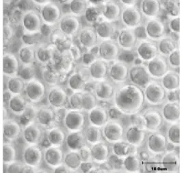
Figure 1. Phase contrast picture of the control C#15 PGC line, on the Day 50th of cultivation.
Scale bar: 10𝜇𝜇m
The Figure 1 shows the morphology the cells of the control embryo derived PGC line (C#15) on day 50th of cultivation.
At the end of the experiment 26 cell lines were frozen (Table 1).
We tested the cCvh, cPouv, cNanog and cHsp70
expression in the collected RNA samples at Day 23, Day30 and Day50 (Figure 2). We could find increasing level of the expression of stem cell specific (cPouv, cNanog), PGC specific (cCvh,
(chicken vasa homologue) and heat shock related
cHsp70 in PGC culture during long term
cultivation.
Examination the expression level of heat shock protein cHsp70 monitored the effect of heat stress (Figure 2). The highest cHsp70 expression was observed in heat stressed PGCs at 50th day and lowest on 23th day. Similarly, the expression level of pluripotency markers cPouv and cNanog was observed maximum at day 50th and minimum in the 23-day-old cultures. No significant difference in the expression level of the PGC specific markers cCVH was observed. The analysis of all collected PGC samples will be carried out in the next period.
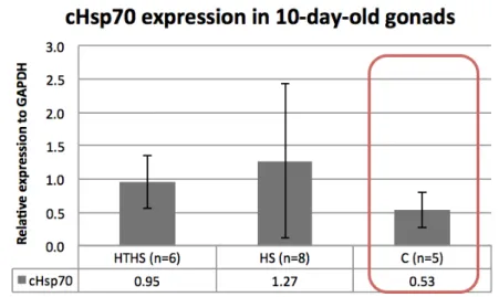
Hsp70 expression in 10-day-old embryonic
gonads
The Figure 3 is demonstrating the cHsp70
expressions in 10-day-old embryonic gonads. We collected left and right gonads from 10-day-old female and male chicken embryos. The expression of the cHsp70 was considerably higher in the gonads of HTHS and HS embryos than in the control (C) group (Figure 3). Statistical analysis could not revelled significant difference in the relative expression level of cHsp70 between the heat-treated and heat stressed (HTHS) and non-heat treated (HS) (p=0.53); non-heat-treated and non-heat stressed (HTHS) and control (C) (p=0.07); heat stressed (HTHS) and control (C) (p=0.19) samples. We suppose that the high levels of calculated standard deviation were derived because of the different expression level of
cHsp70 in the left and right side and female and
Table 1. Number of established and frozen PGC cultures
Treatments HTHS HS C Sum
Number of eggs incubated 47 47 47 141
Number of good quality embryos 22 14 20 56
Number of good PGC cultures on day 10 16 11 12 39
Number of good PGC cultures on day 20 12 8 8 28
Number of good PGC cultures on day 40 11 7 8 26
Number of frozen PGC cultures 11 7 8 26
HTHS: heat-treated and heat stressed; HS: non-heat treated, but heat stressed; C: control non-treated, non-stressed samples
Figure 2. Expression of stem cell specific (cPouv, cNanog), PGC specific (cCvh) and heat shock related cHsp70 in C#15 PGC culture on Day 23, Day 30 and Day 50 relative to housekeeping gene cGapdh.
Figure 3. Expression of stem cell specific cHSP70 in gonads of 10-day-old embryos
4. Conclusions
Based on our preliminary results we can conclude that the expression of cHsp70 heat shock protein was detectable in the PGC cultures and also in the gonads. High level of the cHsp70 expression was detected in heat stressed animal derived PGCs and gonads. Chicken subjected to heat stress showed varied expression of cHsp70 and also improved thermo tolerance. The expression of cHsp70 was minimum in control gonads. This proves that fact that due to heat stress there is induced cHsp70
expression. Heat stress leads to apoptosis, which may result in cell death of gonad cells, but increased level of HSP70 can prevent the gonads from degradation. HSP70 has anti-apoptotic function and prevents heat stress induced apoptosis.
In the future the results of the comparison of indigenous breeds and intensive lines allow elaborating new lines, which can adapt easily to the extreme weather conditions.
Acknowledgements
This project was supported by CGIAR CCAFS project (Ministry of Agriculture).
References
1. Al-Zghoul, M. B., Dalab, A. E., Ababneh, M. M., Jawasreh, K. I., Al Busadah, K. A., Ismail, Z. B., Thermal manipulation during chicken embryogenesis results in enhanced Hsp70 gene expression and the acquisition of thermotolerance, Res Vet Sci., 2013, 95(2) 502-507
2. Xie, J., Tang, L., Lu L, Zhang, L., Xi, L., Liu, H., C., Odle, J., Luo, X., Differential expression of heat shock transcription factors and heat shock proteins after acute and chronic heat stress in laying chickens (Gallus gallus), PLoS One, 2014, 9(7), e102204
3. Rupik, W., Jasik, K., Bembenek, J., Widłak, W., The expression patterns of heat shock genes and proteins and their role during vertebrate's development, Comp Biochem Physiol A Mol Integr Physiol, 2011, 159(4) 349-366