Darure. World Journal of Pharmaceutical and Life Sciences
TRANSDERMAL DRUG DELIVERY SYSTEM
Pranali S. Darure*
Shri Ambabai Talim Sanstha’s Diploma in Pharmacy College, Miraj.
Article Received on 08/09/2017 Article Revised on 29/09/2017 Article Accepted on 19/10/2017
INTRODUCTION
The method by which a drug is delivered can have a significant effect on its efficacy. Some drugs have an optimum concentration range within which maximum benefit is derived, and concentrations above or below this range can be toxic or produce no therapeutic benefit at all 1. On the other hand, the very slow progress in the efficacy of the treatment of severe diseases, has suggested a growing need for a multidisciplinary approach to the delivery of therapeutics to targets in tissues. From this, new ideas on controlling the pharmacokinetics, pharmacodynamics, non-specific toxicity, immunogenicity, biorecognition, and efficacy of drugs were generated. These new strategies, often called drug delivery systems (DDS), which are based on interdisciplinary approaches that combine polymer science, pharmaceutics, bioconjugate chemistry, and molecular biology. To minimize drug degradation and loss, to prevent harmful side-effects and to increase drug bioavailability and the fraction of the drug accumulated in the required zone, various drug delivery and drug targeting systems are currently under development.[1] Transdermal drug delivery systems are topically administered medicaments in form of patches that deliver the drug for systemic effects at a predetermined controlled rate. A transdermal drug delivery device, which may be of an active or a passive design, is a device which provides an alternative route for administering medication. These devices allow pharmaceuticals to be delivered across the skin barrier. A drug is applied in a relatively high dosage to the inside of a patch, which is worn on the skin for an extended period of time. Through a diffusion process, the drug enters the
bloodstream directly through the skin. Since there is high concentration on the patch and low concentration in the blood, the drug will keep diffusing into the blood for a long period of time, maintaining the constant concentration of drug in the blood flow. The best mixture is about fifty percent of the drug being each hydrophilic and lipophillic. This is because “Lipid-soluble substances readily pass through the intercellular lipid bi- layers of the cell membranes whereas water-soluble drugs are able to pass limiting steps in transdermal drug delivery system. Sweat ducts and hair follicles are paths of entry, but they are considered rather insignificant.[2]
Definition
A transdermal patch is defined as medicated adhesive patch which is placed above the skin to deliver a specific dose of medication through the skin with a predetermined rate of release to reach into the bloodstream.[3]
Advantages
1) Avoidance of first pass metabolism.
2) Avoidance of gastro-intestinal incompatibility. 3) Predictable and extended duration of activity. 4) Minimizing undesirable side effects.
5) Provides utilization of drugs with short biological half-lives, narrow therapeutic window.
6) Improving physiological and pharmacological response.
7) Avoiding the fluctuation in drug levels. 8) Inter and intra-patient variations.
9) Maintain plasma concentration of potent drugs. 10) Termination of therapy is easy at any point of time.
Review Article ISSN 2454-2229
wjpls, 2017, Vol. 3, Issue 9, 118-128
World Journal of Pharmaceutical and Life Sciences
WJPLS
www.wjpls.org SJIF Impact Factor: 4.223
*Corresponding Author: Pranali S. Darure
Shri Ambabai Talim Sanstha’s Diploma in Pharmacy College, Miraj.
ABSTRACT
The method by which a drug is delivered can have a significant effect on its efficacy. Some drugs have an optimum concentration range within which maximum benefit is derived, and concentrations above or below this range can be toxic or produce no therapeutic benefit at all 1. On the other hand, the very slow progress in the efficacy of the treatment of severe diseases, has suggested a growing need for a multidisciplinary approach to the delivery of therapeutics to targets in tissues. From this, new ideas on controlling the pharmacokinetics, pharmacodynamics, non-specific toxicity, immunogenicity, biorecognition, and efficacy of drugs were generated.
Darure. World Journal of Pharmaceutical and Life Sciences
11) Greater patient compliance due to elimination of multiple dosing profiles.[4]
Disadvantages
1) The drug must have some desirable physicochemical properties for penetration through stratum corneum and if the drug dose required for therapeutic value is more than 10 mg/day, the transdermal delivery will be very difficult.
2) Only relatively potent drugs are suitable candidates for TDDS because of the natural limits of drug entry imposed by the skin’s impermeability.
3) Some patients develop contact dermatitis at the site of application for one or more of the system components, necessitating discontinuation.
4) Clinical need is another area that has to be examined carefully before a decision is made to develop a transdermal product.
5) The barrier function of the skin changes from one site to another on the same person, from person to person and with age.[5]
Ideal properties drug for TDDS Half-life <10hrs
Therapeutic index low
Oral bioavailability low
PH between 5-9
Skin permeability coefficient 0.5×10⁻³
Drug solution in direct contact with release liner for successfully developing transdermal delivery system, the drug should be chosen with great care. The following are some of the desirable properties of a drug for transdermal delivery.[6]
(a) Physiochemical Properties
1) The drug should have are molecular weight less than approximately 500 Daltons.
2) The drug should have affinity for both lipophilic and hydrophilic phases. Extreme partitioning characteristics are not conductive to successful drug delivery via the skin.
3) The drug should have a low melting point.
(b) Biological Properties
1. The drug should be potent with a daily dose of the order of a few mg per day.
2. The half-life (t1/2) of the drug should be short. 3. The drug must not induce a cutaneous or allergic
response.
4. Drug which degrade in the G.I tract or inactivated by hepatic first pass effect are suitable candidates for transdermal delivery.
5. Tolerance to the drug must not develop under the near zero order release profile of transdermal delivery.
6. Drugs which have to be administered for a long period of time or which cause adverseeffects to non-target tissues can also be formulated for transdermal delivery.[7]
Skin Layers
Cellular level elements (Level 1) form the three different skin layers: epidermis, dermis and subcutis.
These layers are composed of different types of cellular level elements. Hence, they are very different in terms of structure and function. As a result, they exhibit different types of light propagation.
The epidermis is composed of four sublayers: stratum corneum, stratum granulosum, stratum spinosum and stratum basale. In the sole and the palm, there is an additional layer called stratum lucidum underneath stratum corneum.
1) Epidermis
The epidermis is the outermost layer of skin. There are no veins and capillaries in this layer. Its thickness is about 0.2mmon average and this thickness varies depending on the location on the body. Furthermore, the thickness also varies according to the volume of water that the epidermis holds. The epidermis is further divided into five sublayers. From the bottom (innermost), these sublayers are stratum basale (basal cell layer), stratum
spinosum (prickle cell layer), stratum granulosum
(granular cell layer), stratum lucidum (clear layer) and
stratum corneum (horny cell layer).
The epidermis is a metabolically active tissue. Keratinocytes produced in stratum basale move upward to the outer surface. This process is called turn-over. During this turn-over, keratinocytes change their structures and physiological functions. One cycle of this turn-over process takes about 28 days. In the following, we will describe the physio-anatomical properties of each sublayer.
a) Stratum basale (basal cell layer) is the deepest
sublayer of the epidermis and is composed of a single layer of basal cells. This sublayer forms the boundary to the dermis. Keratinocytes are produced in this sublayer. It holds approximately 8% of the water in the epidermis. With aging, this layer becomes thinner and loses the ability to retain water. Melanocytes, which were mentioned in the previous section, also lie in this layer.
b) Stratum spinosum (prickle cell layer) refers to the
Darure. World Journal of Pharmaceutical and Life Sciences
c) Stratum granulosum (granular cell layer) is
composed of 2 to 4 granular cell layers. The typical thickness is 3 μm. In this sublayer, cornification called keratinization of keratinocytes begins. In this process, organelles such as nuclei and mitochondria start to resolve. Cells are increasingly filled with keratin fibers and contain less moisture than basal and prickle cell layers. The shape of these cells becomes much flatter during this process.
d) Stratum lucidum (clear layer) can only be found in
soles and palms. It is a highly refractive sublayer. Its cells become flatter and more densely packed during turn-over.
e) Stratum corneum (horny cell layer) is the exterior
sublayer of the epidermis. Its thickness ranges from 8 to 15 μm. This sublayer is composed of several layers of hexagonal-shaped flat and hard cells named horny cells or corneocytes. These are dry dead cells without organelles and filled with keratin fibers. This sublayer prevents excessive dehydration of the skin tissue and usually contains 10 to 15% of the mass of water in the epidermis, depending on the skin condition. Horny cells are surrounded by intercellular lipids. A principal constituent is
ceramide, which plays a crucial role in water
retention. Horny cells also contain special chemical compounds called natural moisturizing factor (NMF) that also plays an important role in retaining skin moisture. NMF is composed of sodium PCA, sphinolipids and ceramides, phospholipids, fatty acids, glycerol, squalane and cholesterol. Skin that lacks NMF and ceramide tends to be very dry.
2) Dermis The dermis is the second layer of skin,
beneath the epidermal layer. This layer is much thicker than the epidermis (usually 1 to 4 mm). The main components of the dermis are collagen and elastin fibers. Compared to the epidermis, there are much fewer cells and much more fibers in the dermis. Dermis has the following two sublayers.
a) Papillary layer is the upper sublayer of the dermis that is clearly demarcated from the epidermis. This sublayer is a loosely connected tissue and includes a large amount of nerve fibers, capillaries, water and cells (e.g fibroblasts). In this sublayer, collagen fibers form a finer network than those of the reticularm layer.
b) Reticular layer constitutes the lower part of the
dermis and represents a continuous transition to the 15 subcutis. This sublayer has a denser and thicker network than the papillary layer and includes fewer nerve fibers and capillaries. In this sublayer, collagen fibers are aggregated into thick bundles which are mostly aligned parallel to the surface of skin.
The micro-anatomical complexity of skin, particularly in Level 1, makes quantitative analysis of the optical
properties of skin difficult. However, it can be significantly simplified by considering the physioanatomical characteristics of each skin layers. As mentioned above, the epidermis and the dermis are very different in composition, thickness and functions. Hence, these two layers can be considered to be independent of each other in terms of optical behaviors. Indeed, the epidermis and the dermis are viewed as independent optical media in many early studies on skin optics. For example, melanin is present only in the epidermis. On the other hand, hemoglobin is found only in the dermis since there are no veins and capillaries in the epidermis. Hence, the epidermis can be essentially viewed as a
melanin layer and the dermis can be viewed as a
hemoglobin layer when analyzing the absorption
properties of skin.
3) Subcutis
Subcutis, or hypodermis in histology, is the third layer beneath the dermis. It is important to note that it is not categorized as another skin layer. Subcutis is an elastic layer and includes a large amount of fat cells that work as a shock absorber for blood vessels and nerve endings. The thickness of this layer is reported to be 4 to 9 mm on average. However, the actual thickness differs from person to person and also depends on the body region.[8]
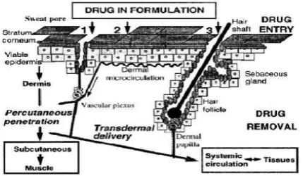
Kinetics of Transdermal Permeation
For a systemically active drug to reach a target tissue, it has to possess some physicochemical properties which facilitate the sorption of the drug through the skin and enter the microcirculation.
Fig. 3: Absorption across the skin can occur through sweat ducts (1), intercellular regions of the stratum corneum (2) and through the hair follicles (3).
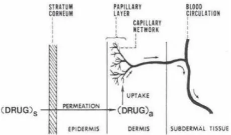
The release of a therapeutic agent from a TDDS applied to the skin surface and its transport to the systemic circulation involves the following steps: i. Dissolution within and release from the formulation,
1) Partitioning into the outermost layer of the skin, SC, 2) Diffusion through the SC,
3) Partitioning from the SC into the aqueous viable epidermis,
4) Diffusion through the viable epidermis and into the upper dermis
Darure. World Journal of Pharmaceutical and Life Sciences
Fig. 4: A multilayer skin model showing sequence of Transdermal permeation of drug for systemic delivery.
Knowledge of skin permeation kinetics is vital to the successful development of transdermal systems. This permeation can be possible if the drug possesses certain physico-chemical properties. The rate of permeation across the skin (dQ/ dt) is given by: dQ∕dt=Ps (Cd-Cr)……(1)
Where, Cd = concentration of skin penetrant in the donar compartment (e.g., on the surface of stratum corneum)
Cr = concentration in the receptor compartment (e.g., body) respectively Ps = the overall permeability constant of the skin tissue to the penetrant Ps =KsDss/hs…….(2)
Where, Ks is the partition coefficient for the interfacial partitioning of the penetrant molecule from a solution medium or a transdermal therapeutic system onto the stratum corneum, Dss is the apparent diffusivity for the steady state diffusion of the penetrant molecule through a thickness of skin tissues and hs is the overall thickness of skin tissues. As Ks, Dss and hs are constant under given conditions, the permeability coefficient (Ps) for a skin penetrant can be considered to be constant.
From Eq.1 it is clear that a constant rate of drug permeation can be obtained only when Cd>>Cr i.e., the drug concentration at the surface of the stratum corneum (Cd) is constistently and substantially greater than the drug concentration in the body (Cr) then Eq. 1 becomes:
dQ∕dt =PsCd….. (3)
Permeability coffiecient = KsDss / hs = 1/ resistance Resistance has many components Vehicle
Stratum corneum (usually most significant) Epidermis
Dermis
The resistance occurs one after another ‘in series’ Rtotal =
1 1 1 1 --- --- --- --- Rvehicle + Rstratum corneum + Repidermis + Rdermis
Total permeability = Rvheical + Rstratum corneum + R epidermis + R dermis
The membrane limited flux (J) under steady state condition is described by equation:
Where,
J = Amount of drug passing through membrane system per unit area per unit time.
D = Diffusion coefficient with in the membrane h = Membrane thickness
K = Membrane / vehicle partition coefficient C = Concentration gradient across the membrane[9]
Types of T. D. D. S
1) Single-layer Drug-in-Adhesive
The Single-layer Drug-in-Adhesive system is characterized by the inclusion of the drug directly within the skincontacting adhesive. In this transdermal systemdesign, the adhesive not only serves to affix the system to the skin, but also serves as the formulation foundation, containing the drug and all the excipients under a single backing film. The rate of release of drug from this type of system is dependent on the diffusion across the skin.
2) Multi-layer Drug-in-Adhesive
The Multi-layer Drug-in-Adhesive is similar to the Single-layer Drug-in-Adhesive in that the drug is incorporated directly into the adhesive. However, the multi-layer encompasses either the addition of a membrane between two distinct drug-inadhesive layers or the addition of multiple drug-in-adhesive layers under a single backing film
3) Drug Reservoir-in-Adhesive
The Reservoir transdermal system design is characterized by the inclusion of a liquid compartment containing a drug solution or suspension separated from the release liner by a semi-permeable membrane and adhesive. The adhesive component of the product responsible for skin adhesion can either be incorporated as a continuous layer between the membrane and the release liner or in a concentric configuration around the membrane.
4) Drug Matrix-in-Adhesive
The Matrix system design is characterized by the inclusion of a semisolid matrix containing a drug solution or suspension which is in direct contact with the release liner. The component responsible for skin adhesion is incorporated in an overlay and forms a concentric configuration around the semisolid matrix.(10)
Basic Components of TDDS 1. The drug
2. Polymer matrix 3. Permeation enhancers 4. Adhesive
Darure. World Journal of Pharmaceutical and Life Sciences
1. Drug
The drug is in direct contact with release liner. Ex: Nicotine, Methotrexate, and Oestrogen. Some of the desirable properties of a drug for transdermal delivery: 1. The drug molecule should possess an adequate
solubility in oil and water.
2. The drug should have a molecular weight less than approximately 1000 daltons.
3. The drug should have low melting point.
4. The drug molecule would require a balanced partition coefficient to penetrate the stratum corneum.
2. Polymer Matrix
These polymers control the release of the drug from the drug reservoir.
Natural polymers: shellac, gelatin, waxes, gums, starch etc., Synthetic polymers: polyvinyl alcohol, polyamide, polyethylene, polypropylene, Polyurea, polymethylmethacrylate etc.
3. Permeation Enhancers
Substances exist which temporarily diminish the impermeability of the skin are known as accelarants or sorption promoters or penetration enhancers. These include water, pyrolidones, fatty acids and alcohols, azone and its derivatives, alcohols and glycols, essential oils, terpenes and derivatives, sulfoxides like dimethyl sulfoximide and their derivatives, urea and surfactants.
Surfactants are proposed to enhance polar pathway
transport especially of hydrophilic drugs. The ability of a surfactant to alter penetration is a function of the polar head group and the hydrocarbon chain length.
1) Anionic surfactants: sodium lauryl sulphate,
Decodecylmethyl sulphoxide etc.
2) Nonionic surfactants: Pluronic F 127, Pluronio
F68, etc.
Enhancer actions can be classified by lipid-proteinpartitioning concept. This hypothesis suggests that enhancers act by one or more ways selected from three main possibilities.
Lipid action the enhancer interacts with the organized intracellular lipid structure of the stratum corneum so as to disrupt it and make it more permeable to drug molecules. Very many enhancers operate in this way. Some solvents act by extracting the lipid Components and thus make the horny layer more permeable.
Protein modification Ionic surface active molecules in particular tend to interact well with the keratin in the corneocytes, to open up the dense keratin structure and make it more permeable. The intracellular route is not usually prominent in drug permeation, although drastic reductions to this routes resistance could open up an alternative path for drug penetration.
Partitioning promotion many solvents can enter the
stratum corneum, change its solvent properties and thus increase the partitioning of a second molecule into the horny layer. This molecule may be a drug, a
coenhancer or a cosolvent. For example ethanol has been used to increase the penetration of the drug molecules nitroglycerin and estradiol.
4. Adhesive
Serves to adhere the patch to the skin for systemic delivery of drug.
Ex: Silicones, Polyisobutylene.
5. Backing Layer
Backing layer protects patch from outer environment. Ex: Cellulose derivatives, Polypropylene silicon rubber.[11]
Various Methods for Preparation of TDDS 1. Asymmetric TPX membrane method
A prototype patch can be fabricated for this a heat sealable polyester film (type 1009, 3m) with a concave of 1cm diameter will be used as the backing membrane. Drug sample is dispensed into the concave membrane, covered by a TPX {poly (4-methyl-1 pentene)} asymmetric membrane, and sealed by an adhesive. [(Asymmetric TPX membrane preparation): These are fabricated by using the dry/wet inversion process. TPX is dissolved in a mixture of solvent (cyclohexane) and nonsolvent additives at 60°c to form a polymer solution. The polymer solution is kept at 40°C for 24 hrs and cast on a glass plate to a pre-determined thickness with a Gardner knife. After that the casting film is evaporated at 50°C for 30 sec, then the glass plate is to be immersed immediately in coagulation bath [maintained the temperature at 25°C]. After 10 minutes of immersion, the membrane can be removed, air dry in a circulation oven at 50°C for 12 hrs].
2. Circular Teflon mould method: Solutions
Darure. World Journal of Pharmaceutical and Life Sciences
3. Mercury substrate method: In this method drug is
dissolved in polymer solution along with plasticizer. The above solution is to be stirred for 10-15 minutes to produce a homogenous dispersion and poured in to a leveled mercury surface, covered with inverted funnel to control solvent evaporation.
4. By using “IPM membranes” method: In this
method drug is dispersed in a mixture of water and propylene glycol containing carbomer 940 polymers and stirred for 12 hrs in magnetic stirrer. The dispersion is to be neutralized and made viscous by the addition of triethanolamine. Buffer pH 7.4 can be used in order to obtain solution gel, if the drug solubility in aqueous solution is very poor. The formed gel will be incorporated in the IPM membrane.
5. By using “EVAC membranes” method: In order to
prepare the target transdermal therapeutic system, 1% carbopol reservoir gel, polyethylene (PE), ethylene vinyl acetate copolymer (EVAC) membranes can be used as rate control membranes. If the drug is not soluble in water, propylene glycol is used for the preparation of gel. Drug is dissolved in propylene glycol; carbopol resin will be added to the above solution and neutralized by using 5% w/w sodium hydroxide solution. The drug (in gel form) is placed on a sheet of backing layer covering the specified area. A rate controlling membrane will be placed over the gel and the edges will be sealed by heat to obtain a leak proof device.
6. Aluminium backed adhesive film method:
Transdermal drug delivery system may produce unstable matrices if the loading dose is greater than 10 mg. Aluminium backed adhesive film method is a suitable one for preparation of same, chloroform is choice of solvent, because most of the drugs as well as adhesive are soluble in chloroform. The drug is dissolved in chloroform and adhesivematerial will be added to the drug solution and dissolved. A custammade aluminium former is lined with aluminium foil and the ends blanked off with tightly fitting cork blocks.
7. Preparation of TDDS by using Proliposomes: The
proliposomes are prepared by carrier method using film deposition technique. From the earlier reference drug and lecithin in the ratio of 0.1:2.0 can be used as an optimized one. The proliposomes are prepared by taking 5mg of mannitol powder in a 100 ml round bottom flask which is kept at 60-70°c temperature and the flask is rotated at 80-90 rpm and dried the mannitol at vacuum for 30 minutes. After drying, the temperature of the water bath is adjusted to 20-30°C. Drug and lecithin are dissolved in a suitable organic solvent mixture, a 0.5ml aliquot of the organic solution is introduced into the round bottomed flask at 37°C, after complete drying second aliquots (0.5ml) of the solution is to be added. After the last loading, the flask containing proliposomes are connected in a lyophilizer and subsequently drug loaded mannitol powders (proliposomes) are placed in
desiccators overnight and then sieved through 100 mesh. The collected powder is transferred into a glass bottle and stored at the freeze temperature until characterization.
8. By using free film method: Free film of cellulose acetate is prepared by casting on mercury surface. A polymer solution 2% w/w is to be prepared by using chloroform. Plasticizers are to be incorporated at a concentration of 40% w/w of polymer weight. Five ml of polymer solution was poured in a glass ring which is placed over the mercury surface in a glass petri dish. The rate of evaporation of the solvent is controlled by placing an inverted funnel over the petri dish. The film formation is noted by observing the mercury surface after complete evaporation of the solvent. The dry film will be separated out and stored between the sheets of wax paper in a desiccator until use. Free films of different thickness can be prepared by changing the volume of the polymer solution.[12]
Factors Influencing Transdermal Drug Delivery The effective transdermal drug delivery can be formulated by considering three factors as Drug, Skin, and the vehicles. So the factors affecting can be divided in to classes as biological factors and physicochemical factors.
A. Biological factors
1. Skin condition: Acids and alkalis, many solvents like
chloroform methanol damage the skin cells and promote penetration. Diseased state of patient alters the skin conditions. The intact skin is better barrier but the above mentioned conditions affect penetration.
2. Skin age: The young skin is more permeable than
older. Children are more sensitive for skin absorption of toxins. Thus, skin age is one of the factors affecting penetration of drug in TDDS.
3. Blood supply: Changes in peripheral circulation can
affect transdermal absorption.
4. Regional skin site: Thickness of skin, nature of
stratum corneum, and density of appendages vary site to
site. These factors affect significantly penetration.
5. Skin metabolism: Skin metabolizes steroids,
hormones, chemical carcinogens and some drugs. So skin metabolism determines efficacy of drug permeated through the skin.
6. Species differences: The skin thickness, density of appendages, and keratinization of skin vary species to species, so affects the penetration.
B. Physicochemical factors
1. Skin hydration: In contact with water the
Darure. World Journal of Pharmaceutical and Life Sciences
most important factor increasing the permeation of skin. So use of humectants is done in transdermal delivery.
2. Temperature and pH: The permeation of drug
increase tenfold with temperature variation. The diffusion coefficient decreases as temperature falls. Weak acids and weak bases dissociate depending on the pH and pKa or pKb values. The proportion of unionized drug determines the drug concentration in skin. Thus, temperature and pH are important factors affecting drug penetration.
3. Diffusion coefficient: Penetration of drug depends
on diffusion coefficient of drug. At a constant temperature the diffusion coefficient of drug depends on properties of drug, diffusion medium and interaction between them.
4. Drug concentration: The flux is proportional to the concentration gradient across the barrier and concentration gradient will be higher if the concentration of drug will be more across the barrier.
5. Partition coefficient: The optimal K, partition
coefficient is required for good action. Drugs with high
K are not ready to leave the lipid portion of skin. Also, drugs with low K will not be permeated.
6. Molecular size and shape: Drug absorption is
inversely related to molecular weight; small molecules penetrate faster than large ones. Because of partition coefficient domination, the effect of molecular size is not known.[13]
Recent Technology Used In Transdermal Drug Delivery System
1) Iontophoresis
This method involves the application of a low level electric current either directly to the skin or indirectly via the dosage form in order to enhance permeation of a topically applied therapeutic agent19, 20. Increased drug permeation as a result of this methodology can be attributed to either one or a combination of the following mechanisms: Electro-repulsion (for charged solutes), osmosis (for uncharged solutes) and electro-pertubation (for both charged and uncharged). Several iontophoretic systems are currently under commercial development including the Phoresor device developed by Iomed Inc. and the Vyteris and E-TRANS devices developed by Alza Corp.
2) Electroporation
This method involves the application of high voltage pulses to the skin which has been suggested to induce the formation of transient pores. High voltages (100 V) and short treatment durations (milliseconds) are most frequently employed. Other electrical parameters that affect permeation rate include pulse properties such as waveform, rate and number. The technology has been successfully used to enhance the skin permeability of
molecules with differing lipophilicity and size (i.e. small molecules, proteins, peptides and oligonucleotides) including biopharmaceuticals with molecular weights greater than 7kDA.23
3) Microneedle-based Devices
The very first microneedle systems, described in 1976, consisted of a drug reservoir and a plurality of projections (microneedles 50 to 100 mm long) extending from the reservoir, which penetrated the stratum corneum and epidermis to deliver the drug. The ALZA Corp. has recently commercialized a microneedle technology named Macroflux which can either be used in combination with a drug reservoir or by dry coating the drug on the microprojection array24, the latter being better for intracutaneous immunization.
4) Abrasion
The abrasion technique involves the direct removal or disruption of the upper layers of the skin to facilitate the permeation of topically applied medicaments. Some of these devices are based on techniques employed by dermatologists for superficial skin resurfacing (e.g. microdermabrasion) which are used in the treatment of acne, scars, hyperpigmentaion and other skin blemishes.
5) Needle-less Injection
This is reported to involve a pain-free method of administering drugs to the skin. Over the years, there have been numerous examples of both liquid (Ped-O-Jet, Iject, Biojector2000, Medi-jector and Intraject) and powder (PMED device formerly known as Powderject injector) systems. The latter device has been reported to successfully deliver testosterone, lidocaine hydrochloride and macromolecules such as calcitonin and insulin. of ultrasonic energy to enhance the transdermal delivery of solutes either simultaneously or via pre-treatment and is frequently referred to as sonophoresis or phonophoresis. The SonoPrep device (Sontra Medical Corp.) uses low frequency ultrasound (55 kHz) for an average duration of 15 seconds to enhance skin permeability. This battery-operated, handheld device consists of a control unit, ultrasonic horn with control panel, a disposable coupling medium cartridge, and a return electrod.
The Pharma Innovation – Journa
6) Laser Radiation
This method involves direct and controlled exposure of a laser to the skin which results in the ablation of the stratum corneum without significantly damaging the underlying epidermis. Removal of the stratum corneum using this method has been shown to enhance the delivery of lipophilic and hydrophilic drugs.[6]
Evaluation Parameters
1. Interaction studies: Excipients are integral
Darure. World Journal of Pharmaceutical and Life Sciences
excipients. The drug and the excipients must be compatible with one another to produce a product that is stable, thus it is mandatory to detect any possible physical or chemical interaction as it can affect the bioavailability and stability of the drug. If the excipients are new and have not been used in formulations containing the active substance, the compatibility studies play an important role in formulation development. Interaction studies are commonly carried out in Thermal analysis, FT-IR, UV and chromatographic techniques by comparing their physicochemical characters such as assay, melting endotherms, characteristic wave numbers, absorption maxima etc.,
2. Thickness of the patch: The thickness of the drug loaded patch is measured in different points by using a digital micrometer and determines the average thickness and standard deviation for the same to ensure the thickness of the prepared patch.
3. Weight uniformity: The prepared patches are to be
dried at 60°c for 4hrs before testing. A specified area of patch is to be cut in different parts of the patch and weigh in digital balance. The average weight and standard deviation values are to be calculated from the individual weights.
4. Folding endurance: A strip of specific are is to be cut evenly and repeatedly folded at the same place till it broke. The number of times the film could be folded at the same place without breaking gave the value of the folding endurance.
5. Percentage Moisture content: The prepared films
are to be weighed individually and to be kept in a desiccator containing fused calcium chloride at room temperature for 24 hrs. After 24 hrs the films are to be reweighed and determine the percentage moisture content from the below mentioned formula. Percentage moisture content = [Initial weight- Final weight/ Final weight] ×100. 6. Percentage Moisture uptake: The weighed films are to be kept in a desiccator at room temperature for 24 hrs containing saturated solution of potassium chloride in order to maintain 84% RH. After 24 hrs the films are to be reweighed and determine the percentage moisture uptake from the below mentioned formula
Percentage moisture content = [Initial weight- Final weight/ Final weight] ×100.
6. Percentage Moisture uptake: The weighed films
are to be kept in a desiccator at room temperature for 24 hrs containing saturated solution of potassium chloride in order to maintain 84% RH. After 24 hrs the films are to be reweighed and determine the percentage moisture uptake from the below mentionedformula
Percentage moisture uptake = [Final weight- Initial weight/ initial weight] ×100.
7. Water vapour permeability (WVP) evaluation:
Water vapour permeability can be determined with foam dressing method the air forced oven is replaced by a natural air circulation oven. The WVP can be determined by the following formula WVP=W/A Where, WVP is expressed in gm/m2 per 24hrs, W is the amount of vapour permeated through the patch expressed in gm/24hrs and A is the surface area of the exposure samples expressed in m2.
8. Drug content: A specified area of patch is to be dissolved in a suitable solvent in specific volume. Then the solution is to be filtered through a filter medium and analyse the drug contain with the suitable method (UV or HPLC technique). Each value represents average of three different samples.
9. Uniformity of dosage unit test: An accurately
weighed portion of the patch is to be cut into small pieces and transferred to a specific volume volumetric flask, dissolved in a suitable solvent and sonicate for complete extraction of drug from the patch and made up to the mark with same. The resulting solution was allowed to settle for about an hour, and the supernatant was suitably diluted to give the desired concentration with suitable solvent. The solution was filtered using
0.2m membranes filter and analysed by suitable analytical technique (UV or HPLC) and the drug content per piece will be calculated.
10. Polariscope examination: This test is to be
performed to examine the drug crystals from patch by polariscope. A specific surface area of the piece is to be kept on the object slide and observe for the drugs crystals to distinguish whether thedrug is present as crystalline form or amorphous form in the patch.
11. Shear Adhesion test: This test is to be performed for the measurement of the cohesive strength of an adhesive polymer. It can be influenced by the molecular weight, the degree of crosslinking and the composition of polymer, type and the amount of tackifier added. An adhesive coated tape is applied onto a stainless steel plate; a specified weight is hung from the tape, to affect it pulling in a direction parallel to the plate. Shear adhesion strength is determined by measuring the time it takes to pull the tape off the plate. The longer the time take for removal, greater is the shear strength.
12. Peel Adhesion test: In this test, the force required to
Darure. World Journal of Pharmaceutical and Life Sciences
13. Thumb tack test: It is a qualitative test applied for
tack property determination of adhesive. The thumb is simply pressed on the adhesive and the relative tack property is detected
14. Flatness test: Three longitudinal strips are to be cut
from each film at different portion like one from the center, other one from the left side, and another one from the right side. The length of each strip was measured and the variation in length because of non-uniformity in flatness was measured by determining percent constriction, with 0% constriction equivalent to 100% flatness.
15. Percentage Elongation break test: The percentage
elongation break is to be determined by noting the length just before the break point, the percentage elongation can be determined from the below mentioned formula. Elongation percentage = L1-L2 /L2×100 Where, L1is the final length of each strip and L2 is the initial length of each strip.
16. Rolling ball tack test: This test measures the
softness of a polymer that relates to talk. In this test, stainless steel ball of 7/16 inches in diameter is released on an inclined track so that it rolls down and comes into contact with horizontal, upward facing adhesive. The distance the ball travels along the adhesive provides the measurement of tack, which is expressed in inch.
17. Quick Stick (peel-tack) test: In this test, the tape is
pulled away from the substrate at 90ºC at a speed of 12 inches/min. The peel force required breaking the bond between adhesive and substrate is measured and recorded as tack value, which is expressed in ounces or grams per inch width.
The percentage elongation break is to be determined by noting the length just before the break point, the percentage elongation can be determined from the below mentioned formula. Elongation percentage = L1-L2 /L2×100 Where, L1is the final length of each strip and L2 is the initial length of each strip.
18. Probe Tack test: In this test, the tip of a clean probe with a defined surface roughness is brought into contact with adhesive, and when a bond is formed between probe and adhesive. The subsequent removal of the probe mechanically breaks it. The force required to pull the probe away from the adhesive at fixed rate is recorded as tack and it is expressed in grams.
19. In vitro drug release studies: The paddle over disc
method (USP apparatus V) can be employed for assessment of the release of the drug from the prepared patches. Dry films of known thickness is to be cut into definite shape, weighed, and fixed over a glass plate with an adhesive. The glass plate was then placed in a 500-mL of the dissolution medium or phosphate buffer (pH 7.4), and the apparatus was equilibrated to 32± 0.5°C. The
paddle was then set at a distance of 2.5 cm from the glass plate and operated at a speed of 50 rpm. Samples (5-mL aliquots) can be withdrawn at appropriate time intervals up to 24 h and analyzed by UV spectrophotometer or HPLC. The experiment is to be performed in triplicate and the mean value can be calculated.
20. In vitro skin permeation studies: An in vitro
permeation study can be carried out by using diffusion cell. Full thickness abdominal skin of male Wistar rats weighing 200 to 250g. Hair from the abdominal region is to be removed carefully by using a electric clipper; the dermal side of the skin was thoroughly cleaned with distilled water to remove any adhering tissues or blood vessels, equilibrated for an hour in dissolution medium or phosphate buffer pH 7.4 before starting the experiment and was placed on a magnetic stirrer with a small magnetic needle for uniform distribution of the diffusant. The temperature of the cell was maintained at 32 ± 0.5°C using a thermostatically controlled heater. The isolated rat skin piece is to be mounted between the compartments of the diffusion cell, with the epidermis facing upward into the donor compartment. Sample volume of definite volume is to be removed from the receptor compartment at regular intervals, and an equal volume of fresh medium is to be replaced. Samples are to be filtered through filtering medium and can be analyzed spectrophotometrically or HPLC. Flux can be determined directly as the slope of the curve between the steady-state values of the amount of drug permeated (mg cm-2) vs. time in hours and permeability coefficients were deduced by dividing the flux by the initial drug load (mg cm-2).
21. Skin Irritation study: Skin irritation and
sensitization testing can be performed on healthy rabbits (average weight 1.2 to 1.5 kg). The dorsal surface (50cm2) of the rabbit is to be cleaned and remove the hair from the clean dorsal surface by shavingand clean the surface by using rectified spirit and the representative formulations can be applied over the skin.The patch is to be removed after 24 hr and the skin is to be observed and classified into 5 grades on the basis of the severity of skin injury.
22. Stability studies: Stability studies are to be
conducted according to the ICH guidelines by storing the TDDS samples at 40±0.5°c and 75±5% RH for 6 months. The samples were withdrawn at 0, 30, 60, 90 and 180 days and analyze suitably for the drug content.(13)
Transdermal Market
Darure. World Journal of Pharmaceutical and Life Sciences
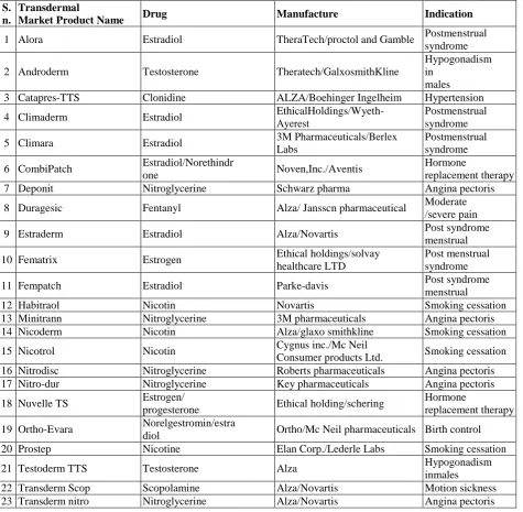
Table 1: gives detail information of the different drugs which are administered by this route and the common names by which they are marketed it also gives the conditions for which the individual system is used 44.
S. n.
Transdermal
Market Product Name Drug Manufacture Indication
1 Alora Estradiol TheraTech/proctol and Gamble Postmenstrual syndrome
2 Androderm Testosterone Theratech/GalxosmithKline
Hypogonadism in
males 3 Catapres-TTS Clonidine ALZA/Boehinger Ingelheim Hypertension
4 Climaderm Estradiol EthicalHoldings/Wyeth- Ayerest
Postmenstrual syndrome
5 Climara Estradiol 3M Pharmaceuticals/Berlex Labs
Postmenstrual syndrome
6 CombiPatch Estradiol/Norethindr
one Noven,Inc./Aventis
Hormone
replacement therapy
7 Deponit Nitroglycerine Schwarz pharma Angina pectoris
8 Duragesic Fentanyl Alza/ Jansscn pharmaceutical Moderate /severe pain
9 Estraderm Estradiol Alza/Novartis Post syndrome
menstrual
10 Fematrix Estrogen Ethical holdings/solvay healthcare LTD
Post menstrual syndrome
11 Fempatch Estradiol Parke-davis Post syndrome
menstrual
12 Habitraol Nicotin Novartis Smoking cessation
13 Minitrann Nitroglycerine 3M pharmaceuticals Angina pectoris 14 Nicoderm Nicotin Alza/glaxo smithkline Smoking cessation
15 Nicotrol Nicotin Cygnus inc./Mc Neil
Consumer products Ltd. Smoking cessation 16 Nitrodisc Nitroglycerine Roberts pharmaceuticals Angina pectoris 17 Nitro-dur Nitroglycerine Key pharmaceuticals Angina pectoris
18 Nuvelle TS Estrogen/
progesterone Ethical holding/schering
Hormone
replacement therapy
19 Ortho-Evara Norelgestromin/estra
diol Ortho/Mc Neil pharmaceuticals Birth control 20 Prostep Nicotine Elan Corp./Lederle Labs Smoking cessation
21 Testoderm TTS Testosterone Alza Hypogonadism
inmales
22 Transderm Scop Scopolamine Alza/Novartis Motion sickness 23 Transderm nitro Nitroglycerine Alza/Novartis Angina pectoris
Future Scope: The future scope of the TDDS includes An insulin patch
Sufentanil patch for chronic cancer pain
Varenicline patch for smoking cessation and a high-dose nicotine patch for fast metabolizers
Estrogen and testosterone patches for post-menopausal women
Selegiline patch for depression in the elderly and cocaine addiction
Clonidine transdermal for the treatment of delerium in trauma patients
Dexamethasone iontophoretic delivery for the treatment of tennis elbow
An iontophoretic sumatriptan patch for migraine treatment, and
Transdermal glyceryl trinitrate for acute stroke therapy, to name a few[14]
CONCLUSION
Darure. World Journal of Pharmaceutical and Life Sciences
delivery system, greater understanding of the different mechanisms of biological interactions, and polymer are required. TDDS realistic practical application as the next generation of drug delivery system.
REFERENCE
1. R. R. Bhagwat* and I.S. Vaidhya, NOVEL DRUG DELIVERY SYSTEMS: AN OVERVIEW, International journal of pharmaceutical science and research, February, 2013; 4(3): 970.
2. Nirav S Sheth , Rajan B Mistry , Formulation and evaluation of transdermal patches and to study permeation enhancement effect of eugenol Journal of Applied Pharmaceutical Science, 2011; 01(03): 96-97.
3. Richa Sachan, Meenakshi Bajpai, and Transdermal Drug Delivery System: A review, International Journal of Research and Development in Pharmacy and Life Sciences, December - January, 2013; 3(1): 748.
4. Tejvir KaurTransdermal drug delivery system: Innovations in skin permeation, Innovations in Pharmaceuticals and Pharmacotherapy, 2017; 5(2): 121-128.
5. Abdul Hafeez, Dr. Upendra Jain, Jagpal Singh, Arun Maurya, Lakhan Rana, Recent Advances in Transdermal Drug Delivery System (TDDS): An Overview, Journal of Scientific and Innovative Research, 10/8/2013; 2(3): 734.
6. Chinmaya Keshari Sahoo1, Prakash Kumar Nayak2, Tanmaya Keshari Sahoo3*, Powshya Dasari4, Santhoshipriya Dandamundi4, A Review of Transdermal drug delivery system. Recent Advances in Pharmaceutical Science Research, 2013; 2(1): 37-38.
7. Takanori Igarashi, Ko Nishino, and Shree K. Nayar, The Appearance of Human Skin, June 2005; 13-16. 8. Mithun Bhowmick*, Tamizharasi Sengodan,
Mechanisms, kinetics and mathematic modeling of transdermal permiation- an updated review, International Journal of Research and Development in Pharmacy and Life Sciences, October - November, 2013; 2(6): 638-639.
9. Nirav S Sheth, Rajan B Mistry, Formulation and evaluation of transdermal patches and to study permeation enhancement effect of eugenol, Journal of applied pharmaceutical scienc, 30-04-2011; 1(3): 98.
10. Latheeshjlal. L, P. Phanitejaswini, Y. Soujanya, U. Swapna, V. Sarika, G. Moulika, Transdermal Drug Delivery Systems: An Overview, International Journal of Pharm Tech Research, Oct-Dec 2011; 3(4): 2143-2144.
11. Garima Verma,Tranasdermal drug delivery system, advanced development and evaluation A Review,International Journal of Pharmaceutical Science and research, 01 February, 2017; 8(2): 392-393.
12. Shikha Deshwal* and Navneet Verma Optimization Techniques in Tansdermal Drug Delivery System,
International journal of pharmaceutical science and research, 2012; 3(08): 2364-2365.
13. Debjit Bhowmik, K. Rao.Pusupoleti2, S.Duraivel2, KP. Sampath Kumar*, Recent Approaches in Transdermal Drug DeliverySystem, the Pharma Innovation – Journal, 2013; 2(3): 105-106.