METHODOLOGY
Visualization of the impatiens downy
mildew pathogen using fluorescence in situ
hybridization (FISH)
Catalina Salgado‑Salazar
1,2, Gary R. Bauchan
3, Emma C. Wallace
1,2,4and Jo Anne Crouch
1*Abstract
Background: Plasmopara obducens is the biotrophic oomycete responsible for impatiens downy mildew, a destruc‑ tive disease of Impatiens that causes high crop loss. Currently, there are no available methods for the microscopic detection of P. obducens from leaves of impatiens, which may be contributing to the spread of the disease. Fluores‑ cence in situ hybridization (FISH) is a sensitive and robust method that uses sequence‑specific, fluorescence‑labeled oligonucleotide probes to detect target organisms from the environment. To study this important pathogen, we developed and standardized a FISH technique for the visualization of P. obducens from Impatiens walleriana tissues using a species‑specific 24‑mer oligonucleotide probe designed to target a region of the rRNA internal transcribed spacer 2 (ITS2).
Results: Since P. obducens cannot be propagated in vitro, we developed a custom E. coli expression vector that tran‑ scribes the P. obducens rRNA‑ITS target sequence (clone‑FISH) for use as a control and to optimize hybridization condi‑ tions. The FISH assay could detect P. obducens sporangiophores, sporangia and oospores, and hyphae from naturally infected I. walleriana leaves and stems. Cross‑reactivity was not observed from plant tissue, and the assay did not react when applied to E. coli with self‑ligated plasmids and non‑target oomycete species.
Conclusions: This FISH protocol may provide a valuable tool for the study of this disease and could potentially be used to improve early monitoring of P. obducens, substantially reducing the persistence and spread of this destructive plant pathogen.
Keywords: Oomycota, Peronosporales, Oospores, Downy mildew, Natural environment
© The Author(s) 2018. This article is distributed under the terms of the Creative Commons Attribution 4.0 International License (http://creat iveco mmons .org/licen ses/by/4.0/), which permits unrestricted use, distribution, and reproduction in any medium, provided you give appropriate credit to the original author(s) and the source, provide a link to the Creative Commons license, and indicate if changes were made. The Creative Commons Public Domain Dedication waiver (http://creat iveco mmons .org/ publi cdoma in/zero/1.0/) applies to the data made available in this article, unless otherwise stated.
Background
Impatiens downy mildew caused by the obligate biotroph
Plasmopara obducens (Oomycota, Peronosporales) is one of the most devastating diseases of the ornamental bedding plant Impatiens walleriana, and also may affect interspecific hybrids and related wild species of Impa-tiens (impatiens; [1–3]). Symptoms of impatiens downy mildew (IDM) include leaf yellowing, stunted growth, leaf drop and stem collapse [4]. As the disease progresses, a white downy-like growth on the underside of leaves can
be observed, corresponding with the emergence of asex-ual fruiting structures (sporangiophores and zoospore-bearing sporangia) from the leaf stomata [5]. Oospores, the pathogen’s primary survival structure, are formed in stems of plants that have recently died, and may serve as a primary inoculum source for subsequent growing sea-sons in landscape settings [6]. Plasmopara obducens spo-rangia are spread by wind currents or by water from rain or irrigation, and under cool and moist conditions, the disease spreads rapidly [6].
Epidemic outbreaks of IDM on I. walleriana were first reported in the early 2000s in the UK, Europe and Aus-tralia [7–10]. In the United States, the first epidemic outbreaks that reached landscape settings started dur-ing the 2011 and 2012 growdur-ing seasons [1, 2, 11–14].
Open Access
*Correspondence: [email protected]
1 Agriculture Research Service (ARS), Mycology and Nematology Genetic
Diversity and Biology Laboratory, U.S. Department of Agriculture, 10300 Baltimore Avenue, Beltsville, MD 20705, USA
Currently, IDM is reported throughout the continental US and the Hawaiian Islands, and continues to limit the health and production of this economically important crop, worth $120 million as of 2014 (USDA-National Agricultural Statistics Service Census of Agriculture 2015 report, https ://www.agcen sus.usda.gov, [3]). Diag-nosis of IDM relies on the presence of typical symptoms that can include leaf yellowing and stunted growth, and at later stages of the disease, is aided by the visible signs of the pathogen’s vegetative and fruiting bodies on the undersurface of infected leaves. However by the time that disease symptoms and visual signs of the pathogen appear, the disease is incurable: plants cannot be treated and losses cannot be avoided. Additionally, plants may be infectious long before detectable symptoms appear. Early detection methods aimed at identifying the infec-tion status of apparently uninfected individuals is key for efficient use of disease control resources [15].
Fluorescence in situ hybridization (FISH) has been widely used as a cultivation-independent tool for direct detection, identification and quantification of microor-ganisms [16]. The cultivation-independent characteristic of this technique is also particularly important for micro-organisms such as obligate biotrophs, as it allows for the direct study of plant pathogens in their natural environ-ment [17, 18]. Although updated techniques, applications and protocol improvements are now available for FISH, the technique is based around four core steps: (1) speci-men fixation and immobilization; (2) permeabilization to increase accessibility of an organism specific-nucleic acid probe to the target; (3) hybridization of the probe; (4) washing to remove unbound probe; and (4) documenta-tion by microscopy or flow cytometry [16, 19, 20]. Typi-cal oligonucleotide probes used for FISH range between 15 and 30 base pairs in length and are labeled with one or more fluorescent dyes [20]. Most FISH applications target ribosomal RNA (rRNA), as these molecules are highly abundant and stable within cells, and possess both variable and highly conserved sequence domains [19]. Single copy genes can also be detected using FISH when coupled with signal amplification techniques such as catalyzed reporter deposition—CARD-FISH [21]. Even though FISH assays using oligonucleotide probes tar-geting rRNA were first introduced almost 30 years ago [22], only few studies have applied this technique for the visualization of oomycete plant pathogens such as Phy-tophthora agathidicida and P. cinnamomi [23–25]. Non-specific fluorescent staining techniques have been used to visualize infection structures and plant cellular growth and response to the grape downy mildew pathogen Plas-mopara viticola [26–28], and to visualize the in planta
development of pathogens such as Peronospora sparsa,
Pe. tabacina, Pseudoperonospora cubensis, and Ps.
humuli, causing rose, tobacco, cucurbit, and hops downy mildew, respectively [28, 29]. However, to date, FISH assays have not been developed for species-specific visu-alization of oomycetes that cause downy mildew diseases. The aim of the current study is to develop a detailed protocol for species-specific FISH visualization of the impatiens downy mildew pathogen. For this, a spe-cific oligonucleotide probe targeting the rRNA inter-nal transcribed spacer (ITS) region of P. obducens was developed and tested using the clone-FISH approach and then validated with P. obducens mycelia, sporan-giophores, sporangia and oospores harvested from I. walleriana, as well as I. walleriana leaves and stems showing symptoms of downy mildew. The development of a FISH probe and hybridization assay allowed the microscopic visualization of P. obducens within I. wal-leriana plant tissues, easily distinguishable from plant cells. This technique could be a useful tool for pathogen detection on non-symptomatic I. walleriana, as well as a tool to study P. obducens life cycle including key cel-lular events such as host penetration and colonization.
Materials and methods
Probe design
The oligonucleotide probe used in this study (rRNA_ ITS_Pob) was designed to specifically target P. obdu-cens. This was achieved by comparing the P. obducens
rRNA ITS 1 and 2 regions (including the 5.8S rRNA gene) [1] with publicly available rRNA ITS sequences retrieved from NCBI GenBank of closely related oomy-cetes in the Peronosporaceae family and oomycete plant pathogens commonly found inhabiting soil. The rRNA_ITS_Pob probe (5′-ACC AAA CTG GTC GCC GAC TTG TTA -3′) has a melting temperature of 59 °C, G–C content 45.8%, phosphorothioate bonds (to be resistant to nuclease degradation), and was synthe-sized commercially (IDT, Coralville, IA, USA). Pre-liminary assessments of autoflorescence emitted by I. walleriana leaves, stems and flowers revealed broad emissions in the green spectra (data not shown), which precluded the use of green spectra fluorophores such as GFP. Two versions of the probe within the blue and red emission spectra were synthesized: one probe with an AlexaFluor350 fluorophore (Invitrogen, Rockville, MD) at the 5′ end (excitation = 346 nm, emission = 442 nm
[blue]) and a second probe with an AlexaFluor594 fluorophore at the 5′ end (excitation = 590 nm,
emis-sion = 617 nm [red]). The spectral properties of these
Preparation of control materials: clone‑FISH cells and non‑target oomycetes
In the absence of pure cultures of P. obducens that could be used as controls to evaluate the rRNA_ITS_Pob probe before its application on impatiens samples, a clone-FISH protocol was used to develop E. coli cells express-ing the P. obducens target sequence, following the general method of Schramm et al. [30]. DNA from P. obducens
sample DE14.2.9 [3] was extracted using the Omni-Prep kit (G-Biosciences, St. Louis, MO, USA) and puri-fied using the Zymo DNA Clean and Concentrator kit (Zymo Research, Irvine, CA, USA). The ~ 1 kb P. obdu-cens target rRNA ITS 1 and 2 regions (including the 5.8S rRNA gene) was amplified by PCR using the DC6-PL-OB-3′-699 primer set (DC6: 5′-GAG GGA CTT TTG GGT AAT CA-3′; PL-OB-3′-699: 5′-TTA GAA GAC CAA GCA ACT CG-3′) with cycling conditions as follows: initial denaturation at 95 °C for 5 min, followed by 35 cycles of 95 °C for 30 s, 51 °C for 45 s and 72 °C for 45 s, and final extension at 72 °C for 10 min. The PCR product was puri-fied using the Wizard SV Gel and PCR Clean-up System (Promega, Madison, WI, USA), ligated into the pCR 2.1-TOPO vector and inserted into Escherichia coli TOP10F’ competent cells (Invitrogen, Carlsband, CA, USA). Forty-eight clones were randomly picked and the insertion and direction of ligated PCR product was checked by Sanger sequencing using the M13 forward–reverse primer set included in the TOPO® TA Cloning Kit (Invitrogen, Carlsband, CA, USA). One E. coli colony containing plas-mid constructs with the insert in the correct orientation was purified using the QIAprep Spin Miniprep Kit (Qia-gen, Germantown, MD, USA) and used to transform E. coli BL21 Star (DE3) One Shot cells (Invitrogen, Carls-band, CA, USA) containing a genomic copy of IPTG-inducible T7 RNA polymerase to generate sufficient transcript for hybridization purposes. One E. coli colony with plasmids containing the insert in the opposite direc-tion and one E. coli colony carrying a self-ligated plasmid (without the insert) were used as negative controls and used to transform E. coli DE3 cells. Cells were cultivated in fresh LB broth in a shaking incubator at 37 °C until they reached a mid-log phase (OD600 of 0.3–0.4). IPTG (isopropyl-β-d-thiogalactopyranoside) in a final concen-tration of 0.5 mM was added to the cultures and allowed to incubate for 1 h to induce transcription of the insert sequence. Chloramphenicol (170 g L−1) was added to the cell culture for 4 h and cells were then fixed overnight in 4% formaldehyde solution at 4 °C. After fixation, clones were pelleted, washed in 1 × PBS (phosphate-buffered
saline) buffer twice, and stored in 1 × PBS/absolute etha-nol (1:1) at − 20 °C until needed for FISH treatment.
Slide cultures of Phytophthora infestans, Phy. sojae and
Pythium irregulare were prepared according to Riddell
[31] to test for cross reactivity between the P. obducens
FISH probe and other oomycetes that commonly reside in agricultural soil. For this, mycelia of Phy. infestans,
Phy. sojae and Py. irregulare was collected from 1-week old cultures on rye agar medium supplemented with 2% glucose (Phy. infestans and Phy. sojae) and potato dex-trose agar (PDA, Py. irregulare) and inoculated at the edge of agar blocks on glass slides following the method described by Riddell [31]. The samples on the resulting slide cultures were processed immediately.
Collection and preparation of Plasmopara obducens samples for imaging
Mycelia, sporangia and sporangiophores of P. obdu-cens were obtained from naturally infected I. walleriana
plants collected in Montgomery County, MD. Mycelia, sporangia and sporangiophores were collected from the underside of the leaves using an entomological needle and with the aid of a Zeiss dissecting microscope Discov-ery V20 (Carl Zeiss Microscopy, Thornwood, NY, USA). Oospores were obtained by inoculating surface-disin-fested (10% commercial bleach for 60 s) stem segments of healthy I. walleriana with a suspension of freshly col-lected sporangia (1 × 10⁵ spores mL−1). Inoculated stems were kept in moist chambers consisting of moist filter paper in a 90 mm Falcon sterile polystyrene petri plates (Becton–Dickinson Labware, Franklin Lakes, NJ, USA) and incubated with a 14 h photoperiod for approximately 1 month at 20 °C to allow the oospores to develop and mature.
Fluorescence in situ hybridization
In preparation for hybridization, samples of E. coli, Phy. infestans, Phy. sojae, P. obducens and Py. irregulare were heat fixed onto glass microscope slides by incubating the slide on the surface of a 60 °C hotplate for 15 s. After incubation, a 65 µL capacity Frame-seal© (BIO-RAD Lab-oratories, Hercules, CA, USA), was placed on the slides around the samples.
Fresh leaves and stems of I. walleriana naturally infected with P. obducens were prepared for hybridiza-tion by fixing overnight in 4% formaldehyde soluhybridiza-tion at 4 °C. After fixation, the leaves and stems were washed in 1 × PBS buffer twice and tissue cleared using a solution of
ethanol 95%:acetic acid:glycerol (75:15:10 v/v). The fixed plant material can be stored in 1 × PBS/absolute ethanol
(1:1) at 4 °C for up to 6 months. Hybridization of leaves and stems was performed in sterile polystyrene petri plates (Falcon 35 × 10 mm; Becton–Dickinson Labware,
Franklin Lakes, NJ, USA).
of formamide with non-toxic organic solvents, increas-ing the hybridization rate and reducincreas-ing the temperature required to perform the denaturation and hybridization steps [32, 33]. Briefly, samples were dehydrated through a graded series of ethanol (2 min 70% ethanol, 2 min 85% ethanol, 2 min 96% ethanol) and air-dried. Dehydrated samples were incubated with hybridization buffer (15% v/v ethylene carbonate, 20% v/v dextran sulfate, 600 mM NaCl, 10 mM citrate buffer, 2 ng L−1 fluorescent probe, pH 6.2) at 67 °C for 10 min, followed by incubation at 45 °C for 1 h. 50 µL of hybridization buffer was used on slides and approximately 200 µL was used for incuba-tion of leaf and stem tissue. After hybridizaincuba-tion, samples were washed three times: an initial 10 min wash at 65 °C in buffer 1 (0.05 M Tris–HCl, 0.3 M NaCl, 0.1% Tween 20, pH 7.6), followed by two room temperature washes in buffer 2 (0.05 M Tris–HCl, 0.15 M NaCl, 0.1% Tween 20, pH 7.6) for 3 min. Washed samples were dehydrated through a graded series of ethanol (2 min 70% ethanol, 2 min 85% ethanol, 2 min 96% ethanol) and air-dried. Samples mounted on glass microscope slides had their Frame-seal© removed and a glass cover slip was added to the samples with a drop of ProLong® Diamond Antifade
Mountant (ThermoFisher, Carlsband, CA, USA) and cured overnight at 4 °C in the dark.
Microscopy and image acquisition
Fluorescence microscopy was performed with a Zeiss Axio Imager.M2 microscope equipped with an HXP120V fluorescent light source and filter sets 49 (excitation G365, emission BP 445/50) and 64 HE (exci-tation BP587/25, emission 647/70) (Carl Zeiss Micros-copy, Thornwood, NY, USA). Images were acquired in gray scale using an Axiocam 506 Mono digital camera (Carl Zeiss Microscopy) and processed using Zen 2 Pro Software (Carl Zeiss Microscopy). Lower magnifica-tion images were obtained using a Zeiss Axio Zoom. V16 steromicroscope system (Carl Zeiss Microscopy). The exposure time for each channel was auto-balanced prior to image acquisition in order to avoid oversatura-tion of signal or bleaching of the probe. Exposure times obtained for the fluorescent channels using a positive sample resulting from the clone-FISH assay and P. obdu-cens mycelia were subsequently used for the evaluation of the probes hybridized against I. walleriana infected leaves and negative controls (other oomycetes and E. coli
colony carrying a self-ligated plasmid). Confocal images of oospores were obtained using a Zeiss LSM710 con-focal laser scanning microscopy (CLSM) system (Carl Zeiss Microscopy). CLSM images were generated using a Zeiss Axio Observer inverted microscope with 10 × 0.45 NA and 25 × 0.8 NA Plan-Apochromat objectives. Two lasers were used, 488 nm argon laser and 561 nm diode-pumped solid state laser with a pin hole of 33 μm passing through a MBS 488 or MBS 561 beam splitter filter with limits set between 490 and 530 nm for detection using the 488 nm laser and 565–650 nm for detection using the 561 nm laser. Zeiss Zen 2012 (Carl Zeiss Microscopy) software was used to obtain 20–30 Z-stack images and a maximum intensity projection was used to develop the final 2D image.
Results and discussion
Validation of rRNA ITS‑targeted oligonucleotide probe
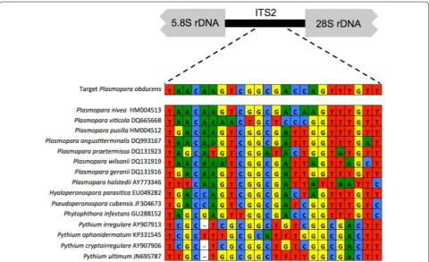
The 24-nucleotide long rRNA_ITS_Pob probe designed in this study was based on the nucleotide divergence between P. obducens ITS rRNA region and that of other
Plasmopara species and Peronosporaceae taxa (Fig. 1). The region used to develop the probe was located within the ITS 2 region and possessed ideal nucleo-tide composition for use as a probe (data not shown). Additional probe regions in the ITS rRNA regions with enough species-specific nucleotides were not identified. BLASTn searches against NCBI GenBank showed that ITS sequences from two samples of the lettuce downy mildew pathogen Bremia lactucae (host =Hemistepta lyrata) shared significant similarity with the probe, with 100% identity over 79% of the sequence (e-value = 2.3; accessions DQ235793, DQ235794). Other members of the genus Plasmopara shared a maximum of 96% identity over 88% of the probe sequence (e.g. P. nivea
EF553508, host =Aegopodium podagraria; P. angusti-terminalis DQ993167, host =Xanthium strumarium; e-value = 1e − 04). However, given the fact that none of these Peronosporaceae organisms are known to inhabit impatiens, cross-reactivity with these organisms from impatiens samples is extremely unlikely.
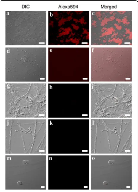
Exposure times for images of clone-FISH and P. obducens positive samples using the Alexa-350 and Alexa-594-labeled probes varied according to the sam-ple assayed, with clone-FISH cells requiring exposure times an order of magnitude longer (2.6–5.2 s expo-sure; Fig. 2a–c) than P. obducens mycelia and oospores (50–500 ms exposure) or pathogen-infected leaf tissue (5–20 ms exposure). By reusing the fluorescent channel exposure time of positive samples, no fluorescence signal
was detected from E. coli DE3 strains carrying self-ligated plasmids (Fig. 2d–f), slide preparations of Phy. infestans,
Phy. sojae and Py. irregulare (Fig. 2g–o) or from healthy I. walleriana leaf tissue (not shown).
FISH assay of P. obducens mycelia and I. walleriana
infected leaves and stems (oospores).
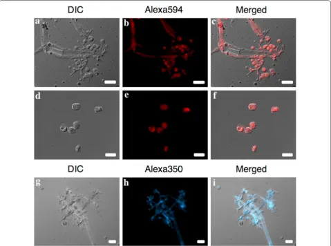
The FISH assay directly applied to P. obducens sporan-giophores and sporangia fixed onto glass slides showed the rRNA_ITS_Pob probe exhibited strong fluorescence, suggesting good permeability of the probe into the cells of P. obducens, without the need for additional cell pre-treatment (Fig. 3a–i). The rRNA_ITS_Pob probe labeled with the Alexafluor 350 (blue) dye generally gave a less
Fig. 2 Probe validation using clone‑FISH and evaluation of cross‑reactivity with non‑target oomycetes. a–c Positive control:
Escherichia coli DE3 cells expressing the Plasmopara obducens target sequence; d–f negative control: E. coli DE3 cells carrying a self‑ligated plasmid without the P. obducens target sequence insert; g–i negative controls: Phytophthora infestans mycelia and spore; j–lPhy. sojae
intense signal than the probe labeled with Alexafluor 594 (red), but still provided specific labeling of P. obducens
sporangiophores and sporangia (Fig. 3g–i).
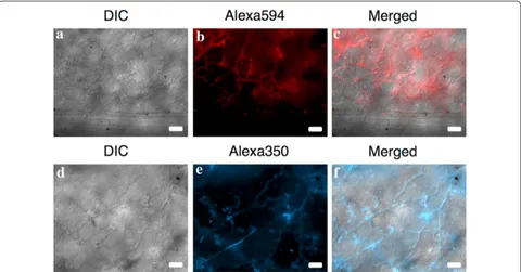
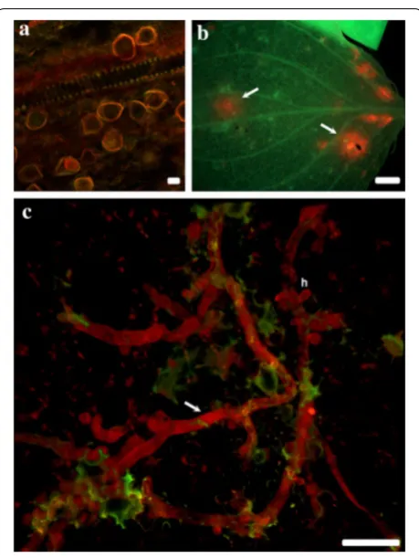
Naturally infected I. walleriana leaves that were cleared previous to the FISH treatment produced fluorescence signal that corresponded to P. obducens hyphae growing within the palisade and spongy mesophyll (Figs. 4a–f,
5c). Fluorescence appeared especially intense on some areas of the I. walleriana leaves (Fig. 5b), mostly due to accumulated overlaying invading hyphae, which are com-mon beneath the pathogen penetration sites and the presence of haustoria in mesophyll cells [34]. Based on these results, it appears that the probe has the ability to penetrate several layers of I. walleriana cells. This would allow for the application of this assay on a variety of I.
walleriana plant tissues, without the need of histologi-cal sectioning. Mycelia was not observed within vascular tissue (Fig. 4a–c), however mature oospores within the stem cortex were detected, with the fluorescence local-ized in the area between the wall of the oospore and the oogonium (Fig. 5a).
The FISH probe developed in this study targets tran-scribed target rRNA, however its successful application relies on the presence of intact RNA molecules in the sample. The rRNA content of microorganisms has been found to be correlated the active growth and it is often used as an indicator of ongoing cellular activity [35]. The FISH method applied to a single dried herbarium speci-men of I. walleriana infected with P. obducens failed to emit detectable fluorescence (data not shown). It is likely Fig. 3 Plasmopara obducens sporangiophores and sporangia hybridized to probes labeled with Alexa595 (red, a–f) or Alexa350 (blue, g–i)
that fresh samples previously fixed using paraformalde-hyde or any other preservation methods are better suited for the application of this FISH method.
Conclusion
A novel probe was developed for FISH-based visualiza-tion of P. obducens on I. walleriana. FISH is a reliable tool to detect and localize a wide variety of microorganisms in different growing stages and from different sources. This is the first time this technique has been success-fully applied for the visualization of plant pathogens in the Peronosporaceae that cause downy mildew disease. This technique is anticipated to be valuable in the early detection of infected I. walleriana plant material before the appearance of typical symptoms or the presence of pathogen structures on the exterior of the plant host, which are both indicative of advanced stages of disease
overall improved understanding of IDM are essential to reduce the effect of this devastating disease.
Authors’ contributions
CS‑S and JAC conceived and designed experiments. CS‑S, GB, ECW performed experiments. CS‑S, GB, ECW and JAC analyzed data. CS‑S wrote the manu‑ script. GB, ECW and JAC critically commented and revised the manuscript. All authors read and approved the final manuscript.
Author details
1 Agriculture Research Service (ARS), Mycology and Nematology Genetic
Diversity and Biology Laboratory, U.S. Department of Agriculture, 10300 Balti‑ more Avenue, Beltsville, MD 20705, USA. 2 ARS Research Participation Program,
Oak Ridge Institute for Science and Education, MC‑100‑44, P.O. Box 117, Oak Ridge, TN 37831, USA. 3 Agriculture Research Service, Electron and Confocal
Microscopy Unit, U.S. Department of Agriculture, 10300 Baltimore Avenue, Beltsville, MD 20705, USA. 4 Present Address: Department of Plant Pathol‑
ogy and Environmental Microbiology, The Pennsylvania State University, 120 Buckhout Lab, University Park, PA 16802, USA.
Acknowledgements
We are grateful for the contributions of Aaron Palmateer, Karen Rane, and Margery Daughtrey who provided diseased plant samples, and to Margery Daughtrey and Richard Jones who provided Phytophthora and Pythium cultures.
Competing interests
The authors declare that they have no competing interests.
Commercial endorsement disclaimer
Mention of trade names or commercial products in this publication is solely for the purpose of providing specific information and does not imply recom‑ mendation or endorsement by the USDA.
Equal opportunity statement
USDA is an equal opportunity provider and employer.
Funding
This work was supported by funds from the USDA‑APHIS Farm Bill 10007 program, USDA‑ARS Project 8042‑22000‑298‑00‑D, and by the appointment of C. Salgado‑Salazar and E. Wallace to the ARS Research Participation Program administered by the Oak Ridge Institute for Science and Education (ORISE) through an interagency agreement between the U.S. Department of Energy (DOE) and the USDA. ORISE is managed by ORAU under DOE Contract No. DE‑AC05‑06OR23100.
Publisher’s Note
Springer Nature remains neutral with regard to jurisdictional claims in pub‑ lished maps and institutional affiliations.
Received: 22 August 2018 Accepted: 19 October 2018
References
1. Crouch JA, Ko MP, McKemy JM. First report of impatiens downy mildew outbreaks caused by Plasmopara obducens throughout the Hawaiian Islands. Plant Dis. 2014;98:696.
2. Palmateer AJ, Lopez P, Seijo TE, Peres NAR. Severe outbreak of downy mil‑ dew caused by Plasmopara obducens on Impatiens walleriana in Florida. Plant Dis. 2013;97:687.
3. Salgado‑Salazar C, LeBlanc N, Ismaiel A, Rivera Y, Warfield C, Crouch JA. Genetic variation of the pathogen causing impatiens downy mildew pre‑dating and including 21st century epidemics on Impatiens walleriana. Plant Dis. 2018. https ://doi.org/10.1094/PDIS‑01‑18‑0077‑RE.
4. Warfield CY. Downy mildew of impatiens. GrowerTalks. 2012;75:78–86. 5. Eskandari F, Shishkoff N. Systemic infection of Impatiens balsamina
through inoculation of roots with vegetative sporangia of the impa‑ tiens downy mildew (Plasmopara obducens) (Abstr.). Phytopathology. 2017;107:S4.3.
6. Shishkoff N. Evidence points to homothally in the Impatiens downy mildew (Abstr.). Phytopathology. 2016;106:S3.4.
7. Bulajić A, Vučurović A, Stanković I, Ristić D, Jović J, Stojković B, Krstić B. First report of Plasmopara obducens on Impatiens walleriana in Serbia. Plant Dis. 2011;95:491.
8. Cunnington JH, Aldaoud R, Loh M, Washington WS, Irvine G. First record of Plasmopara obducens (downy mildew) on impatiens in Australia. Plant Pathol. 2008;57:371.
9. Lane CR, Beales PA, O’Neill TM, McPherson GM, Finlay AR, David J, Constantinescu O, Henricot B. First report of impatiens downy mildew (Plasmopara obducens) in the UK. Plant Pathol. 2005;54:243.
10. Petróczy M, Csejk G, Palkovics L. First report of Plasmopara obducens causing downy mildew on Impatiens walleriana in Hungary. Plant Dis. 2012;96:148.
11. Baysal‑Gurel F, Taylor NJ, Chatfield J, Miller SA. First report of impatiens downy mildew caused by Plasmopara obducens in Ohio. Plant Dis. 2012;96:1699.
12. Conner KN, Olive J, Hagan AK, Zhang L, Bloodworth ME. First report of impatiens downy mildew caused by Plasmopara obducens in Alabama. Plant Dis. 2014;98:1006.
13. McGinnis E, Kinzer K, LeBoldus J. First report of impatiens downy mildew caused by Plasmopara obducens in North Dakota. Plant Dis. 2015;99:1039. 14. Ward NA, Dixon E, Amsden B. First report of impatiens downy mildew
caused by Plasmopara obducens in Kentucky. Plant Dis. 2013;97:428. Fig. 5 Plasmopara obducens oospores and mycelia colonizing
•fast, convenient online submission
•
thorough peer review by experienced researchers in your field
• rapid publication on acceptance
• support for research data, including large and complex data types
•
gold Open Access which fosters wider collaboration and increased citations maximum visibility for your research: over 100M website views per year
•
At BMC, research is always in progress.
Learn more biomedcentral.com/submissions
Ready to submit your research? Choose BMC and benefit from: 15. Thompson RN, Gilligan CA, Cunniffe NJ. Detecting presymptomatic infec‑
tions is necessary to forecast major epidemics in the earliest stages of infectious disease outbreaks. PLoS Comput Biol. 2016;12:e1004836. 16. Wagner M, Haider S. New trends in fluorescence in situ hybridization for
identification and functional analyses of microbes. Curr Opin Biotechnol. 2012;23:96–102.
17. Ellison MA, McMahon MB, Bonde MR, Palmer CL, Luster DG. In situ hybridization for the detection of rust fungi in paraffin embedded plant tissue sections. Plant Methods. 2016;12:37.
18. Kubota K. CARD‑FISH for environmental microorganisms: technical advancement and future applications. Microbes Environ. 2013;28:3–12. 19. Amann R, Fuchs BM. Single‑cell identification in microbial communi‑
ties by improved fluorescence in situ hybridization. Nat Rev Microbiol. 2008;6:339–48.
20. Moter A, Göbel UF. Fluorescence in situ hybridization (FISH) for direct visualization of microorganisms. J Microbiol Methods. 2000;41:85–112. 21. Zwirglmaier K. Fluorescence in situ hybridization (FISH)—the next gen‑
eration. FEMS Microbiol Lett. 2005;246:151–8.
22. DeLong EF, Wickham GS, Pace NR. Phylogenetic stains: ribosomal RNA‑based probes for the identification of single cells. Science. 1989;243:1360–3.
23. Bellgard SE, Padamsee M, Probst CM, Lebel T, Williams SE. Visualizing the early infection of Agathis australis by Phytophthora agathidicida using microscopy and fluorescent in situ hybridization. For Pathol. 2016;46:622–31.
24. Crone M, McComb JA, O’Brien PA, Hardy GESJ. Survival of Phytophthora cinnamomi as oospores, stroma, and thick‑walled chlamydospores in roots of symptomatic and asymptomatic annual and herbaceous peren‑ nial plant species. Fungal Biol. 2013;117:112–23.
25. Li AY, Crone M, Adams PJ, Fenwick SG, Hardy GESJ, Williams N. The micro‑ scopic examination of Phytophthora cinnamomi in plant tissues using fluorescent in situ hybridization. J Phytopathol. 2014;162:747–57. 26. Díez‑Navajas AM, Greif C, Poutaraud A, Merdinoglu D. Two simplified fluo‑
rescent staining techniques to observe infection structures of the oomy‑ cete Plasmopara viticola in grapevine leaf tissues. Micron. 2007;38:680–3.
27. Díez‑Navajas AM, Wiedemann‑Merdinoglu S, Greif C, Merdinoglu D. Non‑ host versus host resistance to the grapevine downy mildew, Plasmopara viticola, studied at the tissue level. Phytopathology. 2008;98:776–80. 28. Kortekamp A. Growth, occurrence and development of septa in
Plas-mopara viticola and other members of the Peronosporaceae using light‑ and epifluorescence‑microscopy. Mycol Res. 2005;109:640–8. 29. Salgado‑Salazar C, Shishkoff N, Daughtrey ML, Palmer C, Crouch JA.
Downy mildew: a serious disease threat to rose health worldwide. Plant Dis. 2018. https ://doi.org/10.1094/PDIS‑12‑17‑1968‑FE.
30. Schramm A, Fuchs BM, Nielsen JL, Tonolla M, Stahl DA. Fluorescence in situ hybridization of 16S rRNA gene clones (clone‑FISH) for probe vali‑ dation and screening of clone libraries. Environ Microbiol. 2002;4:713–20. 31. Riddell RW. Permanent stained mycological preparations obtained by
slide culture. Mycologia. 1950;42:265–70.
32. Matthiesen SH, Hansen CM. Fast and non‑toxic in situ hybridization without blocking of repetitive sequences. PLoS ONE. 2012;7:e40675. 33. Moffitt JR, Hao J, Wang G, Chen KH, Babcock HP, Zhuang X. High‑
throughput single‑cell gene‑expression profiling with multiplexed error‑robust fluorescence in situ hybridization. Proc Natl Acad Sci USA. 2016;113:11049–51.
34. Lu Y‑J, Schornack S, Spallek T, Gelder N, Chory J, Schellmann S, Schu‑ macher K, Kamoun S, Robatzek S. Patterns of plant subcellular responses to successful oomycete infections reveal differences in host cell repro‑ gramming and endocytic trafficking. Cell Microbiol. 2012;14:682–97. 35. Blazewicz SJ, Barnard RL, Daly RA, Firestone MK. Evaluating rRNA as an
indicator of microbial activity in environmental communities: limitations and uses. ISME J. 2013;7:2061–8.
36. Prieto P, Navarro‑Raya C, Valverde‑Corredor A, Amyotte SG, Dobinson KF, Jesús M‑B. Colonization process of olive tissues by Verticillium dahlie and its in planta interaction with the biocontrol root endophyte Pseudomonas fluorescens PICF7. Microb Biotechnol. 2009;2:499–511.