R E S E A R C H
Open Access
Non-invasive neuromodulation to improve gait in
chronic multiple sclerosis: a randomized double
blind controlled pilot trial
Mitchell E Tyler
1,2, Kurt A Kaczmarek
2, Kathy L Rust
2, Alla M Subbotin
2, Kimberly L Skinner
1*and Yuri P Danilov
2Abstract
Background:This study sought to examine the effect of targeted physical therapy with and without cranial nerve non-invasive neuromodulation (CN-NINM), on the walking ability of people with MS who exhibited a dysfunctional gait. We hypothesized that subjects who received electrical stimulation would have greater improvement than those who had a control device after a 14-week intervention. Gait disturbance is a common problem for people with multiple sclerosis (MS). Current management may include exercise, pharmacology, functional electrical stimulation, compensatory strategies, use of assistive devices, and implanted electrical devices. We have developed an effective rehabilitative strategy using neuromodulation of the cranial nerves via electrical stimulation of the tongue to enhance the plasticity of the brain.
Methods:The study is a within-subject blinded randomized control design. Twenty chronic MS subjects with an identified gait disturbance were assigned to either an active or control group. Both groups completed a 14-week intervention program using a standardized combination of exercise and a device that provided electrical stimulation to the tongue. Those in the active group received electrical stimulation on the tongue that they could perceive. Those in the control group used a device that did not provide a physiologically significant stimulus and was not perceivable. Subjects were assessed with the Dynamic Gait Index (DGI).
Results:The DGI scores improved for both groups. There were significant between-group differences, with the active group showing statistically greater improvement than the control group mean.
Conclusion:People with MS demonstrated improved gait with CN-NINM training in a pilot randomized controlled trial. This study suggests that tongue-based neurostimulation may amplify the benefits of exercise for improving gait in people with chronic MS.
Keywords:Multiple sclerosis, Neuromodulation, Balance, Gait, Tongue, Neurostimulation, Non-invasive, Electrical stimulation
Background
Individuals with moderate multiple sclerosis (MS) present with an array of symptoms, with walking impairment being among the most common. Gait speed, cadence, stride length and time spent on double-limb support are fre-quently affected, and correlate with reduced independence and productivity, impacting overall quality of life [1]. It is a major factor in estimates of disease progression [2-4], and
approximately 40% of MS patients will need some form of walking assistance within 15 years of disease onset [5]. Consequently, interventions targeting gait disturbance in patients with MS are in demand. Currently these include rehabilitation therapy and pharmacological management.
While therapeutic exercise has long been believed to increase symptoms, more recent evidence has demon-strated that exercise and increased physical activity are beneficial for people with MS, and are becoming more commonly included as part of treatment interventions [6-10]. The optimal type or mode of exercise, intensity, frequency, duration and maintenance of exercise training
* Correspondence:[email protected] 1
Department of Biomedical Engineering, University of Wisconsin, Madison, WI 53706, USA
Full list of author information is available at the end of the article
to manage the progression of balance and gait dysfunc-tion in MS has, however, yet to be established through randomized trials [3].
Rehabilitation therapy includes occupational, physical and speech therapies as part of the comprehensive man-agement of symptoms [11,12]. Compensatory strategies such as energy conservation, use of adaptive equipment, and environmental adaptations are often utilized in these interventions [13-20]. Medical devices and physical mo-dalities may also be used in multidisciplinary interven-tions [21]. A recent Cochrane review of multidisciplinary rehabilitation for adults with MS did not find strong evi-dence of interventions that resulted long-term improve-ments [12].
Pharmacological management may slow disease pro-gress or reduce motor symptoms. For example, Ampyra (Acorda Therapeutics) was approved after it was shown to be effective in improving walking speed by 20% when compared to a placebo group that improved 8% [2]. This outcome, however, was observed in only 35% of those tested, and many participants reported significant side effects. Additionally, there is little evidence that these drugs will prevent the mobility disability of persons with 2nd stage MS (Kurtzke Expanded Disability Status Scale (EDSS) score of 4.0 or greater) [1,3,22,23].
The medical devices and physical modalities utilized in treatment for people with MS vary. Functional electrical stimulation, as delivered through electrodes attached to the skin, has been shown to be effective in improving gait, as long as the electrodes are attached and the stimulation unit is on [24-29]. Other contemporary forms of neurosti-mulation (aimed at induced neuromodulation) are inva-sive, expensive and have the potential for adverse effects. For example, deep brain stimulation and vagus nerve stimulation, which use implanted pacemaker-like electrical devices, are indicated for decreasing tremors in MS, but carry surgical risks [30-33]. These therapies have not been widely attempted in MS rehabilitation. Neurostimulation that directly stimulates the peripheral or central nervous systems to improve motor impairments is still being inves-tigated. Non-invasive neurostimulation that has been tried in people with MS are typically large clinic-based devices that employ powerful electromagnetic stimulation of the brain’s cortex (transcranial magnetic stimulation) [34,35], or electrodes that pass electric current through the skull (transcranial direct current brain stimulation) [36,37]. Intermittent theta burst stimulation of the motor cortex has been demonstrated to reduce spasticity in people with MS [38], and when combined with exercise therapy, to decrease fatigue [35]. Success with these inter-ventions has, however, been limited. Consequently, there continues to be a need for alternative approaches that have the potential to help improve mobility in people with MS.
Previous studies have shown that the tongue can be used as an effective interface for sending electrical signals to the central nervous system [39-45], for example sensory substitution in balance-impaired or blind individuals [43,46-49,50]. Individuals with primary vestibular disor-ders who trained using electrical stimulation through the tongue coupled to head-position information dem-onstrated balance improvements that were sustained for weeks beyond the final stimulation session [46,51].
Using a different methodology that delivers tongue stimulation which, like the present study, is devoid of in-formation linked to head position or any other exogen-ous variable, our recent functional MRI results indicated that the improvements that occurred with the neuromo-dulation training (using electrical stimulation on the tongue) are likely related to modulation of neural activ-ity within structures of the brain that control balance and movement [44,45,52].
Objective
This study sought to examine the effect of targeted physical therapy, with and without cranial nerve non-invasive neuromodulation (CN-NINM), on the walking ability of people with MS who exhibited a dysfunctional gait. We hypothesized that subjects who received elec-trical stimulation on the tongue would have greater im-provement than those who had a control device after a 14-week intervention.
Methods
Study design
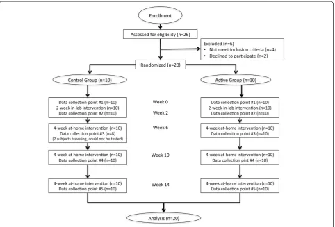
The study was a randomized, double blind controlled trial. Twenty subjects (males and females) with identified gait deficits due to the effects of MS were distributed into 2 treatment groups: 10 used an“Active”CN-NINM device, or Portable Neuromodulation Stimulator (PoNS™), and 10 used a“Control”(very low stimulation) device. The study consisted of two phases: 2 weeks of twice-daily gait train-ing in the laboratory while ustrain-ing the PoNS, followed by 12 weeks of the same daily routine at home. All subjects performed the same gait training over the period of the study. All subjects were tested at the beginning and end of the first phase, and every 4 weeks during the second phase. The structure of the study is depicted in Figure 1.
Subjects
the study; EDSS scores of 3.5 to 6.0; no changes in medi-cation within 3 months of enrollment; and ability to walk 20 minutes on a treadmill (with handrail support as needed) without rest. The EDSS is a rating system used for classifying the condition of people with MS, and em-phasizes walking ability. Scores in the range of 3.5 to 6.0 on the EDSS represent people who are ambulatory and have a few functional systems affected to those more sig-nificantly affected, requiring intermittent or constant uni-lateral assistance (e.g., a cane, crutch, or brace) to walk 100 meters with or without resting.
Exclusion criteria were: major co-morbidities, espe-cially other neurological disorders, uncontrolled pain, hypertension, diabetes, or oral health problems. A sum-mary of the subjects’general characteristics is presented in Table 1. Three subjects (2 Control, 1 Active) were on neurostimulating medication (e.g. fampridine) that might alter motor function. Five subjects were on anti-inflammatory medications, six on antispasmodics, and six had non-narcotic prescriptions to manage chronic pain. Four candidates were not enrolled because they did not meet the inclusion criteria, and two did not par-ticipate because they could not meet the time commit-ments of the study.
The University of Wisconsin-Madison Health Sciences Institutional Review Board approved this study and all subjects gave written consent before participation.
Randomization and blinding
Subjects were randomly assigned to either the control or active group by the primary investigator (PI) as they en-rolled in the study. Ten subjects were assigned to each group, with no regard given to age, gender, individual EDSS score, disease state, functional status, or chronicity of MS. The PI provided each subject with the proper Figure 1Detailed flow chart of the study intervention.
Table 1 Subject characteristics at baseline
Active Control
Subjects 10 10
Men/Women 4/6 2/8
Mean SD Mean SD p
Age 55.40 8.73 51.90 9.31 0.40
Years with MS 24.10 11.03 13.10 6.72 0.01*
EDSS 5.25 0.98 4.60 1.05 0.17
DGI 8.90 2.85 11.95 4.04 0.07
*Indicates significant difference.
neuromodulation device and instructed the subject in its use. The subjects in the active group used a device that provided electrical stimulation on the tongue that they could perceive. Those in the control group used a device that provided a stimulus that was not perceivable. Subjects were instructed that this was controlled study investigat-ing the effects of stimulus level so they may or may not feel the stimulation. To avoid deception, all subjects in both groups were assured that they were in fact receiving stimulation, whether or not they could feel the stimu-lus. The subjects and the therapists providing the inter-vention and testing were not informed which group a subject was assigned. In order to maintaining blinding, both subjects and therapists were instructed to not dis-cuss any details of the stimulus sensation with each other. Additionally, all subjects were instructed to not adjust the stimulus intensity in the presence of the therapist. All questions about device use or the stimulation were to be addressed only to the PI. A summary of subject distribu-tion in the two cohorts is presented in Table 2.
Experimental procedures Intervention
The goal of the training was to develop a more normal gait pattern. The twice-daily intervention was identical for each subject, and progressed in two phases: a 2-week twice daily in-lab phase, followed by a 12-week at-home phase where subjects performed the same training as instructed in the laboratory. The structure and progres-sion of both the in-lab and at-home interventions, as well as the assessments, are shown in Figure 1. Subjects were telephoned weekly during the at-home phase to en-sure compliance with the protocol. In order to maximize any potential benefit from participation, the degree of challenge in each subject’s training program was in-creased according to the progress they made at each 4-week follow-up visit for testing and retraining.
Device
Electrical stimulation to the tongue was delivered via the Portable Neuromodulation Stimulator (PoNS™) device shown in Figure 2. The PoNS™device was held in place lightly by the lips and teeth around a rectangular tab
that goes into the mouth and rests on the anterior, su-perior part of the tongue. The tab has 144 exposed gold-plated circular electrodes (1.5 mm diam., on 2.3-mm centers) on a 3 cm × 3 cm square matrix on a rigid printed circuit board that is coated with a biocompatible epoxy (Epotech 302-3 M, Epoxy Technology, Billerica, MA). The 12x12 array is divided into nine 4x4 sectors. Only one electrode in each sector is pulsed at any given time; the remaining electrodes serve as the return current path. The stimulation on each electrode is a triplet of 50μs-wide positive pulses delivered at 200 pulses/s every 20 ms. Capacitive coupling ensures zero net direct current to minimize the possibility of tissue irritation. Electrode and waveform parameters were derived from earlier re-search aimed at developing electrotactile stimulation that is maximally comfortable and controllable [53-56], and implemented using circuitry similar to that in the earlier Tongue Display Unit [51].
Device function is user-controlled by buttons for ‘On’ and ‘Off,’ and subjects could adjust the stimulus level (pulse amplitude) from 0 to 17 volts by manipulating a knob on the device (see Figure 2). The“Control”version of the device was physically identical to the Active de-vice, but delivered a stimulus at approximately 1/1,000 the minimum perceivable level. Here, adjustment of the intensity knob did not change this level.
To avoid bias or deception, subjects in both groups were provided with identical instructions to adjust the intensity level by turning the knob. They were told that they may or may not feel the stimulation. If they could feel the stimulus, they were to adjust the intensity until it was strong but not uncomfortable. We allowed individual ad-justment rather than setting a fixed amplitude across all subjects, because of known inter-subject differences in tongue sensitivity, with the rationale that it is more im-portant to hold invariant the result of the stimulation Figure 2Portable Neuromodulation Stimulator (PoNS™) device, top and bottom view.
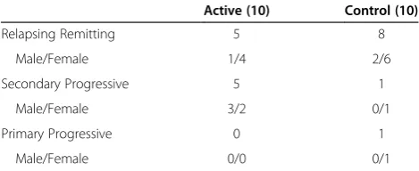
Table 2 Multiple sclerosis subgroups
Active (10) Control (10)
Relapsing Remitting 5 8
Male/Female 1/4 2/6
Secondary Progressive 5 1
Male/Female 3/2 0/1
Primary Progressive 0 1
rather than its physical level [54]. From our previous re-search, we have observed that with experience, subjects set the intensity level at between 50 and 80% of the max-imum dynamic range of sensation [43-48,50-52]. If sub-jects could not feel it, they were reassured that they were still receiving stimulation but it was below their individual threshold of perception.
CN-NINM training
Instruction and implementation of the 14-week interven-tion was identical for both groups. Subjects trained in the lab for 5 consecutive days (Monday through Friday) for 2 consecutive weeks working one-on-one with a therapist researcher for all in-laboratory training sessions. Subjects then continued the same training independently at home for the next 12 weeks. Subjects returned to the lab every 4 weeks for assessment, training review, and exercise pro-gram progression. Compliance was monitored daily in the lab, and weekly via self-reports by phone when the sub-jects trained at home.
The in-lab training consisted of two appointments per day. Within each appointment, subjects performed movement isolation exercises without the device, 20 minutes of gait training with the device, 20 minutes of balance training with the device, and 20 minutes of re-laxation training with the device. The training was tar-geted to the specific ability of each individual, and they were provided with rest periods as needed. Subjects who were unable to complete the training components in the lab due to fatigue were allowed to delay that component until later in the day after they had had an opportunity to rest. Subjects were also expected to incorporate an additional relaxation session with the device at home each evening one hour before bed.
Subjects were instructed in movement isolation exer-cises (without device) at the beginning of each training session. These were geared to the ability of that individual. These exercises were designed to change abnormal move-ment patterns and re-train movemove-ments for improved neuromuscular control and mobility. Sample exercises were chin circles, shoulder circles, and hip circles. The emphasis was placed on quality of movements, not speed, and mirrors were used to provide visual feedback as needed. As the individual demonstrated competency with an exercise, new exercises were introduced.
During gait training subjects walked on a treadmill at progressive speeds and challenges designed to re-establish appropriate dynamic balance and gait patterns. Particular attention was given to symmetry of the gait pattern, in-cluding dynamic weight transfer, stride length, kinematics of the hip, knee, and ankle flexions/extensions, and bilat-eral symmetry of the stance and swing phases. In-lab gait training was performed on the treadmill for the first week, and both over ground and on the treadmill during the
second week. Subjects were required to use a treadmill for at least 50% of the at-home training phase to control and monitor their training progress.
Gait training sessions were 20 minutes in overall dur-ation. The first 5 minutes were performed at a comfort-able pace. In the next two 5-minute periods the challenge was increased by changing a gait variable. The last 5-minute period was performed at a comfortable in-tensity but greater than that for the first 5-minute period. The variables were:
Speed: from very slow to fast walking, focusing on
maintaining gait kinematics without deterioration of performance;
Grade: an incline increases effort and affects the
relative involvement of the ankle, knee and hip joints;
Support: use of handrails provides greater stability,
but prevents arm swing and full weight transfer during the gait cycle. Challenge was created by decreasing hand contact time and force on the rails, with the goal of achieving arm swing commensurate with normal gait.
The therapist worked with the subject to determine an appropriate starting point relative to the subject’s baseline. The objective of each session of gait train-ing was to start at a higher level than the previous session. The therapist used verbal and tactile cues as needed to correct posture and abnormal movement patterns during gait.
Balance training (with device) was performed by hav-ing the subjects stand on the floor or on foam with eyes closed, depending on their ability. As in gait training, balance training was targeted to each subject, advancing their challenge as they improved. To increase the bal-ance challenge, subjects could change their stbal-ance width, foot position, or stand barefoot. Subjects stood close to a table for support if needed, and were guarded against falls by the therapist providing stand-by assist if needed.
Relaxation training (with device) was performed in an unsupported sitting position while wearing headphones and listening to theta-wave based sound tracks. Subjects were instructed in diaphragmatic breathing and main-taining relaxed attention.
Assessments
a total of 5 assessment points. Subjects were also instructed to provide daily (in-lab) and weekly (at-home) written documentation of their at-home training program in order to monitor compliance with the protocol.
The EDSS is a clinical tool for assessing and com-paring patients’ global neurological disability and was not used to assess any changes in gait. It was used only for entrance criteria for the study. The final score reflects the status of many functional systems and may not be impacted by changes in gait. It has been shown to lack responsiveness to change and is therefore not recommended for research as an assessment tool for measuring change.
Data analysis
Statistical analyses were completed with Systat version 8.0 (SPSS, Inc.). The demographic data presented in Table 1 were examined by descriptive statistics and the differences between active and control groups were compared using unpaired, two-tailed t-tests. DGI data summarized in Table 3 were also examined with descriptive statistics. DGI differences from baseline (week 0) were calculated for the 2, 6, 10, and 14-week test points and subjected to analysis of variance, separately for active and control groups; multiple comparisons for these analyses were per-formed with a Tukey HSD test and are shown in Table 3. Finally, the DGI differences from baseline were compared for the active and control groups using unpaired, two-tailed t-tests, separately for the 2, 6, 10, and 14 week data points. Although the DGI scale is technically categorical, it has been shown to have good psychometric properties [13,58-60]. As a precaution we repeated the latter analyses using the non-parametric Wilcoxon Rank Sum Test.
Results
Twenty people with MS participated in this study. The subjects were randomized into active and control groups as they enrolled. The groups were similar across age and EDSS scores (see Table 1). There was a difference be-tween the groups for mean number of years with a diag-nosis of MS (p = 0.01) and for baseline DGI, although
the latter did not reach statistical significance. All sub-jects completed the 14-week intervention and the 5 data collection points except for 2 subjects in the control group who were unavailable for data collection point #3 due to travel.
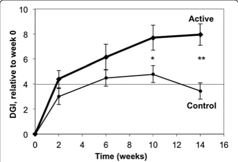
Subjects in the Active group achieved both statistically significant (p< 0.05) and clinically significant (DGI change of at least 4 points [13]) improvements in gait by the 6-week test point and these improvements continued through the 14-week test point (Figure 3 and Table 3). Sub-jects in the control group did not achieve a statistically-significant improvement in gait at any test points, although the apparent improvement at Week 10 would be consid-ered clinically significant.
When the pairwise differences in DGI change from baseline (Table 3) are compared for the two groups, the active group shows a statistically-significant greater im-provement than the control group at 10 weeks (p= 0.027) and 14 weeks (p < 0.001), using the independent t-test. (The Correspondingpvalues for the Wilcoxon test were 0.034 and 0.002).
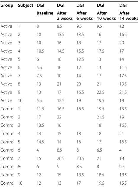
The raw DGI data from this study are shown in Table 4a. It can be seen that every Active group subject (100%), and all but one in the Control group (90%) exhibited some im-provement in their score between baseline and the end of the study at 14-weeks. The major difference is the magni-tude of both the individual and mean group change be-tween the two treatment groups. On average, the Active group improved by 7.95 points, while the Control group exhibited a mean change of 3.45 points.
All subjects reported an increase in salivation at the outset of the study due to the presence of the device in the mouth. With instruction, each was able to develop
Table 3 DGI Mean Scores
Active Control
Week N Mean (SD) Diffa pa N Mean (SD) Diffa pa
0 10 8.90 (2.85) 10 11.95 (4.04)
2 10 13.30 (3.92) 4.40c 0.056 10 14.95 (4.29) 3.00 0.610
6 10 15.05 (3.53) 6.15c 0.003b 8 15.63 (4.73) 3.68 0.471
10 10 16.60 (3.95) 7.70c <0.001b 10 16.75 (5.20) 4.80c 0.166
14 10 16.85 (3.40) 7.95c <0.001b 10 15.40 (5.03) 3.45 0.745
a
Difference from baseline (week 0).
b
Statistically significant difference (p < .05). c
Clinically significant difference (> = 4).
swallowing strategies to manage saliva volume while maintaining the device in their mouth. Five subjects also reported mild headaches and temporo-mandibular joint pain during the first few days of participation. These symptoms were mitigated by the end of the 2-week in-lab training period by instructing them to not bite the array, relax their jaw, press the array firmly with the tongue, and breathe uniformly. Two RRMS subjects in the Active group experienced relapses requiring suspen-sion of their participation, one for a period of 5 weeks, the others for 3 months. When they were able to resume training at the same level they had achieved prior to the relapse they were reintegrated into the study without adverse effect. Three subjects (2 Control, one Active group) experienced minor illnesses that required suspen-sion of training for approximately 2 weeks each. Each was able to resume training without complication, and their testing schedules adjusted accordingly. Finally, sev-eral subject experienced fatigue after completion of the morning training session. They were encouraged to practice relaxation exercises to help them prepare for performing the afternoon sessions. This proved to be a useful skill for them to develop in managing their energy levels so that they could consistently complete their daily participation in the study.
Discussion
This study investigated whether individuals with mul-tiple sclerosis could improve their gait using CN-NINM intervention, a training program that incorporates exer-cises combined with noninvasive electrical stimulation of the tongue. The results show that subjects in the active group had greater improvements in their gait relative to the control group, and the improvements were clinically and statistically significant.
The sample size of this study was small (10 subjects per group), which may limit the external validity of the results. The sample size estimation for this study was based on the robust results of our earlier non-controlled proof of concept studies.
In considering sources of the induced improvement, our previous studies have demonstrated through fMRI that neuromodulation training using electrical stimulation of the tongue, combined with movement exercises in balance-impaired individuals, induces activity of the cerebellum and brainstem nuclei, structures of the brain that process balance and movement [44,45,52]. Additionally, Mori, et al. showed that using transcranial magnetic stimulation combined with exercise therapy resulted in a reduction in spasticity and fatigue [35]. These findings, combined with known afferent neural pathways from the tongue, sug-gest that tongue stimulation preferentially predisposes certain cerebellum and brainstem nuclei to beneficial neu-roplastic effects resulting from movement exercises de-pendent on these nuclei, with the end result of improved movement control.
We hypothesize that CN-NINM induces neuroplasti-city by noninvasive stimulation of two major cranial nerves: trigeminal, CN-V, and facial, CN-VII. This stimu-lation excites a flow of action potentials (AP’s) to the brainstem (pons varolli and medulla) and cerebellum via the lingual branch of the cranial nerve (CN-Vc), and chorda tympani branch of CN-VII. This effect of the stimulation extends to the corresponding nuclei of the brainstem –at least in the sensory and spinal nuclei of trigeminal nuclear complex and the caudal part of the nucleus tractus solitarius [44,45,52]. We postulate that the intensive activation of these structures initiates a se-quential cascade of changes in neighboring and/or con-nected nuclei by direct collateral connections, brainstem interneuron circuitry and/or passive transmission of bio-chemical compounds in the intercellular space. There is evidence from related research that has observed changes in both neurotransmitter and other neuroactive compounds in response to chronic stimulation. Each AP in the trigeminal nuclei and brainstem releases up to 23 biologically active compounds including neurotransmit-ters that affect synaptic transmission, and the brainstem has the highest glia-to-neuron density anywhere in the CNS (50:1) [61]. The full function of the glial network is Table 4 Raw DGI scores
Group Subject DGI DGI DGI DGI DGI
Baseline After 2 weeks
After 6 weeks
After 10 weeks
After 14 weeks
Active 1 8 8.5 9.5 9.5 12
Active 2 10 13.5 13.5 16 16.5
Active 3 10 16 18 17 20
Active 4 10.5 14.5 15.5 17.5 17
Active 5 6 10 12.5 13 14
Active 6 5.5 10 12 13 11.5
Active 7 7.5 10 14 17 17.5
Active 8 13 21 20 21 19.5
Active 9 13 17 16.5 22.5 21.5
Active 10 5.5 12.5 19 19.5 19
Control 1 11.5 16.5 18.5 19.5 15.5
Control 2 17 22 21.5 19
Control 3 13.5 16 18 16.5
Control 4 14 15 18 18 21
Control 5 14.5 14 16 17 16.5
Control 6 4 8.5 8 6.5 4
Control 7 15 20.5 20.5 21 18
Control 8 6 9 8.5 8 9.5
Control 9 12 15 18.5 18.5 18.5
unknown, although it is generally agreed that it plays a vital role in the regulation of neural behavior through management of the chemical environment at the synap-tic gap. Consequently, we believe the stimulation directly activates not only the neuronal network by electrical impulses (AP’s) but also the glial network by neuro-chemical impact. The net effect of this upregulation of neuroactive compounds is to potentiate the networks in-volved within the CNS and set the stage for sustained focal and global changes in brain behavior [62-68].
Improvement is also common in patients receiving a new intervention with a therapist [7,69]. A better indica-tion of efficacy is if the patient improves when inde-pendently training at home. Here we see that both groups continued to improve during the at-home phase of the study. In a related study, Di Fabio, et al., found that patients with progressive MS who continued with an extended home outpatient rehabilitation over the course of 1 year experienced a lower rate of decline in physical function when compared with subjects who did not exercise [70,71]. Our study did not compare subjects who had performed the extended exercise protocol with any who had exercised and stopped. Consequently, to investigate this phenomenon, the natural progression of this study would be to repeat it using a longer interven-tion period.
In our study, though all subjects appeared to demon-strate improvements initially, only the active group con-tinued to improve over the length of the study. It is likely that the early improvement in both groups was due to the intense involvement of the subjects with the trainers for the first 2 weeks of the study. It can been seen in Table 3 that improvements in performance for the active group continued to accumulate as subjects trained at home after the initial 2-week training phase. Subjects who trained using exercise only without stimu-lation (control group) continued to improve for the first month at home and then exhibited a plateau or even a decrease in performance. This provides preliminary evi-dence that the intervention is effective when performed independently at home.
We observed a significant difference between groups for number of years with MS, with those in the active group having had MS for a longer period of time. Be-cause the progression of the disease varies from person to person, however, we did not feel that chronicity (years with MS) was as important as symptom presentation. We based our inclusion criteria on symptom presenta-tion, specifically gait dysfuncpresenta-tion, and EDSS score. We also observed that the mean initial (baseline) DGI score was lower in the active group (although this did not ap-proach statistical significance), raising the concern that this may bias the results. Because of this apparent differ-ence, we chose to analyze data using a simple DGI
difference from baseline, rather than a % change as sug-gested by other groups which would have unduly favored the active group [60]. Similar analysis of % DGI change yielded identical conclusions.
We did not investigate whether the subtype of MS had an effect on the reported outcomes. It is acknowledged that because subject assignment was purely random there is some possibility that the disease subtype could affect sensitivity to the intervention and therefore the sults. We note, however, that these sub-classifications re-late primarily to the progression of the disease and not to their particular state during participation in the study. Furthermore, we excluded all candidates that had any changes in symptoms or medication in the 3 months prior to participation to ensure that their presentation was as stable as possible. Nonetheless, it would be bene-ficial to repeat this study using only MS patients of one particular type, such as PPMS or SPMS to determine if the rate or magnitude of change in performance differs as a function of the disease sub-classification.
The remaining question that may be posed is whether CN-NINM training could be practically deployed in a re-habilitation setting. The in-lab training for this study was admittedly time intensive (approximately 2 to 3 hours per day, per subject), involving far more time for therapy than a typical clinical setting would allow. This was an intentional departure from most therapeutic models for rehabilitation, derived from our prior experience with treating vestibular disorders [43,46]. Given the rigors of the intervention, and the nature of this neurodegenerative disease, this intensive in-lab phase was designed to ensure that subjects understood and could reliably perform the training program before using the device at home. The sults suggest that this new paradigm for home-health re-habilitation, particularly for disorders previously deemed untreatable, is efficacious. Additional studies are necessary to determine if an abbreviated intervention would be as effective as the model presented here.
Conclusions
previously reported. This leads us to speculate whether people with other types of neurological conditions would benefit from this type of intervention to improve motor control. We suggest that further studies are warranted to investigate the breadth of applicability that this form of therapeutic intervention may have for meaningful neurorehabilitation.
Endnotes a
While not formally approved, half-point scores (0.5) of the DGI are commonly used. The author of the DGI (Shumway-Cook) has acknowledged the limitations of the scale as originally constructed, and conceded that, if employed consistently by the clinician, the use of 0.5 interval is an acceptable interpolation of the defined condition on conventional scale.
Abbreviations
(ANOVA):Analysis of variance; (CN-NINM): Cranial nerve non-invasive neuromodulation; (DGI): Dynamic gait index; (DGIB): Dynamic gait index baseline; (EDSS): Expanded disability status scale; (fMRI): Functional magnetic resonance imaging; (MA): Massachusetts; (MS): Multiple sclerosis;
(RRMS): Relapsing remitting multiple sclerosis; (PoNS™): Portable
neuromodulation stimulator; (PPMS): Primary progressive multiple sclerosis; (SPMS): Secondary progressive.
Competing interests
Danilov, Kaczmarek, and Tyler have a financial interest in Advanced NeuroRehabilitation, LLC and in NeuroHabilitation Corp., which both have intellectual property rights in the field of use reported in this article.
Authors’contributions
MT, YD, KK, KR, AS, KS. 1 Study design. 2 Draft manuscript. 3 Statistical analysis. 4 Implementation of the intervention/study protocol. 5 Critical revision of manuscript. All authors read and approved the final manuscript.
Authors’information
All authors are affiliated with the University of Wisconsin, Madison. MT teaches in the Department of Biomedical Engineering at the University of Wisconsin, Madison. He is a co-inventor of the PoNS device and is a contributor to the development of the CN-NINM training method. YD is a co-inventor of the PoNS device and is a major contributor to the development of the CN-NINM training method. KK is an expert in electrotactile stimulation and a co-inventor of the PoNS device. KR is an Occupational Therapist with an interest in human subjects rehabilitation research. AS is a psychiatrist who specializes in cognitive function and emotional processing. KS is a Physical Therapist with an interest in the application of neuromodulation for rehabilitation and is a contributor to the development of the CN-NINM training method.
Acknowledgements
We gratefully acknowledge Chris Luzzio, MD, for advice on both the clinical and experimental assessments, and for referring human subjects; and Scott Hetzel, MS (Dept. of Biostatistics and Medical Informatics, University of Wisconsin, Madison) for assistance with, and independent verification of the statistical analyses. Funding for this study was provided by the University of Wisconsin Foundation.
Author details
1
Department of Biomedical Engineering, University of Wisconsin, Madison, WI 53706, USA.2Department of Orthopedics and Rehabilitation, University of Wisconsin, Madison, WI 53706, USA.
Received: 25 June 2013 Accepted: 24 April 2014 Published: 1 May 2014
References
1. Panitch H, Applebee A:Treatment of walking impairment in multiple sclerosis: an unmet need for a disease-specific disability.Expert Opin Pharmacother2011,12:1511–1521.
2. Goodman AD, Brown TR, Krupp LB, Schapiro RT, Schwid SR, Cohen R, Marinucci LN, Blight AR, Fampridine MSFI:Sustained-release oral fampridine in multiple sclerosis: a randomised, double-blind, controlled trial.Lancet2009,373:732–738.
3. Motl RW:Physical activity and irreversible disability in multiple sclerosis.
Exerc Sport Sci Rev2010,38:186–191.
4. Snook EM, Motl RW:Effect of exercise training on walking mobility in multiple sclerosis: a meta-analysis.Neurorehabil Neural Repair2009, 23:108–116.
5. Myhr K, Riise T, Vedeler C, Nortvedt MW, Gronning M, Midgard R, Nyland HI: Disability and prognosis in multiple sclerosis: demographic and clinical variables important for the ability to walk and awarding of disability pension.Mult Scler2001,7:59–65.
6. Pilutti LA, Lelli DA, Paulseth JE, Crome M, Jiang S, Rathbone MP, Hicks AL: Effects of 12 weeks of supported treadmill training on functional ability and quality of life in progressive multiple sclerosis: a pilot study.
Arch Phys Med Rehabil2011,92:31–36.
7. Rietberg MB, Brooks D, Uitdehaag BMJ, Kwakkel G:Exercise therapy for multiple sclerosis.Cochrane Database Syst Rev2004,3:1–35. 8. Wiles CM:Physiotherapy and related activities in multiple sclerosis.
Mult Scler2008,14:863–871.
9. Garrett M, Coote S:Multiple sclerosis and exercise in people with minimal gait impairment-a review.Phys Ther Rev2009,14:169–180.
10. Hogan N, Coote S:Therapeutic interventions in the treatment of people with multiple sclerosis with mobility problems: a literature review.
Phys Ther Rev2009,14:160–168.
11. Rasova K, Feys P, Henze T, van Tongeren H, Cattaneo D, Jonsdottir J, Herbenova A:Emerging evidence-based physical rehabilitation for Multiple Sclerosis - Towards an inventory of current content across Europe.Health Qual Life Outcomes2010,8:76–82.
12. Khan F, Pallant J, Brand C, Kilpatrick T:Effectiveness of rehabilitation intervention in persons with multiple sclerosis: a randomized controlled trial.J Neurol Neurosurg Psychiatry2008,79(11):1230–1235.
13. Shumway-Cook A, Woollacott M:Motor Control: Translating Research into Clinical Practice.3rd edition. Lippincott, Williams, and Wilkins: Philadelphia; 2007.
14. Thompson AJ:The effectiveness of neurological rehabilitation in multiple sclerosis.J Rehabil Res Dev2000,37:455–461.
15. Patti F, Ciancio MR, Reggio E, Lopes R, Palermo F, Cacopardo M, Reggio A: The impact of outpatient rehabilitation on quality of life in multiple sclerosis.J Neurol2002,249:1027–1033.
16. Freeman JA, Langdon DW, Hobart JC, Thompson AJ:The impact of inpatient rehabilitation on progressive multiple sclerosis.Ann Neurol
1997,42:236–244.
17. Khan F, Pallant JF, Zhang N, Turner-Stokes L:Clinical practice improvement approach in multiple sclerosis rehabilitation: a pilot study.Int J Rehabil Res2010,33:238–247.
18. Paltamaa J, Sarasoja T, Leskinen E, Wikstrom J, Malkia E:Measures of physical functioning predict self-reported performance in self-care, mobility, and domestic life in ambulatory persons with multiple sclerosis.Arch Phys Med Rehabil2007,88:1649–1657.
19. Grasso MG, Troisi E, Rizzi F, Morelli D, Paolucci S:Prognostic factors in multidisciplinary rehabilitation treatment in multiple sclerosis: an outcome study.Mult Scler2005,11:719–724.
20. Khan F, Gray O:Disability management and rehabilitation for persons with multiple sclerosis.Neural Regeneration Res2010,5:301–309. 21. Fregni F, Imamura M, Chien HF, Lew HL, Boggio P, Kaptchuk TJ, Riberto M,
Hsing WT, Battistella LR, Furlan A:Challenges and recommendations for placebo controls in randomized trials in physical and rehabilitation medicine A report of the international Placebo symposium working group.Am J Phys Med Rehabil2010,89:160–172.
22. Compston A, Coles A:Multiple sclerosis.Lancet2008,372:1502–1517. 23. Katrych O, Simone TM, Azad S, Mousa SA:Disease-modifying agents in the
treatment of multiple sclerosis: a review of long-term outcomes.Cns Neuro Disord Drug Targets2009,8:512–519.
25. Taylor NF, Dodd KJ, Prasad D, Denisenko S:Progressive resistance exercise for people with multiple sclerosis.Disabil Rehabil2006,28:1119–1126. 26. Paul L, Rafferty D, Young S, Miller L, Mattison P, McFadyen A:The effect of
functional electrical stimulation on the physiological cost of gait in people with multiple sclerosis.Mult Scler2008,14:954–961. 27. Esnouf JE, Taylor PN, Mann GE, Barrett CL:Impact on activities of daily
living using a functional electrical stimulation device to improve dropped foot in people with multiple sclerosis, measured by the Canadian Occupational Performance Measure.Mult Scler2010, 16:1141–1147.
28. Barrett CL, Mann GE, Taylor PN, Strike P:A randomized trial to investigate the effects of functional electrical stimulation and therapeutic exercise on walking performance for people with multiple sclerosis.Mult Scler
2009,15:493–504.
29. Courtney AM, Castro-Borrero W, Davis SL, Frohman TC, Frohman EM:Functional treatments in multiple sclerosis.Curr Opin Neurol2011,24:250–254. 30. Fahy BG:Intraoperative and perioperative complications with a vagus
nerve stimulation device.J Clin Anesth2010,22:213–222.
31. Spuck S, Tronnier V, Orosz I, Schonweiler R, Sepehrnia A, Nowak G, Sperner J:Operative and technical complications of vagus nerve stimulator implantation.Neurosurgery2010,67:489–494.
32. Hu X, Jiang X, Zhou X, Liang J, Wang L, Cao Y, Jin A, Yang P:Avoidance and management of surgical and hardware-related complications of deep brain stimulation.Stereotact Funct Neurosurg2010,88:296–303. 33. Chang EF, Cheng JS, Richardson RM, Lee C, Starr PA, Larson PS:Incidence
and management of venous air embolisims during awake deep brain stimulation surgery in a large clinical series.Stereotact Funct Neurosurg
2011,89:76–82.
34. Zeller D, Dang SY, Stefan K, Biller A, Bartsch A, Saur D, Bendszus M, Rieckmann P, Toyka KV, Classen J:Functional role of ipsilateral motor areas in multiple sclerosis.J Neurol Neurosurg Psychiatry2011,82:578–583. 35. Mori F, Ljoka C, Magni E, Codeca C, Kusayanagi H, Monteleone F, Sancesario
A, Bernardi G, Koch G, Foti C, Centonze D:Transcranial magnetic stimulation primes the effects of exercise therapy in multiple sclerosis.
J Neurol2011,258:1281–1287.
36. Mori F, Codeca C, Kusayanagi H, Monteleone F, Buttari F, Fiore S, Bernardi G, Koch G, Centonze D:Effects of anodal transcranial direct current stimulation on chronic neuropathic pain in patients with multiple sclerosis.J Pain2010,11:436–442.
37. Cogiamanian F, Ferrucci R, de Riz M, Vergari M, Tadini L, Ciocca M, Dilena R, Marceglia S, Mameli F, Fumagalli M, Mrakic-Sposta A, Scarpini E, Barbieri S, Priori A:Transcranial direct current stimulation (tDCS) for fatigue in multiple sclerosis.Neurology2009,72:A82–A82.
38. Mori F, Codeca C, Kusayanagi H, Monteleone F, Boffa L, Rimano A, Bernardi G, Koch G, Centonze D:Effects of intermittent theta burst stimulation on spasticity in patients with multiple sclerosis.Eur J Neurol2010,17:295–300. 39. Bach-y-Rita P, Kaczmarek KA, Meier K:The tongue as a man–machine
interface: A wireless communication system.InProc Int Symp Info Theory & Apps (ISITA’98); Oct. 14–16.Mexico City: IEEE; 1998:79–81.
40. Bach-y-Rita P, Kercel SW:Sensory substitution and the human-machine interface.Trends Cogn Sci2003,7:541–546.
41. Chebat DR, Rainville C, Kupers R, Ptito M:Tactile‘visual’acuity of the tongue in early blind individuals.Neuroreport2007,18:1901–1904. 42. Sampaio E, Maris S, Bachy-y-Rita P:Brain plasticity:‘visual’acuity of blind
persons via the tongue.Brain Res2001,908:204–207.
43. Tyler M, Danilov Y, Bach-y-Rita P:Closing an open-loop control system: Vestibular substitution through the tongue.J Integr Neurosci2003,2:159–164. 44. Wildenberg JC, Tyler ME, Danilov YP, Kaczmarek KA, Meyerand ME:
Sustained cortical and subcortical neuromodulation induced by electrical tongue stimulation.Brain Imaging Behav2010,4:199–211. 45. Wildenberg J, Tyler M, Danilov Y, Kaczmarek K, Kuo J, Meyerand ME: High-Resolution FMRI detects activity and neuromodulation of individual brainstem nuclei.J Neurosurg2011,115:A438–A439. 46. Danilov YP, Tyler ME, Skinner KL, Hogle RA, Bach-y-Rita P:Efficacy of
electrotactile vestibular substitution in patients with peripheral and central vestibular loss.J Vestib Res2007,17:119–130.
47. Vuillerme N, Cuisinier R:Sensory supplementation through tongue electrotactile stimulation to preserve head stabilization in space in the absence of vision.Invest Ophthalmol Vis Sci2009,50:476–481.
48. Vuillerme N, Pinsault N, Fleury A, Chenu O, Demongeot J, Payan Y, Pavan P: Effectiveness of an electro-tactile vestibular substitution system in
improving upright postural control in unilateral vestibular-defective patients.Gait Posture2008,28:711–715.
49. Kaczmarek KA:Sensory augmentation and substitution.InCRC Handbook of Biomedical Engineering.Edited by Bronzino JD. Boca Raton, FL: CRC Press; 2000:143.1-143.10.
50. Badke MB, Sherman J, Boyne P, Page S, Dunning K:Tongue-based biofeedback for balance in stroke: results of an 8-week pilot study.
Arch Phys Med Rehabil2011,92:1364–1370.
51. Kaczmarek KA:The tongue display unit for electrotactile spatiotemporal pattern presentation.Scientia Iranica D2011,18:1476–1485.
52. Wildenberg JC, Tyler ME, Danilov YP, Kaczmarek KA, Meyerand ME:Electrical tongue stimulation normalizes activity within the motion-sensitive brain network in balance-impaired subjects as revealed by group independent component analysis.Brain Connectivity2011,1:255–265.
53. Kaczmarek KA, Webster JG, Radwin RG:Maximal dynamic range electrotactile stimulation waveforms.IEEE Trans Biomed Eng1992,39:701–715.
54. Tyler ME, Braun JG, Danilov YP:Spatial mapping of electrotactile sensation threshold and intensity range on the human tongue: Initial results. InProc IEEE Eng Med Biol Soc.Minneapolis, MN; 2009:559–562.
55. Bach-y-Rita P, Kaczmarek KA, Tyler ME, Garcia-Lara M:Form perception with a 49-point electrotactile stimulus array on the tongue: A technical note.
J Rehab Res Dev1998,35:427–430.
56. Kaczmarek KA, Tyler ME:Effect of electrode geometry and intensity control method on comfort of electrotactile stimulation on the tongue. InProc ASME Dyn Sys Contr Div.Orlando, Florida: ASME; 2000:1239–1243. 57. McConvey J, Bennett SE:Reliability of the dynamic gait index in individuals
with multiple sclerosis.Arch Phys Med Rehabil2005,86:130–133. 58. Cattaneo D, Jonsdottir J, Repetti S:Reliability of four scales on balance
disorders in persons with multiple sclerosis.Disabil Rehabil2007,29:1920–1925. 59. Cattaneo D, Regola A, Meotti M:Validity of six balance disorders scales in
persons with multiple sclerosis.Disabil Rehabil2006,28:789–795. 60. Erratum:Comparing exercise in Parkinson’s disease-The Berlin LSVT(R)BIG
study.Mov Disord2010,25:2478.
61. Kandel ER, Schwartz JH, Jessell TM: InPrinciples of Neural Science.Third edition. Edited by Kandel ER, Schwartz JH, Jessel TM. New York, NY: McGraw-Hill; 1991:22–32.
62. Abraham WC, Bear MF:Metaplasticity: the plasticity of synaptic plasticity.
Trends Neurosci1996,19:126–130.
63. Bliss TV, Gardner-Medwin AR:Long-lasting potentiation of synaptic transmission in the dentate area of the unanaestetized rabbit following stimulation of the perforant path.J Physiol1973,232:357–374. 64. Bliss TV, Lomo T:Long-lasting potentiation of synaptic transmission in
the dentate area of the anaesthetized rabbit following stimulation of the perforant path.J Physiol1973,232:331–356.
65. Buchs PA, Muller D:Induction of long-term potentiation is associated with major ultrastructural changes of activated synapses.Proc Natl Acad Sci U S A1996,93:8040–8045.
66. Nudo RJ, Plautz EJ, Frost SB:Role of adaptive plasticity in recovery of function after damage to motor cortex.Muscle Nerve2001,24:1000–1019. 67. Zucker RS:Calcium- and activity-dependent synaptic plasticity.Curr Opin
Neurobiol1999,9:305–313.
68. Zucker RS, Regehr WG:Short-term synaptic plasticity.Annu Rev Physiol
2002,64:355–405.
69. Wiles CM, Newcombe RG, Fuller KJ, Shaw S, Furnival-Doran J, Pickersgill TP, Morgan A:Controlled randomised crossover trial of the effects of physiotherapy on mobility in chronic multiple sclerosis.J Neurol Neurosurg Psychiatry2001,70:174–179.
70. DiFabio RPSJ, Choi T, Hansen CR, Schapiro RT:Extended outpatient rehabilitation: Its influence on symptom frequency, fatigue, and functional status for persons with progressive multiple sclerosis.Arch Phys Med Rehabil1998,79:141–146.
71. Schapiro RT, Soderberg J, Hooley M, Terry G, Ruhsam S, Linroth R, Hatgidakis J, Hall S:The Multiple Sclerosis Achievement Center: a maintenance rehabilitation approach toward a chronic progressive form of the disease.J Neuro Rehabil1988,2:21–23.
doi:10.1186/1743-0003-11-79