R E S E A R C H
Open Access
Placenta-specific epimutation at
H19
-DMR
among common pregnancy complications:
its frequency and effect on the expression
patterns of
H19
and
IGF2
Yuko Yamaguchi
1,2, Chiharu Tayama
1, Junko Tomikawa
1, Rina Akaishi
3, Hiromi Kamura
1, Kentaro Matsuoka
4,7,
Norio Wake
2, Hisanori Minakami
5, Kiyoko Kato
2, Takahiro Yamada
6, Kazuhiko Nakabayashi
1*and Kenichiro Hata
1*Abstract
Background:H19andIGF2genes are imprinted and involved in regulating fetal and placental growth. TheH19
differentially methylated region (DMR) is paternally methylated and maternally unmethylated and regulates the imprinted expression ofH19andIGF2. Epimutation at theH19-DMR in humans results in congenital growth disorders, Beckwith-Wiedemann and Silver-Russell syndromes, when erroneously its maternal allele becomes methylated and its paternal allele becomes unmethylated, respectively. AlthoughH19andIGF2have been assessed for their involvement in pregnancy complications including fetal growth restriction (FGR) and pregnancy-induced hypertension (PIH)/ hypertensive disorder of pregnancy (HDP) intensively in the last decade, it is still not established whether epimutation at theH19-DMR in the placenta results in pathogenic conditions in pregnancy. We aimed to assess the frequency of H19-DMR epimutation and its effects on the allelic expression patterns ofH19andIGF2genes among normal and abnormal pregnancy cases.
Results:We enrolled two independently collected sets of placenta samples from normal pregnancies as controls and common pregnancy complications, FGR and PIH (HDP). The first set consisted of 39 controls and 140 FGR and/or PIH cases, and the second set consisted of 29 controls and 62 cases. For these samples, we initially screened for DNA methylation changes atH19-DMR andIGF2-DMRs by combined bisulfite restriction analysis, and further analyzed cases with methylation changes for their allelic methylation and expression patterns. We identified one case each of FGR and PIH showing hypomethylation ofH19-DMR andIGF2-DMRs only in the placenta, but not in cord blood, from the first case/control set. For the PIH case, we were able to determine the allelic expression pattern ofH19to be biallelically expressed and theH19/IGF2expression ratio to be highly elevated compared to controls. We also identified a PIH case with hypomethylation atH19-DMR andIGF2-DMRs in the placenta from the second case/control set.
Conclusions:Placental epimutation atH19-DMR was observed among common pregnancy complication cases at the frequency of 1.5% (3 out of 202 cases examined), but not in 68 normal pregnancy cases examined. Alteration ofH19/IGF2 expression patterns due to hypomethylation ofH19-DMR may have been involved in the pathogenesis of pregnancy complications in these cases.
Keywords:Genomic imprinting, DNA methylation, Epimutation, Placenta,H19,IGF2, Fetal growth restriction, Pregnancy-induced hypertension, Hypertensive disorder of pregnancy
© The Author(s). 2019Open AccessThis article is distributed under the terms of the Creative Commons Attribution 4.0 International License (http://creativecommons.org/licenses/by/4.0/), which permits unrestricted use, distribution, and reproduction in any medium, provided you give appropriate credit to the original author(s) and the source, provide a link to the Creative Commons license, and indicate if changes were made. The Creative Commons Public Domain Dedication waiver (http://creativecommons.org/publicdomain/zero/1.0/) applies to the data made available in this article, unless otherwise stated.
* Correspondence:[email protected];[email protected]
1Department of Maternal-Fetal Biology, Research Institute, National Center for
Background
Genome imprinting is an epigenetic mechanism whereby only one of the two parental alleles is expressed, and has been known to be critical for proper development of the fetus and the placenta. Imprinted genes tend to be clus-tered in the genome. Allelic expression patterns of a cluster of imprinted genes have been shown to be regu-lated in cis by an imprinting control region (ICR). ICRs overlap with a differentially methylated region (DMR) that exhibits parental allele-specific DNA methylation inherited from gametes (sperm and oocyte) and
main-tained throughout subsequent development. ICRs
(germline DMRs) are essential in establishing additional somatic DMRs and allele-specific histone modifications within imprinted domains [1].H19 and IGF2 genes are imprinted (maternally and paternally expressed,
respect-ively) and regulated by H19-DMR, the ICR for these
genes being located at 2.5 kb upstream of the H19 pro-moter region.H19andIGF2are known to be crucial for fetal and postnatal growth [2]. Abnormal DNA methyla-tion (epimutamethyla-tion) atH19-DMR occurring in gametes or early development leads to specific imprinting disorders.
Hypermethylation atH19-DMR causes
Beckwith-Wiede-mann syndrome (BWS), a congenital overgrowth syn-drome occasionally associated with embryonal tumors (such as Wilms tumor, hepatoblastoma, adrenal
carcin-oma, and neuroblastoma) [3, 4]. Hypomethylation at
H19-DMR causes Silver-Russell syndrome (SRS), which
is characterized by prenatal and postnatal growth restric-tion with addirestric-tional dysmorphic features [5]. Increased and decreased expression levels of theIGF2 gene encod-ing insulin-like growth factor 2 due to ICR epimutation are considered to be the leading causes of BWS and SRS, respectively [3,5].
Previous studies using mouse models have shown the
involvement of H19 and Igf2 in the development and
functions of the placenta.H19deletion in mice has been shown to result in increased Igf2 expression and fetal/ placental overgrowth [6, 7] and reduced nutrient trans-port capacity [8].Igf2deletion in mice is shown to result in fetal/placental growth restriction [9, 10] and altered diffusional exchange properties [11]. These lines of evi-dence from mouse studies suggested that imprinting
de-fects at the H19/IGF2 gene cluster in the human
placenta could also cause placental dysfunction and be involved in pregnancy complications. Accordingly, the
H19-IGF2 imprinted gene cluster has been intensively characterized by itself or together with other key imprinted genes in the placentas from common preg-nancy complications such as FGR and PIH in the last decade [12–18]. Despite past efforts to gain knowledge of its etiological aspect, the cause of PIH is still unclear [19]. The etiologies of FGR are considered to be multi-factorial (maternal, fetal, placental, and environmental)
[20]. Therefore, the etiologies of all PIH cases and the majority of FGR cases are unknown. These pregnancy complications are considered to be heterogeneous con-ditions for which a variety of genetic and environmental factors contribute to their onset [21]. It should be noted that PIH has been recently renamed as hypertensive dis-order of pregnancy (HDP). Gene expression and DNA methylation properties assessed in the studies of
H19-IGF2 locus in FGR and PIH cases [12–18] include gene expression levels and loss of imprinting (LOI)
frequen-cies of H19 and IGF2, and DNA methylation levels of
H19 germline DMR, H19 promoter, and IGF2 somatic
DMRs in the placentas with or without pregnancy com-plications. The extent of consistency among the results from independent studies varies depending on the prop-erty examined as described below.
LOI, biallelic expression of an imprinted gene due to the reactivation of the normally repressed allele, ofH19 at the third trimester placentas was initially reported to occur only in PIH but not in normal pregnancy cases [14]. However, in later studies [16, 18], LOI of H19 in the third trimester and full-term placentas was detected regardless of the presence/absence of obstetric complica-tions. LOI of IGF2 in the placentas was frequently ob-served (5 out of 16 cases) in one study [13], but not in another study (none in 92 cases) [18]. It should be noted that the criteria for LOI (or relaxation of imprinting, ROI) were significantly different between these two stud-ies (> 3% and > 25%, respectively). In these LOI studstud-ies [13, 14, 18], LOI of H19 and IGF2 in the placenta did not correlate with the gene expression levels. Gene ex-pression levels ofH19andIGF2were shown to be corre-lated (with statistical significance) both in the placenta (n= 36) and the cord blood (n= 100) in one study [18], although such a correlation was not observed in the pla-centa (n= 34) in another study [12]. Further evaluation of the gene expression patterns of H19and IGF2 in the placenta is important to validate these previous findings.
Whereas two studies [15,17] reported statistically sig-nificantly lower levels of DNA methylation ofH19-DMR in FGR placentas compared to normal placentas, such
association was not detected by another study [12].
Small sample sizes in these studies may explain the dis-crepant results. Whereas Guo et al. [12] identified a small for gestational age (SGA) case exhibiting
hypome-thylation at H19-DMR accompanied with the biallelic
expression of H19 in the placenta among the 22 SGA
cases, Tabano et al. [16] did not find such H19-DMR epimutation cases among the placenta samples of the 66 FGR cases. Additional and larger-scale studies may help further establish the roles of the H19/IGF2gene cluster in the pathogenesis of pregnancy complications.
In this study, we aimed to determine whether and how
placentas from common pregnancy complications, FGR and PIH, and also assess some of the previously sug-gested features of gene expression and methylation
pat-terns at the H19/IGF2 imprinted gene cluster in our
sample collections.
Results
Identification of FGR and PIH placentas with H19-DMR hypomethylation
To screen for cases with DNA methylation abnormality
at H19-DMR among FGR and/or PIH placentas, we
measured the DNA methylation levels of H19-DMR in
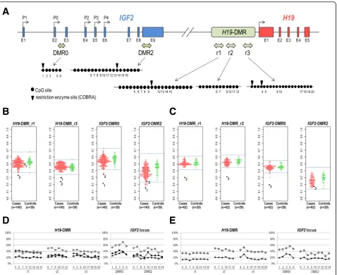
placenta samples by combined bisulfite restriction ana-lysis (COBRA). We analyzed two subregions (r1 and r3 in Fig. 1a) withinH19-DMR. Both subregions have pre-viously been shown to be differentially methylated (pa-ternally methylated) in human fetal tissues [22, 23]. The r1 region includes the sixth CTCF-binding site, which has been suggested to be a key regulatory domain for the imprinted expression of H19 and IGF2 genes [22]. The r3 region corresponds to a promoter-proximal re-gion (H421), which was previously reported to be pater-nally methylated [23]. We initially subjected a total of 140 cases and 39 controls (case/control set I, Table1) to COBRA. Genomic DNA was isolated from the chorionic plate of placentas for the case/control set I. The mean and standard deviation (SD) of the methylation levels of the 39 control samples were 52.2% (SD 4.8%) and 44.8%
(SD 3.6%) for H19-DMR_r1 and H19-DMR_r3,
respect-ively. We considered methylation levels over/under the range of ± 3SD of the mean as aberrant methylation. Two cases (designated as case 1 and case 2 hereafter)
were found to be aberrantly hypomethylated at H19
-DMR. Case 1, an FGR case, was hypomethylated atH19
-DMR_r3 (30.1%, −4.1SD), but not at H19-DMR_r1
(50.1%, −0.4SD). Case 2, a PIH case, was
hypomethy-lated at both H19-DMR_r1 (33.2%, –4.0SD) and H19
-DMR_r3 (24.5%, −5.5SD) (Fig. 1b). To screen for add-itional cases with aberrant methylation atH19-DMR, we conducted COBRA for another set of cases and controls, which were collected independently from the first case/ control set. The second case/control set consisted of 62 FGR and/or PIH cases and 29 controls (case/control set
II in Table 1). Genomic DNA was isolated from
chori-onic villi of the placentas of the case/control set II. The means and SD of the methylation levels in control group
II were 51.2% (SD 4.1%) and 52.7% (SD 2.8%) for H19
-DMR_r1 and H19-DMR_r3, respectively. One PIH case
(designated as case 3) was identified to be
hypomethy-lated at H19-DMR_r1 (29.4%, −5.3SD) and H19-DMR_
r3 (31.4%,−7.6SD) (Fig.1c). We validated the observed hypomethylation at H19-DMR in cases 1–3 (Fig. 1b, c) by pyrosequencing analysis (for 23 CpG sites within
H19-DMR, 6 CpG sites withinIGF2-DMR0, and 11 CpG
sites withinIGF2-DMR2 as depicted in Fig.1a). Consist-ent with the results obtained by COBRA assays, the r1 subregion was confirmed to be hypomethylated in case 2 and case 3, and the r2 and r3 subregions were hypo-methylated in three cases (Fig. 1d, e). The methylation
levels at IGF2-DMR0 and IGF2-DMR2 were also found
to be lower than the mean of the controls in all cases (Fig. 1d, e). For case 2, we also analyzed a second chori-onic plate sample and observed similar DNA methyla-tion levels. These results of hypomethylamethyla-tion of H19
-DMR accompanied with hypomethylation of IGF2
-DMRs suggest the regulatory role of H19-DMR in the
somatic establishment of differential methylation at
IGF2-DMR0 andIGF2-DMR2 in the human placenta, as
has been shown for the mouse H19/Igf2 imprinted
do-main [24]. Summarizing the above, we identified three
cases with aberrant DNA hypomethylation atH19-DMR
out of 202 placentas from FGR and/or PIH cases.
Correlation and association analyses of DNA methylation levels among loci and tissues and with pregnancy complications
We assessed correlation of DNA methylation levels of
H19-DMRs and IGF2-DMRs. Within a tissue, H19
-DMR_r1 and H19-DMR_r3 were most highly correlated
in the cord blood (correlation coefficient r= 0.80) and moderately correlated (r= 0.50) in the placentas (chori-onic plate and villi) (Additional file1: Figure S1).IGF2
-DMR0 and IGF2-DMR2 were moderately and weakly
correlated in the chorionic plate (r= 0.47) and the cord blood (r= 0.26), respectively, but not in chorionic villi (r= 0.15) (Additional file1: Figure S1). DNA methylation
levels of H19-DMR and IGF2-DMRs were moderately
correlated only in chorionic villi (r= 0.41 forH19-DMR_
r3 vs IFG2-DMR0), but not in chorionic plate or cord
blood. Between the chorionic plate and the cord blood,
whereas DNA methylation levels ofH19-DMR were not
correlated (r= 0.18 for r1 and r= 0.20 for r3), those of
IGF2-DMRs were moderately and weakly correlated (r= 0.39 for DMR0 andr= 0.26 for DMR2) (Additional file1: Figure S1).
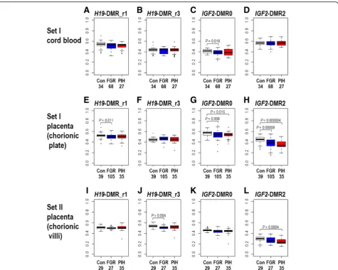
We also assessed the differences in DNA methylation levels in the FGR and PIH groups compared to controls (Fig.2and Additional file3: Table S1). In the cord blood samples (of set I), the methylation levels of H19-DMR were not significantly different in 105 FGR cases without PIH phenotype or in 29 PIH cases with or without FGR phenotype compared with 39 controls. On the other hand, in the placental samples, the methylation levels of
both H19-DMR_r1 and H19-DMR_r3 were statistically
previous report by Bourque et al. [15]. The methylation
levels of IGF2-DMR0 were statistically significantly
lower in FGR cases in both the cord blood and placentas and in PIH cases only in the placentas of set I (Fig.2c, g). Whereas the statistical significance of the
abovemen-tioned lower levels of methylation at H19-DMR and
IGF2-DMR0 tended to be marginal to the statistical
threshold (p= 0.05), methylation levels at IGF2-DMR2 were more significantly lower both in FGR and PIH cases in the placentas of set I (Fig. 2h, p= 0.00008 and
p= 0.000004, respectively) and in PIH cases in the pla-centas of set II (Fig.2l,p =0.0004). These results suggest
A
B
C
D
E
Fig. 1DNA methylation analyses forH19-DMR andIGF2-DMRs by COBRA and pyrosequencing assays.aLocation of CpG sites withinH19-DMR,
IGF2-DMR0, andIGF2-DMR2 analyzed. Exons ofIGF2andH19genes are shown as blue and red squares, respectively. Transcriptional start sites of
IGF2(P0-P4) andH19are indicated by arrows. The approximate positions of the genomic intervals subjected to DNA methylation analyses within
H19-DMR,IGF2-DMR0, andIGF2-DMR2 are shown by light green two-headed arrows. The genomic intervals ofIGF2-DMR0 andIGF2-DMR2 were described previously [17]. Closed circles represent CpG sites included in the bisulfite-PCR amplicons subjected to COBRA, pyrosequencing, and bisulfite sequencing analyses. Downward arrowheads in the r1 and r3 subregions withinH19-DMR indicate TaqI sites used in COBRA assays. The CpG sites whose methylation level was assessed by pyrosequencing are numbered underneath the closed circles.b,cDNA methylation levels of
that the DNA methylation status ofIGF2-DMR2 may be used as a placental biomarker for pregnancy complica-tion phenotypes (in particular, PIH) and placental dysfunctions.
Hypomethylation at H19-DMR in FGR and PIH cases is accompanied with hypomethylation of IGF2-DMRs and is confined to the placenta
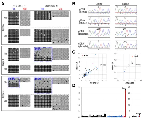
We subsequently aimed to determine the allelic methyla-tion patterns ofH19-DMR in the placenta and the cord blood of cases 1–3 by bisulfite sequencing. We screened
for heterozygous SNPs within H19-DMR to distinguish
paternal and maternal alleles and identified such SNPs for cases 1 and 2. Case 3 was excluded from the allelic bisulfite sequencing analysis due to the absence of
het-erozygous SNPs within H19-DMR. In a sample from
control group I, the paternal allele was nearly completely methylated (100% methylated) and the maternal allele was (nearly) completely unmethylated in both regions (r1 and r3) examined in the placenta (chorionic plate) as well as in the corresponding cord blood (Fig.3a). In case 1, paternal hypomethylation was observed in the r3 re-gion but not in the r1 rere-gion in the placenta. In case 2, paternal hypomethylation was observed in both regions in the placenta. These patterns of paternal hypomethyla-tion in the placenta of case 1 and case 2 are well
consist-ent with the results of COBRA (Fig. 1b) and
pyrosequencing analysis (Fig. 1d). However, in the cord blood of case 1 and case 2, the paternal allele was nearly completely methylated (Fig. 3a). These results
demon-strate that hypomethylation at H19-DMR in case 1 and
case 2 occurred only in the placenta, but not in the fetus. The observed partial loss of paternal methylation
at H19-DMR in these cases (34.9 to 44.4% of paternal
methylation retained) suggests the possibility of epigen-etic mosaicism atH19-DMR in these placentas.
Biallelic expression of H19 in the placenta with H19-DMR hypomethylation
We tried to determine the allelic expression pattern of
H19in the placentas of cases 1–3. However, due to the unavailability of total RNA for case 1 and due to the ab-sence of heterozygous SNPs in the exonic regions of
H19in case 3, we were able to assess the allelic expres-sion pattern ofH19only for case 2. We carried out dir-ect sequencing of RT-PCR amplicons that include
rs2067051 (a transcribed SNP at the H19 locus).
Whereas H19 was found to be exclusively expressed
from the maternal (G) allele in a control chorionic plate sample, it was apparently biallelically expressed (A/G) in the chorionic plate of case 2 (Fig.3b). Such a loss of im-printing ofH19 was not observed in 20 additional con-trol placenta samples with a normal level of DNA
methylation at H19-DMR (data not shown). These
re-sults suggest that paternal hypomethylation at H19
-DMR led to the expression ofH19 from its paternal lele, which is normally silent. We could not assess the
al-lelic expression pattern of IGF2 because no
heterozygous SNP was found in the exons of IGF2 in
cases 1–3.
Expression levels of H19 and IGF2 in the placentas with H19-DMR epimutation (cases 2 and 3)
Hypomethylation at H19-DMR on the paternal allele is
expected to result in the expression of H19 from the
normally repressed paternal allele (as was confirmed in the placenta of case 2 (Fig. 3b)) and the suppression of
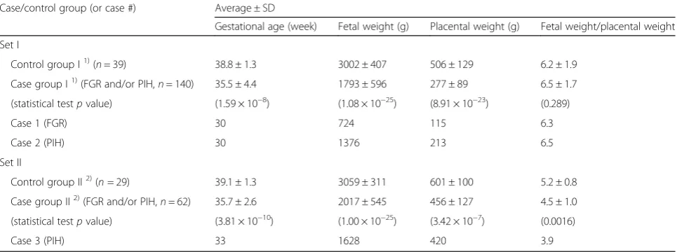
IGF2 expression [25,26]. To validate these expected ex-pression patterns, we quantitatively measured the Table 1Summary of clinical information for case and control groups enrolled in this study
Case/control group (or case #) Average ± SD
Gestational age (week) Fetal weight (g) Placental weight (g) Fetal weight/placental weight
Set I
Control group I1)(n= 39) 38.8 ± 1.3 3002 ± 407 506 ± 129 6.2 ± 1.9
Case group I1)(FGR and/or PIH,n= 140) 35.5 ± 4.4 1793 ± 596 277 ± 89 6.5 ± 1.7
(statistical testpvalue) (1.59 × 10⁻⁸) (1.08 × 10⁻²⁵) (8.91 × 10⁻²³) (0.289)
Case 1 (FGR) 30 724 115 6.3
Case 2 (PIH) 30 1376 213 6.5
Set II
Control group II2)(n= 29) 39.1 ± 1.3 3059 ± 311 601 ± 100 5.2 ± 0.8
Case group II2)(FGR and/or PIH,n= 62) 35.7 ± 2.6 2017 ± 545 456 ± 127 4.5 ± 1.0
(statistical testpvalue) (3.81 × 10⁻¹⁰) (1.00 × 10⁻²⁵) (3.42 × 10⁻⁷) (0.0016)
Case 3 (PIH) 33 1628 420 3.9
1)
Chorionic plate tissues were collected for all cases and controls. Cord blood samples were collected for subset of cases and controls
2)
expression levels ofH19and IGF2 in our placenta sam-ples. Consistent with previous reports [12, 18], the
pla-cental expression levels of H19 and IGF2 were diverse
among samples irrespective of the presence/absence of pregnancy complications. We observed positive correla-tions between the intra-individual expression levels of
H19 and IGF2 in both sets of placenta samples
(chori-onic plate samples from case/control set I [n= 27, R2= 0.55] and chorionic villi samples from case/control set II [n= 7,R2= 0.90]) (Fig. 3c). We could not include case 1 in this analysis due to the unavailability of its RNA as aforementioned. It should be noted that the correlation coefficients were calculated for only the samples without
H19-DMR hypomethylation (i.e., case 2 and case 3 were excluded). In case 2, theH19/IGF2ratio was 22.3, which was extraordinarily higher than those of the control
chorionic plate samples (n= 27) and the cases without
H19-DMR epimutation (n= 20) (ratios ranging from 0.6 to 2.7) (Fig. 2d, left), suggesting the increase of H19 ex-pression due to its exex-pression from the normally re-pressed paternal allele and the resultant reciprocal
decrease in IGF2 expression from the paternal allele.
However, the H19/IGF2 ratio in case 3 was similar to
those in the control chorionic villi samples (n= 6) exam-ined (Fig.3d, right).
Absence of copy number alteration at H19/IGF2 locus suggests epimutation as a cause of H19-DMR
hypomethylation
Confined placental mosaicism (CPM), the presence of trisomic cells predominantly in the placenta, was re-cently shown to be present in approximately 10% of
FGR cases [27]. In the situation of confined placental tri-somy of chromosome 11 due to excess of a maternal
copy, the overall methylation levels of H19-DMR and
IGF2-DMRs, which are paternally methylated, are
expected to be lower (one of three copies is methylated) than those in the normal diploid placenta (one of two copies is methylated). Duplication of a genomic segment
containing maternally derived H19 and IGF2 genes
A
B
C
D
would also result in lower methylation levels of H19
-DMR andIGF2-DMRs. Therefore, when only the overall
methylation level is available, hypomethylation of H19 -DMR could be interpreted in the following two ways: as a consequence of loss of methylation on the paternal al-lele and as a consequence of copy number change of the
H19locus. To further evaluate whether hypomethylation atH19-DMR in the placentas of cases 1–3 is due to epi-mutation, we carried out chromosomal copy number analysis for the placental genomic DNA samples as well as available parental blood genomic DNA samples. In all three cases, we detected neither copy number changes at
H19/IGF2 locus nor aneuploidy of chromosome 11 (Additional file2: Figure S3). We also confirmed the bi-parental inheritance of chromosome 11 in all three cases by comparing SNP genotypes between each placenta and corresponding parent(s). These results further suggest
that hypomethylation ofH19-DMR observed in the
pla-centas of cases 1–3 is due to epigenetic alteration, but not copy number alteration.
DNA methylation analysis of other imprinted DMRs and LINE1 by COBRA
To gain further insight into possible mechanisms
under-lying the epimutation at H19-DMR in the placentas of
cases 1–3, we examined the methylation levels of 16
imprinted DMRs (other than H19-DMR) and LINE1
re-petitive elements in the placentas of cases 1–3 and con-trols by COBRA assays. Considering the gestational weeks of cases 1–3 (30, 30, and 33 weeks, respectively,
Table 1), we chose control samples both from preterm
birth (24–34 weeks) and normal-term birth (37–40
weeks). At H19-DMR (the r1 and r3 subregions), the
distribution of methylation levels was similar between normal-term controls and preterm controls in both of the chorionic plate sample set (Additional file 1: Figure S2A) and the chorion sample set (Additional file 1: Fig-ure S2B). The methylation levels of other DMRs and
LINE1 elements in the placentas of cases 1–3 were
found to be within the normal range (mean ± 3SD of control data) with the following few exceptions:
methyla-tion levels ofARHI-DMR in case 2 and ofZAC-DMR in
case 3 were slightly out of the normal range (0.4% and 1.0% lower than“average−3SD”). However, their extent of hypomethylation is much smaller than those observed forH19-DMR (e.g., 17.3% lower than“average−3SD”at
H19-DMR_r1 in case 2). These results suggest that the placental epimutation in cases 1–3 occurred locally at
H19-DMR, but not in a genome-wide manner.
Discussion
In this study, we demonstrated that epimutation at
H19-DMR could occur in a placenta-specific manner.
Among 202 FGR and/or PIH cases in total, we
identified three cases, one FGR case (case 1) and two
PIH cases (cases 2 and 3), showing H19-DMR
hypome-thylation in the placenta. For cases 1 and 2, we also re-vealed that the hypomethylation on the paternal allele
of H19-DMR is specific to placenta, but not observed
in cord blood (Fig. 3a). IGF2 DMRs were also
hypo-methylated in the placentas of cases 1–3. We
demon-strated that H19 was biallelically expressed and the
relative H19/IGF2 expression ratio was 22.3 times
higher in the placenta of case 2 than that of the control. In a precedent study conducting methylation analysis ofH19-DMR in the placentas, Guo et al. [12] examined 22 SGA cases and identified one hypomethylation case accompanied withH19biallelic expression, but without the elevation inH19/IGF2expression ratio. It is not ex-plicitly mentioned whether the authors examined the
methylation status of H19-DMR in the corresponding
cord blood. Two FGR cases in Koukoura et al. [17] and
one FGR case in Bourque et al. [15] also showed
ex-tremely low (≈0%) and notably low (< 25%) methylation
levels at H19-DMR in the placenta, but they were not
characterized further. On the other hand, Tabano et al.
[16] did not find any H19-DMR epimutation cases
among 66 FGR placentas [13]. It should be also
men-tioned that Rancourt et al. [18] examined the DNA
methylation levels of H19 DMR in 114 placentas from
normal pregnancy cases and identified one case
show-ing hypermethylation at H19-DMR (presumably due to
the gain of methylation on the maternal allele) without
apparent alterations of H19 and IGF2 gene expression
levels. Considering the results of these studies and ours together, extreme hypomethylation (i.e., epimutation) at
H19-DMR in the placenta has been observed only in
the pregnancies with complications. We clearly
demon-strated that epimutation at H19-DMR could occur in a
placenta-specific manner and is accompanied with
hy-pomethylation at IGF2 DMRs. The frequency of H19
-DMR hypomethylation in the placenta was 1.5% (3/ 202) among the FGR and/or PIH pregnancy cases.
It is well established that germline epimutations at
H19-DMR cause two growth disorders with opposite
phenotypes: an overgrowth disorder, Beckwith-Wiede-mann syndrome (when hypermethylated), and a growth retardation disorder, Silver-Russell syndrome (when hypomethylated). Increased and decreased expression levels of IGF2 due to epimutation are considered to be the primary cause of BWS and SRS, respectively [3, 5]. In contrast, it remains unknown whether the placenta-specific epimutation atH19-DMR identified in this study and by Guo et al. [12] is responsible for the associated phenotypes such as FGR (SGA) and PIH. Considering that a variety of genetic and environmental influences can contribute to the risk of these common pregnancy
epimutation at H19-DMR does not necessarily diminish its potential significance. PIH and FGR are considered to be multifactorial diseases, and their causal genetic and /or epigenetic factors are scarcely known [19, 20]. In such a situation, it is important to identify candidate genes involved in their etiologies even for a subset of cases. Recent independent studies have revealed critical functions ofH19lincRNA as a key regulator in multiple physiological processes and cell types [28–31]. Such regulatory roles of H19lincRNA in various tissues also suggest a potential impact of the dysregulation of H19 expression in the placenta. However, evidence is cur-rently lacking to link the observed epimutation with pla-cental dysfunction and FGR/PIH phenotypes. Recently
established CRISPR/dCas9-based epigenome editing
technologies [32] could be applied to create cell line and
mouse models of placenta-specific H19-DMR
epimuta-tion to elucidate its funcepimuta-tional outcomes at the cellular and organ levels.
Consistent with a previous study [18], we observed the correlation ofH19andIGF2expression levels in placen-tas (Fig.3c, d). Furthermore, we revealed an extreme
ele-vation of H19/IGF2 expression ratio in case 2 with
placenta-specific epimutation at H19-DMR, implying an increase ofH19expression and decrease ofIGF2 expres-sion due to the disruption of imprinted regulation. These results suggest the possibility that H19and IGF2 expression levels vary among the human population pre-sumably due to genetic diversity, that the appropriate
balance of H19 and IGF2 expression levels is required
for normal development, and that the extreme disturb-ance of theH19/IGF2expression ratio in the placenta of case 2 may have impaired placental development and functions. However, it should be also mentioned that
this dysregulated pattern of H19 and IGF2 expression
was not observed in case 3 in our study and the H19
-DMR epimutation case identified by Guo et al. [12]. It is
plausible that the outcome ofH19-DMR epimutation on
gene expression of H19and IGF2 differs between chori-onic plate (case 2) and chorichori-onic villi (case 3 and #8928 in Guo et al. [12]) and also differs depending on the de-velopmental stages. Correlations of the expression levels
of imprinted genes such as IGF2 and IGF2R and fetal
weight have been detected in a manner dependent of the developmental stages of placenta [33].
Yamazawa et al. demonstrated that H19-DMR
hypo-methylation affects the imprinted expression ofH19and
IGF2 in both fetus and placenta in several SRS cases
[34], strongly suggesting the germline origin of H19 -DMR epimutation in the SRS cases examined. On the other hand, we have previously reported two BWS cases
whose methylation levels of H19-DMR were discordant
in embryo-derived somatic tissues (blood and skin) and
placenta, suggesting that the aberrant gain of
methylation on the maternally derived allele occurred after implantation in these cases [35]. The extent of gain
of methylation at H19-DMR was higher in blood and
skin than that in the placenta. The epimutation at H19 -DMR was specific to the placenta in cases 1 and 2 and was not detected in their cord blood. Therefore, the de-velopmental timing of epimutation in these cases is speculated not to be of germ-cell origin, but of post-zyg-otic origin. Cases 1 and 2 were confirmed to have devel-oped normally at their clinical examination at 3 years old. Case 3 was recorded to be healthy at 6 years old through an interview to its mother.
Both PIH and FGR have been considered to be associ-ated with impaired maternal uteroplacental perfusion secondary to defective extravillous trophoblast invasion [36–38].H19and IGF2 have been shown to be involved in placental development and trophoblast cell prolifera-tion, differentiaprolifera-tion, and migration. In cell culture
models, IGF2has been shown to prevent apoptosis and
enhance proliferation and migration/invasion of human trophoblast cells [reviewed in [39]], differentiation of murine ectoplacental cone trophoblast [40], and migra-tion of ovine trophoblast cells [41]. H19 has also been shown to promote the migration and invasion of a hu-man extravillous trophoblast cell line [42]. In a mouse model,H19and Igf2genes are demonstrated to contrib-ute to the control of placental mass and the normal dif-ferentiation of giant cells [43]. Disruption of imprinting leading to the biallelic expression of H19and repression
of Igf2 is considered to impair placental development
and trophoblast differentiation. The placental
hypome-thylation at H19-DMR was associated with PIH in two
cases (cases 2 and 3) and with FGR in one case (case 1) in this study. Common pregnancy complications repre-sented by FGR and PIH are heterogeneous conditions for which a variety of genetic and environmental influ-ences can contribute to risk [21]. Therefore, the same
epimutation (i.e., placental hypomethylation at H19
-DMR) can be associated with two different phenotypes, PIH and FGR, depending on the genetic and the envir-onmental factors the fetus and the mother possess and get exposed to.
Despite the efforts by previous and our current stud-ies, many of the findings reported regarding the
alter-ation of DNA methylalter-ation at H19-DMR are not
to collect genomic, transcriptomic, and epigenomic data in an unbiased manner from mother and baby (soma and placenta) of normal and abnormal pregnancies at a larger scale than ever before. Clinical follow-up of en-rolled individuals is important to assess the effects of placental functions on individual postnatal phenotypes.
Conclusions
Our study demonstrated that H19-DMR epimutation
could occur in a placenta-specific manner, presumably post-zygotically, and potentially be associated with preg-nancy complications, but at a low frequency (1.5%).
Al-teration of the H19/IGF2 expression ratio due to
hypomethylation of H19-DMR may have been involved
in the pathogenesis of pregnancy complications. Further characterization of theH19/IGF2imprinted gene cluster in normal pregnancy and pregnancy complication cases at a larger scale and conducted in a more comprehensive manner will help elucidate its involvement with placen-tal development and relevant diseases in humans.
Methods Subjects
This study was approved by the Institutional Review Board Committees at the National Center for Child Health and Development (NCCHD) of Japan, Kyushu University Hospital, and Hokkaido University Hos-pital, and written informed consent was obtained from all participants. Placenta, corresponding umbil-ical cord blood, and parental blood were collected for normal, FGR, and PIH pregnancy cases. A total of 140 cases (105 FGR, 12 PIH, and 23 FGR/PIH cases) and 39 controls were obtained from NCCHD and Kyushu University Hospital as case group I and con-trol group I, respectively. A total of 62 cases (27 FGR, 26 PIH, and 9 FGR/PIH cases) and 29 controls were obtained from Hokkaido University Hospital as case group II and control group II, respectively. Oga-wa’s birthweight percentile tables [44] were used for
the diagnosis of FGR. Singleton newborns at 37–41
weeks of gestation with birthweight > 10th percentile without any complications were enrolled as controls. Pregnancy with birthweight < 10th percentile was di-agnosed as FGR (gestational age at birth and sex were considered). Pregnancy with hypertension (sBP > 140 mmHg and/or dBP > 90 mmHg) with or without pro-teinuria (> 300 mg/day) after 20 weeks was diagnosed as PIH. Newborns with karyotypic abnormality and multiple conceptions were excluded. The mean of gestational ages, fetal and placental weights for case and control groups, and individual data for the cases
that exhibited hypomethylation at H19-DMR (case 1,
case 2, and case 3) are provided in Table 1.
Sample collection and preparations
We dissected out the chorionic plate from the placentas of case group I and control group I collected at NCCHD and Kyushu University (set I) and chorionic villi from the placentas of case group II and control group II col-lected at Hokkaido University Hospital (set II). It should be noted that the difference in the tissue types of pla-centa collected between set I and set II was not
ration-ally planned. We initiated the placental sample
collection for set I at NCCHD and chose the chorionic plate as the target tissue. When we sought for an inde-pendent set of placental samples from common preg-nancy complications and controls, only the sample set of chorionic villi (set II) was available. A piece of chorionic plate tissue was divided into two pieces (one for gen-omic DNA extraction and the other for RNA extraction) and washed with 1× PBS. Chorionic plate samples for
genomic DNA extraction were stored at −80 °C until
genomic DNA extraction. Chorionic plate samples for RNA extraction were soaked in RNAlater (Qiagen) over-night at 4 °C and stored at −80 °C. Chorionic villi were collected, washed with 1× PBS repeatedly, divided into two tubes, and stored at−80 °C until genomic DNA and total RNA extraction. Genomic DNA was extracted using either the standard phenol/chloroform extraction method or QIAamp DNA Blood Midi Kit (Qiagen). Total RNA was extracted using TRIzol Reagent (Invitro-gen). Complementary DNA (cDNA) synthesis was car-ried out using QuantiTect Reverse Transcription Kit (Qiagen).
DNA methylation analyses
ForH19-DMR_r1, the PCR primers described previously
[45] were used in COBRA and pyrosequencing analyses. ForH19-DMR_r3, a pair of primers that amplify a 256-bp fragment and another pair of primers that amplify a 476-bp fragment were designed for COBRA and pyrose-quencing, respectively. A 195-bp interval located in
be-tween r1 and r3 regions was designated asH19-DMR_r2
and subjected to pyrosequencing analysis. A 352-bp re-gion within IGF2-DMR0 [46, 47] and a 337-bp region withinIGF2-DMR2 [12,46] were also subjected to pyro-sequencing analysis. Bisulfite-PCR primers for COBRA
were designed using the MethPrimer (
http://www.uro-gene.org/methprimer/index1.html). Bisulfite-PCR and sequencing primers for pyrosequencing were designed using the PyroMark Assay Design 2.0 (Qiagen). Detailed information for the primers is listed in Additional file4: Table S2.
One microgram of genomic DNA was bisulfite-treated and purified using the EpiTect Bisulfite Kit (Qiagen).
PCR amplification for COBRA assays for H19-DMR
20 pmol each primer, dNTPs (2.5 mM each), 1.5 U ExTaq HS (Takara), and supplied buffer. The standard thermal cycling conditions used were as follows: initial denatur-ation at 94 °C for 5 min, followed by 35 cycles of de-naturation at 94 °C for 30 s, annealing for 30 s, and extension at 72 °C for 30 s (the annealing temperature for each primer pair is listed in Additional file 4: Table S2). The PCR products were purified using the Illustra GFX 96 PCR purification kit (GE Healthcare). One fourth of the purified PCR products were digested with 6 U of TaqI (NEB) in a 15-μL scale reaction for 2 h. One
out of 15μL was electrophoresed using the MultiNA
Microchip Electrophoresis System and DNA-1000 re-agents kit (Shimadzu, Japan). The methylation percent value for each sample was calculated as described
previ-ously [48]. COBRA assay conditions for LINE1 and 16
imprinted DMRs other than H19-DMR were described
previously [49,50].
Pyrosequencing was carried out using the Pyro-MarkQ24 system (Qiagen) and PyroMark Gold Q24
Re-agents (Qiagen) according to the manufacturer’s
instructions. In brief, the biotinylated PCR products were purified with the Streptavidin Sepharose HP beads (GE Healthcare). The purified PCR products were washed, denatured, and then annealed with a sequencing primer. Quantification of methylated cytosine was per-formed using PyroMark Q24 software (Qiagen).
For bisulfite sequencing, PCR products were cloned using the StrataClone PCR Cloning Kit (Agilent Tech-nologies) and transformed into StrataClone Competent Cells. Single colonies were picked up and used as start-ing material to amplify plasmid DNA within them usstart-ing the TempliPhi DNA Amplification Kit (GE Healthcare). Sequencing reactions for individual amplified clones were conducted using the BigDye Terminator version 3.1 Cycle Sequencing kit (Applied Biosystems) with the M13 Rev primer. Sequence data were obtained using ABI3130xl Genetic Analyzer and analyzed using the QUMA website (http://quma.cdb.riken.jp/) [51]. SNPs,
rs2107425 (A/G) within the H19-DMR_r1, and
rs2251312 (C/G) and a T/G SNP at chr11:2,019,694 (hg19) within theH19-DMR_r3 were used to distinguish parental alleles.
Allelic expression analysis for H19
First-strand cDNA was synthesized from 1μg of placen-tal toplacen-tal RNA using QuantiTect Reverse Transcription Kit (Qiagen). For the allelic expression analysis of H19, an exonic SNP rs2067051 (A/G) was used to distinguish parental alleles. RT-PCR was performed in a total vol-ume of 20μL containing one tenth of the RT reaction, 20 pmol each primer, dNTPs (2.5 mM each), 1.5 U ExTaq HS (Takara), and supplied buffer. The RT-PCR products were treated with ExoSAP- IT (GE Healthcare) and
subjected to direct sequencing using one of the PCR primers as a sequencing primer. The details for the primers are listed in Additional file4: Table S2.
Quantitative RT-PCR
First-strand cDNAs synthesized using the QuantiTect Reverse Transcription Kit were subjected to real-time PCR analysis using the SYBR Premix Ex Taq (RR420A, Takara) on an ABI7500fast system (Thermo-Fisher Sci-entific). The thermal cycling conditions used were the initial denaturation at 95 °C for 1 min, followed by the 40 cycles of 95 °C for 15 s and 60 °C for 60 s. The se-quences of the PCR primers used are listed in Add-itional file 4: Table S2. The relative expression levels of
H19 and IGF2 were calculated by the delta-delta cycle
threshold (Ct) method. Delta Ct values were calculated
using the Ct values of ACTB as the normalization
con-trol, and delta-delta Ct values corresponding to relative expression levels were calculated using a placenta sam-ple of normal pregnancy as the reference.
Chromosomal copy number analysis
HumanCytoSNP-12 BeadChip data were obtained as de-scribed previously [52] and were visualized using Kar-yoStudio Data Analysis Software version 1.4.3.0 Build 37 (Illumina). Copy number analysis was conducted using CNV Plugin V3.0.7.0 (Illumina) with its default parame-ters for copy number gain/loss and copy neutral loss of heterozygosity.
Additional files
Additional file 1:Figure S1.Correlation plots for DNA methylation levels ofH19-DMR andIGF2-DMRs in the placenta and cord blood samples of case/control set I and in the placenta samples of case/control set II.Figure S2.Methylation levels of 18 imprinted DMRs and LINE1 repetitive elements in the placentas of cases 1–3 and controls determined by COBRA assays. (PDF 333 kb)
Additional file 2:Figure S3.Absence of copy number alterations at the
H19-IGF2 imprinted gene cluster in the placenta and cord blood samples of cases 1, 2, and 3. (PDF 395 kb)
Additional file 3:Table S1.Mean methylation levels of four CpG sites within H19-DMR and IGF2-DMRs measured by COBRA in case and control groups. (XLSX 13 kb)
Additional file 4:List of primers. (XLSX 12 kb)
Abbreviations
DMR:Differentially methylated region; FGR: Fetal growth restriction; HDP: Hypertensive disorder of pregnancy; ICR: Imprinting control region; LINE1: Long interspersed nuclear element 1; LOI: Loss of imprinting; PIH: Pregnancy-induced hypertension; ROI: Relaxation of imprinting
Acknowledgements
We thank Naoko Sugahara for her technical assistance for COBRA assays.
Authors’contributions
wrote the manuscript draft. KN and KH reviewed and finalized the manuscript. All authors read and approved the final manuscript.
Funding
This research was supported by AMED (Grant number 18ek0109278h0002) and JSPS KAKENHI (Grant numbers JP17K08689 and JP17K19535).
Availability of data and materials
All data generated and analyzed throughout this study are included in this published article and its supplementary information files.
Ethics approval and consent to participate
This study was approved by the Ethics Committee at the National Center for Child Health and Disease (NCCHD), the Institutional Review Board for Human Genome/Gene Research at Kyushu University, and the Ethics Committees at Hokkaido University Graduate School of Medicine. Written informed consent was obtained from the participants.
Consent for publication
Not applicable.
Competing interests
The authors declare that they have no competing interests.
Author details
1Department of Maternal-Fetal Biology, Research Institute, National Center for
Child Health and Development, Tokyo 157-8535, Japan.2Department of Obstetrics and Gynecology, Graduate School of Medical Sciences, Kyushu University, Fukuoka 812-8582, Japan.3Center of Maternal-Fetal, Neonatal and Reproductive Medicine, National Center for Child Health and Development, Tokyo 157-8535, Japan.4Department of Pathology, National Center for Child Health and Development, Tokyo 157-8535, Japan.5Department of Obstetrics
and Gynecology, Hokkaido University Graduate School of Medicine, Sapporo 060-8638, Japan.6Clinical Genetics Unit, Kyoto University Hospital, Kyoto
606-8507, Japan.7Present Address: Department of Pathology, Dokkyo Medical University, Saitama Medical Center, Koshigaya, Japan.
Received: 10 January 2019 Accepted: 22 July 2019
References
1. Arnaud P. Genomic imprinting in germ cells: imprints are under control. Reproduction. 2010;140:411–23.
2. Delaval K, Wagschal A, Feil R. Epigenetic deregulation of imprinting in congenital diseases of aberrant growth. Bioessays. 2006;28:453–9. 3. Weksberg R, Shuman C, Beckwith JB. Beckwith-Wiedemann syndrome. Eur J
Hum Genet. 2010;18:8–14.
4. Mussa A, Molinatto C, Baldassarre G, Riberi E, Russo S, Larizza L, et al. Cancer risk in Beckwith-Wiedemann syndrome: a systematic review and meta-analysis outlining a novel (epi)genotype specific histotype targeted screening protocol. J Pediatr. 2016;176:142–149.e1.
5. Gicquel C, Rossignol S, Cabrol S, Houang M, Steunou V, Barbu V, et al. Epimutation of the telomeric imprinting center region on chromosome 11p15 in Silver-Russell syndrome. Nat Genet. 2005;37:1003–7. 6. Leighton PA, Ingram RS, Eggenschwiler J, Efstratiadis A, Tilghman SM.
Disruption of imprinting caused by deletion of the H19 gene region in mice. Nature. 1995;375:34–9.
7. Eggenschwiler J, Ludwig T, Fisher P, Leighton PA, Tilghman SM, Efstratiadis A. Mouse mutant embryos overexpressing IGF-II exhibit phenotypic features of the Beckwith-Wiedemann and Simpson-Golabi-Behmel syndromes. Genes Dev. 1997;11:3128–42.
8. Angiolini E, Coan PM, Sandovici I, Iwajomo OH, Peck G, Burton GJ, et al. Developmental adaptations to increased fetal nutrient demand in mouse genetic models of Igf2-mediated overgrowth. FASEB J. 2011;25:1737–45. 9. DeChira TM, Efstratiadis A, Robertson EJ. A growth-deficiency phenotype in
heterozygous mice carrying an insulin-like growth factor II gene disrupted by targeting. Nature. 1990;296:78–80.
10. Lopez MF, Dikkes P, Zurakowski D, Villa-Komaroff L. Insulin-like growth factor II affects the appearance and glycogen content of glycogen cells in the murine placenta. Endocrinology. 1996;137:2100–8.
11. Coan PM, Fowden AL, Constancia M, Ferguson-Smith AC, Burton GJ, Sibley CP. Disproportional effects of Igf2 knockout on placental morphology and diffusional exchange characteristics in the mouse. J Physiol. 2008;586:5023–32.
12. Guo L, Choufani S, Ferreira J, Smith A, Chitayat D, Shuman C, et al. Altered gene expression and methylation of the human chromosome 11 imprinted region in small for gestational age (SGA) placentae. Dev Biol. 2008;320:79–91. 13. Diplas AI, Lambertini L, Lee MJ, Sperling R, Lee YL, Wetmur J, et al.
Differential expression of imprinted genes in normal and IUGR human placentas. Epigenetics. 2009;4:235–40.
14. Yu L, Chen M, Zhao D, Yi P, Lu L, Han J, et al. The H19 gene imprinting in normal pregnancy and pre-eclampsia. Placenta. 2009;30:443–7. 15. Bourque DK, Avila L, Peñaherrera M, von Dadelszen P, Robinson WP.
Decreased placental methylation at the H19/IGF2 imprinting control region is associated with normotensive intrauterine growth restriction but not preeclampsia. Placenta. 2010;31:197–202.
16. Tabano S, Colapietro P, Cetin I, Grati FR, Zanutto S, Mandò C, et al. Epigenetic modulation of the IGF2/H19 imprinted domain in human embryonic and extra-embryonic compartments and its possible role in fetal growth restriction. Epigenetics. 2010;5(4):313–24.
17. Koukoura O, Sifakis S, Zaravinos A, Apostolidou S, Jones A, Hajiioannou J, et al. Hypomethylation along with increased H19 expression in placentas from pregnancies complicated with fetal growth restriction. Placenta. 2011;32:51–7. 18. Rancourt RC, Harris HR, Barault L, Michels KB. The prevalence of loss of
imprinting of H19 and IGF2 at birth. FASEB J. 2013;27:3335–43.
19. Mol BWJ, Roberts CT, Thangaratinam S, Magee LA, de Groot CJM, Hofmeyr GJ. Pre-eclampsia. Lancet. 2016;387(10022):999–1011.
20. Nardozza LM, Caetano AC, Zamarian AC, Mazzola JB, Silva CP, Marçal VM, Lobo TF, Peixoto AB, Araujo Júnior E. Fetal growth restriction: current knowledge. Arch Gynecol Obstet. 2017;295:1061–77.
21. Manokhina I, Del Gobbo GF, Konwar C, Wilson SL, Robinson WP. Review: placental biomarkers for assessing fetal health. Hum Mol Genet. 2017;26(R2): R237–45.
22. Takai D, Gonzales FA, Tsai YC, Thayer MJ, Jones PA. Large scale mapping of methylcytosines in CTCF binding sites in the human H19 promoter and aberrant hypomethylation in human bladder cancer. Hum Mol Genet. 2001; 10:2619–26.
23. Vu TH, Li T, Nguyen D, Nguyen BT, Yao XM, Hu JF, et al. Symmetric and asymmetric DNA methylation in the human IGF2-H19 imprinted region. Genomics. 2000;64:132–43.
24. Lopes S, Lewis A, Hajkova P, Dean W, Oswald J, Forné T, et al. Epigenetic modifications in an imprinting cluster are controlled by a hierarchy of DMRs suggesting long-range chromatin interactions. Hum Mol Genet. 2003;12:295–305. 25. Bell AC, Felsenfeld G. Methylation of a CTCF-dependent boundary controls
imprinted expression of the Igf2 gene. Nature. 2000;405(6785):482–5. 26. Singh P, Lee DH, Szabó PE. More than insulator: multiple roles of CTCF at
the H19-Igf2 imprinted domain. Front Genet. 2012;3:214.
27. Robinson WP, Peñaherrera MS, Jiang R, Avila L, Sloan J, McFadden DE, et al. Assessing the role of placental trisomy in preeclampsia and intrauterine growth restriction. Prenat Diagn. 2010;30:1–8.
28. Wan P, Su W, Zhang Y, Li Z, Deng C, Li J, et al. LncRNA H19 initiates microglial pyroptosis and neuronal death in retinal ischemia/reperfusion injury. Cell Death Differ. 2019; [Epub ahead of print] PubMed PMID: 31127201.
29. Cao T, Jiang Y, Wang Z, Zhang N, Al-Hendy A, Mamillapalli R, et al. H19 lncRNA identified as a master regulator of genes that drive uterine leiomyomas. Oncogene. 2019; [Epub ahead of print] PubMed PMID: 31089260.
30. Zhang L, Yang Z, Huang W, Wu J. H19 potentiates let-7 family expression through reducing PTBP1 binding to their precursors in cholestasis. Cell Death Dis. 2019;10:168.
31. Zhou J, Xu J, Zhang L, Liu S, Ma Y, Wen X, et al. Combined single-cell profiling of lncRNAs and functional screening reveals that H19 is pivotal for embryonic hematopoietic stem cell development. Cell Stem Cell. 2019;24: 285–298.e5.2.
32. Morita S, Noguchi H, Horii T, Nakabayashi K, Kimura M, Okamura K, et al. Targeted DNA demethylation in vivo using dCas9-peptide repeat and scFv-TET1 catalytic domain fusions. Nat Biotechnol. 2016;34:1060–5.
34. Yamazawa K, Kagami M, Nagai T, Kondoh T, Onigata K, Maeyama K, et al. Molecular and clinical findings and their correlations in Silver-Russell syndrome: implications for a positive role of IGF2 in growth determination and differential imprinting regulation of the IGF2-H19 domain in bodies and placentas. J Mol Med. 2008;86:1171–81.
35. Higashimoto K, Nakabayashi K, Yatsuki H, Yoshinaga H, Jozaki K, Okada J, et al. Aberrant methylation of H19-DMR acquired after implantation was dissimilar in soma versus placenta of patients with Beckwith-Wiedemann syndrome. Am J Med Genet A. 2012;158A:1670–5.
36. Sones JL, Davisson RL. Preeclampsia, of mice and women. Physiol Genomics. 2016;48:565–72.
37. Mifsud W, Sebire NJ. Placental pathology in early-onset and late-onset fetal growth restriction. Fetal Diagn Ther. 2014;36:117–28.
38. Kaufmann P, Black S, Huppertz B. Endovascular trophoblast invasion: implications for the pathogenesis of intrauterine growth retardation and preeclampsia. Biol Reprod. 2003;69:1–7.
39. Sferruzzi-Perri AN, Sandovici I, Constancia M, Fowden AL. Placental phenotype and the insulin-like growth factors: resource allocation to fetal growth. J Physiol. 2017;595:5057–93.
40. Kanai-Azuma M, Kanai Y, Kurohmaru M, Sakai S, Hayashi Y. Insulin-like growth factor (IGF)-I stimulates proliferation and migration of mouse ectoplacental cone cells, while IGF-II transforms them into trophoblastic giant cells in vitro. Biol Reprod. 1993;48:252–61.
41. Kim J, Song G, Gao H, Farmer JL, Satterfield MC, Burghardt RC, et al. Insulin-like growth factor II activates phosphatidylinositol 3-kinase-protooncogenic protein kinase 1 and mitogen-activated protein kinase cell Signaling pathways, and stimulates migration of ovine trophectoderm cells. Endocrinology. 2008;149:3085–94.
42. Zuckerwise L, Li J, Lu L, Men Y, Geng T, Buhimschi CS, et al. H19 long noncoding RNA alters trophoblast cell migration and invasion by regulating TβR3 in placentae with fetal growth restriction. Oncotarget. 2016;7:38398–407.
43. Kawahara M, Wu Q, Yaguchi Y, Ferguson-Smith AC, Kono T. Complementary roles of genes regulated by two paternally methylated imprinted regions on chromosomes 7 and 12 in mouse placentation. Hum Mol Genet. 2006; 15:2869–79.
44. Ogawa Y, Iwamura T, Kuriya K, Nishida H, Takeuchi H, Takeda M, et al. Birth size standards by gestational age for Japanese neonates. Acta neonat Jap. 1998;34:624–32.
45. Sasaki K, Soejima H, Higashimoto K, Yatsuki H, Ohashi H, Yakabe S, et al. Japanese and North American/European patients with Beckwith-Wiedemann syndrome have different frequencies of some epigenetic and genetic alterations. Eur J Hum Genet. 2007;15:1205–10.
46. Moore T, Constancia M, Zubair M, Bailleul B, Feil R, Sasaki H, et al. Multiple imprinted sense and antisense transcripts, differential methylation and tandem repeats in a putative imprinting control region upstream of mouse Igf2. Proc Natl Acad Sci U S A. 1997;94:12509–14.
47. Murrell A, Ito Y, Verde G, Huddleston J, Woodfine K, Silengo MC, Spreafico F, et al. Distinct methylation changes at the IGF2-H19 locus in congenital growth disorders and cancer. PLoS One. 2008;3:e1849.
48. Brena RM, Auer H, Kornacker K, Hackanson B, Raval A, Byrd JC, et al. Accurate quantification of DNA methylation using combined bisulfite restriction analysis coupled with the Agilent 2100 Bioanalyzer platform. Nucleic Acids Res. 2006;34:e17.
49. Yang AS, Estécio MR, Doshi K, Kondo Y, Tajara EH, Issa JP. A simple method for estimating global DNA methylation using bisulfite PCR of repetitive DNA elements. Nucleic Acids Res. 2004;32:e38.
50. Yamazawa K, Nakabayashi K, Kagami M, Sato T, Saitoh S, Horikawa R, et al. Parthenogenetic chimaerism/mosaicism with a Silver-Russell syndrome-like phenotype. J Med Genet. 2010;47:782–5.
51. Kumaki Y, Oda M, Okano M. QUMA: quantification tool for methylation analysis. Nucleic Acids Res. 2008;36(Web Server issue):W170–5. 52. Miyata T, Sonoda K, Tomikawa J, Tayama C, Okamura K, Maehara K, et al.
Genomic, epigenomic, and transcriptomic profiling towards identifying omics features and specific biomarkers that distinguish uterine leiomyosarcoma and leiomyoma at molecular levels. Sarcoma. 2015;2015: 412068.
Publisher’s Note