Eukaryon
Eukaryon
Volume 2 Engaged Scholars in and out of the
Classroom
Article 18
1-1-2006
Remember the Protofibrils
Remember the Protofibrils
Michael White
Lake Forest College
Follow this and additional works at:
https://publications.lakeforest.edu/eukaryon
Part of the
Molecular and Cellular Neuroscience Commons
Disclaimer:
Eukaryon is published by students at Lake Forest College, who are solely responsible for its
content. The views expressed in Eukaryon do not necessarily reflect those of the College.
Articles published within Eukaryon should not be cited in bibliographies. Material contained
herein should be treated as personal communication and should be cited as such only with the
consent of the author.
Eukaryon, Vol. 2, January 2006, Lake Forest College
Review Article
Remember the Protofibrils
Michael White* Department of Biology Lake Forest College Lake Forest, Illinois 60045
[Role playing: Peter Lansbury, Jr. Center for Neurologic Diseases,
Brigham and Women’s Hospital, and Department of Neurology,
Harvard Medical School,
65 Landsdowne St., Cambridge, Massachusetts 02139]
Summary
The highly prevalent neurodegenerative diseases Alzheimer’s (AD), Parkinson’s (PD), and Amyotrophic Lateral Sclerosis (ALS) have managed to elude countless efforts directed at understanding their causative agents and pathogenesis. Each of these diseases is characterized by abnormal protein aggregation under an unknown modi operandi we have determined to illuminate. AD pathology consists of neurofibrillary tangles and β-amyloid plaques in numerous brain regions, most importantly the hippocampus. Our studies have focused on β-amyloid plaques, and we have demonstrated that a protofibrillar intermediate exists in the pathway from monomeric β-amyloid to fibril and plaque formation. Our studies of the α -Synuclein aggregates characteristic of PD, known as Lewy Bodies, have revealed a protofibrillar species that may be critical in PD pathogenesis. Our studies of ALS have focused on superoxide dismutase-1 (SOD1) because previous studies have demonstrated that the A4N mutation in SOD1 leads to increased susceptibility to disease and forms aggregates within spinal motor neurons but not fibrils. We have shown that the SOD1 functions properly as a dimer and forms aggregates when it dissociates into monomers. We subsequently found a molecule that stabilized the dimer and p r e v e n t e d a g g r e g a t i o n . T h e s e t h r e e neurodegenerative diseases all refer to common theme, which is abnormal protein aggregation. Our research has led us to support the hypothesis that, in the cases of PD and AD, a toxic protofibrillar intermediate is the causative agent in each disease. Treatment methods aimed at blocking protofibril formation may prove to be most effective.
Introduction
The neurodegenerative diseases Parkinson’s (PD), Alzheimer’s (AD) and Amyotrophic Lateral Sclerosis (ALS) are three of the most prevalent and heavily studied. Alzheimer’s alone affects 4.5 million Americans, 50% of individuals over 85 years old, and received an estimated $647 billion of funding in 2005 from the American Government1. Parkinson’s disease
*
This paper was written for BIO346 Molecular Neuroscience. In this assignment, Michael White role-played a noted biologist, Peter Lanzbury, Jr., and wrote a state–of-the-art review article on Dr. Lansbury’s research field, as if he were Dr. Siegel himself. He then presented a PowerPoint seminar as Dr. Lansbury in an annual public student research conference “NeuroFrontiers” held at Lake Forest College.
affects 1.5 million Americans, and 1 in 100 individuals over 60 years of age are diagnosed with the disease2 . Amyotrophic Lateral Sclerosis affects an estimated 30,000 Americans and has an incidence of 2 in 100,000 individuals3. Together, these diseases represent prime research targets.The defining characteristics of AD are dementia and memory loss due to atrophy of hippocampal neurons located in the medial temporal lobe. These neurons are crucial for embedding memories in cortical regions of the frontal lobe, and retrieving memories from these long-term storage sites. Upon autopsy, the AD brain is characterized by increased size/depth of sulci and widening of the lateral ventricles. Familial Alzheimer’s Disease (FAD) accounts for roughly 10% of all cases and is the result of the Aβ40 and Aβ42 mutations in the amyloid pre-cursor protein (APP)4.
The PD Brain consists of neurofibrillary tangles composed primarily of α-synuclein in structures called Lewy Bodies4. These fibrillar deposits are specific to dopaminergic neurons of the substantia nigra. PD is characterized by a motor initiation deficiency in which the patient has postural deficits and resting tremor in the hands and other limbs that develop when substantia nigra atrophy nears ~ 60% 4,6.
ALS is the result of spinal neuron atrophy7. Degeneration of these motor neurons leads to the degeneration of skeletal muscles and death within five years7. ALS contrasts AD and PD because it progresses more rapidly and is often found in adolescents. The protein involved is known to be the superoxide dismutase-1 (SOD1) enzyme4. Similar to AD and PD, ~20% of ALS cases are the result of SOD1 mutations. Our research is concentrated on these mutations and their affects4,7,8.
In common between AD, PD, and ALS is that all three demonstrate abnormal protein aggregation or fibrillization. Through an unknown mechanism, α -synuclein, β-amyloid, and SOD1 form fibrils and aggregates that have come to define each disease. Our lab has set out to determine if it is the protein aggregates/fibrils, an intermediate in aggregate/fibril formation, or a deficiency in the degradation of the relevant protein that causes these diseases.
Alzheimer’s Disease
Biological Basis
The definitive biological basis of AD remains to be elucidated. However, Dr. Alzheimer noted the presence of extra-cellular neurofibrillary plaques that were later found to be composed of β-amyloid which is a product of the cleavage of Alzheimer’s Pre-Cursor Protein (APP) 4. In addition, he noted intra-cellular tangles that have recently been found to contain Tau protein. These two features of AD occur in conjunction with disease progression5. Genetically, two
β-amyloid mutants, Aβ42 and Aβ40, have been implicated in a causal role in FAD and are the focus of many of our studies4.
Prior to our research, a definitive biological mechanism for AD remained unknown, and research focused mainly on the β-amyloid plaques and neurofibrillary tangles as the likely disease causing agents5. Furthermore, the
pathway had not been discovered. It had been determined that amyloid plaque formation was dependent on the monomeric concentration of Aβ5. Thus, in order for Aβ fibrils to form, a critical concentration had to be reached5. Most importantly, a modus operandi had not been established for plaque/fibril formation and no intermediates had been identified. Our early research focused specifically on illuminating the pathway to fibril formation.
Model System of AD
Several model systems are currently referred to for the study of AD but none express all features characteristic of human AD. A transgenic mouse model expressing one of two known (FAD) mutants, Aβ42 and Aβ40, produces plaques but not tangles and demonstrates memory deficiency4. A second mouse model that was transgenic for wild type (WT) human β-amyloid has a non-fibrillar phenotype but retains memory deficits4. The WT transgenic model provides strong evidence for a protofibrillar intermediate as the causative agent of AD that we will subsequently discuss4.
Illuminating the Protofibril
In order to support the current (1993) hypothesis that fibrils were the AD causative agent, a detailed understanding of the pathway to fibril formation had to be achieved5 Therefore, our 1993-1997 experiments focused on understanding this pathway. Through atomic force microscopy (AFM), we were able to observe the transition from monomer to fibril of Aβ40, Aβ42, and WT in vitro10. In this experiment, we discovered the Aβ protofibril10. Prior to fibril formation, there was a stable intermediate that elongated over time to form a fibril, which we named the protofibril10. Significantly, as protofibril concentration decreased, fibril concentration increased10. In addition, A
β40 was found to form fibrils at an increased rate compared to Aβ4210. Future research would contradict this difference in formation speed9.
Following our discovery of the Aβ protofibril10, we gathered more evidence to support its existence. In vitro, a solution containing Aβ40 protofibrils was used to study the transition from protofibril to fibril because Aβ40 had been found to fibrillize slower that Aβ425,11. Addition of fibrils to the protofibrillar solution led to immediate formation of amyloid fibrils11. In contrast, addition of protofibrils to the protofibrillar solution did not yield any fibrils for more than 15 days11. This experiment showed that fibril formation from protofibrils could only be seeded by pre-formed fibrils11. More importantly, we have demonstrated that protofibrils are incorporated into fibrils, refracting criticism that suggests the protofibril is formed as a byproduct of fibril formation or numerous other alternatives10,11.
The Protofibril
Atomic force microscopy (AFM) provides a three-dimensional image of a substrate by detecting surfaces changes on mica or compounds or similar compounds. W e u s e d A F M t o s t u d y p r o t o f i b r i l assembly/disassembly which are both related to concentration, temperature, and ionic environment9. Aβ40 protofibrils elongate as Aβ40 monomer is added to the ends. This can be accelerated with an increase in Aβ40 monomer concentration9. In contrast to our previous study, we found Aβ40 and Aβ42 protofibril elongation to be similar9,10. Disassembly of the A
β40 protofibrils occurred upon dilution of the protofibrillar solution9. We also found an increase in temperature
and NaCl concentration to coincide with an increase in protofibril elongation 9.
Quantifying β-Amyloid Pre-Autopsy
The relationship between Aβ-amyloid fibrillization and AD associated cognitive decline was our most recent topic of research13. We hypothesized that a molecule could be designed to bind Aβ fibrils and be detected by single proton computed tomography (SPECT), allowing this relationship to be understood13. Rhenium complexes were synthesized containing the dye Congo Red and found to bind amyloid plaques13. This could be a potential break-through in the understanding of AD pathogenesis and diagnosis because it is low cost and detectable readily detectable13.
Compilation of our AD studies with those of competitors has led us to a four-step process for amyloid fibril formation9. A competitor found the first step by determining a small amount, possibly less that 20 Aβ monomer units, spontaneously interacted to form protofibrils12. Second, the protofibril elongates by interacting with other small protofibrils9. Third, the protofibril to fibril conversion is due to interaction among elongated protofibrils9. Fourth, the A
β fibril is elongated when placed in solution containing Aβ monomer11. This new model projects much focus upon the Aβ protofibril as a possible AD causative agent.
Parkinson’s Disease
Biological Basis
The biological basis of PD has yet to be elucidated but two mutations α-synuclein, A30P and A53T have been linked to Familial Parkinson’s Disease (FPD) 4. In addition, the S18Y mutant in the UCH-L1 enzyme and mutations in the enzyme parkin are both linked to FPD4. α-synuclein is known to be the primary component of the neurofibrillary inclusions called Lewy Bodies4. The enzymes UCH-L1 and parkin are known to aid in degradation of α-synuclein but an exact role has yet to be determined4. Our research over the past several years has focused on understanding the biological basis of PD and the relationship between Lewy Bodies and pathogenesis4. Prior to our research, the two mutant forms of α-synuclein, A30P and A53T had been linked to PD and known to be a component of Lewy Bodies, but the pathway to fibril formation was unknown as well as any intermediate species16. Pre-2000 research focused on the “old model” of disease causation until we discovered a protofibrillar intermediate (Figure 1) 14.
Model Systems of PD
Several model systems for the study of PD exist but some are significantly more beneficial than others. There is a transgenic mouse model that over-expresses human WT α-synuclein4. These WT transgenic mice suffer atrophy of substantia nigra neurons and have motor initiation deficiency4. They also contain
α -synuclein aggregates but they do not conform into a fibrillar structure4. A line of mice has been established that expresses both β-synuclein and α-synuclein resulting in no signs of PD4. A drosophila model has been established that is transgenic and expresses WT, A30P, or A53T4. The transgenic drosophila phenotype consists of movement initiation deficiency, atrophy of substantia nigra neurons, and α-synuclein fibrils4.
A30P & A53T Fibrillization
determined16. We set out to achieve this through in
vitro analysis of the two α-synuclein mutants and human WT16. We found all to have a disordered conformation in monomeric form in dilute solution16. Increasing the α-synuclein concentration for either type led to formation of fibrils16. Notably, the A53T mutation was most rapid at fibril formation16. This finding led us to hypothesize that A53T accelerated fibril formation was the cause for FAD16.
The α-Synuclein Protofibril
In vitro, we studied A53T, A30P, and WT in relation to fibril formation14. We found monomeric consumption of A30P to be more rapid than WT and A53T to be more rapid than both14. However, a mixture of WT and A30P to fibrillize slower than unmixed WT and A30P14. Protofibrillar intermediates were separated from a solution containing monomers and Fibrils using gel-filtration chromatography14. These protofibrils were analyzed using AFM and determined to have a spherical conformation14. Since the mutations cause changes in the transition from monomer to protofibril and fibril formation, it is likely that some property such as resisting transition from protofibril to fibril (A30P) that PD results14.
Toxicity of the Protofibril
In vitro, we analyzed the rate of fibril formation between mouse α-synuclein, human WT, A53T, and A30P17. Mouse α-synuclein formed fibrils most rapidly when it was alone but upon addition of human WT the process was dramatically slowed17.
As stated earlier, the transgenic mouse expressing human WT α-synuclein did not develop fibrils but demonstrated symptoms of PD4,15. Thus, this experiment supports the hypothesis that the toxic agent of PD is the protofibril17. It is possible that the transgenic mouse was unable to form protective fibrils in the presence of WT and accumulated toxic protofibrils15,17,. We next decided to study of the
α -synuclein protofibril in hopes of illuminating the role of the potentially toxic agent18. We purified
α-synuclein protofibrils to study its reactivity with synthetic membranes in vitro and found that the protofibrils bind the membranes as predominantly β-sheet rich spheres18. Pores were then formed in the membranes that resembled those of bacterial toxins18. Introduction of monomeric α-synuclein to membranes did not yield any pores18. The same result was observed for fibrils and membrane18. This study filled several gaps in knowledge18. First, the fibril formation pathway was discovered to contain a protofibril14,18. Second, a role of α-synuclein had been demonstrated at the protofibrillar level (Figure 1)18.
Remember the Substantia Nigra
Neuronal atrophy of PD is localized to the substantia nigra for unknown reasons4. We hypothesized that a library of various molecules could be screened for one that inhibited the protofibril transition to fibril19. Our search resulted in finding 15 molecules that were theoretically capable of stabilizing the protofibril of which 14 were catecholamines similar to dopamine19. Specifically, an oxidized form of dopamine
Monomer
Protofibril
Fibril
Parkinson’s
Disease
Alzheimer’s
Disease
AD and PD?
α
-Synuclein
β
-amyloid
New
Old
Old
AD image courtesy of www.urmc.rochester.edu/neuros lides/slides/slide194.jpg
PD Image courtesy of
http://medic.med.uth.tmc.edu/edprog/Path/neu ro2/neu3-74.jpg
Figure 1: Diagrammatic pathway showing monomeric α-Synuclein and β-amyloid, and the old versus new model for AD and PD pathogenesis.
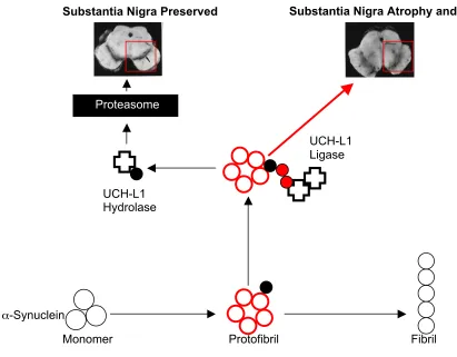
Figure 2: UCH-L1 Activity and a Model Relating it to the Toxic Protofibril Hypothesis. Under the toxic protofibril hypothesis, the disease causing agent is the α-synuclein protofibril, shown here as red circles. UCH-L1 (Cross shape) is part of the ubiquityl/proteasomal
α-synuclein degradative pathway. UCH-L1 ligase activity is dependent on a dimeric conformation of the enzyme which adds addition molecules of ubiquitin (small red dots) to α-synuclein. Monomeric UCH-L1 (single cross) demonstrates hydrolase activity by removing/recycling ubiquitin molecules bound to α-synuclein before it enters the Proteasome. Increase in ligase activity may promote PD pathogenesis while a i n c r e a s e i n hydrolase activity may prevent PD. Images courtesy of http://alzheimer.wustl.edu/adrc2/Research/Neuropathology/images/cerad_sn.bw.gif, retrieved on 22 November 2005
bound the α-synuclein protofibril and prevented the formation of fibrils19. It is reasonable to hypothesize that the dopaminergic substantia nigra neurons are specific to PD because of their dopamine producing capabilities19.
Remember the Proteasome
The parkin enzyme functions as part of the ubiquityl/proteasomal pathway for several intracellular and membrane bound proteins, including α -synuclein4,20. Parkin is a ubiquityl ligase that adds ubiquitin to O-glycosylated α-synuclein destined for proteasomal degradation20. Numerous p a r k i n mutations have been identified but a direct effect has yet to be seen20.
Ubiquitin C-hydorlase–L1 (UCH-L1) functions as part of the ubiquityl/proteasomal degradative pathway of α-synuclein420,21,4. UCH-L1 mutants I93M and S18Y had been identified prior to our research. I93M was implicated in a significant increase in FPD development and S18Y a decrease20. Our research built off the known UCH-L1 hydrolyzing role of removing/recycling ubiquitin from α-synuclein allowing for its degradation20,21 (Figure 2).
We studied UCH-L1 in the I93M and S18Y mutants on α-synuclein in vitro21. I93M was known to increase FAD susceptibility whereas S18Y appeared to protect against FAD21,20. Our findings revealed a dual role of UCH-L1 that directed related to the type of mutation assessed21. We found UCH-L1 to have a dimeric conformational ubiquityl ligase activity, and a monomeric ubiquityl hydrolase activity21. An increase in dimeric ligase function was found to correlate with increased levels of α-synuclein and the I93M mutant21. The S18Y mutant was found to reduce dimerization, decrease ligase activity, and increase hydrolase activity, resulting in a decrease in α- s y n u c l e i n concentration21. This study leads to several hypothesis concerning the role of α-synuclein monomer, protofibril or fibril degradation and PD4,20,21 (Figure 2).
Return to the Toxic Protofibril
Following our elucidation of the PD protofibril and its potentially toxic functions, a detailed study of protofibrillar structure and functions had yet to be accomplished. Use of AFM allowed us to research these unknown protofibrillar characteristics and shed some light onto a dimly lit species.
α
-Synuclein
Proteasome
Substantia Nigra Atrophy and PD
Substantia Nigra Preserved
UCH-L1
Ligase
UCH-L1
Hydrolase
As mentioned in our 2001 paper, protofibrillar α -synuclein was found to bind membranes and form potentially pathogenic pores18,22. We elaborated on that study and made several discoveries in protofibril structure and function. The spherical and annular protofibrils were studied in order to determine their interaction with membranes. Spherical protofibrils (A50P, A53T, and WT) bound membranes but annular forms, monomers, and fibrils did not22. Most significantly, a solution containing membrane and spherical protofibrils led to membrane pore formation that was imaged with AFM22. We noted that the pores formed by the β-sheet rich spherical protofibrils resembled those of bacterial toxins22. This provides further evidence that the spherical protofibril is indeed to PD causative agent22. Our observations of the spherical protofibril have led us to hypothesize that the PD pathogenesis could result from increased spherical protofibril concentration resulting from a decrease in spherical to annular to fibril formation22.
Protofibrils & Cytoplasmic Crowding
Protofibril formation is known to be accelerated in the A30P and A53T mutants but the majority of PD cases are sporadic4,16,23. These early researches lead us to the later hypothesis that there must be some other mechanism responsible for accelerating protofibril formation in WT23. To fill this gap in knowledge, we hypothesized that protofibril formation could be accelerated by crowding the cell with inert material23. In
vitro, we found an increase in WT protofibril formation occurred with an increase in concentration of an inert polymer23. As mention in our earlier studies, a lag time exists between monomer to protofibril formation to fibril formation23. Our data showed that the lag time for protofibril formation decreased from 3 days to 1 day when 5% PEG 20000 (inert polymer) was added23. An increase in the magnitude of crowding comparable to that in our study could feasibly occur in a cellular environment that had decreased protein degradation, such as that seen in UCH-L1 dysfunction23.
α & β Synuclein Demonstrate Profound Interactions
The α-synuclein homologue β-synuclein is known to be 78% identical to α-synuclein24. It has been previously demonstrated that a double-transgenic mouse expressing both human α-synuclein and β-synuclein did not develop any Parkinsonian symptoms indicating that β-synuclein possibly inhibits the toxic agent of PD24. We wanted to further support the hypothesis that the protofibril was the toxic species24. We hypothesized that in vitroβ-synuclein would inhibit α-synuclein protofibril formation. We initially found that both monomeric homologues formed protofibrils but β -synuclein protofibrils did not bind and form pores in vesicles24. We then mixed monomeric solutions of
β -synuclein and A53T α-synuclein, resulting in inhibition of A53T protofibril formation24. This finding further supported the toxic protofibril hypothesis because the non-PD phenotype demonstrated in the double-transgenic mouse aligned with our in vitro data showing β-synuclein to inhibit A53T protofibril formation24,25.
Remember the N-Terminus
α-synuclein is known to have a seven-conserved amino acid sequence on its N-terminus containing both A30P and A53T mutations26. We studied a plus 2 and minus 2 sequence mutant to elucidate the evolutionary conservation of these seven amino acids26. We found the minus 2 mutant to favor β-sheet formation over α -helix and the plus 2 mutant to favor α-helical structure. As demonstrated in earlier studies, the spherical, pore forming protofibrils are rich in β-sheet, thus the
conserved amino acid sequence may have been evolutionarily conserved for its α-helical, non-PD favoring characteristics14,22,26
.
Amyotrophic Lateral Sclerosis
Biological Basis
The biological basis of Amyotrophic Lateral Sclerosis (ALS) has yet to be discovered4. The pathology of the disease consists of spinal motor neuron atrophy with skeletal muscle degeneration in parallel4. Similar to PD and AD, Familial ALS is characterized by mutations in the superoxide dismutase-1 (SOD1) enzyme4,7,8. SOD1 normally converts superoxides to hydrogen peroxide4. The protein aggregates found in ALS neurons contain copious amounts mutant SOD14. It remains
unknown if WT SOD1 forms aggregates, and if fibrils are formed4.
The primary model system for ALS research is the mouse4. The most important of these models was a mouse transgenic for a disabled active-site SOD1 enzyme demonstrated that ALS was not the result of SOD1 enzymatic activity because the mouse developed ALS4.
Prior to our research, the SOD1 mutant had been identified in Familial ALS and the function of the enzyme was known4,7,8. However, the mechanism for mutant SOD1 aggregate formation remained elusive4,27. SOD1 was known to function as a dimer but further detailing had not been accomplished4,7. Our research was directed at mutant SOD1 and establishing a pathway for its presence in aggregates7,8.
SOD1 & Aggregation
We studied the he Familial ALS associated mutant SOD1 A4V which is known to be extremely virulent. WT SOD1 dimer is more stable than the mutant leading us to hypothesize that dissociation of the dimer into monomeric form was somehow involved in ALS aggregate formation and pathology7. In order to test this hypothesis, we introduced a disulfide bridge at V148C to stabilize the dimer through covalent modification7. We found (in vitro) the unmodified A4V mutant to contain a lesser dimeric concentration compared to WT indicating that its stability was decreased7. Introduction of the disulfide bridge stabilized the A4V mutant dimer and prevent its breakdown into monomers and subsequent aggregate formation7.
Building on our previous finding that aggregate formation was the result of an unstable A4V dimer, we screed banks of molecules for ones that could possibly stabilize the dimer8,7. We found 15 molecules that stabilized the dimer, four of which conferred near identical stability to the WT8. These two discoveries are potentially critical in the understanding of ALS8. We have identified the loss of dimeric function in SOD1 as a result of the A4N mutation of Familial ALS and found the resulting monomer to aggregate. In relation to AD and PD, pore like aggregates were observed in A4V mutant SOD1, suggest a possible link between the three diseases8. We have also demonstrated a novel approach toward stabilization of mutant SOD17,8.
Treatments
the phenotypical Aβ plaques and α-synuclein Lewy Bodies27,28. Parkinson’s treatment may take aim at several key processes. Inhibition of UCH-L1 ligase activity by preventing dimerization would increase hydrolase activity and increase α- s y n u c l e i n degradation. In addition, over expression of β -synuclein could provide another means for protofibril repression24. ALS treatments that focused on using the compounds noted in our research could stabilize the SOD1 dimer and prevent dissociation and subsequent aggregation7,8. It must be noted that we believe the pharmaceutical industry to be slowing the progression of treatment formation. We believe this is due to our lack of a complete understanding of the modi operandi underlying AD, PD, and ALS27. Penicillin and aspirin were developed before their effective mechanism had been developed. The same applies for AD, PD, and ALS27.
Conclusion
Alzheimer’s, Parkinson’s, and Amyotrophic Lateral Sclerosis represent the most prevalent neurodegenerative disorders4. Despite all of our research efforts over the past decade, each disease remains terminal. Our research has closely portrayed AD and PD in that a potentially toxic protofibril was identified and the beginnings of its functions illuminated4. All of our AD and PD studies have returned to the protofibril its β-sheet rich pore forming structure4. Though we identified an aggregate structure similar to that of the AD and PD protofibrillar pores, more research will need to be done to determine the properties of this the SOD1 aggregate7,8. Early 1990’s AD and PD research began with the assumption that the toxic agent was the plaque or Lewy Body because they were found upon autopsy5. In the years since this hypothesis was widely accepted, a new toxic protofibril hypothesis has emerged and future research appears to be in that direction4.
Acknowledgements
I would like to thank Dr. Debburman for his guidance through out the research. Thanks to Katrina Brandeis for reviewing the manuscript and anyone else not mentioned who contributed.
Note: Eukaryon is published by students at Lake Forest College, who are solely responsible for its content. The views expressed in Eukaryon do not necessarily reflect those of the College.
References
1. Statistics provided by the Alzheimer’s Association, http://www.alz.org/AboutAD/statistics.asp, retrieved on 18 November 2005
2. Statistics provided by the National Parkinson’s F o u n d a t i o n , I n c . http://www.parkinson.org/site/pp.asp?c=9dJFJLPwB&b=7 1117 retrieved on 18 November 2005
3. ALS Association, www.alsa.org, information retrieved on 18 November 2005
4. Lansbury, Peter Jr., Protofibrils, Pores, Fibrils, and NEURODEGENERATION: Separating the Responsible Protein Aggregates from The Innocent Bystanders, Annu. Rev. Neuroscience, volume 26, pages 267-298, 2003
5. Lansbury, Peter Jr. and Jarrett, Joseph., Seeding “One-Dimensional Crystallization of Amyloid: A Pathogenic Mechanism in Alzheimer’s Disease and Scrapie? Cell,
6 . N a t i o n a l P a r k i n s o n ’ s F o u n d a t i o n , http://www.parkinson.org/site/pp.asp?c=9dJFJLPwB&b=7 1354 information retrieved on 21 November 2005.
7. Lansbury, Peter Jr. et al., An Intersubunit Disulfide Bond Prevents in Vitro Aggregation of a Superoxide Dismutase-1 Mutant Linked to Familial Amytrophic Lateral Sclerosis,
Biochemistry, volume 43, pages 4899-4905, 2004
8. Lansbury, Peter, Jr. et al., Small-molecule-mediated stabilization of familial amyotrophic lateral sclerosis-linked superoxide dismutase mutants against unfolding and aggregation, PNAS, volume 102, no. 10, pages 3639-3644, 2005.
9. Lansbury, Peter, Jr., Assembly of Aβ Amyloid Protofibrils: An in Vitro Model for a Possible Early Event in Alzheimer’s Disease, Biochemistry, volume 38, pages 8972-8980, 1999.
10. Lansbury, Peter Jr., et al., Observation of metastable Aβ
amyloid protofibrils by atomic force microscopy,
Chemistry and Biology, volume 4, pages 119-125, 1997.
11. Atomic force microscopic imaging of seeded fibril formation and fibril branching by the Alzheimer’s disease amyloid-β protein, Chemistry and Biology, volume 4, pages 951-959, 1997.
12. Teplow, David., et al., Amyloid β-Protein Fibrillogenesis: DETECTION OF A PROTOFIBRILLAR INTERMEDIATE, Journal of Biological Chemistry, volume 272, no. 35, pages 22364-22372.
13. Lansbury, Peter., Jr., Synthesis and Amyloid Binding Properties of Rhenium Complexes: Preliminary Progress Toward a Reagent for
14. Lansbury, Peter Jr., Acceleration of oligomerization, not fibrillization, is a shared property of both α-synuclein
mutations linked to early-onset Parkinson’s disease: Implications for pathogenesis and therapy, PNAS, volume 97, no. 2, pages 571-576, 2000.
15. Lansbury, Peter Jr., and Goldberg, Matthew., Is there a cause-and-effect relationship between α-synuclein fibrillization and Parkinson’s disease?, Nature Cell Biology, volume 2, pages E115-E119, 2000.
16. Lansbury, Peter Jr., et al., Accelerated in vitro fibril formation by a mutant α-synuclein linked to early-onset Parkinson disease, Nature Medicine, volume 4, No. 11, pages 1318-1320, 1998.
17. Lansbury, Peter Jr., et al., Inhibition of Fibrillization and Accumulation of Prefibrillar Oligomers in Mixtures of Human and Mouse α-synuclein, Biochemistry, volume 39,
pages 10619-10626
18. Lansbury, Peter., et al., Vesicle Permeabilization by Protofibrillar α-Synuclein: Implications for the Pathogenesis and Treatment of Parkinson’s Disease,
Biochemistry, volume 40, pages 7812-7819, 2001.
19. Lansbury, Peter Jr., et al., Kinetic Stabilization of the α -Synuclein Protofibril by a Dopamine-α-Synuclein Adduct,
Science, volume 294, pages 1346-1349, 2001.
20. Lansbury, Peter Jr. and Brice, Alexis., Genetics of Parkinson’s disease and biochemical studies of implicated gene products, Current Opinion in Cell Biology, volume 14, pages 653-660, 2002.
21. Lansbury, Peter Jr., et al., The UCH-L1 Gene Encodes Two Opposing Enzymatic Activities that Affect α -Synuclein Degradation and Parkinson’s Disease Susceptibility, Cell, volume 111, pages 209-218, 2002.
10209-23. Lansbury, Peter Jr., et al., Molecular Crowding Accelerates Fibrillization of α-synuclein: Could an Increase in the Cytoplasmic Protein Concentration Induce Parkinson’s Disease?, Biochemistry, volume 41, pages 3855-3860, 2002.
24. Lansbury, Peter Jr., Park, June-Young., β-Synuclein Inhibits Formation of α-Synuclein Protofibrils: A Possible Therapeutic Strategy against Parkinson’s Disease,
Biochemistry, volume 42, pages 3696-3700, 2003.
25. Lansbury, Peter Jr., and Volles, Michael J., Zeroing in on the Pathogenic Form of α-Synuclein and Its Mechanism of Neurotoxicity in Parkinson’s Disease, Biochemistry, volume 42, no. 26, pages 7871-7878, 2003
26. Lansbury, Peter Jr., et al., The N-Terminal Repeat Domain of α-Synuclein Inhibits β-Sheet and Amyloid Fibril Formation, Biochemistry, volume 42, pages 672-678, 2003.
27. Lansbury, Peter Jr., Back to the future: the ‘old-fashioned’ way to new medications for neurodegeneration, Nature Neuroscience Reviews, pages S51-S57, 2004