Expression of Hepatitis B Virus Core Antigen in Native and
Fusion Forms in E. coli
Mohammad Azizi
*1,2, Alexis Musacchio
2, Orlando Pardo
2, Nelvis Figueroa
2and
Verena Muzio
21Dept. of Biotechnology, Pasteur Institute of Iran, Tehran 13164, Iran,2Vaccine Division, CIGB, P.O. Box 10600,
Havana, Cuba
ABSTRACT
DNA coding for the core antigen from hepatitis B Virus (HBcAg) was amplified, cloned and propagated in E. coli. The core protein was expressed in E. coli and the product was readily detected by Western blot. This protein can be used as a diagnostic material in serum screening tests. To increase the level of expression of this antigen in bacteria, two plasmids were constructed in which the gene coding for N-terminal part of the human IL-2 has been fused to 5'-terminus of the core antigen gene under control of the tryptophan promoter. Although the expression level of core antigen from hepatitis B Virus in E. coli was increased in fusion forms, but the size of fusion partner could affect the antigenicity, particle formation and assembly of the core antigen. The native and the two fusion forms of the core antigens from hepatitis B Virus were evaluated as a diagnostic material in serum screening.Iran. Biomed. J. 4: 37-43, 2000
Keywords: Hepatitis B core antigen, HBcAg, HBV, Expression
INTRODUCTION
Expression of the gene coding for HBcAg in E.
coli leads to highly efficient synthesis of capsids that
are morphologically and immunologically indistinguishable from viral core particle [1]. This protein is a 21.5 kDa polypeptide that spon-taneously assembles into characteristic 27-nm particles [2] when expressed in a wide range of systems including bacteria [3] and yeast [4]. In addition to the application of recombinant HBcAg for diagnostic purposes, the usefulness of this polypeptide as a carrier for foreign oligopeptide sequences has been suggested [1, 5, 6]. The core particle is known to be highly immunogenic as a result of its polymeric nature and the presence of a number of well defined helper T-cell epitops within HBcAg sequence [7]. However, the cellular immune response to the nucleocapsid antigens is thought to be an important factor in viral clearance because it elicits strong HLA class I and class II-restricted T-cell responses in the infected patients [8]. Previously, it has been shown that the native form of HBcAg is poorly expressed in E. coli [1]. Therefore, attempts were made to express this protein in fusion
forms to improve the expression level. In this study, the expression of HBcAg was undertaken in order to use the expressed protein as a detecting antigen for an ELISA system for diagnostic purposes.
MATERIALS AND METHODS
Bacterial strains. E. coli strains XL-1 Blue and
HB101 were used for DNA manipulation and expression of recombinant HBcAg, respectively.
Enzymes. Restriction enzymes were purchased
from New England BioLabs and Boehringer Mannheim A.G.
Construction of expression vector for HBcAg.
Manipulation of the bacterial strains and plasmids followed routine protocol [9]. Synthetic oligo-nucleotides including restriction enzyme recognition sites were designed on computer software (Gene Runner 3.02, Hasting Software Inc., USA). The core gene of HBV was amplified using these primers: F1 5'-GGCCATGGACATTGACCC-TTAT, R1 ACGGATCCTAACATT GAGATT-CCC, F2
CCTCTAGACATTGACC CTTATAA. The pHBV plasmid that contains the whole genome of hepatitis B Virus subtype adw2 was used as a template for PCR reaction [10]. The PCR products were run on 1.5% LGT agarose gel and the desired band was purified with phenol extraction. The purified band was cloned into pMOSBlue plasmid (Amersham UK) and then subcloned into the expression vectors, pFP15 [11] or pILc-4 [12]. These plasmids contain tryptophan (trp) promoter and gene coding for human interlukin 2 (hIL2). Cloning the gene of interest in XbaI and BamHI sites of pFP15 or pILc-4 resulted in fusion proteins with 60 or 26 amino acids partner from hIL2, respectively.
Expression of recombinant HBcAg. The plasmid
constructs were transformed into E. coli strain HB101. Bacteria harboring expression vectors were grown until the optical density at 620 nm (OD620)=1
in LB medium containing Ampicillin (50 m g/ml) and Tryptophan (200 µg/ml). Then the bacterial culture was inoculated onto fresh medium containing Ampicillin (but not Tryptophan) at initial OD620= 0.05 and incubated at 37°C over night.
SDS-PAGE and Western blot. The optical density
of the cultures was measured at 620 nm and the same cell content of bacteria was taken for SDS-PAGE. The bacteria were spun down and washed with TE buffer (1mM Tris, 6mM EDTA). The bacterial pellets were lysed by boiling in 2X sample buffer (4% SDS, 125mM Tris-Hcl, pH 6.8, 10% 2-mercaptoethanol, 10% glycerol and 0.002% bromophenol blue). SDS-PAGE was performed with a discontinuous gel system, using Tris/glycine buffer [13]. The protein bands were stained with Coomassie Blue or electroblotted to 0.45 µm pore size nitrocellulose membrane (BioRad, USA). The electroblotting onto nitrocellulose filter was performed using a solution containing 0.025M Tris/HCl, 0.15 M Glycine, 20% methanol, pH 8.3, for 1 h at room temperature. The immunodetection was done as previously described [14]. The protein concentration was determined as described by Lowry et al. [15], using bovine serum albumin as a standard. A mouse monoclonal antibody against truncated HBcAg (aa 1-149) and the human antiserum from infected patient were used for Western blot.
Purification of recombinant HBcAg. The bacteria
were harvested and sonicated. The cell lysates were centrifuged for 15 min at 18,000 g and the soluble fraction was separated from precipitant. The sample was dialyzed against 0.01M carbonate-bicarbonate buffer (CB), pH 10.6. Differential ammonium sulfate (AS) precipitation was done [16] and the protein contaminants were precipitated at 10% AS. The recombinant proteins were later precipitated at 30% AS. For further purification steps, the pellets were washed with 1 mM phosphate buffer, pH 6.8 and applied on Q-Sepharose Fast Flow column (Pharmacia, Sweden). Recombinant proteins were eluted using a linear gradient of NaCl (0.1 - 0.8 M) in 1 mM phosphate buffer, pH 6.8 [17].
In the sample where the recombinant HBcAg remained in the insoluble fraction after cell disruption, the protein was purified by washed pellet procedure. Briefly, pellets were washed with CB buffer and 4M urea in the same solution. The pellets were washed again with CB buffer, prior to the extraction of the recombinant HBcAg. For extraction, saturated guanidinium hydrochloride in 70% acetic acid was used. Further, purification was performed in a Butyl 650 (S) column (TosoHaas, Japan). The extracted protein was diluted 1:3 with 0.1% trifluoroacetic acid (TFA), applied to the column and purified using H2O-acetonitrile
(AcN)/TFA buffer system [18] with a linear gradient (10-60% AcN: 0-60 min). The eluted protein was freeze-dried, and kept at 4°C until used. Later, the freeze-dried protein was resuspended in 2% sarcosyl in CB buffer, and dialyzed back to CB buffer to remove the existing detergent.
ELISA. Maxisorp plates (Nunc, Denmark) were
coated with 5 µg/ml of HBcAg preparations (native and fusion forms) in 0.1M Tris/HCl pH 8.5 with 0.02% sodium azide, 3 h at 37°C. Plates were washed 4 times with PBS containing 0.05% Tween 20 (PBST) before serum dilutions were added. Sera were diluted 1:20 in PBST and incubated 1 h at 37°C in precoated plates. After washing, bound antibodies were detected by incubation for 1 h at 37°C with alkaline phosphatase labeled anti-human conjugate, diluted 1:1,000. The substrate p-nitrophenyl phosphate (1 mg/ml) in 0.5 M diethanolamine buffer pH 9.8, was used to develop the antigen-antibody reaction, and absorbance was read after 20-45 min at 405 nm. A panel of 18 known sera for anti-HBcAg antibody was used for
the evaluation of reactivity of the antigen-antibody as a diagnostic material.
Sequencing of 5' end of HBcAg gene. The 5' end
of HBcAg gene in each construct was sequenced to defined in-frame sequence by Sanger method [19]. It was done using oligonucleotides matching to the upstream and downstream of HBcAg gene (sequencing kit and 32P-Labelled dATP form Amersham UK).
RESULTS
Construction of expression vectors for HBcAg.
Three expression vectors were constructed according to (Fig. 1) under the control of trp promoter. The
plasmid pHBc180 coding for native core antigen was resulted from subcloning of HBcAg gene in expression vector (pFP15). Plasmid pHBcSF8 and pHBcLF7 were obtained after the subcloning of HBcAg gene in two types of expression vectors (pILc-4 or pFP15). These vectors contain some parts of hIL2 gene and coded for short and long fusion HBcAg, respectively (Fig. 1).
Expression of HBcAg in native and fusion forms.
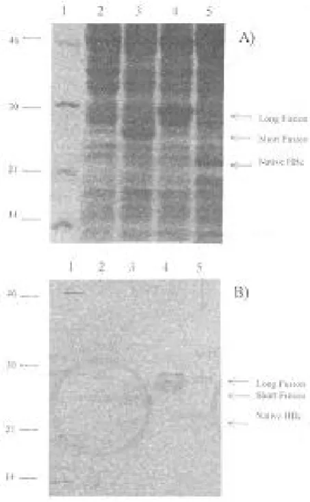
The constructed plasmids expressed the HBcAg in native (185 amino acids) or fusion forms in E. coli (Fig. 2). The fusion forms of core antigen contain 26 (short) or 60 (long) amino acids of hIL2 as a partner. The expression products were recognized as a
Fig.1. Scheme for construction of plasmids expressing HBcAg in the native and fusion forms. Plasmid A expresses the native form of HBcAg and plasmids B & C express the long & short forms of the fusion HBcAg protein, respectively.
single band with expected molecular masses in the Western blot by staining with anti-truncated HBcAg specific monoclonal antibody (Fig. 2). The expression level of native core antigen from pHBc180 plasmid was less than 1% of total cell protein, whereas it was increased to 10% and 12% for short and long fusion forms, respectively.
Purification of recombinant HBcAg. E. coli cells
expressing various forms of HBcAg were disrupted and centrifuged. Following centrifugation the native
Fig. 2. A) SDS-PAGE, B) Western blot analysis of E. coli strain HB101 with and without the following expression vectors. Lane 1, molecular weight marker; lane 2, bacteria without plasmid; lane 3, plasmid pHBcSF8 (Short fusion); lane 4, plasmid pHBcLF7 (long fusion); lane 5, plasmid pHBc180 (Native).
and the short fusion form of HBcAg remained in the soluble fraction. The soluble proteins were purified up to 70% after differential precipitation with ammonium sulfate. Soluble recombinant HBcAg was precipitated in 30% ammonium sulfate and then eluted with 0.4-0.5 N NaCl in Q-Sepharose Fast Flow column chromatography. Precipitation followed by anion exchange chromatography resulted in up to 90% purity of target proteins. The long fusion form of HBcAg was insoluble after cell disruption and separation from host soluble proteins. This resulted in 70% purity in one step. The purity was increased up to 85% by washing the pellets with 4M urea in CB buffer pH 10.6. Finally, passage through a Butyl 650 (S) column gave a purity of up to 95%.
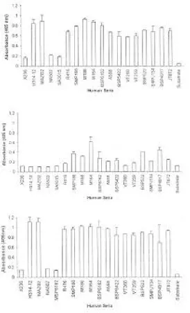
ELISA. The evaluation of reactivity of each
antigen with human anti-sera was performed using ELISA. Antigens were coated on microtitre plate and a monoclonal antibody specific for truncated HBcAg (aa 1-149) was used as a positive control. All the positive sera from infected patients showed similar reactivity with the recombinant proteins except for the long fusion form. The validity of the test was assessed with a panel of 18 sera with known titers for anti-HBcAg antibody. The native and the short fusion form of the recombinant HBcAg clearly discriminated between known positive (OD405=
1.5-2) and negative (OD405= 0.1-0.2) sera (Fig. 3).
DISCUSSION
The cloning of HBV core antigen gene in the expression vector and transformation into E. coli led to the synthesis of recombinant HBcAg. E. coli harboring these plasmids were analyzed for the expression of the HBcAg by SDS-PAGE and Western blotting. As the fusion form of HBcAg could react with antiserum in Western blot, therefore it is valuable for screening of HBcAg positive sera. Further work is required to determine the specificity and sensitivity of the reaction by using a larger number of antisera from different HBV infected patients.
The purified HBcAg was used as a diagnostic antigen for the detection of anti-HBc antibody in human sera by ELISA system. The validity of the
Fig. 3. ELISA analysis using the HBcAg in forms of A) native, B) long fusion and C) short fusion. Sera coded X236, NA502 and MSP6162 were known as negative sera for anti-HBc antibody.
test was confirmed using a panel of positive sera detected by commercial kit. The presence of anti-HBc in these sera was detected by a home-made ELISA, using recombinant HBcAgs. In this study, recombinant native and short fusion forms of HBcAg were recognized in the ELISA system equally well by both anti-HBc monoclonal antibody and sera obtained from infected individuals (Fig. 3). The expression of HBcAg in E. coli was first reported by Pasek et al. [20]. Then, Cohen and Richmond [2] demonstrated that this protein which
is produced by recombinant DNA technology has a particle structure and antigenicity similar to the native HBcAg. For this reason, the recombinant HBcAg has often been used to investigate the antigenicity, particle forming ability and RNA binding capacity of HBcAg [3]. When a large amount of recombinant proteins are to be prepared in E. coli different methods may be utilized. The fusion of the gene for a highly expressed protein in
E. coli to the N-terminal or C-terminal of the
recombinant protein is one of these methods [21].
Furthermore, when examining the antigenicity and particle forming ability of the HBcAg, it is important to avoid the influence of fusion partner on HBcAg antigenic properties. For this purpose, a protein with the same primary sequence as that of the wild type HBcAg must be prepared. However, it is difficult to obtain a high level expression of the unfused HBcAg in E. coli for protein purification.
A number of explanations can be offered for the various levels of expression of HBcAg in the native and fusion forms. Most of which are related to differences in the nucleotide sequence and secondary structure of mRNA that lead to alterations in the efficiency of translation [22]. The variations may also be simply due to differences in the nucleotide sequence preceding the initiation codon of the HBcAg gene, which alter the efficiency of ribosome binding and initiation of protein synthesis, and needs further investigation.
Furthermore, a recombinant HBcA gencom-passing the complete precore sequence produced in yeast was also shown to assemble into particles [23]. Further evidence for the sequence–specific influence of precore (fusion) residues comes from the observation that a number of N-terminal extensions fused to HBcAg for vaccine purpose have not prevented particle assembly [23, 24]. Recent studies have revealed that the cysteine disulfide bridges within HBcAg particles stabilize the molecule but are not essential for particle formation [25]. It was observed that the exchange of cysteine at aa position 7 in precore region is essential to prevent particle assembly probably by forming disulfide bridges [26].
Several groups have reported that the addition of heterologous sequences to the termini of the HBcAg protein maintain the property of spontaneous self-assembly presenting the foreign peptide on its surface at a high density [23]. Sequences as long as 40 amino acids have been successfully inserted with no deterimental effect on particle assembly [6]. Furthermore, other groups have shown that it is also possible to add heterologous sequences to the carboxy terminus of the molecules next to the amino acid 144 [1] and retain particle structure. Therefore, there is a great potential for the development of multivalent HBcAg based particle containing sequences from the same or different pathogens at the amino- and or carboxy-termini of the protein [27].
This analysis revealed that the inclusion of 26 aa
from hIL2 at the N-terminal of HBcAg precluded spontaneous assembly into particulate structures and resulted in a molecule that exhibits HBc antigenicity. However, the addition of 60 aa from hIL2 resulted in insolubility of the protein with lower antigenicity as it was obtained from the ELISA results (Fig. 3). It should be added that such differences were not observed while analyzing these combination with Western blot (Fig. 2). Therefore, the results obtained in this study indicated that addition of limited numbers of aa at the N-terminal could not affect the biological activity of the protein. Similar observation was noted by Brown, A.L., et al. [6]. Further experiments in the particle formation and mRNA structure analysis of these different forms are underway.
ACKNOWLEDGEMENTS
The authors thank Dr. S. Bouzari and Dr. M. Mohammadi for the critical review of this manuscript, and Mr. D. Palenzuela for the generous contribution of anti-HBc monoclonal antibody.
REFERENCES
1. Borisova, G.P., Berzins, I., Pushko, P.M., Pumpen, P., Gren, E.J., Tsibinogin, V.V., Loseva, V., Ose, V., Ulrich, R. and Siakkou, H. (1989) Recombinant core particles of hepatitis B virus exposing foreign antigenic determinants on their surface. FEBS Lett.
259 (1): 121-124.
2. Cohen, B.J. and Richmond, J.E. (1982) Electron microscopy of hepatitis B core antigen synthesized in
E. coli. Nature 296 (5858): 677-679.
3. Stahl, S., MacKay, P., Magazin, M., Bruce, S.A. and Murray, K. (1982) Hepatitis B virus core antigen: Synthesis in Escherichia coli and application in diagnosis. Proc. Natl. Acad. Sci. USA 79 (5):
1606-1610.
4. Kniskern, P.J., Hagopian, A., Montgomery, D.L., Burke, P., Dunn, N.R., Hofmann, K.J., Miller, W.J. and Ellis, R.W. (1986) Unusually high-level expression of a foreign gene (hepatitis B virus core antigen). In: saccharomyces cerevisiae. Gene 46 (1):
135-141.
5. Chambers, M.A., Dougan, G., Newman, J., Brown, F., Crowther, J., Mould, A.P., Humphries, M.J., Francis, M.J., Clarke, B., Brown, A.L. and Rowlands, D. (1996) Chimeric hepatitis B virus core particles as probes for studying peptide-integrin interactions. J.
Virol. 70 (6): 4045-4052.
6. Brown, A.L., Francis, M.J., Hastings, G.Z., Parry, N.R., Barnett, P.V., Rowlands, D.J. and Clarke, B.E. (1991) Foreign epitopes in immunodominant regions of hepatitis B core particles are highly immunogenic and conformationally restricted. Vaccine 9 (8):
595-601.
7. Chisari, F.V. and Ferrari, C. (1995) Hepatitis B virus immunopathogenesis. Annu. Rev. Immunol. 13:29-60. 8. Mondelli, M., Vergani, G.M., Alberti, A., Vergani, D., Portmann, B., Eddleston, A.L. and Williams, R. (1982) Specificity of T lymphocyte cytotoxicity to autologous hepatocytes in chronic hepatitis B virus infection: evidence that T cells are directed against HBV core antigen expressed on hepatocytes. J.
Immunol. 129 (6): 2773-2778
9. Sambrook, J., Fritsch, E.F. and Maniatis, T. (1989) Molecular cloning: A laboratory manual, 2nd Ed., Cold Spring Harbor Laboratory, Cold Spring Harbor. NY.
10. Valenzuela, P., Quiroga, M., Zalvidar, J., Gray, P. and Rutter, W.J. (1980) The nucleotide sequence of the hepatitis B viral genome and the identification of the major viral genes. In: Animal Virus Genetics. (Fields, B.N., Jaenisch, R. and Fox, C.F. eds.), Academic Press, New York. pp. 57-70.
11. Novoa, L.I., Machado, J.A., Fernلndez, J.R., Benيtez, J.V., Narciandi, R.E., Rodrguez, J.L., Estrada, M.P., Garcيa, J. and Herrera, L.S. (1995) Methods for the expression of heterologous proteins produced in fuse form in E. coli, use thereof, expression vector and recombinants strains.
European Patent Application: (90202108.8) .
12. Vلzquez, D., Montero, M., Duarte, C. and Menéndez, A. (1997) Influence of the stabilizing sequence on the expression of multi-epitope polypeptides of HIV-1 in Escherichia coli. Biorecnologيa Aplicada, 14: 91-96.
13. Hames, B.D. and Rickwood, D. (1990) Gel electrophoresis of proteins: A practical approach. Oxford University Press, New York.
14. Towbin, H., Staehelin, T. and Gordon, J. (1979) Electrophoretic transfer of proteins from polyacrylamide gels to nitrocellulose sheets: procedure and some applications. Proc. Natl. Acad.
Sci. USA 76: 4350-4354.
15. Lowry, O.H., Rosebrough, N.J., Farr, A.L. and Randall, R.J. (1951) Protein measurment with the Folin-Phenol reagent. J. Biol. Chem. 193: 265-275. 16. Deutscher, M.P. (1990) Methods in Enzymology:
Guide to protein purification. 1st ed., Academic press, Inc. 285-296.
17. Pharmacia LKB Biotechnology. (1993) Ion-exchange chromatography: principles and methods. 4th ed. Pharmacia Bioprocess Technology.
18. Snyder, L.R. and Kirkland, J.J. (1979) Bonded-Phase Chromatography. In: Introduction to Modern Liquid
Chromatography, John Wiley & Sons, USA. pp.
232-269.
19. Sanger, F., Nicklen, S. and Coulson, A.R. (1977) DNA sequencing with chain-terminating inhibitors.
Proc. Natl. Acad. Sci. USA 74 (12): 5463-5467.
20. Pasek, M., Goto, T., Gilbert, W., Zink, B., Schaller, H., MacKay, P., Leadbetter, G. and Murray, K. (1979) Hepatitis B virus genes and their expression in
E. coli. Nature 282 (5739): 575-579.
21. Nagai, K. and Thogersen, H.C. (1987) Synthesis and sequence-specific proteolysis of hybrid proteins produced in Escherichia coli. Methods Enzymol. 153:
461-481.
22. Miyanohara, A., Imamura, T., Araki, M., Sugawara, K., Ohtomo, N. and Matsubara, K. (1986) Expression of hepatitis B virus core antigen gene in Saccharomyces cerevisiae: synthesis of two polypeptides translated from different initiation codons. J. Virol. 59 (1): 176-180.
23. Clarke, B.E., Newton, S.E., Carroll, A.R., Francis, M.J., Appleyard, G., Syred, A.D., Highfield, P.E., Rowlands, D.J. and Brown, F. (1987) Improved immunogenicity of a peptide epitope after fusion to hepatitis B core protein. Nature 330 (6146):381-384. 24. Schodel, F., Moriarty, A.M., Peterson, D.L., Zheng,
J.A., Hughes, J.L., Will, H., Leturcq, D.J., McGee, J.S. and Milich, D.R. (1992) The position of heterologous epitopes inserted in hepatitis B virus core particles determines their immunogenicity. J.
Virol. 66 (1): 106-114.
25. Zheng, J., Schodel, F. and Peterson, D.L. (1992) The structure of hepadnaviral core antigens. Identification of free thiols and determination of the disulfide bonding pattern. J. Biol. Chem. 267(13): 9422-9429. 26. Schodel, F., Peterson, D., Zheng, J., Jones, J.E.,
Hughes, J.L. and Milich, D.R. (1993) Structure of hepatitis B virus core and e-antigen. A single precore amino acid prevents nucleocapsid assembly. J. Biol.
Chem. 268 (2): 1332-1337.
27. Borisova, G., Borschukova, O., Skrastina, D., Dislers, A., Ose, V., Pumpens, P. and Grens, E. (1999) Behavior of a short preS1 epitope on the surface of hepatitis B core particles. Biol. Chem. 380
(3): 315-324.