EFFECTS OF THE ROOT BARK EXTRACT OF Monotheca buxifolia(FALC.) A. DC ON ANTIOXIDANT POTENTIAL AND SERAL PROPERTIES OF RABBIT
(Oryctolagus cuniculus)
Khan Sher1, Ali Hazrat2, Muhammad Nisar2 and Asim Muhammad4
https://doi.org/10.28941/24-4(2018)-11
ABSTRACT
This activity was aimed to examine the phytomedicinal effects of the crude extract of the bark of the root of Monotheca buxifolia (Falc.) A. DC on the serum biochemistry and antioxidant potential of the in-vitro lipids present in various tissues of the rabbits and the overall correlation of the extract and serum lipids. The extract was administered orally to the rabbits in different doses (0, 25, 75 and 150 mg) in triplicate daily for 10, 20 and 30 days respectively. The data showed that the weights of rabbits and their different organs were reduced. Serum cholesterol level of the experimental animal decreased, while no variation in the serum glucose was observed. The histopathology results showed that the central vein had normal endothelial lining with no evidence of pericentral fibrosis. The hepatic cords in almost all cases were well formed. The portal tracts showed branches of hepatic artery, portal veins and cholengioles with normal morphology. A significant (P < 0.05) reduction was observed in the RSA values of the brain, liver and muscles of the subject animals. Increasing the dose reduced the RSA values in all the tissues. It was concluded that this plant has the potency for decreasing weight of the whole body and also decrease the cholesterol level without significantly affecting other parameters. Hence, there are implications for further detailed studies on this plant.
Keywords: Anti-Oxidant, Monotheca buxifolia (Falc.) A. DC, LDL-c level, HDL-c level, total cholesterol.
Citation:Effects of the root bark extract ofMonotheca buxifolia (Falc.) A. DC on
antioxidant potential and seral properties of rabbit (Oryctolagus cuniculus). Pak. J. Weed Sci. Res. 24 (4):429-438.
1Department of Botany, Shaheed Benazir Bhutto University, Dir Upper, Khyber
Pakhtunkhwa,, Pakistan.
INTRODUCTION
Monotheca buxifolia (Falc.) A. DC. Vern. Gurgurah belongs to family Sapotaceae. It has short thorns either axillary or terminal in position and alternate leaves. Pedicelate flowers of about 5 mm in diameter are produced in April to May. The filaments of the flower are longer than the petals. The fruit of M. buxifolia are round in shape and of about 1 cm in diameter. The fruits are eaten by local people and are famous for their delicious taste. M. buxifolia is one of the numerous species of wild fruits and nuts found in the different areas of Pakistan
(
Nasir and Ali, 1972), Afghanistan, Oman, North of Somalia and Southern Ethiopia (Ahmed et al.,2010).It has been investigated that it contains hydrocarbons (heptacosane, penta triacontane) and alcoholic sterols (Nasir & Ali, 1972). Different parts of M. buxifolia contains high quantity of antioxidant compounds like anthraquinones, flavonoids, terpenoids, tannins, saponins, and cardiac glycosides (Rehman et al., 2013; Ullah et al., 2016). The antioxidants from these natural sources have strong effect on serum lipid profile (HDL, LDL, and cholesterol), body weight (Georgiev et al., 2011), atherosclerosis (Peng et al., 2011) and against the liver diseases (Fontanari et al., 2012).
Medicinally, fruits and other parts of the plant are laxative, used against gastric and in urinary tract diseases (Rashid and Marwat, 2006). Extracts of the various portions of plant are used extensively in conventional medicine, especially for the preparation of different remedies like treatment of cough, laryngitis and liver diseases. In South Asia, plant extracts are used as a separate branch of medicine from the very ancient time (Dahanukar et al., 2000; Habib ul Hassan et al.,2014).
This activity was aimed to examine the phytomedicinal effects of the crude extract obtained from the bark of the root of Monotheca buxifolia on the serum biochemistry and antioxidant potential of
the in-vitro lipids present in various tissues of the rabbits and the overall correlation of the extract and serum lipids.
MATERIALS AND METHODS
Extraction of Metabolic contents of M. buxifolia: The plant was collected from the Hindu Kush range of lower Dir, Khyber Pakhtunkhwa Pakistan. After complete shade drying the bark was grinded to a fine powder. The powder was then put in 50 % aqueous methanolic solution in a beaker and was placed on a shaker for five days. After complete shaking of the methanolic solution was filtered out and then put on rotary vacuum evaporator. This activity was continued till all the methanolic and aqueous portion got separated from the crude extract. The extract was then reserved in an oven for some time at a high temperature to make it completely dry (Chellan et al., 2008). Total Phenolic Contents Assay
The total phenolic content (TPC) of the extracts was determined by using Folin-Ciocalteu’s reagent. Rendering to the method of Slinkard and Singleton (1977) Gallic acid was used as a reference. After incubation at 27℃ for 1 hr. the absorbance was measured at 765 nm with
a Shimadzu UV/Vis-1700
spectrophotometer (Shimadzu, Japan). Animals Feeding
Twelve numbers of rabbits of the local strains were selected as experimental animals. The animals were classified into four groups (S, P, Q and R) triplicate in each. All the rabbits were kept in uniform conditions of temperature and fodder feeding. Group S were control hence no extract was given, group P was fed with 25 mg, group Q with 75 mg of extract and group R were fed 150 mg extract given orally for one month on daily basis.
Blood Biochemical and hematological Analyses
biochemical analysis. The same were then subjected to centrifugation to get the serum. The serum was then analyzed for HDL-c, TC, LDL-c, TG, Glucose (G) contents and ALT values using Shimadzu UV-vis-1700 spectrophotometer (Shimadzu, Japan). Likewise blood was also taken in EDTA tube (Guangzhou Medical Instrument Co. Ltd Guangzhou, China for hematological analysis) and were analyzed for Haemoglobin level, TLC (total leucocytes count), Total RBCs (red blood cells), Mean Corpuscular Volume (MCV), Mean Corpuscular Haemoglobin (MCH), Mean Corpuscular Hemoglobin Concentration (MCHC) and Platelet Count. Liver Histopathology
After completion of the one month trial the rabbits were slaughtered to obtain the different organs (kidney, brain and liver) for further analyses. Liver from each triplicate was taken. Sections were prepared by using microtome and stain on microscopic slides. The images were analyzed for any possible change produced by the extract using CC TV fitted binocular microscope.
Lipid Extraction from Tissues
Liver, muscles, and brain were collected from the rabbits on autopsy. These were preserved in formalin and used for the lipid extraction. A 10 gram of each tissue was used. The tissues were homogenized separately and chloroform-methanol mixture (2:1) was added. After complete shaking for 72 h the homogenized was separated by using various instruments like filter paper and separating funnel. The obtained filtrate was then subjected to rotary vacuum evaporator for the separation of lipids from the filtrate (Folchet al., 1957). Radical Scavenging Assay (RSA)
The Radical Scavenging Assay of the extracts and lipids obtained from different tissues like brain, muscles and liver of the rabbits were calculated using Shimadzu UVvis-1700 spectrophotometer (Shimadzu, Japan).
Data Analysis
For data analysis Graph Pad Prism 5 for windows version 5.03 (Graph Pad Software, Inc, 2009) was used. Analysis of variance (ANOVA) and multiple comparisons were carried out. The necessary data are presented in Tables and Figures.
RESULTS AND DISCUSSION
Effects on the Rabbits Body and organs Weight
Increase in body weight or obesity are related to a number of diseases including cardiovascular, diabetic, hypertension, gall bladder and cancer (Akil and Ahmad, 2011). This result showed that the root bark extracts of the plant reduced the total body weight of the rabbits. Similarly the weight of liver, kidney and brain were also reduced due to the intake of extracts after feeding for one month. In contrast to the varying treatments, no change in total body weight was observed in control group (S). Increasing the dose decreased the total weight effectively to 1.05 ± 0.022 at the end of 4th week in R group as shown in Table-1b. Similarly a reduction in weight of liver, kidney and brain were also observed upon feeding of M. buxifolia root extracts (Table-1a). Comparable results were also communicated by Eweka and Enogieru (2011). The researchers in their work concluded that natural extracts reduce the lipid per-oxidation in the tissues and thus decreasing the weight of the subject animals and the tissues related.
Blood Biochemical Analyses
10th day of group S. No significant change in serum triglycerides (TG) was recorded in the present findings. All the values were almost at the same range as shown in Table 2. However, a significant increase in HDL-c level was observed when rabbits were fed with M. buxifolia extracts. Comparing the values of 10th, 20th and 30thday it was found that the extracts had the ability to elevate the level of HDL-c effectively) [Table-2]. It is concluded from the current data that M. buxifolia root extract decreased the TC level, while increased glucose and HDL-c level. But there was no or less effect in case of serum TG level.
Higher level of total Cholesterol (TC) in blood is often related to many pathological conditions, including cardiovascular diseases (CVD) and arthrosclerosis (Pearson et al., 2003). Similarly any abnormality in the blood glucose may lead to the condition of hypoglycemia or hyperglycemia. The most prominent example of hyperglycemia is Diabetes mellitus, which is related to failure of blood sugar regulation. The blood also contains TG that is mainly responsible for energy requirement of the body. The HDL cholesterol is a type of lipoproteins. The higher concentrations of LDL are atherogenic, whereas HDL high concentrations are negatively related with the risk for CVD (Kannel, 1992).
The current results showed that M. buxifolia root extract decreased the TC level, while increased glucose and HDL-c level. But, there was no or less effect in case of serum TG level (Table-2). The results are in support with Uhl et al. (1992) who also recorded a significant reduction in TC level. But the study of Meralet al. (2001) deviates from the current findings, they showed that plant extracts have no effect on serum glucose concentration. However the study of
Murugaiahet al.(1999) and Yokozawa et al. (1996) show that plant root extracts increased HDL-c level and thus reducing the TC burden of the body.
Blood hematological Analyses
Blood hematology is another important parameter to study the effect of a particular compound on animal physiology. In this activity the M. buxifolia root extract decreased the TLC level significantly (P < 0.05). The dose of 150 mg decreased the TLC value to 6950.0 ± 464.2 cmm as compared to control group (13600.3 ± 3200.9 cmm). But there was no significant change in Hb, TRBC, HCT, MCH and MCHC concentration. However, a significant change (P < 0.05) in platelets count was observed. The dose of 75 mg increased the platelets up to 538166.7 ± 40707.6 cmm, but 150 mg dose again reduced the value to 478166.7 ± 6604.3 cmm as shown in Table-3. The current results show that the root extract has a significant effect (P < 0.05) on some hematological indices like the level of TLC decreased, while the platelets level was increased. However no significant change in Hb, TRBC, HCT, MCH and MCHC was deciphered (Table-3).
Table-1a. Change in organ weight with the oral administration of extract. Group Dose (mg day
-1) Liver Organs weight (g)Kidney Brain
S 0 0.0533± 0.0097 0.0130± 0.0014 0.0067± 0.0012
P 25 0.0417± 0.0016 0.0090± 0.0014 0.0067± 0.0019
Q 75 0.0367± 0.0123 0.0100± 0.0007 0.0057± 0.0002
R 150 0.0423± 0.0040 0.0090± 0.0000 0.0057± 0.0012
Table-1b. Change in body weight with the oral administration of extract.
Group Dose Weight Change (kg)
(mg day-1) 1stweek 2nd week 3rdweek 4th week
S 0 1.41± 0.09 1.47± 0.13 1.25± 0.04 1.35± 0.14
P 25 1.39 ± 0.11 1.36 ± 0.06 1.20 ± 0.06 1.20 ± 0.05
Q 75 1.56 ± 0.08 1.50 ± 0.14 1.34 ± 0.12 1.22 ± 0.12
R 150 1.15 ± 0.001 1.13 ± 0.022 1.10 ±
0.047 1.050.022 ±
Table-2. Effect of M. buxifoliaroot extracts on serum biochemistry. Param
eter Days Control (p group)25 mg (q group)75 mg (r group)150mg
TC 10 220.7 ± 31.8
(a) 203.9 ± 1.4 (a) 203.0 ± 17.6(a) 164.2 ± 25.1(a)
20 216.8 ± 30.5
(a) 143.1 ± 31.8(a) 180.5 ± 56.2(a) 245.6 ± 23.3(a)
30 218.4 ± 30.3
(a) 153.7 ± 41.3(b) 129.7 ± 15.6(c) 128.3 ± 18.9(d) GL 10 16.9 ± 0.2 (a) 53.8 ± 1.4 (b) 35.1 ± 7.8 (a) 32.8 ± 19.8 (a)
20 49.0 ± 20.6
(a) 48.1 ± 16.6 (a) 49.1 ± 19.8 (a) 24.3 ± 10.6 (a) 30 40.2 ± 1.9 (a) 34.0 ± 6.6 (a) 26.7 ± 11.2 (a) 56.76 ± 2.8 (b)
TG 10 244.5 ± 5.5
(a) 251.3 ± 13.8(a) 255.8 ± 6.2 (a) 250.4 ± 17.0(a)
20 253.3 ± 2.8
(a) 157.9 ± 2.1 (b) 192.7 ± 1.4 (c) 143.8± (d) 22.0
30 288.5 ± 1.4
(a) 266.5 ± 3.5 (b) 251.3 ± 5.5 (c) 244.0 ± 4.8 (d)
HDL 10 119.1 ± 9.3
(a) 64.0 ± 37.5 (b) 87.4 ± 22.9 (a) 196.1 ± 12.2(c)
20 124.3 ± 4.29
(a) 42.1± 13.7 (b) 67.8 ± 37.8 (c) 33.7 ± 18.3 (d)
30 129.2 ± 5.5
LIVER HISTOLOGY
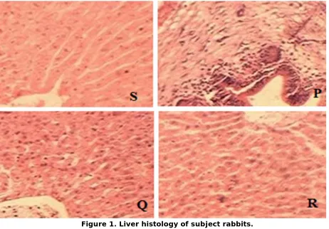
Liver is an important organ of the body and hence plays enormous roles in energy and nutrient metabolism. Feeding different compounds to subject animal changes the Liver functions (Tres et al., 2009). The current result in Figure 1 showed that S group has normal endothelial lining with no evidence of pericentral fibrosis and showing normal hepatocytes architecture. Similar results were also investigated for Q and R groups. However in P group there was few
eosinophils along with mild infiltrate of lymphocytes. Some zenoparasites at the interface of portal tracts showed hepatocytes degeneration in the same group. Similarly in R group the supporting tissues of the portal tracts showed mild infiltrate of lymphocytes and scattering of a few neutrophils is shown. In this connection the study of Bartels et al. (2007) is quite important. They showed that plant extracts contains natural antioxidants that has beneficial role on liver histology
Table-3. Effect of M. buxifolia plant extracts on the hematological indices of the rabbit.
Parameter Groups
S P Q R
Hb (g/dl) 11.3 ± 2.2 12.3 ± 2.6 12.8 ± 1.0 12.9± 1.0
TLC (cmm) 13600.3±3200.9 13358.3 ± 3520.9 6733.3
±1000.3 6950.0464.2 ± TRBC
(mil/cmm) 6.2 ± 0.1 6.6 ± 0.3 6.9 ± 1.9 6.3 ± 0.3
HCT 42.5 ± 1.2 46.5 ± 1.6 46.5 ± 1.6 42.9 ± 1.1
MCV (fl) 62.3 ± 1.1 72.3 ± 1.6 69.3 ± 1.5 69.4 ± 2.4
MCH (pg) 19.1 ± 0.1 19.1 ± 0.1 19.5 ± 0.1 19.0 ± 0.1
MCHC (g/dl) 27.1 ± 0.1 27.1 ± 0.1 27.5 ± 0.9 27.8 ± 0.5
Platelets
(cmm) 401166.7 ± 19388.2 391166.7±112388.2 538166.7±40707.6 478166.7±6604.3
Alanine amino transferase (ALT) Administration of root bark extracts significantly reduced (P < 0.05) the ALT level in P, Q and R groups. The effect was most dominant in R group because of high dose (150 mg) of the test plant root extracts. At t30th day the ALT value was 60.4 ± 52.91 mg/dL for P group, 52.7 ± 11.07 mg/dL for Q and 51.5 ± 10.03 mg/dL for R group, respectively as shown in Table-4. Thus it is concluded from the current findings that root extracts of the plant under study reduce the ALT level and have a pronounced effect on liver biochemistry.
1991). The present results of ALT level in the serum of rabbits administered with M. buxifolia root extracts significantly reduced the ALT level in P, Q and R groups (P < 0.05). Increasing the dose effectively decreased the ALT level in the subject animals. Similar results were
investigated by Zahra et al. (2012). They analyzed various enzymes of the blood serum including ALT to find out the hepatocurative and hepatoprotective effects of Momordica charantia. They concluded that natural extracts significantly reduce the ALT level.
Figure 1. Liver histology of subject rabbits.
Table-4. ALT value of the serum and RSA in brain of the blood after extract administration.
Treatment Treatment days Brain RSA
10 20 30
Control 70.1 ± 0.06 70.09 ± .074 70.1 ± 0.05 6.79 ± 3.42
P 112.8 ± 50.4 71.91 ± 12.31 60.4 ± 52.91 6.60 ± 3.48
Q 59.6 ± 13.6 55.74 ± 10.21 52.7 ± 11.07 4.27 ± 2.03
Radical Scavenging Assay (RSA)
In this work a significant reduction (P < 0.05) was observed in the RSA values of the brain, liver and muscles tissues of the subject animals. Increasing the dose reduces the RSA values in all the tissues. A dose of 150 mg reduced the RSA value to 0.28 ± 0.50 in brain, 1.23 ± 0.68 in liver and 1.66 ± 0.79 in muscles respectively as shown in Table-4. This result showed that brain was most vulnerable to the effect ofM. buxifoliaroot extracts.
An antioxidant is naturally used to prevent the oxidation of other compounds and molecules. In Oxidation reaction free radicals are produced that lead to a chain reactions. This chain reaction finally leads to cell damage (Halliwell, 1994). Antioxidants from natural sources fight free radicals and protect us from various diseases. They act upon either by scavenging the reactive oxygen species or protecting the antioxidant defense
mechanisms. Similar results were observed by Gonzalez-santiago et al. (2006). They concluded that free radicals in the body reduce the RSA activity of the subject animals significantly.
CONCLUSIONS
It is concluded from the current study that M. buxifolia root extract decreased the body and organs weight of the subject animals. A significant decrease in TC and ALT was also observed. However, there was increase in HDL-c level, thereby decreasing the risks of CVD. The extracts have also beneficial effect on liver morphology and biochemistry. Similarly the Administration of root bark extracts significantly reduced the ALT value. That needs further investigation. However, there is no significant effect in RSA value. Finally it can be concluded that the body of the animals got improvement in some of the physiological functions upon the administration of root extracts.
REFERENCES CITED
Ahmad, I., M.J. Akhtar, H.N. Asghar and Ahmed, B.W., T. Koikea, Y. Yua, T. Inouea, T. Aokia, E. Liub and J. Fan. 2010. High-fat diet without excess calories induces metabolic disorders and enhances atherosclerosis in rabbits. Atheroscler., 213:148–155.
Akil, L. and H. A. Ahmad 2011 Relationships between Obesity and Cardiovascular Diseases in Four Southern States and Colorado. J Health Care Poor Underser., 22 (4 Suppl.): 61–72.
Bartels, M., A. M. Z. Vilsendorf, W. T. Kassahun, B. V. Gerstenbergk, K. Engelhart, E. M. Z. Vilsendorf, S. Faber and H. K. Biesalski. 2007. Protective effect of antioxidative vitamins against lipid peroxidation in liver ischemia and reperfusion – an animal experimental study. EXCLI J., 6:152-166.
Chellan, N., D. De Beer, C. Muller, E. Joubert and J. Louw. 2008. A toxicological assessment of Athrixia phylicoides aqueous extract following sub-chronic ingestion in a rat model. Human Exp. Toxicol., 27:819–825.
Dahanukar, S. A., R. A. Kulkarni and N. N. Rege. 2000. Pharmacology of medicinal plants and natural products. Ind. J. Pharmacol., 32:81-118.
Eweka, A. O. and H. B. Enogieru. 2011. Effect of oral administration of Phyllanthus amarus leaf extract on the kidneys of adult wistar rats- a histological study. Afr. J. Trad. Compl. Alternat. Med., 8(3):307-311.
lipids from animal tissues J. Biol. Chem., 226:497-509
Fontanari, G. G., J. P. Batistuti, R. J. Da Cruz, P. H. N. Saldiva and J.A. G. Areas. 2012. Cholesterol-lowering effect of whole lupin (Lupinus albus) seed and its protein isolate. Food Chem., 132:1521–1526. Georgiev, I. P., T. M. Georgieva, V.
Ivanov, S. Dimitrova, I. Kanelov, T. Vlaykova, S. Tanev, Zaprianova. D, Dichlianova. E, G. Penchev, L. Lazarov, E. Vachkova and A. Roussenov, 2011. Effects of castration-induced visceral obesity and antioxidant treatment on lipid profile and insulin sensitivity in New Zealand white rabbits. Res. Vet.Sci., 90(2):196-204.
Gonzalez-santiago, M., E. Martin-bautista, J. J. Carrero, J. Fonoll, C. L. Baro, M. V. Bartolom, P. Gil-loyzaga, and E. Lopez-huertas. 2006. One-month administration of hydroxytyrosol, a phenolic antioxidant present in olive oil, to hyperlipemic rabbits improves blood lipid profile, antioxidant status and reduces atherosclerosis development. Atheroscler., 188:35–42.
Habib ul Hassan, W. Murad, A. Tariq and A. Ahmad. 2014. Ethnoveterinary study of medicinal plants in Malakand Valley, District Dir (Lower), Khyber Pakhtunkhwa, Pakistan. Ir. Ir Vet J.
2014; 67(1): 6.
doi: 10.1186/2046-0481-67-6 Halliwell, B. 1994. Free Radicals,
Antioxidants, and Human Disease: Curiosity, Cause, or Consequence. Lancet, 344:721-724.
Kannel, W. B. 1992. Efficacy of lipid profile in prediction of coronary disease. Heart J. 124: 768-774.
Meral, I., Z. Yener, T. Kahraman and N. Mert. 2001. Effect of Nigella sativa on glucose concentration, lipid
peroxidation, anti-oxidant defense system and liver damage in experimentally-induced diabetic rabbits. J. Vet.Med. Ser., 48:593– 599.
Murugaiah, J. S., N. Nalini, V. P. Menon and N. Namasivayam. 1999. Effect of ginger (Zingiber officinale R.) on lipids in rats fed atherogenic diet. J. Clinical Biol. Nutri., 27:79-87. Nasir, E. and S.I. Ali, 1972. Flora of West
Pakistan. Published under P. L. 480, Research Project of USAID with coordination of A.R.C. Pakistan pp.1-1028.
Owoyele, B.V., O.T. Oyelowo, S.A. Biliaminu, O.N. Alaran, S.A. Alimi and R.S. Saliu. 2011. Hematological and biochemical studies on Parquetina nigrescens root extract in albino rats. J. Appl. Pharma. Sci., 01 (10):176-179. Pearson, T. A., G. A. Mensah, R. W.
Alexander, J. L. Anderson, R. O. Cannon 3rd et. al. 2003. Markers of inflammation and cardiovascular disease: application to clinical and public health practice. Circulation, 107(3):499-511.
Peng, L., J. R Du, M. X. Zhang, X. Li. Y. J. Kuang, Y. S. Chen and Y. He. 2011. Aqueous extract of Gleditsia sinensis (Lam.) fruits improve serum and liver lipid profiles and attenuates atherosclerosis in rabbits fed a high-fat diet. J. Ethnopharmacol., 137: 1061– 1066.
Rashid, A. and S. K. Marwat. 2006. Ethnobotanical study of important wild plants of Bahadurkhel tract (Tehsil Banda Daud Shah) in Karak District. Gomal Univ. J. Res., 2(2): 165-172.
Rehman, J., I.U. Khan, S. Farid, S. Kamal
and N. Aslam. 2013.
Biotechnol. Pharmaceut. Res., 4(4):54-60.
Sherman, K. E. 1991. Alanine aminotransferase in clinical practice. Arch. Int. Med.,151: 260-265.
Slinkard, K. and V. L. Singleton. 1977. Total phenol analyses: automation and comparison with manual methods. Am. J. Enol. Viticult., 28:49-55.
Stephens, S.G. 1961. Species differentiation in relation to crop improvement. Crop Sci., 1: 1-5. Tres, A., R. Bou, R. Codony and F.
Guardiola. 2009. Dietary n-6- or n-3-rich vegetable fats and a-tocopheryl acetate: effects on fatty acid composition and stability of rabbit plasma, liver and meat. Animal 3 (10):1408–1419.
Uhl, H.S., R. C. Dysko and R.W. St Clair. 1992. EDTA reduces liver cholesterol content in cholesterol-fed rabbits. Atheroscler., 96 (2– 3):181–188.
Ullah, I., J. A. Khan, M. S. Shahid, A. Khan, A. Adhikari, P. A.
Hannan, I. Javed , F.
Shakeel and U. Farooq. 2016. Pharmacological screening
of Monotheca buxifolia(Falc.) A. DC. for ntinociceptive, anti-inflammatory and antipyretic activities, BMC Compl. Altern.
Med., 16: 273
doi: 10.1186/s12906-016-1257-z. Yokozawa, T., A. Tsuji, M. Hattori, H.
Oura, T. Kaji, Y. Fujiwara, K. Wakaki, F. Koizumi and M. Shimizu. 1996. Effect of a Chinese medical preparation, Hokoei-to, on lipids in blood and involvement of cells of vascular origin. Phytoth. Res., 10:224-227.