Marta Tanasiewicz
Possibility of Magnetic Resonance
Imaging Application in Teaching Preclinical Dentistry
– Endodontic and Prosthetic Treatment Prognosis
Możliwości zastosowania obrazowania magnetyczno-rezonansowego
w nauczaniu stomatologii przedklinicznej – planowanie postępowania
endodontyczno-protetycznego
Department ofConservative Dentistry and Endodontics, Medical University of Silesia, Katowice, Poland
Abstract
Background. The necessary condition for successful both endodontic and prosthetic reconstruction treatment is the precise mapping of the shape of dental cavities. The aim of this work is an elaboration and verification of the possibility of using 3D Spin Echo MRI techniques in teaching preclinical dentistry both in endodontic and pros-thetics specialty.
Objectives. Author’ aim was to obtain an elaboration and a verification, whether there exists a possibility to use, at the level of in vitro analysis, techniques of the Magnetic Resonance Imaging, which are based on the 3D sequence of the Spin Echo that may in the future find employment in the teaching of preclinical dentistry, clinical dental therapy and diagnostics within the scope of: a dimensional imaging of the inner topography of teeth and spatial structure of a chamber and root canals of teeth for the therapeutic and didactic aims; introduction of a non-destructive and a non-impressional method of reconstruction of the topography of the inner spaces of the human teeth for the purposes of the reconstructive dentistry.
Material and Methods. 6 extracted molar teeth were used for measurements without additional preparation, after endodontic and prosthetic preparation. MR measurements were carried out on a 4.7 T research MRI system equipped with Maran DRX console.
Results. Figures show 3D images of outer surface, inner space of the teeth before and after endodontic preparation and internal tooth fixation constructed using both classical methods (polymer mass impression) and non-impres-sional methods (MRI representation). The sizes of the presented volumes were calculated. Internal tooth volumes were determined before and after endodontic treatment; total tooth volumes were also measured. Research pro-ceedings made it possible to compare the quality of internal tooth space after preparation for inner root canals fixations constructed using both classical methods and non-impressional MRI method.
Conclusions. The results that has been achieved, indicate at the possibility of employing techniques of the MRI, that are based on the 3D sequence of the Spin Echo as well as on the Single Point Imaging, for the dimensional mapping of the outer topography of teeth as well as a structure of the root canals for the therapeutic and didactical purposes. A numerical model of prepared root canals obtained with the method of the magnetic resonance visual-ization may constitute in the future a basis for a non-impressional technique of imaging, useful for reconstructive dentistry and for teaching preclinical dentistry in endodontic and prosthetic speciality (Adv Clin Exp Med 2010, 19, 6, 697–707).
Key words: Magnetic Resonance Imaging, preclinical dentistry, tooth, endodontic treatment, prosthetic treat-ment.
Streszczenie
Wprowadzenie. W dentystyce, podczas kompleksowego postępowania leczniczego, mającego na celu odtworzenie uszkodzonych zębów lub ich fragmentów, ważne jest prawidłowe zobrazowanie topografii kanałów korzeniowych oraz precyzyjne ich odwzorowanie. W klasycznej metodzie odwzorowania w celu otrzymania dokładnej kopii kanałów korzeniowych wykonuje się negatywowe odwzorowanie polimerowe, a następnie model gipsowy, który służy jako matryca do odlania metalowego umocowania wewnątrzkanałowego. Prawidłowe i precyzyjne
odwzoro-Adv Clin Exp Med 2010, 19, 6, 697–707 ISSN 1230-025X
ORIGINAL PAPERS
In dentistry, during a complex medical pro-cedure aimed at restoring damaged teeth, it is ex-tremely important to create a correct and precise topographical image of the root canals. The neces-sary condition for a successful restorative procedure in dentistry is the precise mapping of the shape of dental cavities. Typically, impressional methods are used for that purpose. However, during the course of mapping, copying and storage, the impression material may change to some extent its shape and size, leading to imperfections in the process. Visu-alisation of the root canals topography may help in estimation of the quality of mapping process. In the clinical practice visualisation has been re-stricted so far to X-ray and optical imaging. Mag-netic Resonance Imaging has been used in research of healthy and decayed teeth during last decade. Several papers were presented showing usefulness of spin echo (SE) and gradient echo (GE) imaging, Single Point Imaging (SPI), SPRITE and STRAFI techniques for visualization of the dental surface geometry as well as for distinction between soft tis-sue (pulp) and mineralized tistis-sue (enamel, dentine and root cement) in the extracted teeth [1–3]. In conventional Magnetic Resonance Imaging teeth are visible in the third lower quadrant of the head image and have not been the main purpose of im-aging until recently [4–6]. The enamel and dentine should appear as dark streaks on all cross-sections, the pulp appears as a white or gray structure [7, 8]. Conventional techniques based on magnetic reso-nance do not allow for a realistic imaging of the
internal tooth structure – the chamber and root ca-nal [9]. The pioneer publication treating about the application of magnetic resonance microscopy to image the structure of the tooth and the surround-ing tissue is dated at 1992 [10]. Magnetic Resonance Microscopy (MRM, µMRI) is Magnetic Resonance Imaging (MRI) at a microscopic level. A strict defi-nition is MRI having voxel resolutions of better than 100 µm³. Typical medical MRI resolution is about 1 mm³. MRM chambers are usually small, typically less than 1 cm³ [7, 9].
The obtained micro images (100 µm3) made
it possible to visualize the pulp and root canals, cavities and tooth structure. During the follow-ing years there appeared publications presentfollow-ing attempts at using magnetic resonance microscopy to determine the geometry of the tooth surface, root canals, the location of cavities, and to com-pare tooth structure of younger and older patients [11]. The techniques used for this type of imaging are generally referred to as spin echo and gradient echo methods and they facilitate the imaging of soft tissue or a hydrated medium [12–19].
The aim of this work has been an elaboration and a verification, whether there exists a possibil-ity to use, on the level of in vitro analysis, those techniques of the Magnetic Resonance Imaging, which are based on the 3D sequence of the Spin Echo that may in the future find employment in the teaching of preclinical dentistry, endodontic and prosthetic therapy and diagnostics within the scope of:
wanie kształtu oraz objętości przestrzeni wewnątrzzębowych pozwala na transpozycję uzyskanego odwzorowania topograficznego na ostateczne umocowanie wewnątrzkanałowe. Trudnością w uzyskaniu prawidłowego odwzo-rowania metodami klasycznymi jest występowanie zakrzywień korzeni i ich rozbieżności, szczególnie w zębach wielokorzeniowych. Pojawiają się również niedoskonałości w czasie wykonywania złożonych prac odtwórczych, związane z niestabilnością parametrów mas impresyjnych.
Cel pracy. Opracowanie i sprawdzenie możliwości wykorzystania na poziomie analiz in vitro technik obrazowania magnetyczno-rezonansowego opartego na sekwencji 3D echa spinowego (Spin Echo 3D SE) i SPI (Single Point Imaging) mogących mieć późniejsze zastosowanie w dydaktyce przedklinicznej, terapii i diagnostyce dentystycznej, z zakresu: obrazowania przestrzennego topografii zewnętrznej zębów i przestrzennej struktury komory i kanałów korzeniowych zęba do celów terapeutycznych oraz dydaktycznych; wprowadzenia niedestrukcyjnej i nieimpresyj-nej metody odwzorowania topografii przestrzeni wewnętrznych zębów ludzkich do potrzeb dentystyki odtwórczej; wyznaczania objętości przestrzeni wewnętrznych zębów, podczas postępowania endodontycznego oraz planowania postępowania endodontycznego.
Materiał i metody. Materiał badawczy stanowiły trzonowe zęby ludzkie, opracowane pod umocowania wewnątrz-kanałowe. Posłużono się tomografem badawczym o polu 4,7 T z konsolą pomiarową Maran DRX.
Wyniki. Uzyskano wizualizację przestrzeni wewnętrznych zębów przed i po opracowaniu endodontycznym, odwzorowań negatywowych i kopii gipsowych oraz wartości liczbowe określające odpowiednie objętości.
Wnioski. Otrzymane wyniki wskazują na możliwość wykorzystania w przyszłości techniki obrazowania magnetycz-no-rezonansowego opartego na echu spinowym do przestrzennego odwzorowania topografii zewnętrznej zębów oraz przebiegu i struktury kanałów korzeniowych zębów do celów terapeutycznych i dydaktycznych. Numeryczny model opracowanych kanałów korzeniowych otrzymany metodą obrazowania magnetyczno-rezonansowego może stanowić w przyszłości podstawę nieimpresyjnej techniki odwzorowywania. Wyznaczenie dzięki zastosowanym technikom obrazowania magnetyczno-rezonansowego parametrów pomiarowych może usprawnić planowanie postępowania endodontycznego (Adv Clin Exp Med 2010, 19, 6, 697–707).
– a dimensional imaging of the inner topog-raphy of teeth and spatial structure of a chamber and root canals of teeth for the didactic aims,
– determining the internal volume of the teeth during endodontic procedures,
– introduction of a destructive and a non-impressional method of reconstruction of the to-pography of the inner spaces of the human teeth for the purposes of the reconstructive dentistry.
Material and Methods
Measurements were carried out in the Radio-spectroscopy Laboratory of the Institute of Nuclear Physics of the Polish Academy of Sciences in Cra-cow. Human molar teeth removed upon orthodon-tic recommendation were used during research. MR measurements were carried out on a 4.7 T research MRI system equipped with Maran DRX (Resonance Instruments Ltd.) console and home built actively shielded gradient coils and rf probe head. One ad-vantage of the birdcage coil is that it produces an ex-tremely uniform B1 radiofrequency field over most of the coil’s volume, which results in images with a high degree of uniformity. A second advantage is that nodes with zero voltage occur 90° awal from the driven part of the coil, thus enabling a second signal in quadrature, which produces a circularly polarized radio frequency field. Echo time TE was 26 ms. The dimensions of 3D data matrix varied from 64 × 64
× 64 (low resolution image) to 512 × 256 × 64 (high resolution image) in read, phase 1 and phase 2 di-rections, respectively. In the first case it corresponds to a uniform resolution of 300 µm in all three direc-tions, in the second case to a resolution of 35 × 63
× 300 µm. The maximum resolution of 35 µm was
achieved in read direction because increase of the number of echo sampling points does not increase the total measurement time. High resolution image was acquired with two scans for every phase line in k-space to increase signal to noise ratio. Repetition time, which was 1.5 s for low resolution image, was possible to decrease to 0.6 s for the high-resolution case without decreasing the signal amplitude due to short T1 of CuSO4 solution. The total time of data
acquisition was 1.5 hours for low resolution image and 5.5 hours for high-resolution image [15].
The measurements consisted following phases: tooth measurements before endodontic treatment, tooth measurements after endodontic treatment, comparison of the quality of internal tooth space after preparation for inner root canals fixations constructed using both classical methods (polymer mass impression) and non-impressional methods (MRI representation).
The repetition time of the measurements was
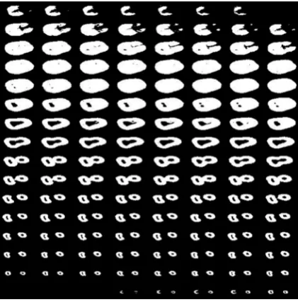
1.2 s and added up to a total of approximately 6 hours. The program 3D Segmentation was used to visualize the three dimensional structure of the teeth. Acquired data were analysed using dedicated program developed for this purpose under IDL 5.5 (Research Systems) programming environment on 1.2 GHz Pentium PC with Windows 2000 operating system. First, a median filter was applied to the 3D data to decrease noise without smoothing borders of the image regions with different intensities. Filtered data were then binarised (i.e. voxels intensities were set 0 or 1 depending on their values) and segmented into separated, compact 3D regions. Algorithm for automatic segmentation of the 3D data is based on comparison of the absolute intensity of the neigh-boroughing voxels. The program allows also visu-alisation of the data and the resulted segments in a form of 2D cross-sections or pseudo-3D volumes, as well as calculation of the segment’s volumes (by calculating number of voxels in segments) (Figs. 1, 2) [15]. The segmentation algorithm was tested on numerical phantom of the tooth before applying it to the MR data. The phantom consists of 4 different segments mimicking the root canals, the tooth solid tissue, the outer liquid forming cylindrical volume, and the rest of the 3D cube. The two root canals were connected in its upper part only by few voxels. The algorithm properly divided the 3D data set into simulated segments and found their volumes [15].
Stages of Preparation
of the Analyzed Material
Preceding the First
Stage of Measurement
Prior to the experiment all teeth were degassed in order to ensure uniform filling of the tooth chambers with the solution and to remove air microbubbles. The degassing process is carried out by decreasing air pres-sure over the sample placed in a liquid. Gas bubbles increase their volume and float to the surface. After pumping them out atmospheric pressure is restored and the container (with internal diameter of 12,7 mm) in which teeth have been degassed is sealed, then mea-surements are carried out. Degassing helps to elimi-nate sources of artifacts created by the difference in magnetic susceptibility between air and the solution.
Stages of Preparation
of the Analyzed Material
Preceding Second Phase
of the Measurements
The pulp was removed from interior of the tooth chamber and root canals, the chamber and root canals were widened and treated using manual endodontic tools. The process of degassing sam-ples was carried out directly before measurements. Preparing the images of internal tooth space after preparation for inner root canals fixations was constructed using both classical methods (polymer mass impression) and non-impressional methods (MRI representation).
Statistical Procedures
Acquired data regarding tooth volume, inter-nal tooth volume and amount of pulp in chamber and root canals before and after preparation and images of internal tooth space after preparation
for inner root canals fixations were collected in samples with normal distribution. The samples’ normality was verified with the Shapiro-Wilk test. The analyzed data was characterized using arith-metic mean, standard deviation and extreme val-ues (minimum and maximum). The estimation of average values was given with 95% confidence lev-el. Samples including data regarding internal tooth volume obtained after pulp removal and treatment of inner tooth were treated as dependent. Mean in-ternal tooth volumes after pulp removal and treat-ment were compared using the one-way ANOVA
test (one-factor analysis of variance) carried out for the model with multiple measurements. The sphericity assumption was verified using Mauch-ley’s test. The statistically significant result of the
ANOVA test permitted post hoc comparisons, which were carried out using the Newman-Keuls test. The correlations’ strength was evaluated us-ing Pearson’s correlation coefficient. All tests were carried out at significance level α = 0.05 using Sta-tistica v 6.0 software (SUM, Katowice, Poland).
Results
The first research stage made it possible to obtain a three dimensional visualization of the in-ternal and exin-ternal tooth structure and numerical values describing the internal volume of the cham-bers and root canals before and after endodontic treatment (Figs. 3–10).
The volume of the internal tooth (chamber and root canals) was determined before and after end-odontic treatment, the total tooth volume was also determined. Statistical analysis showed the useful-ness of the obtained numerical results purposed to planning endodontic treatment which could help improve root canal treatment.
Based on the total tooth volume determined before treatment it is possible to predict the
vol-Fig. 1. 2D cross-sections of the tooth as a result of visualization in program 3D Segmentation: a – before binarization, b – after binarization
Ryc. 1. Dwuwymiarowe przekroje zębów poddanych badaniu uzyskane w programie rekonstrukcyjnym
3D Segmentation: a – przed binaryzacją, b – po binaryzacji
a b
Fig. 2. The output data obtained in the process of seg-mentation
Fig. 3. Tooth A2 before experiment Ryc. 3. Ząb A2 przygotowany do badania
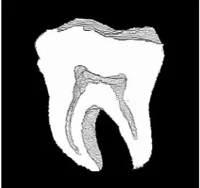
Fig. 4. Reconstructed 3D representation external struc-ture of A2 teeth
Ryc. 4. Trójwymiarowe rekonstrukcje zewnętrznych powierzchni zęba A2
Fig. 5. Reconstructed representation external and inner space of A2 teeth
Ryc. 5. Rekonstrukcja powierzchni zewnętrznych i przestrzeni wewnętrznych zęba A2
Fig. 6. Reconstructed 3D representation chamber and canals of A2 teeth
Ryc. 6. Trójwymiarowe rekonstrukcje komory oraz kanałów korzeniowych zęba A2 przed opracowaniem endodontycznym
Fig. 7. Tooth A2 after endodontic preparation Ryc. 7. Ząb A2 po opracowaniu endodontycznym
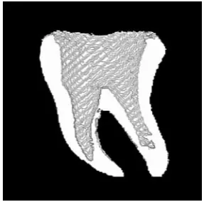
Fig. 8. Reconstructed 3D representation structure of A2 teeth after endodontic preparation
Ryc. 8. Trójwymiarowe rekonstrukcje komory oraz kanałów korzeniowych zęba A2 po opracowaniu endo-dontycznym
ume the internal tooth should obtain after end-odontic treatment and (based on the same initial parameter and on the internal tooth volume before treatment) the amount of hard tooth tissue which should remain after correct endodontic treatment (author’s own model of endodontics treatments prediction) [19]. These values, presented in a
sim-ple and comprehensible form, may be an extremely valuable guideline for a clinical doctor during end-odontic proceedings. Internal tooth volumes were determined before and after endodontic treatment; total tooth volumes were also measured. Numeri-cal data are presented in Tables 1 and 2.
chambers and root canals their volumes increase. Before treatment the internal tooth volume aver-ages at 55.59 mm3. After pulp removal and
inter-nal tooth treatment the inner volumes grow from 55.59 mm3 to a mean value of 125.46 mm3. The
difference between mean values before and after
treatment is statistically highly significant (p = 0,0002). It should be noted that results regarding internal tooth volumes before pulp removal and treatment are more scattered (variation coefficient 26.49%), while pulp removal and treatment sig-nificantly improve uniformity of results, which is shown by a very small value of the variation coef-ficient, equal to 6.85%.
As a result of pulp removal and treatment of the internal tooth the amount of hard tooth tis-sue is decreased from 854.40 mm3 to an average
level of 784.53 mm3. The difference between mean
values measured before and after treatment is sta-tistically highly significant (p = 0.0002). It should be noted that results regarding the amount of hard tooth tissue before pulp removal and treat-ment and after treattreat-ment are characterized by low variance (variance coefficient before treatment is 7.24%, after treatment it’s 6.89%).
A strong correlation was determined between total tooth volume and internal tooth volume af-ter tissue removal and treatment (r = 0.9568; p = 0.0028). The positive correlation coefficient sign means that the larger the total tooth volume, the larger the expected internal volume. Predictions of the situation after pulp removal and internal tooth treatment based on the initial state (before the treat-ment process) were made based on linear regression models regarding the correlation between:
1) total tooth volume and internal tooth vol-ume after treatment,
2) total tooth volume and amount of tissue left after treatment [19].
Ad. 1. The correlation between total tooth vol-ume and internal tooth volvol-ume after endodontic treatment. Based on the own, author’s constructed model a prognosis was made for the internal tooth volumes after treating based on certain initial tooth volumes. Examples of total tooth volumes before treatment and the predicted volumes, determined using the model y = 5.225 + 0.132 · x, as well as internal tooth volumes after treatment in a 95% confidence interval, are presented in Table 3.
Fig. 9. Reconstructed representation external and inner space of A2 teeth after endodontic preparation
Ryc. 9. Rekonstrukcja zewnętrzna oraz komory oraz kanałów korzeniowych zęba A2 po opracowaniu endo-dontycznym
Fig. 10. Reconstructed 3D representation canals of A2 teeth after endodontic preparation
Ryc. 10. Rekonstrukcja komory oraz kanałów korze-niowych zęba A2 po opracowaniu endodontycznym
Table 1. Total volume of teeth and internal tooth volumes determined before endodontic treatment
Tabela 1. Objętości przestrzeni wewnętrznych zębów przed opracowaniem endodontycznym oraz objętości całkowite zębów Teeth before endodontic
treatment
(Zęby przed opracowaniem)
Total volume of teeth [mm3]
(Całkowita objętość zęba)
Volume of inner space before endodontic treatment [mm3]
(Objętość wewnętrzna przed opracowaniem endodon- tycznym)
A1 A2 A3 A4 A5 A6
849.70 983.09 877.59 928.76 977.47 843.30
Table 2. Volume of inner space of teeth after endodontic treatment
Tabela 2. Objętości przestrzeni wewnętrznych po opracowaniu endodontycznym
Teeth before endodontic treatment
(Zęby po opracowaniu endodontycznym)
Volume of inner space of teeth after endodontic treatment [mm3]
(Objętość przestrzeni wewnętrznych zębów po opracowaniu endodontycznym)
Relative difference [%] (Różnica względna)
A1 A2 A3 A4 A5 A6
114.10 135.46 125.46 127.28 133.50 116.94
0 0 0 0 0 0
Table 3. Examples of total tooth volumes before treatment and the predicted volumes, determined using the model y = 5.225 + 0.132 · x, as well as internal tooth volumes after treatment in a 95% confidence interval
Tabela 3. Przewidywana objętość przestrzeni wewnętrznych zęba po opracowaniu dla wybranych objętości całkowitych zęba przed opracowaniem z wykorzystaniem modelu y = 5,225 + 0,132 · x z wykorzystaniem 95% przedziału prognozy
Total volume of teeth before endodontic treatment [mm3]
(Objętość całkowita zęba
przed opracowaniem endodontycznym)
Predicted volume of inner space of teeth after endodontic treatment [mm3]
(Przewidywana objętość przestrzeni wewnętrznej zęba po opracowaniu endodontycznym)
95% Confidence interval (95% przedział prognozy)
800 825 850 875 900 925 950 975 1000
110.93 114.29 117.53 120.83 124.14 127.44 130.74 134.05 137.35
100.54–121.31 104.60–123.86 108.51–126.56 112.23–129.44 115.74–132.4 119.02–135.86 122.07–139.41 124.91–143.18 127.58–147.12
Table 4. The predicted volumes of tissue remaining after treatment – for certain total tooth volumes before treatment – obtained using the model y = –5.225 + 0.868 · x
Tabela 4. Wyniki prognozowania przewidywanej wartości objętości tkanki pozostałej po opracowaniu dla wybranych objętości całkowitych zęba przed opracowaniem z użyciem modelu y = –5,225 + 0,868 · x
Total volume of teeth [mm3]
(Objętość całkowita zęba) Predicted volume of hard tissue of teeth after endodontic treatment [mm3]
(Przewidywana wartość objętości tkanki pozostałej po opracowaniu endodontycznym)
95% confidence interval (95% przedział prognozy)
800 825 850 875 900 925 950 975 1000
689.08 710.77 732.47 754.16 775.86 797.56 819.26 840.95 862.65
678.70–699.46 701.14–720.40 723.44–741.50 745.56–762.77 767.46–784.26 789.14–805.98 810.58–827.93 831.82–850.09 852.88–872.42
Ad. 2. Correlation between total tooth volume and amount of tissue remaining after endodontic treatment. Based on the constructed model a prog-nosis was made regarding the volume of tissue remaining after treatment based on certain total
Fig. 12. Reconstructed 3D representation of negative polymer representations of inner space of tooth A2 Ryc. 12. Przestrzenna rekonstrukcja odwzorowania polimerowego
Fig. 13. The cast of chambers and root canals of tooth A2 Ryc. 13. Model gipsowy przestrzeni wewnętrznych zęba A2 uzyskany na podstawie odwzorowania poli- merowego
Fig. 14. Reconstructed 3D representation of the cast of chambers and root canals of tooth
Ryc. 14. Rekonstrukcja przestrzenna uzyskana z mode-lu gipsowego przestrzeni wewnętrznych zęba A2 Fig. 11. Negative polymer representations of inner
space of tooth A2 after endodontic preparation Ryc. 11. Polimerowe odwzorowanie przestrzeni
wewnętrznych zęba A2 po opracowaniu endodontycznym
ter preparation for inner root canals fixations (the polymer negative and cast) constructed using both classical methods (polymer mass impression) and non-impressional methods (MRI representation). The phase of MR-based visualization of teeth after endodontic treatment, of negative polymer rep-resentations and of plaster models, demonstrated imperfections in the classical method of represen-tation. Visual assessment of the analyzed material revealed imperfections in the impression-based method. Especially visible were deformations of the root canals, created in the process of making plaster models. The imprecise adherence of poly-mer resin to inner tooth surfaces after endodontic treatment should also be noted. During prepara-tions for negative polymer representation, polymer resin residue inside the canals made it difficult to image accurately the root canals. This made it im-possible to obtain an accurate image, and in con-sequence would have prevented the construction of an intracanal fixation fulfilling clinical require-ments. Research proceedings made it possible to create a spatial visualization and obtain numerical values characterizing the inner volume of cham-bers and root canals after treatment, the volume of negative polymer representations, and the inner volume of plaster models of chambers and root canals. Three dimensional reconstructions of the treated tooth, its negative polymer representation and plaster model are shown in Figures 11–14. The next research proceedings made it possible
af-Table 5. Volume of inner space of teeth after endodontic treatment, negative polymer representations and casts of chambers and root canals of teeth
Tabela 5. Objętości przestrzeni wewnętrznych zębów po opracowaniu endodontycznym, objętości odwzorowań polimero-wych i modeli gipsopolimero-wych
Variable
(Zmienna) Number of test (Liczba prób)
Average [mm3]
(Średnia)
Standard Deviation (Odchylenie stan-dardowe)
Ratio of vari-ability V [%] (Współ- czynnik Zmienności)
Range of value (min-max) [mm3]
(Zakres wartości)
95% partition of trust (95% przedział ufności)
–95% +95%
Volume of inner space of teeth after endodontic treatment
(Objętość przestrzeni wewnętrznych zębów po opracowaniu endodonty-cznym)
6 125.46 8.60 6.85 114.10–135.46 116.43 134.48
Volume of negative poly-mer representations of inner space of teeth (Objętość odwzorowań polimerowych przestrzeni wewnętrznych zębów)
6 120.97 11.46 9.47 102.75–130.54 108.94 132.99
Volume of models cast of chambers and root canals of teeth
(Objętość modeli gip-sowych przestrzeni wewnętrznych zębów)
6 120.07 9.91 8.26 103.28–130.0 109.64 130.45
Numerical data is presented in Table 5. Mean vol-umes of negative polymer representations and in-ner volumes of plaster models are statistically sig-nificantly different from the mean inner volume of teeth after endodontic treatment (p = 0.0259 for polymer negative, p = 0.0255 for plaster models). The difference between mean values of volumes of polymer representations and mean values of inner volumes of plaster models is not statistically sig-nificant (p = 0.6029).
Discussion
The presented results show the possibility of using MR Microscopy for visualisation of the root canals topography. The quality of Magnetic Reso-nance visualisation does not depend on compli-cation in the shape of the canals. In contrast, the traditional impressional methods used in dentistry may have difficulties during precise mapping of multiple root canals with complicated curvature. Additionally, due to the fact that parameters of the impression materials depend on variable environ-mental conditions, like temperature or humidity, dimensions of the mapped structures may differ
from the original ones. Thus, Magnetic Resonance visualisation may be used to check the quality of impressional mapping or as an alternative non-impressional 3D mapping method. Research re-sults presented in this article show the possibility of applying MR Imaging to visualize the structure of teeth at the level of in vitro analysis. After con-ducting the experiments, three dimensional visu-alisations of the internal structure of human teeth were obtained. A non-impressional representation of the tooth chambers and root canals was made before treatment and after carrying out endodon-tic treatment. The measured parameters included: total tooth volume, internal tooth volume before and after endodontic treatment.
in the form of simple and apprehensible diagrams, could be a very valuable guideline for clinicians undertaking endodontic work.
Second stage of the research proceedings made it possible to compare the quality of representations of the inner tooth after treatment made using both classical methods (polymer mass impression) and non-impressional methods (MRI). Results show a discrepancy between values of parameters char-acterizing volume. The difference between volumes of the polymer negative and cast is visibly smaller (lower than 3%) than the difference between vol-umes of the polymer negative and the inner tooth after treatment. This suggests that the transposition of inner tooth representation to the polymer nega-tive leads to larger errors and might be responsible for potential imperfections of fixations constructed based on this technique, using the classical method. In most cases, the volumes of polymer negatives and plaster models are slightly smaller than the inner volume of treated teeth. Classical impression mate-rials (polymer mass) are subject to environmental changes and the durability of representations made with their use is dependent on external factors such as temperature, air humidity, transport and storage methods [20]. Imperfections encountered in com-plex reconstructions are a result, among other fac-tors, of the instability of parameters of impression mass, which – due to storage and preparation con-ditions – undergoes changes in linear dimensions within a range normatively determined to reach a maximum level of 1.5% and deformations caused by pressure ranging from 0.8 to 20% [20]. Another reason besides the difficulty of representation using classical methods is the topography and curvature of root canals in multiradicular teeth. Thanks to the developed Magnetic Resonance visualization meth-od a numerical mmeth-odel of the inner tooth was ob-tained which could, in the future, be also used in an automatic three-dimensional modeling technique using an appropriate computer aided design pro-gram designed for precise application of complex spatial models in reconstruction work. There cur-rently exist described and applied systems of build-ing three dimensional models representbuild-ing cavities which make it possible to reconstruct lost fragments of crowns using an insertion with a designed and finely molded shape. Spatial representation of a cav-ity is obtained by scanning its shape with the use of industrial computed tomography. Numerical data are then transferred to a system which designs the prosthetic restoration [21, 22]. In future clinical op-erations the intracanal fixation could be milled from an appropriate block of any material based on an high-precision representation obtained using the Magnetic Resonance method.
An important aspect of the MR Imaging
mea-surements is to obtain high contrast between the cavities and the tooth. This may be achieved by filling cavities with water or water solution of para-magnetic ions having high MR signal. Another im-portant factor is maximal space resolution possible to achieve in reasonable time. In the used sequence (3D SE) increasing twice the 3D resolution requires four time longer experimental time (assuming that lower S/N does not require additional signal accu-mulation), while the necessary disk space increases 8 times. For extracted teeth the time of the measure-ments is not the most critical problem. However, to implement this technique in vivo, it is necessary to decrease it substantially. Using the solution with the appropriate T1 and T2 values for filling cavities may
decrease the total measurement time. However, a more promising solution might lie in the use of fast acquisition techniques. Sequences based on the train of the echoes instead of the single echo are es-pecially promising. Another possibility – sequences based on the gradient echo, seem to be less useful due to their sensitivity to the magnetic susceptibil-ity artifacts. In order to improve the image resolu-tion and get a three dimensional image a three di-mensional sequence of spin echo was used [13–15], which meant that a long measurement time was necessary [16, 17, 23].After correction MRI tech-nique would be safety and screening requirements in order to be eligible candidates if this procedure would be implemented in vivo. It should also be remembered that in MR Microscopy the values of the used magnetic field are higher (in the presented experiment a 4.7 T magnet was used) than in clas-sical magnetic resonance imaging (0.5–3.0 T). For clinical procedures planned in the future it will be important to strive to lower the value of the field without loss of quality of the obtained representa-tion. Visualization of the surface is obtained due to the contrast between the strong signal coming from the solution and a lack of signal from the tooth. This type of measurement is currently only possible in in vitro conditions [11, 18, 23].
Realization of the established target and achievement of satisfactory results, that may have a practical implications in the future, enabled to formulate the following conclusions:
The results that have been achieved, indicate the possibility to employ techniques of the Magnetic Res-onance Imaging, that are based on the 3D sequence of the Spin Echo as well as on the Single Point Imag-ing, for the dimensional mapping of the outer topog-raphy of teeth as well as a structure of the root canals for the therapeutic and didactical purposes.
A numerical model of prepared root canals ob-tained with the method of the magnetic resonance visualization may constitute in the future a basis for a non-impressional technique of imaging, use-ful for reconstructive dentistry.
A numerical model of the prepared root ca-nals obtained with the method of the magnetic
resonance visualization imaging combined with the method of images analysis, may in the future constitute a basis for an automatic 3D model-ing of the inner root canals fixations, supported with computer applications dedicated for the designing purposes (CAD – computer aided design).
References
[1] Wang MQ, Shibata T, Zhang Y: Correlation of coronal form of condyle and disc on MR imaging. J Dent Res 2002, SpecIss A81, 1740.
[2] Araki Y: TMJ space in patients with unilateral posterior crossbite. J Dent Res 2002, SpecIss A81, 1586.
[3] Webber RL: Oral imaging as a diagnostic tool for assessing osseous changes. J Bone Miner Res 1993, 8, 543–548. [4] Uchiyama K: MRI evaluation in diagnosis of mandibular osteomyelitis J Dent Res 2001, Spec Iss 80, 658. [5] Cevidanes LHS: Precision of 3D craniofacial landmark data using magnetic resonance scans. J Dent Res 2002,
SpecIssA, 3128.
[6] Gray CF, Redpath TW, Smith FW: Pre-surgical dental implant assessment by magnetic resonance imaging. J Oral Implantol 1996, 2, 147–153.
[7] Olt S, Jakob P: Contrast-enhanced Dental MRI for visualisation of the teeth and jaw. Magn Reson Med 2004, 2, 174–176.
[8] Shafiei F, Honda E, Takahashi H, Nishimura F, Saasaki T: Minimizing dental alloys artifacts in MR imaging. J Dent Res 2001, Spec Iss 80, 1493.
[9] Appel TR, Baumann MA: Solid-state nuclear magnetic resonance microscopy demonstrating human dental anat-omy. Oral Surg Oral Med Oral Pathol Radiol Endod 2002, 2, 260–261.
[10] Baumann MA, Gross D, LehmannV, Zick K: Magnetic resonance microscopy – new prospects for endodontics. Schw Monat Zahnmed 1993, 103, 1407–1414.
[11] Tanasiewicz MM, Węglarz WP, Kupka TW, Jasiński A: Teeth caries decay in MR Imaging. Polish Seminar on Nuclear Magnetic Resonance and Its Applications, Kraków 2005, 38, 12.
[12] Latta P, Gruwel MLH, Edie E, Shramek M, Tomanek B: Single point imaging with suppressed pressure levels through gradient-shape adjustment. J Magn Reson 2004, 170, 177–185.
[13] Sramek M, Kaufman A: Fast ray-tracing of rectilinear volume data using distance transformers. IEEE Trans Vis Comp Graphics 2000, 6, 236–253.
[14] Ploder O, Patric B, Rand T, Baumann A: Reperfusion of autotransplanted teeth – comparison of clinical mea-surement by means of dental magnetic resonance imaging. Oral Surg Oral Med Oral Pathol Oral Radiol Endod 2001, 92, 333–340.
[15] Węglarz WP, Tanasiewicz M, Kupka T, Skórka T, Sułek Z, Jasiński A: 3D MR imaging of dental cavities – an in vitro study. Solid State NMR 2004, 25, 84–87.
[16] Tanasiewicz M: Mikroskopia rezonansu magnetycznego w diagnostyce endodontycznej. Możliwości nieimpresyj-nego obrazowania jam zębowych dla potrzeb dentystyki odtwórczej. Magazyn Stomatol 2003, 3, 64–68.
[17] Latta P, Gruwel M, Edie E, Shramek M, Tomanek B: SPI with suppressed pressure levels through gradient-shape adjustment. J Magn Reson 2004, 170, 177–185.
[18] Tanasiewicz M, Węglarz WP: Obrazowanie zębów świnki morskiej za pomocą rezonansu magnetycznego techniką echa spinowego w warunkach in vivo. Czas Stomatol 2007, 60, 815–824.
[19] Tanasiewicz M: Nowe obszary zastosowań rezonansu magnetycznego w terapii i diagnostyce dentystycznej. Seria: Rozprawy habilitacyjne – Śląska Akademia Medyczna w Katowicach 2006, 28, 68–70.
[20] Kupka T, Tanasiewicz M, Tanasiewicz R: Odbudowa kombinowana typu żywica/stop z zastosowaniem indy-widualnego umocowania wewnątrzkanałowego. Inż Ortop Prot 2001, 3, 159–166.
[21] Lee M, Yau HT: CAD/CAM use in dental laboratory. Dentistry Today 2006, 9, 92–93.
[22] Wu L, Yan HX, Yang M: The construction of geometric models of dentition effects based on 3-dimensional indus-trial computed tomography image. Shanghai Kou Oiang Yi Xue 2006, 15, 535–538.
[23] Lloyd C, Scrimgeour SN, Chudek JA, Hunter G, MacKay RL: Caries detection with MR technique. Quintessence Int 1998, 3, 145–151.
Address for correspondence:
Marta Tanasiewicz
Department of Conservative Dentistry and Endodontics Medical University of Silesia
Plac Akademicki 17 41-902 Bytom Poland
Tel.: +48 32 282 79 42
E-mail: [email protected]
Conflict of interest: None declared