Citation : Obaid KA. The Diagnosis of Atherosclerosis by Using Digital Image Processing. JJ Med Diagnosis and Image 2019; 5(1): 024.
Original Article
The Diagnosis of Atherosclerosis by Using Digital Image Processing
Kadhim AJeel Obaid
Department of Physics, College of Science, HI Qer University Iraq
*Corresponding author:
Kadhim AJeel Obaid, Department of Physics, College of Science, HI Qer University, Iraq;
Email: [email protected]
Received Date:04-09-2019
Accepted Date: 04-12-2019
Published Date: 04-19-2019
Copyright: © 2019
Kadhim AJeel Obaid
Jacobs Journal of Medical Diagnosis and Medical Imaging
Abstract
Accelerating the pace of scientific progress and that focus in the field of human service requirements is to provide daily health and service. Health aspect is one of the most important themes of science that needs to be searched because it has a direct impact on human existence.
Medical Physics is one of the branches of the Physics Department that deals with the interest of providing theoretical or scientific foundations which had the aims to provide scientific services in the medical field. The subject of the medical im
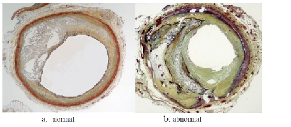
-age processing is one of the applications of this section. It aims to increase the efficiency of medical diagnostic systems to provide an integrated diagnostic system that works to distinguish a heart disease and an atherosclerosis. The main focus of this study depends on medical images that were obtained from patients in Nasiriyah Heart Center, where the researcher was taking images for arteries feeding the heart muscle of persons injured and others to be healthy. Set of operations were performed on these images derived from work areas and the separation of these areas from the rest of unimportant infor
-mation within the image. Then it was reclaimed process recipes, which had been brought into the neural network, the use of 110 images to train the network 60 and 50 were used for the test.
The next step is to count the number of qualities for each image after it has been extracted artery of the image by making some operations on this image. The proposed system has proven successful in the conduct of the diagnostic pro
-cess through the results obtained on the ratio of 100% in the training phase. And 92% in the testing phase.
Keywords: Medical Physics; arteries; abnormal; digital image
Introduction
The increase use of digital images in recent years due to the availability of technology equipment has made the process of dealing with the images of things possible. Conventional cameras are based on physics based which control lighting.
When the user enters the lens to control the amount and direction of light[1]. Not Only the Digital camera re
-quires a simple knowledge by the user, but also the principle of its work depends on converting light into Electrical dis
The enormous development of computer technology and the great advances in digital recording of images helped the emergence of devices that allow access to images without chemical processing. It has raised some of the advantages offered by digital photography, such as the stability of the image quality (regardless of the length of the storage); the time for copies number; and the possibility of computer processing. Also, it has attended many of the workers in the Medical fields to the field of digital photography [3]. The simple computer-aided screenings are methods of comput
-er systems that help physicians in the initial int-erpretation and classification of the medical images. Simple comput
-er-assisted screening is a sub-program of computer-aided diagnostic software (Computer-Aided Detection/Diagnosis –CAD).
The programs “simple sorting systems, comput
-er-aided” work by screening (classification) initial full au
-tomatic for medical diagnostic imaging studies [4]. The sys
-tem was individual unlike the traditional computer-aided diagnosis. However the immediate critical diagnosis deals with such cases. The primary goal of the diagnosis is to help the traditional computer in improving diagnostic accuracy for the user [5].required for the rapid diagnosis of critical conditions that threaten the life of the degree in the medical field, it becomes what is known as (the doctor-mail) which can be reliable and considered highly important factor in the detection of many diseases, that are diagnosed early and which help early healing .In addition it helps to avoid a lot of these diseases, especially diseases such as cancer and
thrombosis . The roles of these systems are increased with the remarkable progress in medical imaging methods of us
-ing the computer which results in a lot of data that need explanation [5, 6].
Symptoms and signs of atherosclerosis disease
There are some signs of atherosclerosis disease as listed below: chest pain as a result of a decrease of blood supply to the heart muscle, fatigue when effortlessly, there is no difference in the measurement of blood pressure [7].
Recognition System Structure
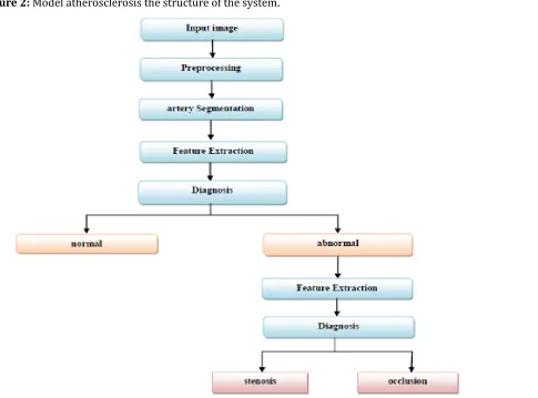
The structure of the atherosclerosis detection system con
-sists of the Following stages 1. Input image
2. Preprocessing 3. Segmentation artery 4. Feature extraction 5. Diagnosis
Figure (2) shows model of the structure to identi
-fy atherosclerosis system. The system starts with the input image after getting it appropriate to capture the source. Then passing the image to the processing for noise removal phase. And then they are feed on an algorithm to recognize discrimination if there is a normal or abnormal then from the features recognize occlusion or stenos is in the artery. Preprocessing
The aim is to consolidation images and removes the
unwanted effects of Treatment. Image processing is the first step in the identification of atherosclerosis, which involves three steps: the first major operations are converting imag
-es to JPEG; the second version of the system is filtering to
remove any additional noise in the image; and the third step contrast enhancement to increase the contrast of the image then to change it into a binary image. Figure (3) shows the main steps of the pre-processing stage.
Figure 2: Model atherosclerosis the structure of the system
.
We input images into the algorithm and apply the main four processes in Preprocessing step.
1. Contrast Enhancement; 2. Remove noise and binary image; 3.Gray image; 4.The separation of the artery. Sep
-aration method is used image as a second step to separate the object of Interest from the background. In this research, the object class is taken the arteries that feed the heart mus
-cle of branch these arteries and the background. Artery is separated from the rest of its branches to extract the fea
-tures of this artery and to recognize the level of the image to extract the gray texture features and with two stages:
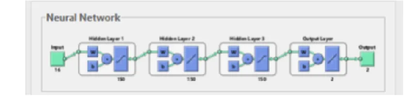
Figure (4) shows the network structure with one input layer, three hidden layers, and four output layer. It is 16×150×150×150×2 network structure. The input vector is sixteen. The output vector is two. This thesis uses the above ANN architecture feed forward back propagation learning algorithm to generate. Train and test the neural network for atherosclerosis type’s diagnosis. MATLAB software with its neural network toolbox is used. Data sets are portioned into two subsets, training set and testing set. The network gives high accuracy when train is equal to 100% and test is equal to 92% with simple training time equals (0.1 seconds) at 60 epochs, with best training performance is 3.97e-015 at epoch 60.
Configurations
The framework of the project is designed by Matlab in 32 bit system with 2.50 GHZ core i3 processor and 4 GB of RAM, run with MS Win.7 operating system. The experi
-ments are implemented on database from Nasiriyah Heart Center to detect the health and the infected arteries and the arteries that contains the injured (stenos is or occlusion ar
-teries).
Conclusions
Through the proposed system and the results that have been reached we realized in the following conclusions: 1. The results showed that it can use statistics (variance,
correlation, energy, harmonies) with different angles as well as the neural network to classify Atherosclerosis disease and determining its kind (occlusion or stenos is arteries) .
2. To purify the image and try to save the image with many details as possible. In this filter system : Palladi
-an of Gaussi-an filter (log) to reduce noise -and filtering the image using image filter . In addition, we modify the image to enhance the clarity of the picture and get the best performance.
3. The proposed algorithms to accurate segmentation of the artery from its image of gray background and deter
-mine where the successful from a healthy artery in gray level images. These algorithms have proven success in most of the images.
4. For recognizing the types of atherosclerosis on the ba
-sis of a steady drumbeat of matrix characteristics they found to be strong and descriptors of the fabric of the level of gray scale images in contrast with, correlation, energy, and homogeneity. The use of all four proper
-ties leads to a correct Recognition (stenos is arteries or occlusion). The proposed system gives good results reached 92%.
5. It showed recognized atherosclerosis which is used in the encouraging results of our research system in terms of the time taken in recognition of the neural network basis, where it took time (less than one minute) for the Diagnosis of pigmented lesion (Normal or abnormal) .
References
1. Rafael C. Gonzalez, Richard E. Woods, Addison-Wes
-ley. Digital image Fundamentals 2010 Sensor Cleaning 2010.
2. T. Young, Jan J. Gerbrands and Lucas J. van Vliet. Funda
-mentals of Image Processing 1995-2007.
3. D. ping Tian. A Review on Image Feature Extraction and Representation Techniques Baoji International Journal of Multimedia and Ubiquitous Engineering 2013; 8.
4. P. Gorgel, A. Sertbas and N. Osman. Feature extraction based wavelet transform inn breast cancer diagnosis
using fuzzy and non-fuzzy classification, International journal of electronics; mechanical and mechatronics engineering 2012; 2: 327-333.
5. G. Dougherty. Digital Image Processing for Medical Ap
-plications, Cambridge university press 200.9
6. M Bergounioux. Mathematical Image Processing, University of Orleans, France, March 29th - April 1st, Springer-Verlag Berlin Heidelberg 2011.
7. S. Hägg Gene. Expression profiling of human atheroscle