Printed in Great Britain © The Company of Biologists Limited 1984
THE VERSATILE SYNAPSE
BY ROBERT M. PITMAN
Department of Physiology and Pharmacology, St Andrew's University, Fife, Scotland
SUMMARY
'Typically' chemical synaptic transmission takes place when an influx of calcium ions during a presynaptic nerve impulse triggers exocytosis of neurotransmitter substance from synaptic vesicles. The neurotransmitter diffuses across the synaptic cleft and occupies receptors embedded in the subsynaptic membrane. This interaction (directly or via a second messen-ger) operates characteristic ion channels and produces an increase in the postsynaptic membrane permeability to particular ions. Depending on the ionic species to which the postsynaptic membrane becomes more perme-able, the physiological response will be an excitatory or an inhibitory post-synaptic potential. The action of neurotransmitters may be terminated either by enzymic inactivation or by cellular uptake mechanisms.
Over the last decade it has become clear that a neurotransmitter substance may exert a number of different actions on a single postsynaptic neurone. These may involve opening or closure of either voltage-independent or voltage-dependent ion channels. It is also possible that in some instances transmitters may act on neuronal biochemical systems to modify the physiology of postsynaptic cells without directly altering their electrical characteristics.
Analysis of the postsynaptic actions of neurotransmitter substances has become further complicated by the increasing body of evidence which in-dicates that more than one transmitter substance (one of which may be a peptide) can be released from a single presynaptic neurone. The significance of such dual transmitter systems has yet to be fully elucidated.
The efficacy of transmission across many synapses may be modified by either presynaptic or postsynaptic mechanisms; both transmitter release and postsynaptic responsiveness may depend on the recent history of a single synapse, on synaptic inputs from other neurones or on circulating neuro-active substances.
INTRODUCTION
Communication between neurones mainly takes place via either electrical or chemi-cal synapses. Electrichemi-cal synaptic transmission is associated with 'gap' junctions possessing intercellular channels which allow small molecules and ions to pass be-tween cells. Movement of ions through these channels allows electrical current to flow between neurones; at some electrical junctions current can flow equally well in either direction, while other junctions show rectification. Generally, electrical synaptic
transmission shows only relatively limited modification under different physiological conditions. Chemical synaptic transmission is more complex than electrical trans-mission, since the presynaptic neurone must possess mechanisms for synthesis, storage and release of transmitter substance, while the postsynaptic neurone must have the ability to respond to released transmitter. Transmitter substances can produce a wide range of different actions on postsynaptic neurones. The efficacy of chemical synaptic transmission may be considerably altered by either presynaptic or postsynaptic changes. The term 'modulation' has become popular in the neuroscience literature to describe a number of different alterations in transmission which cannot be explained in terms of 'classical' synaptic physiology; agents which bring about these changes have been called 'neuromodulators' or simply 'modulators'. Events that have been described as modulation include long-lasting synaptic responses, altera-tions in the effectiveness of transmitters mediated by agents that alone produce only minor effects on synaptic transmission or changes mediated by agents released from distant sites. However, 'modulation' in many cases is achieved by mechanisms which are merely an extension of the basic principles governing transmission across 'conven-tional' synapses.
NEUROTRANSMITTERS
For many years most research concentrated upon the 'classical' neurotransmitters [for example, acetylcholine (ACh), monoamines and amino acids] which are all relatively low molecular weight compounds. However, over the last few years application of immunohistochemical techniques has demonstrated that a wide range of peptides are localized within neurones (Hokfelt et al. 1980). The list of peptides found within the mammalian brain includes a number of hormones which had previously been considered to be exclusively associated with the pituitary or the gastrointestinal tract (Table 1). Immunoreactivity to a number of these vertebrate peptides has also been demonstrated in central neurones of a number of species of invertebrates. However, care is required in the interpretation of data obtained
Table 1. Some peptides present in the mammalian nervous system
Hypothalamic peptides Thyrotropin-releasing hormone (TRH)
Luteinizing hormone-releasing hormone (LHRH) Somatostatin
Vasopressin
Pituitary hormones Adrenocorticotrophic hormone (ACTH) a-Melanocyte-8timulating hormone (a-MSH)
Gut-brain peptides Insulin Glucagon Cholecystokinin
Vasoactive intestinal polypeptide (VIP) Methionine enkephalin
Leucinc enkephalin Substance P Neurotensin
|by immunohistochemistry, since antibodies may react with only one portion of a •peptide, allowing cross-reactions to occur between peptides sharing similar amino acid sequences. This problem makes it particularly difficult to interpret the results of experiments in which antibodies to peptides from one species are used to determine the distribution of immunoreactivity in the nervous system of an un-related species (for example, when antibodies to mammalian peptides are applied to invertebrate nervous systems). Under these circumstances it is therefore impor-tant to demonstrate, by independent techniques, that the peptide of interest is actually present.
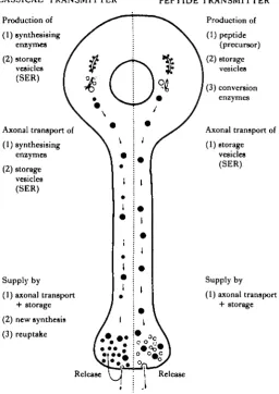
Although peptides may be involved in synaptic transmission it should be remem-bered that, unlike the 'classical' neurotransmitters, they must be synthesized in the cell body, then carried by axonal transport to nerve terminals. Once released at the synapse, it is unlikely that they can be taken back into the presynaptic terminal for recycling (Fig. 1). As a result, it is probable that the supply of peptide in nerve
•CLASSICAL' TRANSMITTER
Production of
(1) synthesising eniymes
(2) storage vesicles (SER)
Axonal transport of
(1) synthesising emymes
(2) storage vesicles (SER)
Supply by
(1) axonal transport + storage
(2) new synthesis
(3) reuptake
Release
PEPTIDE TRANSMITTER Production of
(1) peptide (precursor)
(2)storage vesicles
(3) conversion enzymes
Axonal transport of
(1) storage vesicles (SER)
Supply by
(1) axonal transport + storage
Release
202 R. M. PITMAN
Ion channel
ATP
[image:4.451.44.411.45.250.2]S'AMP
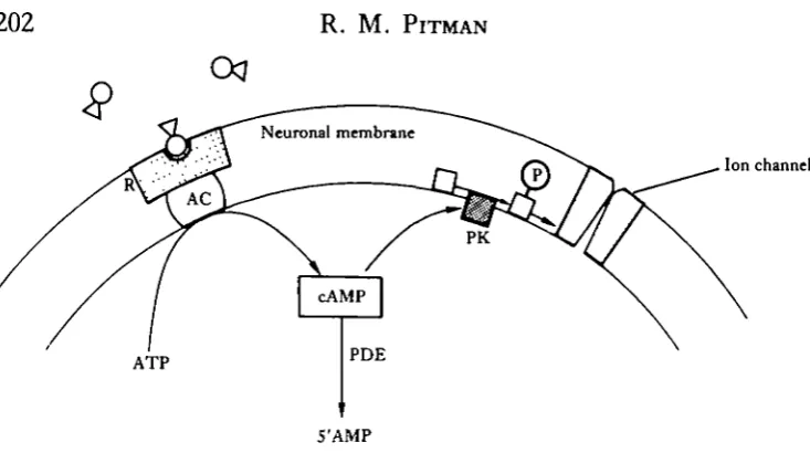
Fig. 2. Diagram showing the main steps by which cyclic AMP may operate as a second messenger in neurones. Transmitter molecules interact with receptors (R) on the neuronal membrane. This interaction stimulates adenylate cyclase (AC) to catalyse synthesis of cyclic AMP from ATP. Cyclic AMP (cAMP) activates a protein kinase (PK) which, in turn, may activate a membrane protein (P) involved in operating ion channels. Cyclic AMP is degraded by the enzyme phosphodiesterase (PDE).
terminals will be more prone to depletion during periods of elevated neuronal activity than the supply of a 'classical' neurotransmitter.
Interaction of transmitter substances with some receptors appears to have a direct effect upon ion channels, while other ion channels are operated indirectly by an intracellular 'second messenger'. One such messenger appears to be cyclic adenosine 3',5'-monophosphate (cyclic AMP), which has been implicated in a number of post-synaptic responses (see Nathanson, 1977; Kupfermann, 1980). The precise mechan-ism by which cyclic AMP brings about its effects in neurones has not been elucidated. It seems, however, that transmitter-receptor interaction activates an adenylate cyclase which converts adenosine triphosphate (ATP) into cyclic AMP. This activates a protein kinase which, in turn, phosphorylates specific proteins. The phosphorylated protein may form part of an ion channel which is operated by the transmitter. Cyclic AMP is degraded by phosphodiesterases (Fig. 2). Besides operating ion channels, cyclic AMP appears to be involved in a number of other cellular functions including the regulation of transmitter synthesis and release. Cyclic guanine 3',5'-monophosphate (cyclic GMP) may also act as a second messenger, however, the biochemical steps through which it operates are not entirely clear.
TRANSMITTER RELEASE
land the squid giant synapse (Llinas, Steinberg & Walton, 1981), although presynaptic &epolarization itself may have an important influence upon the amount of neurotrans-mitter released from crayfish motor neurone terminals (Dudel, Parnas & Parnas, 1983). The supply of transmitter available for release may also influence transmission.
Activity-dependent changes in release
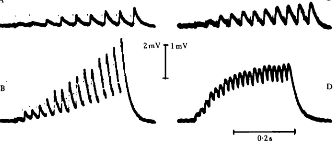
The amount of neurotransmitter released by a single action potential will depend upon the preceding pattern of activity in the neurone. For example, junctional poten-tials in many crustacean muscle fibres show marked facilitation during trains of motor neurone action potentials (Dudel & Kuffler, 1961a) (Fig. 3), while repeated stimula-tion causes depression of postsynaptic responses at synapses made by sensory neurones on follower neurones both in the crayfish tail-flip escape neuronal circuit (Zucker, 1972) and in the gill-withdrawal reflex of Aplysia (Castellucci & Kandel, 1974). In each of the above examples, frequency-dependent alterations in synaptic transmission result from changes in the amount of transmitter released from presynaptic terminals rather than from a reduction in postsynaptic responsiveness (Zucker, 1973). The amount of transmitter released by each presynaptic impulse will depend both upon the number of synaptic vesicles immediately available for release and upon the efficiency of the release process. Both these parameters may be modified during repetitive activity to produce depression or enhancement of transmission. Many preparations show more than one type of frequency-dependent modification in synaptic transmission. At the frog neuromuscular junction there are three different components which contribute to enhancement of transmitter release caused by repetitive stimulation, each of which has a different decay time-constant. ('Facilita-tion' has a decay time-constant of 50—300 ms; 'augmenta('Facilita-tion' a time-constant of about 7s, while 'potentiation' has a time-constant in tens of seconds or minutes.) Neuromuscular transmission is also modified by two other components: these have
2mVTlmV
J # m o
[image:5.451.59.397.437.584.2]0-2s
been termed the 'expression factor', which selectively enhances 'augmentation! without influencing 'potentiation', and the 'time-constant factor', which specifically prolongs the decay of 'potentiation' (Magleby & Zengel, 1976). Presumably, different mechanisms underlie each of the above components. Depression, short-term facilita-tion, frequency facilitation and post-tetanic potentiation have all been observed at a cholinergic synapse on an identified Aplysia neurone (Schlapfer, Woodson, Tremblay & Barondes, 1974; Schlapfer, Tremblay, Woodson & Barondes, 1976). Each com-ponent has a characteristic time-course and may be produced by different patterns of presynaptic activity. These authors have attributed depression to initial depletion of readily-available transmitter. Frequency-facilitation (which develops during a stimulus train) and post-tetanic potentiation (observed after the end of a stimulus train) appear to result both from an increase in the supply of transmitter and an enhancement in the release process.
Although transmitter release may be influenced either by the efficiency of the release process or by the availability of transmitter, in most cases the presynaptic mechanisms underlying frequency-dependent modulation have not been completely elucidated. This is largely because it is not technically possible to make intracellular recordings from nerve terminals in most preparations. It has been suggested that frequency-dependent modulation of the process of transmitter release may be caused by a cumulative increase in intracellular free calcium which could be enhanced by a change in: (1) the amplitude of the presynaptic action potential, (2) the duration of the presynaptic action potential, (3) the point blockade of action potentials within presynaptic terminals.
Katz & Miledi (1965, 1968) obtained evidence that facilitation at the frog neuromuscular junction and the squid giant synapse results from a cumulative in-crease in the intracellular calcium concentration within the presynaptic nerve termi-nal. Although incomplete intracellular calcium buffering alone could account for facilitation observed at some junctions, the features of facilitation observed elsewhere indicate that calcium influx must be progressively enhanced in some way during a train of presynaptic action potentials (Bracho & Orkand, 1970; Lang & Atwood, 1973). In fact, non-linear calcium influx has been directly observed by injecting the calcium-sensitive protein aequorin into the cell bodies of Aplysia neurones (Stinnakre & Tauc, 1973; Fig. 4). In this preparation, enhanced calcium entry is associated with an increase in both action potential amplitude and duration.
20 mV
[image:7.451.87.372.47.412.2]• n i 9
A I
0-5 s ,Fig. 4. Light emission from a molluscan neurone following injection of the calcium-sensitive protein aequorin. (A) There is a progressive increase in the size of the light response (lower trace) to each successive action potential in a burst. During a burst there is also a progressive increase in the spike overshoot and duration. (B) When calcium channels are blocked by adding Co2* to the bathing
medium, the light response is suppressed. (From Stinnakre & Tauc, 1973.)
effects of membrane potential on transmitter release in Aplysia neurones, although ig this case, polarization also caused changes in the action potential amplitude ana duration. By voltage-clamping the presynaptic neurone they were able to demonstrate that transmitter release was modulated by a voltage-dependent steady state calcium current. Membrane depolarization also caused a reduction in outward potassium current; under physiological conditions this would also contribute to increased trans-mitter release by prolonging the action potential thereby increasing calcium influx. It is likely that a similar steady-state voltage-dependent calcium current may be respons-ible for graded transmission (in which transmitter release is altered by relatively small presynaptic membrane potential changes in the absence of action potentials) reported in both spiking (Nicholls & Wallace, 1978; Graubard, Raper & Hartline, 1980, 1983) and non-spiking neurones (Burrows, 1979; Burrows & Siegler, 1976, 1978; Bush & Cannone, 1973; Werblin & Dowling, 1969).
Broadening of successive presynaptic action potentials associated with a progressive increase in the intracellular Ca2+ concentration has been proposed as a possible mechanism underlying frequency-dependent changes in transmitter release in molluscs (Eckert & Lux, 1977; Stinnakre & Tauc, 1973). However, as discussed above, transmitter release from Aplysia neurones can be altered at different holding potentials without any change in the duration of transient presynaptic depolarizations (produced artificially by a voltage-clamp pulse). Zucker & Lara-Estrella (1979) have also shown that facilitation can occur at the crustacean neuromuscular junction under conditions which blocked changes in the duration of the action potential in motor neurone terminals. Nonetheless, although spike broadening may not be the only mechanism involved in frequency-dependent changes in transmitter release, any alteration in the duration of action potentials with a significant calcium component would almost certainly affect synaptic transmission.
Facilitation at the crustacean neuromuscular junction has been attributed to progressive invasion of motor neurone action potentials into motor neurone terminals (Lang & Atwood, 1973). Unfortunately, in this preparation, there is some doubt whether action potentials actually invade presynaptic terminals or whether they undergo pre-terminal block allowing only decremental electrotonic depolarization to reach transmitter release sites. Zucker (1974a,b), also studying the crustacean neuromuscular junction, obtained evidence that action potentials do reach motor neurone terminals. However, this question has been reopened recently, since Dudel (1983) (in patch-clamp recordings from the neuromuscular junctions of three other species of crustacean) reported that although some nerve terminals were electrically excitable, the majority were inexcitable.
The versatile synapse 207
fcteral (DEAL) bundles of the deep abdominal extensor muscles. At low stimulus raquencies (below 40-50 Hz), both the medial and lateral branches of the motor nerve conducted action potentials. At stimulus frequencies above 50 Hz, action poten-tials in the medial motor axon branch were rapidly blocked, while conduction in the lateral branch persisted. Conduction block takes place at branch points and largely results from an extracellular accumulation of potassium ions during action potential trains; this depolarizes the axon and reduces its input resistance and action potential amplitude. Grossman et al. (197%) proposed that the two branches of the motor axon show differential conduction block because the concentration of sodium ions rises more rapidly in the smaller (lateral) branch owing to its larger surface-to-volume ratio. Elevation of the intracellular sodium concentration stimulates the sodium-pump, which will reduce extracellular potassium accumulation around the lateral motor axon branch and so prevent conduction block. If the sodium pump in these axons is electrogenic (Thomas, 1972), it may also enhance conduction through the axon branch by causing membrane potential hyperpolarization.Repetitive stimulation causes synaptic depression by blocking conduction into terminals of mammalian (Krnjevid & Miledi, 1958a, 1959) and crustacean (Hatt & Smith, 1976) motor neurones. Graded synaptic depression appears to result from a progressive increase in the number of terminals which become blocked as the presynaptic impulse frequency rises. Conduction failure occurs at intraganglionic branch-points of leech tactile sensory neurones (T cells) (Yau, 1976). Muller & Scott (1981) demonstrated that this failure causes a marked reduction in the size of post-synaptic responses in neurones to which the T cell is electrically coupled. Thus, branch-point block can allow frequency-dependent modification of transmission at electrical synapses, which normally exhibit only a limited degree of plasticity. In principle, synaptic inputs onto presynaptic areas of low safety factor, by altering membrane conductance, could exert a powerful influence on both chemical and electrical synaptic transmission (Spira, Yarom & Parnas, 1976).
Biochemical changes within the presynaptic terminal may alter the effectiveness of the actual release process as well as altering the supply of transmitter available for release. In some preparations, cyclic nucleotides appear to be involved in regulating transmitter synthesis and release, presumably producing their effects by stimulating phosphorylation of specific proteins (Castellucci et al. 1980; Kaczmarek et al. 1980). In addition to altering the magnitude of ion currents during the action potential (Klein & Kandel, 1978; Deterre, Paupardin-Tritsch, Bockaert & Gerschenfeld, 1981; see below), agents that increase intracellular cyclic AMP (such as cyclic AMP analogues, phosphodiesterase inhibitors) can cause an increase in the frequency of miniature end-plate potentials in vertebrate (Goldberg & Singer, 1969; Onodera, 1973; Statham & Duncan, 1976) and insect muscle (Fahim & Usherwood, 1983), suggesting that cyclic AMP can in some way influence spontaneous transmitter release. However, these data must be viewed with some caution, since theophylline and other methylxanthines cause release of intracellular calcium as well as inhibiting phosphodiesterases (Duncan & Statham, 1977).
Presynaptic receptors and transmitter release
terminals of many neurones (Starke, 1981). The list includes receptors for biogenw amines, acetylcholine, amino acids, prostaglandins, adenosine, ATP and a variety of peptides. Many types of neurone have receptors for their own neurotransmitter on their synaptic terminals (known as autoreceptors). Stimulation of presynaptic recep-tors can, in different preparations, either facilitate or depress transmitter release.
Presynaptic receptors may allow the function of presynaptic terminals to be modified in three ways: (1) by neuro-active agents present in the neuronal environ-ment (for example, hormones entering the nervous system from the circulation and perhaps also substances, released by neurones, which are able to diffuse through the neuropile); (2) by synaptic contacts from other neurones onto presynaptic terminals; or (3) by transmitter released from the terminals themselves.
Circulating adrenalin may enhance skeletal neuromuscular transmission in mam-mals by acting upon presynaptic adrenoreceptors, since both adrenalin and noradrenalin increase the magnitude of end-plate potentials without altering the mag-nitude of muscle fibre responses to iontophoretically applied acetylcholine (Krnjevid & Miledi, 19586). It is also likely that circulating 5-hydroxytryptamine (5-HT) and octopamine presynaptically modulate neuromuscular transmission in crustaceans, where they act as neurohormones (Kravitz et al. 1980). 5-HT, in addition to acting postsynaptically on muscle fibres, enhances transmitter release from both excitatory and inhibitory motor neurone terminals (Dudel, 1965; Glusman & Kravitz, 1982), while octopamine exerts a smaller presynaptic action which is apparently limited to excitatory motor neurones (Fischer & Florey, 1983; Kravitz et al. 1980). Johnston, Kravitz, Meiri & Rahamimoff (1983) have recently shown that adrenocorticotrophic hormone (ACTH) produces (albeit in relatively high concentrations) potentiation of acetylcholine release from frog motor neurone terminals which may last as long as 4 h. The first clear demonstration that presynaptic receptors could alter transmitter release was at the crustacean neuromuscular junction, where it was shown that stimulation of an inhibitory motor neurone, or application of y-amino-butyric acid (GABA), reduced the number of transmitter quanta released from excitatory motor neurone terminals (Dudel & Kuffler, 19616). Since then, presynaptic inhibition has been observed in a number of other preparations; enkephalin, for example, is thought to exert at least some of its effects in the spinal cord by inhibiting the release of substance P from nociceptive sensory nerve terminals (Jessell & Iversen, 1977). Mudge, Leeman & Fischbach (1979) have shown that enkephalin and [D-Ala2] enkephalin (DAEA) depress the release of substance P from chick dorsal root ganglion cells in culture. Although these peptides have no detectable effect upon resting potential or membrane resistance, they cause a reduction in the duration of the action potential in the cell bodies of these neurones. This action of enkephalin and DAEA probably results from suppression of a calcium current. Enkephalin (or an enkephalin-like peptide) released from preganglionic nerve fibres also acts presynaptically to suppress cholinergic transmission through sympathetic ganglia (Konishi, Tsunoo & Otsuka, 1981). Presynaptic receptors on the terminals of some neurones can produce relatively long-term heterosynaptic facilitation of transmitter release; this has been studied extensively as the cellular basis of behavioural sensitization (Kandel, Brunelli, Byrne & Castellucci, 1976; see below).
The versatile synapse 209
Oppression of transmitter release. Although it has been suggested that these receptors Tnay, in some way, regulate neurotransmitter synthesis and release, there is stillconsiderable doubt about their physiological role.
POSTSYNAPTIC ACTIONS
At 'conventional' synapses interaction of neurotransmitter with its postsynaptic receptors causes a rise in the conductance of the postsynaptic membrane by increasing the probability that particular ion channels will be open. The receptor-operated ion channels may be selective for sodium, potassium, calcium or chloride ions or may allow passage of more than one ion species. At some synapses, a single transmitter can interact with more than one type of receptor and produce several different conduc-tance changes each with its own characteristic time-course (e.g. ACh in sympathetic ganglia, Kuba & Koketsu, 1978). A single presynaptic neurone can also generate a wide range of responses on different follower cells (e.g. inAplysia neurones, Kandel &Wachtel, 1968).
Although receptor-operated ion channels act as current generators in neuronal membranes, the voltage shift produced by an EPSP is more important than the actual current which flows, since excitation requires that sufficient depolarization occurs to reach the threshold potential for action potential initiation. The amplitude of an EPSP is approximated by the following equation:
AVEPSP = Ag(EEPSP - Vm)Rm .
Where AVEPSP is the change in membrane potential produced by the EPSP, Ag is
the change in conductance produced by the EPSP, EEPSP is the equilibrium potential
of the EPSP, Vm is the membrane potential, (EEPSP—Vm) corresponds to the driving
force on ions producing the EPSP and Rm is the membrane resistance of the neurone
(Kuno, 1971). Therefore, if the membrane resistance of a neurone falls, the ampli-tude of EPSPs recorded in the neurone will also fall. Conversely, an increase in membrane resistance will be accompanied by an increase in EPSP amplitude. IPSPs can exert their effect on neurones more by virtue of their influence upon membrane resistance than upon their effect on membrane potential; in some neurones, in which the chloride equilibrium potential is less negative than the resting potential, the IPSP may cause a small depolarization. However, since the IPSP is accompanied by a marked reduction in neuronal membrane resistance, it will cause a reduction in the amplitude of EPSPs and so reduce the likelihood that they will produce sufficient depolarization to reach spike threshold. Conversely, some synaptic responses involve an increase in membrane resistance (by causing channel closure). The effect of this is to increase the amplitude of conventional postsynaptic potentials generated through other inputs; summed EPSPs will reach threshold more readily.
Voltage-dependent ion channels
however, a number of receptor-operated ion channels that exhibit voltage sensitivitj Some of these channels contribute to ionic currents underlying the action potential? which, therefore, may be modified by transmitter action.
ACh and glutamate produce excitation when applied to mammalian cortical neurones. The ACh response, which is mediated by muscarinic receptors, is relatively slow by comparison with the action of glutamate. ACh increases the input resistance of cortical neurones, and causes a hump to appear on the falling phase of the action potential (Krnjevid, Pumain & Renauld, 1971). The reversal potential of the ACh response is more negative than the resting potential and, unlike IPSPs, is unaffected by leakage of chloride ions from the recording microelectrode. It has been concluded, therefore, that ACh causes a reduction in potassium conductance. This action may be direct, or may result indirectly from suppression of a calcium current since these neurones may have a calcium-dependent potassium conductance (see Meech, 1976). ACh also causes an increase in input resistance of frog sympathetic ganglion cells by acting upon muscarinic receptors (Weight & Votava, 1970); as in mammalian cortical neurones, ACh slows repolarization of the action potential probably by suppressing potassium conductance (Kuba & Koketsu, 1975, 1976). ACh may suppress sodium and calcium currents in these neurones since it causes a decrease in the rate of rise of sodium and calcium action potentials. Adrenalin and noradrenalin have similar effects on the action potential to those of ACh (Minota & Koketsu, 1977). Although these workers observed no change in the characteristics of action potentials recorded from the cell bodies of sensory neurones following application of ACh or adrenalin (Kuba & Koketsu, 1976; Minota & Koketsu, 1977), Dunlap & Fischbach (1978) reported that y-aminobutyric acid (GABA), noradrenalin and 5-HT could all shorten the plateau on the falling phase of action potentials recorded from cultured dorsal root ganglion cells. This could have resulted from suppression of a calcium current or activation of a potassium current. Baz+, which blocks potassium channels, did not prevent action potential shortening; it was concluded, therefore, that these drugs act directly to close calcium channels.
The increase in input resistance of sympathetic ganglion cells produced by muscarinic agonists is accompanied by an enhancement of repetitive firing attribut-able to suppression of a potassium current (Kuba & Koketsu, 1976; Weight & Votava,
holinergic agents, but can also be operated by luteinizing hormone releasing hor-mone (LHRH); there is evidence that an LHRH-like peptide is released from some preganglionic nerve fibres and produces a long-lasting EPSP in ganglion cells (Jan, Jan & Kuffler, 1979, 1980). Responses of sympathetic neurones, however, may be complex since there is considerable heterogeneity among actions of both muscarinic agents and LHRH observed in different neurones; other currents may be involved in addition to the M-current (Kuffler & Sejnowski, 1983). Despite variations in the characteristics of responses of different neurones, muscarinic and peptidergic res-ponses recorded from the same neurone have a similar voltage-dependence and produce similar conductance changes (even though the time-course of muscarinic and peptidergic EPSPs differ considerably). Although pharmacological evidence in-dicates that muscarinic agents act on different receptors from LHRH and its analogues, responses to LHRH could be blocked by application of muscarinic agon-ists and vice versa. This suggests that ACh and the LHRH-like peptide may operate the same ion channels via different receptors in sympathetic neurones.
The actions of 5-HT have been studied on several different molluscan preparations, where it is able to exert a range of voltage-dependent actions. Pellmar & Wilson (1977) used voltage-clamp to show that S-HT exerted an excitatory effect with two com-ponents when applied iontophoretically to the cell bodies of neurones in the abdomi-nal and buccal ganglia oiAplysta. The first of these actions was a conventioabdomi-nal voltage-independent increase in sodium conductance, which was blocked in sodium-free external solutions. The second component was also depressed by sodium-free bathing solutions, but only developed at membrane potentials more positive than approximately — 30 mV; the magnitude of the response increased with further
- 5 0 - 4 0 - 3 0
Membrane potential (mV)
-20 - 1 0 0 10 20 30
Response amplitude . (nA)
4
-- 6 -- 1
- - 2
- I S m V - S m V OmV 10 mV
2nA
212
R. M. PITMANdepolarization. Since the voltage-dependent component of the 5-HT response w<« unaffected by changes in the external potassium or chloride concentration, it waS initially concluded that 5-HT probably activated a voltage-dependent increase in inward sodium current (Pellmar & Wilson, 1977). Further studies on the same preparation (Pellmar & Carpenter, 1979, 1980), however, showed that cobalt, man-ganese and cadmium ions rapidly and reversibly blocked the voltage-dependent com-ponent of the 5-HT response. Since these ions block calcium currents in other systems, these workers concluded that 5-HT induces a regenerative calcium current which is too slow to contribute to the action potential. The voltage sensitivity of this current is shown in Fig. 5.
Similar ionic currents can be generated synaptically in neurones of the land snails Helixpomatia and Helix aspersa (Cottrell, 1981, \9S2a,b). Activation of an identified 5-HT-containing neurone (the giant serotonin neurone, GSN) produces a voltage-dependent synaptic response in an identified follower neurone (the A neurone) which is mimicked by 5-HT. The response consists of a prolonged small depolarization,
20 mV
' 1
h
10 s
V _ ^ ^ ^ ^
[image:14.451.47.402.273.568.2]mV
GSN
20
[image:15.451.45.409.47.333.2]40 10s
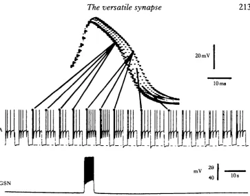
Fig. 7. Effect of activity in a serotonergic molluscan neurone (GSN) upon the duration of the action potential in a follower cell (A). The first A neurone action potential produced by each depolarizing current pulse is shown at a rapid sweep-speed (upper trace); activity in the GSN (lower trace) prolongs the falling phase of the A neurone spike. (From Cottrell, 19826.)
which may be associated with small membrane potential oscillations and some spike activity in the postsynaptic neurone. Voltage-clamp analysis showed that although the response is small at normal resting potential values (about —40 to — 50 mV), the response increases markedly at more positive holding potentials. Like the responses produced by 5-HT in Aplysia neurones (Pellmar & Wilson, 1977; Pellmar & Carpen-ter, 1979, 1980), the currents generated synaptically or by 5-HT in snail neurones are unaltered by removal of sodium or chloride ions from the bathing solution, but are depressed in the presence of cobalt or cadmium. The observations indicate that calcium is probably involved, although it is unclear whether it acts as a charge carrier itself or whether it controls the conductance of some other ion such as potassium. Although the functional significance of this voltage-dependent synaptically-mediated response is not entirely clear, activity in the giant serotonin neurone produces two changes which may be seen in voltage recordings from the postsynaptic A neurone. Firstly, it reverses accommodation of action potentials which occurs during repeated depolarizing current pulses (Fig. 6); this cannot be attributed simply to the depolariz-ing effect of synaptic current as it is not simulated by an equivalent increase in the amplitude of the applied current pulses. Secondly, it causes a prolongation of the falling phase of the action potential (Cottrell, 1982a,b; Fig. 7).
applied to the head. Sensitization results from presynaptic facilitation of transmitted release from the terminals of sensory neurones innervating the siphon. 5-HT mimics the effect of stimulating the sensitizing nerve pathway and appears to be the trans-mitter responsible for sensitization (Brunelli, Castellucci & Kandel, 1976). The effects both of stimulating the sensitizing pathway and of 5-HT are apparently mediated by an elevation in the intracellular concentration of cyclic AMP which, in turn, operates ion channels, by triggering phosphorylation of specific proteins (per-haps the ion channels themselves).
Because nerve terminals are inaccessible to electrophysiological techniques, sen-sory neurone somata have been studied as they appear to serve as a good model for sensory neurone terminals. Changes observed in calcium currents of the cell body action potential are sufficient to account for the changes associated with behavioural sensitization of the gill-withdrawal reflex in Aplysia. Both sensitization and 5-HT cause broadening of the action potential in the cell bodies of sensory neurones in the abdominal ganglion (Klein & Kandel, 1978). Initially this action potential broadening was thought to result from activation of voltage-dependent calcium channels. How-ever, on closer examination, it turned out that 5-HT had no direct action on calcium channels at all, but instead caused a reduction in potassium conductance. The effect of this is to delay repolarization and so prolong the calcium current of the sensory neurone cell body (Klein & Kandel, 1980). More recently, the properties of 5-HT-sensitive potassium channels have been studied under patch-clamp (Siegelbaum, Camardo & Kandel, 1982). These channels are distinct from the four types of potassium channel that have been described in molluscan neurones (Adams, Smith
rl6
- 8 pA
0 - Lo
2-30 I
o
i r r r n r r^>^
1r->.,, r »-mm, „ ,,j, m.
60
| 4PA
[image:16.451.38.413.376.586.2]100 ms
t
; Thompson, 1980) and are the most frequently seen channel type inAplysia sensory eurones; they are only slightly voltage-dependent and are insensitive to changes in the intracellular Ca2+ concentration (determined by using inside-out membrane patches). When 5-HT was applied in the external solution it caused a dose-dependent increase in the input resistance of the cell and a reduction in the number of active channels under the patch, without altering the kinetics of those potassium channels which continued to operate (Fig. 8). The seal at the tip of a patch electrode appears to be sufficiently tight to exclude pharmacological agents applied to the bathing solution from the membrane under the patch. 5-HT was effective when added to the bath during in situ patch experiments, indicating that it must exert its effect on ion channels indirectly via a second messenger, presumably cyclic AMP. This conclusion is supported by the fact that intracellular injection of cyclic AMP mimics the effects of 5-HT on single channels.The molluscan peptide FMRFamide (Phe-Met-Arg-Phe-NH2), like 5-HT, has
voltage-dependent actions upon snail neurones (Cottrell, 1982c). At the resting potential, the peptide produced a hyperpolarizing response, which appears to result from an increase in potassium conductance. At potentials more positive than the resting potential, a second component to the peptide response became apparent; this consisted of an apparent inward current, which progressively increased in size with further depolarization. This was still present when sodium chloride in the external solution was replaced with sucrose, but was abolished when either Co2+ or Ba2+ were present. Injection of Ca2+, on the other hand potentiated the peptide response. These observations suggest that the inward current induced by FMRFamide at relatively depolarized membrane potentials results from suppression of a calcium-dependent potassium current. The outward current observed at more negative membrane poten-tials probably results from opening of a different class of potassium channels. Recent patch-clamp recordings made at depolarized membrane potentials have revealed out-ward current channels. These single channel currents had the properties of potassium channels and appeared less frequently on application of FMRFamide to the surface of the cell body (Cottrell & Green, 1984). The physiological significance of two opposing voltage-dependent conductance changes evoked by a single transmitter is, as yet, unclear, although they would probably render the membrane potential less stable.
216
R. M. PITMANfrom alterations in potassium conductance. Although vasopressin and oxytocin ar| probably not found in molluscs, a peptide component which can induce burst activity has been isolated from the snail nervous system (Ifshin, Gainer & Barker, 1975).
ACh and dopamine can have the opposite effect to vasopressin on some molluscan bursting neurones; both inhibit a persistent slow regenerative inward current and suppress burst firing. Neurones L2 to L$ in the abdominal ganglion of Aplysia are monosynaptically inhibited by the cholinergic neurone L10. This inhibition has a rapid phase which is reversed on hyperpolarization and results from an increase in chloride conductance. This is followed by a slow phase, lasting several seconds, which is not inverted by hyperpolarization. Because the slow phase failed to reverse, it had been attributed previously to synaptic activation of an electrogenic sodium pump, (Pinsker & Kandel, 1969) or to an increase in potassium conductance generated at some distance from the recording site (Kehoe & Ascher, 1970). Voltage-clamp has demonstrated that the slow phase of synaptic inhibition has no effect on the I/V curve of postsynaptic neurones at membrane potentials more negative than —60 mV. How-ever, at more positive membrane potentials it eliminated the region of negative-slope resistance which is characteristic of burster neurones (Wilson & Wachtel, 1978; Fig. 10). The effects of synaptically-mediated inhibition could be mimicked by
AiControl
-40
A2 Vasopressin B O Control
- 3 5
+ 2 0
--OmV
Vasopressin
I I |
- 6 0
I
-20 v +20 mV
[image:18.451.39.414.315.566.2]- 2 0 ->
0 nA
• - 2 0
[image:19.451.123.328.51.212.2]• - 4 0
Fig. 10. Effect of prolonged cholinergic inhibition on the I/V curve of a molluscan burst firing neurone studied under voltage-clamp. Inset, the membrane potential of the burster neurone is held at - 3 5 mV while 10 mV hyperpolarizing command pulses are applied before and just following activity in a cholinergic prcsynaptic neurone (Lio). The first command pulse produces an outward cun-ent (showing the existence of a negative resistance characteristic). The command pulse delivered during prolonged synaptic inhibition results in an inward current (indicating that the negative resis-tance has been suppressed). The graph illustrates the I/V relation of the neurone before (filled circles) and during (open circles) synaptic inhibition; the negative resistance region in the I/V curve is suppressed by cholinergic inhibition. Calibration: horizontal, 8 s; vertical, 30mVfor Vm (membrane
potential), 20 nA for Ic (voltage-clamp current) and 100 mV for Lio (the membrane potential of the
presynaptic neurone). (From Wilson & Wachtel, 1978.)
iontophoretically applying ACh to the axon of the cell. The slow component of the ACh response was unaltered by a three-fold increase in the external potassium con-centration, but was abolished in sodium-free solution or by lowering the temperature. Wilson & Wachtel (1978) concluded that prolonged cholinergic inhibition blocks a slow regenerative inward current that is responsible for the membrane potential oscillations seen in burster neurones (Wilson & Wachtel, 1974). Dopamine has a similar effect upon the slow regenerative current observed in the burster neurone R15 (Wilson & Wachtel, 1978). The slow voltage-dependent inward currents which are modulated by ACh and dopamine may involve an increase in conductance to calcium ions (Lewis & Wilson, 1983).
Co-transmitters
Histochemical and biochemical evidence is accumulating to indicate that a single neurone may contain more than one neurotransmitter. In some instances, the co-transmitters may be biochemically related, but in other instances they are not; thus peptides have been found co-localized with noradrenalin, 5-HT, dopamine or ACh, while some neurones show immunoreactivity to more than one peptide (Hokfelt et al.
1980).
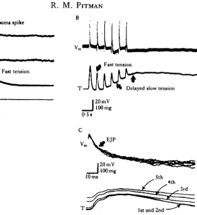
B
Fast tension
0 1 s
[image:20.451.114.402.48.363.2]1st and 2nd '
Fig. 11. Response of an insect muscle to activity in a pcptidc-containing motor neurone. (A) Upper trace, a single action potential in the slow coxal depressor motor neurone of the cockroach (D,) evokes a brief excitatory junctional potential (EJP) in muscle fibres (Vro) and a transient tension response
from the whole muscle (T). Trace I indicate* the current injected into the motor neurone soma. (B) Repetitive motor neurone activity evokes a series of EJPs (trace Vm) without any sustained change
in membrane potential. Each EJP is accompanied by a brief contraction, but muscle tension also shows a slow rise, which is not associated with any change in muscle fibre membrane potential (trace T ) . (C) During a burst of motor neurone spikes, there is a progressive slowing in the relaxation rate of rapid contractions (trace T ) , although the shape of successive EJPs is unaltered (trace Vm). The
fast contractile responses may be produced by the action of glutamate, while the peptide proctolin may cause the slow component of contraction. (From Adams & O'Shea, 1983.)
& O'Shea (1983) have concluded that the transient tension and electrical Responses observed in the coxal depressor muscles are produced by synaptically released glutamate, while the slow tension response is mediated by proctolin acting as a co-transmitter.
The action of proctolin on cockroach muscle has similarities with the effects of 5-HT on buccal muscle of Aplysia (Weiss, Cohen & Kupfermann, 1975, 1978; Kupfermann et al. 1979). When 5-HT is released from peripheral terminals of an identified serotonergic neurone, it potentiates contractions evoked by cholinergic motor neurone activity. Applied or synaptically released 5-HT has no detectable effect upon resting tension, membrane potential or input resistance of muscle fibres, although it does produce a modest increase in amplitude of EJPs. However, the enhancement of EJPs cannot account for the effects of 5-HT, as the time course of the enhancement in contractility is not necessarily the same as that for augmentation of EJPs. In addition, activation of the serotonergic neurone potentiates the contractile response to applied ACh. These observations indicate that 5-HT enhances contrac-tion of Aplysia muscle by a postsynaptic mechanism which does not involve any electrical change in the muscle fibres. It has been suggested that cyclic AMP mediates a direct action of 5-HT upon excitation-contraction coupling (Weiss et al. 1978; Kupfermann et al. 1979). It is possible that similar mechanisms may be involved in the postsynaptic actions of octopamine on locust muscle (O'Shea & Evans, 1979) and of octopamine, 5-HT and proctolin on lobster muscle (Kravitzef al. 1980; Schwarz, Harris-Warrick, Glusman & Kravitz, 1980).
CONCLUDING REMARKS
The complexity of chemically mediated synaptic transmission provides a substrate for considerable flexibility; not only can different synapses have individual properties, but also the characteristics of any single synapse may depend upon its recent history of activity. This versatility enables quite diverse functional requirements to be met. Some synapses, like the vertebrate neuromuscular junction, may be required to trans-mit information rapidly with a large safety factor, while others may perform more subtle tasks over a longer time-scale.
Transmitter release will depend upon both its availability for release and upon the characteristics of the release process. Individual synapses may show differences in the rate at which released transmitter is replenished; this may become important at peptidergic synapses, since peptide neurotransmitters cannot be synthesized in nerve terminals. It may turn out, however, that peptides are released at junctions where the overall transmitter requirement does not fluctuate greatly or that their actions are sufficiently prolonged that short-term adjustments in supply are not required.
integration can take place by means of local graded interactions restricted to limited portions of neurones.
The ability of neurotransmitters to operate voltage-dependent channels has a number of interesting consequences. Operation of such channels provides one mechanism for presynaptic inhibition. Calcium influx into presynaptic terminals may be influenced by direct modulation of calcium channels or may be altered indirectly as a result of an effect upon other channels that themselves alter the configuration of the action potential. If the modification in the calcium current of the action potential is widespread throughout a neurone, it may cause a change in the amount of transmitter released per impulse from spatially separated transmitter release sites. This may be possible, since synaptic activation of voltage-dependent channels can alter the con-figuration of the action potential recorded from the cell body of the neurone. Transmitter-operated voltage-sensitive channels provide the potential for extremely non-linear summation of different synaptic inputs; if a transmitter operates voltage-dependent channels which can only open at membrane potentials more positive than the normal resting potential, this transmitter would, by itself, be inoperative and would require some other input to 'enable' it. Conversely, this voltage-dependent response could be completely 'gated out' by any transmitter which hyperpolarized the membrane potential beyond the range in which channels operate. Transmitter actions upon voltage-dependent channels can also alter the pattern of activity in a neurone. For example, it has been suggested that the potassium M-current in sympathetic neurones may normally limit impulse frequency since it will generate an outward current when it is activated by depolarization. Synaptic inactivation of this potassium current will enhance excitability. Although the transmitter itself does not cause strong excitation, it may prolong excitation generated by another synaptic input. The ability of transmitters specifically to increase or decrease the conductances underlying 'burster' activity may be important in regulating the properties of 'oscillator' circuits. This type of synaptic action could turn a network on or off and may even switch it from one operating mode to another.
R E F E R E N C E S
ADAMS, D. ] . , SMITH, S. J. & THOMPSON, S. H. (1980). Ionic currents in molluscan soma. A Rev. Neurosci. 3, 141-167.
ADAMS, M. E. & O'SHEA, M. (1983). Peptide cotransmitter at a neuromuscular junction. Science, N.Y. 221, 286-289.
BARKER, J. L. & GAINER, H. (1974). Peptide regulation of bursting pacemaker activity in a molluscan neurosecretory cell. Science, N.Y. 184, 1371-1373.
BARKER, J. L., IFSHIN, M. S. & GAINER, H. (1975). Studies on bursting pacemaker potential activity in molluscan neurons. III. Effects of hormones. Brain Res. 84, 501—513.
BARKER, J. L. & SMITH, T . G. (1976). Peptide regulation of neuronal membrane properties. Brain Res. 103, 167-170.
BISHOP, C. A. & O'SHEA, M. (1982). Neuropeptide proctolin immunocytochemical mapping of neurons in the central nervous system of the cockroach Periplaneta americana. J. camp. Neurol. 207, 223—238.
BRACHO, H. & ORKAND, R. K. (1970). Effect of calcium on excitatory neuromuscular transmission in the crayfish. J . Physiol., Land. 206, 61-71.
BROWN, D. A. & ADAMS, P. R. (1980). Muscarinic suppression of a novel voltage-sensitive K+ current in a vertebrate neurone. Nature, Land. 283, 673-676.
BROWN, D. A., CONSTANT!, A. & ADAMS, P. R. (1981). Slow cholinergic and peptidergic transmission in sympathetic ganglia. Fedn Proc. Fedn Am. Socs exp. Biol. 40, 2625-2630.
BURROWS, M. (1979). Synaptic potentials effect the release of transmitter from locust nonspiking interneurones. __ 'Science, N.Y. 204, 81-83.
BURROWS, M. & SIEGLER, M. V. S. (1976). Transmission without spikes between locust interneurones and motoneurones. Nature, Land. 262, 222-224.
BURROWS, M. & SIEGLER, M. V. S. (1978). Graded synaptic transmission between local interneurones and motor neurones in the metathoracic ganglion of the locust. J. Physiol., Land. 285, 231-255.
BUSH, B. M. H. & CANNONE, A. J. (1973). A stretch reflex in crabs evoked by muscle receptor potentials in non-impulsive afferent*. J. Physiol., Land. 232, 95P.
CASTELLUCCI, V. F. & KANDEL, E. R. (1974). A quantal analysis of the synaptic depression underlying habituation of the gill-withdrawal reflex in Aplysia. Proc. natn. Acad. Sci. U.SA 71, 5004-5008.
CASTEJAUCCI, V. F., KANDEL, E. R., SCHWARTZ, J. H., WILSON, F. D., NAIRN, A. C. & GREENGARD, P. (1980). Intracellular injection of the catalytic subunit of cyclic AMP-dependent protein kinase stimulates facilitation of transmitter release underlying behavioral sensitization in Aplysia. Proc. natn. Acad. Sci. U.SA. 77, 7492-7496.
COTTREIX, G. A. (1981). An unusual synaptic response mediated by a serotonin neurone. Q.Jlexp. Physiol. 66, 475-485.
COTTRELL, G. A. (1982a). Physiological role of a slow, voltage-sensitive, synaptic response mediated by an identified serotonin-containing neurone. Q.Jlexp. Physiol. 67, 179—183.
COTTRELL, G. A. (19826). Voltage-dependent actions of endogenous and exogenous serotonin on identified neurones. Comp. Biochem. Physiol. 72C, 271-279.
COTTRELL, G. A. (1982c). FMRFamide neuropeptides simultaneously increase and decrease K+ currents in an
identified neurone. Nature, Land. 296, 87-89.
COTTRELL, G. A. Si GREEN, K. A. (1984). Single-channel analysis of the action of FMRFamide on K channels in Helix neurones. J. Physiol., bond, (in press).
DETKRRE, P., PAUPARDIN-TRITSCH, D . , BOCKAERT, J. & GERSCHENFELD, H. M. (1981). Role of cyclic AMP in a serotonin-evoked slow inward current in snail neurones. Nature, Land. 290, 783—785.
DUDEL, J. (1965). Facilitatory effects of 5-hydroxy-tryptamine on the crayfish neuromuscular junction. Arch. exp. Path. Pharmak. 249, 515-528.
DUDEL, J. (1983). Graded or all-or-nothing release of transmitter quanta by local depolarizations of nerve terminals on crayfish muscle?. Pflugers Arch. ges. Physiol. 398, 155—164.
DUDEL, J. it KUFFLER, S. W. (1961a). Mechanism of facilitation at the crayfish neuromuscular junction. J. Physiol., Land. 155, 530-542.
DUDEL, J. & KUFFLER, S. W. (19616). Presynaptic inhibition at the crayfish neuromuscular junction..?. Physiol., Land. 155, 543-562.
DUDEL, J., PARNAS, I. & PARNAS, H. (1983). Neurotransmitter release and its facilitation in crayfish muscle. VI. Release determined by both, intracellular calcium concentration and depolarization of the nerve terminal. Pflugers Arch. ges. Physiol. 399, 1-10.
DUNCAN, C. J. & STATHAM, H. E. (1977). Interacting effects of temperature and extracellular calcium on spontaneous release of transmitter at the frog neuromuscular junction. J . Physiol., Land. 268, 319-333. DUNLAP, K. & FISCHBACH, G. D. (1978). Neurotransmitters decrease the calcium component of sensory
neurone action potentials. Nature, bond. 276, 837—839.
ECCLES, J. C. (1964). The Physiology of Synapses. Berlin: Springer- Verlag.
ECKERT, R. & Lux, H. D. (1977). Calcium-dependent depression of a late outward current in snail neurons. Science, N.Y. 197, 472-475.
FAHIM, M. A. & USHERWOOD, P. N. R. (1983). Effects of c-AMP, caffeine, theophylline and vinblastine on spontaneous transmitter release at locust nerve-muscle junctions. J. Neurobiol. 14, 391—397.
FISCHER, L. & FLOREY, E. (1983). Modulation of synaptic transmission and excitation-contraction coupling in the opener muscle of the crayfish, Astacus leptodactylus, by 5-hydroxytryptamine and octopamine. J. exp. Biol. 102, 187-198.
GLUSMAN, S. & KRAVITZ, E. A. (1982). The action of serotonin on excitatory nerve terminals in lobster nerve-muscle preparations, J. Physiol., Land. 325, 223-241.
GOLDBERG, A. L. & SINGER, J. J. (1969). Evidence for a role of cyclic AMP in neuromuscular transmission.
Proc. natn. Acad. Sci. U.SA. 64, 134-141.
GRAUBARD, K., RAPER, J. A. & HARTLINE, D. K. (1980). Graded synaptic transmission between spiking neurons. Proc. natn. Acad. Sci. U.SA. 77, 3733-3735.
GRAUBARD, K., RAPER, J. A. & HARTUNE, D. K. (1983). Graded synaptic transmission between identified spiking neurons. J. Neurophysiol. 50, 508-521.
GROSSMAN, Y., PARNAS, I. & SPIRA, M. E. (1979a). Differential conduction block in branches of a bifurcating axon. J. Physiol., Land. 295, 283-305.
GROSSMAN, Y., PARNAS, I. & SPIRA, M. E. (19796). Ionic mechanisms involved in differential conduction of action potentials at high frequency in a branching axon. J. Physiol., Land. 295, 307—322.
HATT, H. & SMITH, D. O. (1976). Synaptic depression related to presynaptic axon conduction block. J. PhysioM bond. 259, 367-393.
HOKFELT, T . , JOHANSSON, O., LJUNGDAHL, A., LUNDBERG, J. M. & SCHULTZBERG, M. (1980). Peptidergic neurones. Nature, Land. 284, 515—521.
HUBBARD, J. I. & WILLIS, W. D. (1962). Hyperpolarization of mammalian motor nerve terminals. J. Pkysioi, band. 163, 115-137.
IFSHIN, M., GAINER, H. & BARKER, J. L. (1975). Peptide factor extracted from molluscan ganglia that modulates bursting pacemaker activity. Nature, bond. 254, 72—73.
JAN, Y. N., JAN, L. Y. & KUFFLER, S. W. (1979). A peptide as a possible transmitter in sympathetic ganglia of the frog. Proc. natn. Acad. Sri. U.SA. 76, 1501-1505.
JAN, Y. N., JAN, L. Y. & KUFFLER, S. W. (1980). Further evidence for peptidergic transmission in sympathetic ganglia. Proc. natn. Acad. Sri. U.SA. 77, 5008-5012.
JESSELL, T . M. & IVERSEN, L. L. (1977). Opiate analgesics inhibit substance P release from rat trigeminal nucleus. Nature, bond. 268, 549-551.
JOHNSTON, M. F., KRAVTTZ, E. A., MEIRI, H. & RAHAMIMOFF, R. (1983). Adrenocorticotropic hormone causes long-lasting potentiation of transmitter release from frog motor nerve terminals. Science, N.Y. 220, 1071-1072.
KACZMAREK, L. K., JENNINGS, K. R., STRUMWASSER, F . , NAIRN, A. C , WALTER, U., WILSON, F . D . & GREENGARD, P. (1980). Microinjection of catalytic subunit of cyclic-AMP-dependent protein kinase enhances calcium action potentials of bag cell neurons in cell culture. Proc. natn. Acad. Sri. U.SA. 77, 7487-7491.
KANDEL, E. R., BRUNELLI, M., BYRNE, J. & CASTELLUCCI, V. (1976). A common presynaptic locus for the synaptic changes underlying short-term habituation and sensitization of the gill-withdrawal reflex in Aplysia. Cold Spring Harb. Symp. quant. Biol. 40, 465-482.
KANDEL, E. R. & WACHTEL, H. (1968). The functional organization of neural aggregates in Aplysia. In Physiological and Biochemical Aspects ofNervous Integration, (ed. F. D . Carlson), pp. 17—65. New Jersey: Prentice-Hall.
KATZ, B. & MILEDI, R. (1965). The effect of calcium on acetylcholine release from motor nerve terminals. Proc. R. Soc. B 161, 496-503.
KATZ, B. & MILEDI, R. (1967). A study of synaptic transmission in the absence of nerve impulses..7. Physiol., bond. 192, 407-436.
KATZ, B. & MILEDI, R. (1968). The role of calcium in neuromuscular facilitation. J. Physiol., bond. 195, 481-492.
KATZ, B. & MILEDI, R. (1977). Suppression of transmitter release at the neuromuscular junction. Proc.R. Soc. B 196, 465-469.
KEHOE, J. & ASCHER, P. (1970). Re-evaluation of the synaptic activation of an electrogenic sodium pump. Nature, bond. 225, 820-823.
KLEIN, M. & KANDEL, E. R. (1978). Presynaptic modulation of voltage-dependent Ca2+ current: mechanism for behavioral sensitization in Aplysia califomica. Proc. natn. Acad. Sri. U.SA. 75, 3512-3516.
KLEIN, M. & KANDEL, E. R. (1980). Mechanism of calcium current modulation underlying presynaptic facilitation and behavioral sensitization in Aplysia. Proc. natn. Acad. Sri. U.SA 77, 6912-6916.
KONISHI, S., TSUNOO, A. & OTSUKA, M. (1981). Enkephalin as a transmitter for presynaptic inhibition in sympathetic ganglia. Nature, bond. 294, 80—82.
KRAVTTZ, E. A., GLUSMAN, S., HARJUS-WARJUCK, R. M., LIVINGSTONE, M. S., SCHWARZ, T . & GOY, M. F. (1980). Amines and a peptide as neurohormones in lobsters: actions on neuromuscular preparations and preliminary behavioural studies. J. exp. Biol. 89, 159-175.
KJINJEVIC, K. & MILEDI, R. (1958a). Failure of neuromuscular propagation in rats. J. Physiol., bond. 140, 440-461.
ICRNJEVIC, K. & MILEDI, R. (19586). Some effects produced by adrenaline upon neuromuscular propagation in rats. J . Physiol., bond. 141, 291-304.
KRNjEVKi, K. & MILEDI, R. (1959). Presynaptic failure of neuromuscular propagation in rats. J. Physiol., bond.
149, 1-22.
KRNJEVIC, K., PUMAIN, R. SC RENAULD, L. (1971). The mechanism of excitation by acetylcholine in the cerebral cortex. J. Physiol, bond. 215, 247-268.
KUBA, K. & KOKETSU, K. (1975). Direct control of action potentials by acetylcholine in bullfrog sympathetic ganglion cells. Brain Res. 89, 166-169.
KUBA, K. & KOKETSU, K. (1976). The muscarinic effects of acetylcholine on the action potential of bullfrog sympathetic ganglion cells. Jap. J. Physiol. 26, 703-716.
KUBA, K. & KOKETSU, K. (1978). Synaptic events in sympathetic ganglia. Prog. Neurobiol. 11, 77-169. KUFFLER, S. W. & SEJNOWSKI, T . J. (1983). Peptidergic and muscarinic excitation at amphibian sympathetic
synapses. 7. Physiol., bond. 341, 257-278.
K U N O , M . ( 1 9 7 1 ) . Quantum aspects of central and ganglionic synaptic transmission in vertebrates. Physiol. Rev.
51, 647-678.
tlPFERMANN, I . , COHEN, J . L . , MANDELBAUM, D . E . , S C H O N B E R G , M . , S U S S W E I N , A . J . & WEISS, K . R . (1979). Functional role of serotonergic neuromodulation in Aplysia. Fedn Proc. FednAm. Socsexp. Biol. 38, 2095-2102.
LANG, F. & ATWOOD, H. L. (1973). Crustacean neuromuscukr mechanisms: functional morphology of nerve terminals and the mechanism of facilitation. Am. Zool. 13, 337-3SS.
LEWIS, D. V. & WILSON, W. A. (1983). Dopamine reduces arsenazo III absorbance change during depolariza-tion of bursting neuron. Soc. Neurotd. Abitr. 9, 75.
LLJNAS, R., STEINBERG, I. Z. & WALTON, K. (1981). Relationship between presynaptic calcium current and postsynaptic potential in squid giant synapse. Biophyt. J. 33, 323—352.
MAGLEBY, K. L. SCZENGEL, J. E. (1976). Stimulation-induced factors which affect augmentation and potentia-tion of transmitter release at the neuromuscular juncpotentia-tion. J. Physiol., Land. 260, 687—717.
MEECH, R. W. (1976). Intracellular calcium and the control of membrane permeability. Symp. Soc. exp. Biol.
30, 67-88.
MENDELL, L. M. &WALL, P. D. (1964). Presynaptic hyperpolarization: a role for fine afferent fibres. J. Physiol., Land. 172, 274-294.
MILEDI, R. & SLATER, S. D. (1966). The action of calcium on neuronal synapses in the squid. J. Physiol., Land.
184, 473-498.
MINOTA, S. & KOKETSU, K. (1977). Effects of adrenaline on the action potential of sympathetic ganglion cells in bullfrogs. Jap. J. Physiol. 27, 353-366.
MUDGE, A. W., LEEMAN, S. E. 8C FISCHBACH, G. D . (1979). Enkephalin inhibits release of substance P from sensory neurons in culture and decreases action potential duration. Proc. natn. Acad. Sri. U.SA. 76, 526-530. MULLER, K. J. & SCOTT, S. A. (1981). Transmission at a 'direct' electrical connexion mediated by an
inter-neurone in the leech. J. Physiol., Land. 311, 565—583.
NATHANSON, J. A. (1977). Cyclic nucleotides and nervous system function. Physiol. Rev. 57, 157-256. NICHOLLS, J. & WALLACE, B. G. (1978). Modulation of transmission at an inhibitory synapse in the central
nervous system of the leech. J. Phytiol., Land. 281, 157-170.
ONODERA, K. (1973). Effects of caffeine on the neuromuscular junction of the frog and its relation to external calcium concentration. Jap. J. Physiol. 23, 587-597.
O'SHEA, M. & EVANS, P. D. (1979). Potentiation of neuromuscular transmission by an octopaminergic neurone in the locust. J. exp. Biol. 79, 169-190.
PAHNAS, I. (1972). Differential block at high frequency of branches of a single axon innervating two muscles. J. Neumphysiol. 35, 903-914.
PPIIMAH T . C. (1981). Transmitter-induced calcium current. Fedn Proc. Fedn Am. Socs exp. Biol. 40, 2631-2636.
PELLMAR, T . C. & CARPENTER, D . O. (1979). Voltage-dependent calcium current induced by serotonin. Nature, Land. 277, 483-484.
PELLMAR, T . C. & CARPENTER, D. O. (1980). Serotonin induces a voltage-sensitive calcium current in neurons of Aplysia califomica.J. Neurophysiol. 44, 423-439.
PELLMAR, T . C. 8C WILSON, W. A. (1977). Unconventional serotonergic excitation in Aplysia. Nature, Land.
269, 76-78.
PINSKER, H. & KANDEL, E. R. (1969). Synaptic activation of an electrogenic sodium pump. Science, N.Y. 163, 931-935.
SCHLAPFER, W. T . , TREMBLAY, J. P., WOODSON, P. B. J. & BASONDES, S. H. (1976). Frequency facilitation and post-tetanic potentiation of a unitary synaptic potential in Aplysia califarnica are limited by different processes. Brain Res. 109, 1-20.
SCHLAPFER, W. T . , WOODSON, P. B. J., TREMBLAY, J. P. & BAJIONDES, S. H. (1974). Depression and frequency facilitation at a synapse in Aplysia califarnica: evidence for regulation by availability of transmitter. Brain Res. 76, 267-280.
SCHWARZ, T . L., HARRIS-WARRICK, R. M., GLUSMAN, S. & KRAVTTZ, E. A. (1980). A peptide action in a lobster neuromuscular preparation. J. Neurobiol. 11, 623-628.
SHAPIRO, E., CASTELLUCCI, V. F . & KANDEL, E. R. (1980). Presynaptic membrane potential affects trans-mitter release in an identified neuron in Aplysia by modulating the Ca2* and K+ currents. Pmc. natn. Acad.
Sri. U.SA. 77, 629-633.
SIEGELBAUM, S. A., CAMARDO, J. S. & KANDEL, E. R. (1982). Serotonin and cyclic AMP close single K+ " channels in Aplysia sensory neurones. Nature, Land. 299, 413—417.
SPIRA, M. E., YAROM, Y. & PARNAS, I. (1976). Modulation of spike frequency by regions of special axonal geometry and by synaptic inputs. J. Neurophysiol. 36, 882-899.
STARKE, K. (1981). Presynaptic receptors. A. Rev. Pharmac. Toxicol. 21, 7-30.
STATHAM, H. E. & DUNCAN, C. J. (1976). Dantrolene and the neuromuscular junction: evidence for intracellular calcium stores. Europ.J. Pharmac. 39, 143-153.
STINNAKRE, J. 8C TAUC, L. (1973). Calcium influx in active Aplysia neurones detected by injected aequorin. Nature, Land. 242, 113-115.
WEIGHT, F. F. & ERULKAR, S. D. (1976). Modulation of synaptic transmitter release by repetitive postsynapl action potentials. Science, N.Y. 193, 1023-1025.
WEIGHT, F. F. JC VOTAVA, J. (1970). Slow synaptic excitation in sympathetic ganglion cells: evidence for synaptic inactivation of potassium conductance. Science, N.Y. 170, 755-758.
WEISS, K. R., COHEN, J. & KUFFERMANN, I. (1975). Potentiation of muscle contraction: a possible modulatory function of an identified serotonergic cell in Aplysia. Brain Res. 99, 381-386.
WEISS, K. R., COHEN, J. L. & KUPFERMANN, I. (1978). Modulatory control of buccal musculature by a serotonergic neuron (metacerebral cell) in Aplysia. J. Neumphysiol. 41, 181-203.
WERBLJN, F. S. & DOWLJNG, J. E. (1969). Organization of the retina of the mudpuppy, Necturus maculosus. II. Intracellular recording. J. Neuwphysiol. 32, 339-355.
WILSON, W. A. & WACHTEL, H. (1974). Negative resistance characteristic essential for the maintenance of slow oscillations in bursting neurons. Science, N.Y. 186, 932—934.
WILSON, W. A. & WACHTEL, H. (1978). Prolonged inhibition in burst firing neurons: synaptic inactivation of the slow regenerative inward current. Science, N.Y. 202, 772—775.
WOJTOWICZ, J. M. & ATWOOD, H. L. (1983). Maintained depolarization of synaptic terminals facilitates nerve-evoked transmitter release at a crayfish neuromuscular junction. J. Neumbiol. 14, 385-390.
YAU, K.-W. (1976). Receptive fields, geometry and conduction block of sensory neurones in the central nervous system of the leech. J . Physiol, Land. 263, 513-538.
ZUCKER, R. S. (1972). Crayfish escape behavior and central synapses. II. Physiological mechanisms underlying behavioral habituation.J. Neumphysiol. 35, 621-637.
ZUCKER, R. S. (1973). Changes in the statistics of transmitter release during facilitation. J. Physiol., Land. 229, 787-810.
ZUCKER, R. S. (1974a). Crayfish neuromuscular facilitation activated by constant presynaptic action potentials and depolarizing pulses. J. Physiol., Land. 241, 69—89.
ZUCKER, R. S. (19746). Excitability changes in crayfish motor neurone terminals. J. Physiol., Land. 241, 111-126.