http://go.warwick.ac.uk/lib-publications
Original citation:Pracharova, J., et al. (2012). Interactions of DNA with a new Platinum(IV) Azide Dipyridine complex activated by UVA and visible light : relationship to toxicity in tumor cells. Chemical Research in Toxicology, 25(5), pp. 1099-1111.
Permanent WRAP url:
http://wrap.warwick.ac.uk/46584 Copyright and reuse:
The Warwick Research Archive Portal (WRAP) makes the work of researchers of the University of Warwick available open access under the following conditions. Copyright © and all moral rights to the version of the paper presented here belong to the individual author(s) and/or other copyright owners. To the extent reasonable and practicable the material made available in WRAP has been checked for eligibility before being made available.
Copies of full items can be used for personal research or study, educational, or not-for-profit purposes without prior permission or charge. Provided that the authors, title and full bibliographic details are credited, a hyperlink and/or URL is given for the original metadata page and the content is not changed in any way.
Publisher’s statement:
This document is the Accepted Manuscript version of a Published Work that appeared in final form in Chemical Research in Toxicology, © American Chemical Society after peer review and technical editing by the publisher. To access the final edited and published work see
http://dx.doi.org/10.1021/tx300057y A note on versions:
The version presented here may differ from the published version or, version of record, if you wish to cite this item you are advised to consult the publisher’s version. Please see the ‘permanent WRAP url’ above for details on accessing the published version and note that access may require a subscription.
Interactions of DNA with a New Platinum(IV) Azide Dipyridine Complex Activated by UVA and Visible Light.
Relationship to Toxicity in Tumor Cells
Journal: Chemical Research in Toxicology
Manuscript ID: tx-2012-00057y.R1 Manuscript Type: Article
Date Submitted by the Author: n/a
Complete List of Authors: Pracharova, Jitka; Faculty of Science, Palacky University, Department of Biophysics
Lenka, Zerzankova; Institute of Biophysics, Academy of Sciences of the Czech Republic, v.v.i.
Stepankova, Jana; Institute of Biophysics, Academy of Sciences of the Czech Republic, v.v.i.
Novakova, Olga; Institute of Biophysics, Academy of Sciences of the Czech Republic, v.v.i.
Farrer, Nicola J.; University of Warwick, Department of Chemistry Sadler, Peter; University of Warwick, Deaprtment of Chemistry
Brabec, Viktor; Institute of Biophysics, Academy of Sciences of the Czech Republic, v.v.i.
Kasparkova, Jana; Institute of Biophysics, Academy of Sciences of the Czech Republic, v.v.i.
1
Interactions of DNA with a New Platinum(IV) Azide
Dipyridine Complex Activated by UVA and Visible Light.
Relationship to Toxicity in Tumor Cells
Jitka Pracharova
†, Lenka Zerzankova
‡, Jana Stepankova
‡, Olga Novakova
‡, Nicola J.
Farrer
§, Peter J. Sadler
c, Viktor Brabec
‡, Jana Kasparkova*
,‡†
Department of Biophysics, Faculty of Science, Palacky University, 17. listopadu 12, CZ-77146
Olomouc, Czech Republic
‡
Institute of Biophysics, Academy of Sciences of the Czech Republic, v.v.i., Kralovopolska 135,
CZ-61265 Brno, Czech Republic
§
Department of Chemistry, University of Warwick, Gibbet Hill Road, CV4 7AL, Coventry, United
Kingdom
TITLE RUNNING HEAD: DNA Interactions of Photoactivated Pt Complex
CORRESPONDING AUTHOR FOOTNOTE:
* To whom correspondence should be addressed. Tel: +420-541517174. Fax: +420-541240499.
E-mail: [email protected].
ACS Paragon Plus Environment
2 Abstract
The PtIV diazido complex trans,trans,trans-[Pt(N3)2(OH)2(pyridine)2] (1) is unreactive in the dark but
is cytotoxic when photoactivated by UVA and visible light. We have shown that 1 when photoactivated
accumulates in tumor cells and binds strongly to nuclear DNA under conditions in which it is toxic to
tumor cells. The nature of the DNA adducts, including conformational alterations, induced by
photoactivated 1 are distinctly different from those produced in DNA by conventional cisplatin or
transplatin. In addition, the observation that major DNA adducts of photoactivated 1 are able to
efficiently stall RNA polymerase II more efficiently than cisplatin suggests that transcription inhibition
may contribute to the cytotoxicity levels observed for photoactivated 1. Hence, DNA adducts of 1 could
trigger a number of downstream cellular effects different from those triggered in cancer cells by DNA
adducts of cisplatin. This might lead to the therapeutic effects that could radically improve
chemotherapy by platinum complexes. The findings of the present work help to explain the different
cytotoxic effects of photoactivated 1 and conventional cisplatin and thereby provide new insights into
mechanisms associated with the antitumor effects of platinum complexes photoactivated by UVA and
visible light.
KEYWORDS: Anticancer platinum; cytotoxicity; photoactivation; DNA damage; RNA polymerase II
ACS Paragon Plus Environment
3 Table of Contents Graphic
ACS Paragon Plus Environment
4 INTRODUCTION
It has been demonstrated that PtIV diazido complexes which are stable and non-cytotoxic in the dark
exhibit significant toxicity in cancer cells upon irradiation with short wavelengths (365 nm) (1). Their
activation with UVA irradiation to produce cytotoxic and reactive PtII analogues does not require oxygen
(1, 2), an advantage over conventional photosensitizers currently used in photodynamic therapy (PDT),
such as those based on tetrapyrrole derivatives. In addition, selective photoactivation of platinum
complexes in cancer cells may help to overcome limitations connected with toxic side effects of
antitumor platinum drugs currently used in the clinic. The first photoactivatable PtIV complexes
exhibiting toxicity in tumor cells could be photoactivated only upon irradiation with wavelengths in
UVA region (1), which is not optimal for clinical applications. Recently, the PtIV diazido complex
trans,trans,trans-[Pt(N3)2(OH)2(pyridine)2] (1) was synthesized which is both highly soluble and stable
in aqueous solution. It can be photoactivated over a range of wavelengths and is toxic towards cancer
cells using low doses of UVA radiation and, crucially, also with visible light (3). The presence of planar
pyridine ligands in 1, which appear to remain strongly bound to platinum, even after photoactivation,
has a critical effect not only on the photoactivation pathways, but also on the biological activity.
Modification of clinically-ineffective transplatin (trans-[PtCl2(NH3)2]) by inclusion of planar ligands
considerably changes the binding mode of the trans-PtII species with DNA, the major pharmacological
target of antitumor platinum complexes (4-15). These transplatin analogues exhibited promising toxic
effects in various human tumor cell lines including those resistant to conventional cisplatin. Thus,
photoactivated 1 can be coclustered with other platinum-pyridines which are not cross-resistant and
which exhibit different mechanisms of action from platinum-based drugs already on the market such as
cisplatin, carboplatin and oxaliplatin.
Cisplatin and its analogues carboplatin and oxaliplatin bind to DNA preferentially at the N7 position
of guanine bases (16, 17), inhibiting replication (18, 19) and transcription (20, 21). The inhibition of
these critical DNA-related processes triggers subsequent intracellular events that activate apoptotic and
ACS Paragon Plus Environment
5 necrotic pathways (22). Hence, similar to other antitumor platinum-pyridine complexes, the different
mechanism of action of photoactivated 1 may also derive from its unique mode of binding to DNA.
To assess the importance of the binding of photoactivated 1 to DNA for the efficacy of its anticancer
action, we describe here an investigation of DNA binding of photoactivated 1 in tumor cells treated with
this metallodrug under conditions where it is non-toxic towards these cells (in the absence of
irradiation). In addition, the fact that photoactivated 1 binds to DNA in cells prompted us to investigate
the modifications induced in DNA in vitro (in cell-free media), including conformational alterations of
DNA and the processing of these modifications by RNA polymerase II. We believe that a deep
understanding of the reactions leading to bifunctional DNA cross-link (CL) formation by photoactivated
1 may provide guidance in future efforts to optimize the rational design of photoactivatable anticancer
metallodrugs.
ACS Paragon Plus Environment
6 EXPERIMENTAL PROCEDURES
Chemicals. Trans,trans,trans-[Pt(N3)2(OH)2(py2)] (1) was synthesized and characterized by the
method described previously (3). Cis- and trans-[Pt(Cl2)(NH3)2] (cisplatin and transplatin) were
obtained from Sigma (Prague, Czech Republic) (purity was 99.9 %).
Chloridodiethylenetriamineplatinum(II) chloride ([PtCl(dien)]Cl) was a generous gift of Prof. G. Natile
from the University of Bari. The stock solutions of platinum complexes were prepared in H2O, their
concentrations determined by flameless atomic absorption spectroscopy (FAAS), and stored in the dark.
Calf thymus (CT) DNA (42% G + C, mean molecular mass ca. 20 000 kDa) was prepared as previously
described (23, 24). Plasmids pUC19 [2686 base pairs (bp)], pSP73 (2464 bp) and pSP73KB (2455 bp)
were isolated according to standard procedures. The Klenow fragment from DNA polymerase I,
restriction endonucleases EcoRI and XbaI, plasmid pCMV-GLuc (5764 bp), T4 DNA ligase and T4
polynucleotide kinase were obtained from New England Biolabs (Beverly, MA). HeLaScribe® Nuclear
Extract in vitro Transcription system kit, T7 and SP6 RNA polymerases and RNasin ribonuclease
inhibitor were purchased from Promega (Mannheim, Germany). Ribonucleotide triphosphates were from
Roche Diagnostics, GmbH (Mannheim, Germany). Sephadex G-50 (Coarse) was from Sigma-Aldrich
(Prague, Czech Republic). Agarose and ethidium bromide (EtBr) were from Serva Electrophoresis
GmbH (Heidelberg, Germany). TbCl3·6 H2O was from Fluka Chemie AG. Acrylamide, bis(acrylamide),
dithiothreitol (DTT) and thiourea (TU) were from Merck (Darmstadt, Germany). The radioactive
products were from MP Biomedicals, LLC (Irvine, CA).
Irradiation. The cells and DNA samples in cell-free media were irradiated using the LZC-4V
illuminator (photoreactor) (Luzchem, Canada) with temperature controller and with UVA tubes (4.3
mW cm-2; λmax = 365 nm). DNA samples in cell-free media were also irradiated using LUXEON Star/O
source (Light Emitting Diode) (Quadica Developments Inc., Brantford, Ontario, Canada ) with optic that
allows to aim the light source onto the sample (65 mW cm-2, λmax = 458 nm).
DNA Platination in Cells Exposed to Photoactivated 1. The human ovarian carcinoma cisplatin
sensitive A2780 cells were kindly supplied by Prof. B. Keppler from the University of Vienna (Austria).
ACS Paragon Plus Environment
7 A2780 cells in 10 cm dishes were treated without or with Pt complexes at the concentration of 24 µM
for 1 h in Earle's Balanced Salt Solution (EBSS). After this time, the cells were further kept in the dark
or irradiated with UVA for 1 h. Cells were then washed and incubated in the drug-free medium for
additional 24 h. At the end of incubation, the floating cells were collected and attached cells were
harvested by trypsinization. Total cells (floating + attached) were washed twice in PBS (4 ºC) and lysed
in DNAzol (DNAzol® genomic DNA isolation reagent, MRC) supplemented with RNAse A (100 µg
mL-1). The genomic DNA was precipitated from the lysate with ethanol, dried and resuspended in water.
The DNA content in each sample was determined by UV spectrophotometry. To avoid interference from
high DNA concentrations on FAAS detection of platinum in the samples, the DNA samples were
digested in the presence of hydrochloric acid (11 M) using an high pressure microwave mineralization
system (MARS5, CEM). Experiments were performed in triplicate and the values are the means ±SD.
Platination of DNA in Cell-free Media. If not stated otherwise, CT DNA was mixed with 1 in
NaClO4 (10 mM) and immediately irradiated (UVA, λmax = 365 nm or visible light, λmax = 458 nm) for
indicated time and then kept at 37 °C in the dark. The ri value was0.05 (ri is defined as the molar ratio of
free platinum complex to nucleotide phosphates at the onset of incubation with DNA). Aliquots were
removed at various time intervals, quickly filtered using a Sephadex G-50 column to remove free
(unbound) Pt. The Pt content in these DNA samples (rb, defined as the number of the molecules of
platinum complex coordinated per nucleotide residue) was determined by FAAS.
DNA Interstrand Cross-linking. Plasmid DNA pSP73KB was linearized by EcoRI and 3'-end
labeled by means of Klenow fragment of DNA polymerase I and [α-32P]dATP. Complex 1 at varying
concentrations (ri values) was mixed with 300 ng of linear pSP73KB in 10 mM NaClO4 and irradiated
(λmax = 365 nm or λmax = 458 nm) for 1 h. The mixture was then incubated in the dark for 23 h. The
amount of interstrand CLs was analyzed by electrophoresis under denaturing conditions on alkaline
agarose gel (1%). After the electrophoresis was completed, the intensities of the bands corresponding to
single strands of DNA and interstrand cross-linked duplex were quantified. The number of interstrand
cross-links (CLs) per adduct (%ICL/Pt) was calculated as %ICL/Pt = XL/4910 × rb (pSP73KB plasmid
ACS Paragon Plus Environment
8 contained 4910 nucleotide residues). XL is the number of interstrand CLs per molecule of the linearized
DNA duplex which was ascertained assuming Poisson distribution of the interstrand CLs as XL = -ln A,
where A is the fraction of molecules running as a band corresponding to the non-cross-linked DNA.
Unwinding of Negatively Supercoiled DNA. Unwinding of closed circular supercoiled pUC19
plasmid DNA was assayed by an agarose gel mobility assay (25). The unwinding angle Φ, induced per
Pt–DNA adduct was calculated by the determination of the rb value at which the complete
transformation of the supercoiled to relaxed form of the plasmid was attained. Samples of pUC19
plasmid were mixed with 1 in NaClO4 (10 mM ) and irradiated (λmax = 365 nm or λmax = 458 nm) for 1 h.
The samples were then incubated at 37 °C in the dark. After 23 h, all samples were redissolved in the
Tris-acetate/EDTA (TAE) buffer and subjected to electrophoresis on 1% agarose gels running at room
temperature with TAE buffer and the voltage set at 35 V. The gels were stained with EtBr, followed by
photography with a transilluminator.
Transcription Mapping of DNA Adducts In Vitro. Transcription of the (NdeI/HpaI) restriction
fragment of pSP73KB DNA treated with 1 under irradiation conditions (λmax = 365 nm or λmax = 458 nm
for 1 h and subsequently 23 h in the dark) with DNA-dependent T7 RNA polymerase and
electrophoretic analysis of transcripts were performed according to the protocols recommended by
manufacturer (Promega Protocols and Applications, 43-46, 1989/90) and described in detail previously
(26, 27). The concentration of DNA used in this assay was 3.9 x 10⁻⁵ M (relative to the monomeric
nucleotide content). The ri values for platination reactions were chosen so as to obtain an rb value of
0.01. The drug not bound to DNA was removed by ethanol precipitation.
Fluorescence Measurements. These measurements were performed on a Shimadzu RF 40
spectrofluorophotometer using a 1-cm quartz cell. Fluorescence measurements of CT DNA modified by
platinum complexes in the presence of EtBr were performed at an excitation wavelength of 546 nm, and
the emitted fluorescence was analyzed at 590 nm. The fluorescence intensity was measured at 25 °C in
NaCl (0.4 M) to avoid secondary binding of EtBr to DNA (28, 29). The concentrations were 0.01
mg mL-1 for DNA and 0.04 mg mL-1 for EtBr, which corresponded to the saturation of all sites of EtBr
ACS Paragon Plus Environment
9 in DNA (29). Terbium fluorescence measurements were performed by adding TbCl3 to modified or
control DNA (8 µg mL-1) at a final concentration equivalent twice the monomeric nucleotide content.
The fluorescence intensity was measured after equilibration for 60 min at 25°C in the dark. The
excitation and emission wavelengths were 290 nm and 546 nm, respectively. Other details of these
measurements can be found in papers published earlier (30-32).
DNA Transcription by RNA Polymerase II In Vitro. The in vitro transcription assay was
performed using HeLaScribe® Nuclear Extract in vitro Transcription system kit. All components for in
vitro transcription from a CMV promoter of plasmid pCMV-GLuc are contained in this system.
Complex 1 at varying concentrations was incubated for 1 h under irradiation conditions (λmax = 365 nm
or λmax = 458 nm for 1 h and subsequently 23 h in the dark) with 100 ng of pCMV-GLuc DNA
linearized by XbaI. The samples were then kept at 37 °C in the dark for 23 h. After modification, the
excess of drug was removed by ethanol precipitation. HeLa nuclear extract supplied with the kit was
used along with the protocol for in vitro transcription assay recommended by the manufacturer with
small modifications. Platinated or nonplatinated linearized pCMV-GLuc DNA was incubated in the
transcription buffer supplemented with MgCl2 (4 mM), rATP (0.4 mM), rCTP (0.4 mM), rGTP (0.4
mM), UTP (16 µM), 10 mCi [α-32P]UTP (3000 Ci/mmol), RNase inhibitor (20 U) and nuclear extract (8
U) in a final reaction volume of 25 mL at 30 °C. After 60 min, the reaction was terminated by the
addition of 175 mL HeLa Extract Stop Solution and phenol-chloroform extraction followed. Then the
transcripts were precipitated by ethanol and the pellet was washed, dried and resuspended in a loading
buffer containing formamide (90%), EDTA (10 mM), xylene cyanol (0.1%) and bromphenol blue
(0.1%). The samples were separated by electrophoresis on a 6% denaturing polyacrylamide (PAA) gel.
The gels were then visualized and the radioactivity associated with bands was quantitated.
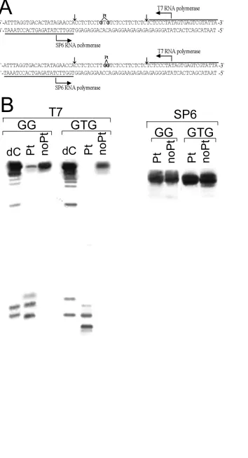
Single-lesion Substrates for DNA Transcription by T7 and SP6 RNA Polymerases In Vitro. The
69-bp templates nonmodified or containing a central, single 1,3-GTG intrastrand CL of photoactivated 1
or 1,2-GG intrastrand CL of cisplatin were assembled from four oligodeoxyribonucleotide strands as
illustrated in Figure 9A. The central oligonucleotide [23 nucleotides (nt)] containing GTG sequence was
ACS Paragon Plus Environment
10 mixed with 1 in their equimolar concentrations, irradiated by UVA for 30 min and subsequently
incubated in the dark for 24 h. The platinated oligonucleotide was purified by using ionic-exchange
HPLC. A product was collected, dialysed against double-distilled water and the unique platinations at
the guanine sites were verified by FAAS and DMS footprinting of platinum bound to DNA, as already
described (33). Cisplatin-modified oligonucleotide containing central GG sequence was prepared by the
same procedure except for the omission of the irradiation step. The central oligonucletides were than
annealed with their bottom strands (69-mers) and two oligonucleotides (two marginal arms, each 23 nt
long) were ligated (one to each side) to these duplexes by T4 DNA ligase. Full-length substrates
(nonmodified, containing the 1,3-GTG intrastrand CL of photoactivated 1 or the 1,2-GG intrastrand CL
of cisplatin) were separated from unligated products on a denaturing 12 % PAA gel, purified by
electroelution, reannealed, and stored in NaClO4 (0.01M).
T7 and SP6 RNA Polymerases Reaction on Single-lesion Templates. T7 and SP6 polymerization
reactions were performed using Riboprobe® In Vitro Transcription Systems Protocol (Promega,
Mannheim, Germany) according the recommended protocol. Briefly, unplatinated or
single-lesion-containing 69 bp templates (2 pmol) were incubated at 37 °C in 20 µL of buffer containingTris-HCl (40
mM, pH 7.9), NaCl (10 mM), MgCl2 (6 mM), DTT (10 mM), spermidine (2 mM), Tween®-20 (0.05%),
0.5 mM each of rATP, rCTP and rUTP, rGTP (0.125 mM) and 0.5µCi [α-32P]rGTP. The reactions were
initiated by the addition of 15 units of T7 polymerase. After 1 h of incubation, reaction mixtures were
precipitated by ethanol and resolved by electrophoresis on 12% PAA/8M urea gel. RNA sequencing
lanes were generated by adding of 3´-dCTP or 3´-dATP into the reactions containing nonplatinated
69-bp constructs as a template. The reaction with SP6 polymerase was performed as described for T7
polymerase (vide supra) except that the concentration of rGTP was 0.5 mM, rUTP 0.125 mM and 0.5
µCi [α-32P]rUTP was used.
Other Physical Methods. Absorption spectra were obtained on a Beckman 7400 DU
spectrophotometer equipped with a thermostated cell holder. The FAAS measurements were carried out
on a Varian AA240Z Zeeman atomic absorption spectrometer equipped with a GTA 120 graphite tube
ACS Paragon Plus Environment
11 atomizer. The gels were visualized on a BAS 2500 FUJIFILM bioimaging analyzer, and the
radioactivity associated with bands was quantified with the AIDA image analyzer software (Raytest,
Germany).
ACS Paragon Plus Environment
12 RESULTS
DNA-bound Platinum in Cells Exposed to Photoactivated 1. Distortions of DNA structure by
metallodrugs often correlate with anticancer activity (34). Hence, it is of great importance to understand
in detail the DNA binding properties of the photoactivatable PtIV complex 1 and the possible
relationship with cytotoxicity in tumor cell lines.
We examined platinated DNA isolated from ovarian carcinoma A2780 cells after exposure to 1 under
irradiation conditions (UVA) or in the dark. Importantly, 1 was potently phototoxic towards a number of
human tumor cell lines including ovarian A2780 cancer cells (3). After the treatment was completed
(see Experimental Procedures), DNA was isolated and the Pt content was determined by FAAS.
Measurements of DNA-bound platinum after 1 h of 24 µM drug exposure under irradiation conditions
(365 nm,4.3 mW cm-2) revealed that the amount of platination by 1 was 700±84fmol Pt/µg DNA. This
amount was approximately 16-fold higher compared to that determined after 1 h treatment with the same
dose of cisplatin in dark (43±8 fmol Pt/µg DNA). Importantly, no Pt bound to DNA was observed if the
cells were treated with 1 in the dark. These results confirm that the binding of 1 to DNA in tumor cells
correlates with the conditions under which it exhibits cytotoxicity – binding to DNA and cytotoxicity
with irradiation, and no binding to DNA or cytotoxicity in the absence of irradiation.
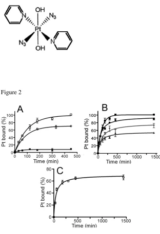
DNA Binding of Photoactivated 1 in Cell-free Media. Kinetics of Binding to Calf Thymus DNA.
The first experiments were aimed at quantifying the binding of 1 to mammalian DNA in cell-free media.
Two samples of double-helical CT DNA (32 µg mL-1) were incubated with 1 at an ri value of0.05 in
NaClO4 (10 mM) at 37 °C. The first sample was irradiated with UVA (365 nm, 4.3 mW cm-2) or visible
(458 nm, 65 mW cm-2) light immediately after addition of 1; the other sample was kept in the dark.
Aliquots of both samples were withdrawn at various time intervals; free, unbound platinum was
removed by gel filtration through Sephadex G-50 coarse column and DNA was assayed for platinum
content by FAAS. Only a very small amount (<5%) of platinum bound to DNA was found in the sample
which was kept in the dark, even after a long time of incubation (24 h; Figure 2A). In contrast, the
ACS Paragon Plus Environment
13 amount of platinum bound to DNA in irradiated samples increased with time. Under continuous UVA
irradiation, 50 % of Pt was bound after 70 min (t50%) and the plateau of the platination was reached after
7.5 h continuous irradiation, when nearly 100% of platinum present in the sample was bound to DNA
(Figure 2A). Similarly, 50 % of Pt was bound after 106 min (t50%) and the plateau of the platination
reaction was reached after 7.5 h continuous irradiation by visible light, when nearly 70% of platinum
present in the sample was bound to DNA (Figure 2C). Interestingly, when 1 was first pre-irradiated by
UVA for 2 h (in the absence of DNA) and only then added to DNA in the dark, the DNA binding rate
was markedly slower and a maximum amount of platinum bound to DNA after 7.5 h was lower as well
compared to the situation when 1 was continuously irradiated in the presence of DNA (Figure 2A).
Unfortunately, extended periods of irradiation in particular by UVA (7.5 h) caused severe DNA
damage which made subsequent analysis of conformational properties of DNA modified by
photoactivated 1 impossible. Therefore, other modes of irradiation were also tested (shown for UVA
irradiation in Figure 2B). DNA was mixed with 1 at ri = 0.05, samples were pre-irradiated for a
preselected time (0.5, 1 or 2 h) and subsequently incubated in the dark. Aliquots were withdrawn and
DNA was assayed for platinum content as described above. Figure 2B shows that the total amount of
platinum bound to DNA after 24 h of post-irradiation incubation increases with the length of the
pre-irradiation (in the presence of DNA). As mentioned above, when 1 was first irradiated for 2 h in the
absence of DNA, then added to DNA and the sample was subsequently incubated in the dark, the
amount of platinum bound to DNA was considerably lower than that when 1 was irradiated for 2 h in the
presence of DNA and the sample was subsequently incubated in the dark (cf. Figures 2A and B).
Qualitatively similar results were obtained if 1 or the mixtures of DNA with 1 were pre-irradiated with
visible light, although yields of DNA binding reactions were lower compared to those when the
mixtures were pre-irradiated by UVA.
Thus, these DNA binding experiments (Figure 2) clearly indicated that 1 was inactive and unable to
bind DNA in the dark, but under irradiation by UVA or visible light it became active and strongly bound
DNA.
ACS Paragon Plus Environment
14 The results of these DNA binding studies in cell-free media indicate that the rates of binding to
natural double-helical DNA, when the reaction mixture was irradiated by UVA or visible light, were
relatively high compared to the binding of non-irradiated conventional cisplatin or transplatin (35). The
binding experiments carried out here also indicate that the modification reactions result in the
irreversible coordination of photoactivated 1 to double-helical DNA, which also facilitates sample
analysis. Hence, it is possible to prepare samples of DNA modified by 1 photoactivated by UVA or
visible light at a preselected value of rb (the number of molecules of the platinum complex bound per
nucleotide residue). The samples of DNA treated with photoactivated 1 and analyzed further by
biophysical or biochemical methods were prepared in NaClO4 (10 mM) at 37 °C. If not stated
otherwise, after 1h of the treatment of DNA with the complex under continuous irradiation by UVA or
visible light, the samples were incubated in the dark for an additional 23 h. After that, the samples were
precipitated in ethanol, dissolved in the medium necessary for a particular analysis and the rb value in an
aliquot of this sample was checked by FAAS. In this way, the analyses described in the present paper
were performed in the absence of unbound (free) platinum complex if not stated otherwise.
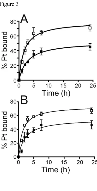
Characterization of DNA Adducts of Photoactivated 1 with Thiourea.Cisplatin, transplatin, and
analogues of these bifunctional platinum compounds coordinate to DNA in a two-step process, forming
first monofunctional adducts preferentially at guanine residues; these monofunctional adducts
subsequently close to bifunctional lesions (35, 36). Considerable evidence suggests that
monofunctionally-bound transplatin and its analogs are labilized by thiourea, whereas bifunctional
adducts are resistant (37, 38). TU is used to labilize monofunctionally bound transplatin and its
analogues from DNA (37). The displacement of transplatin or its analogues is initiated by coordination
of TU trans to the nucleobase. Owing to the strong trans effect of sulfur, the nucleobase nitrogen–
platinum bond is weakened and thus becomes susceptible to further substitution reactions.
Consequently, transplatin or its analogues in monofunctional DNA adducts are effectively removed,
whereas bifunctional adducts of transplatin or its analogues are resistant to TU treatment (37).
ACS Paragon Plus Environment
15 The experiments aimed at the characterization of DNA adducts of photoactivated 1 were conducted
employing TU as a probe for DNA monofunctional adducts formed by this trans-platinum compound.
Double-stranded CT DNA was mixed with 1 at a drug-to-nucleotide ratio of ri =0.05 in NaClO4 (10
mM), irradiated for 1 h by UVA or visible light and then incubated in the dark for an additional 23 h.
During this period the platination reaction was stopped at various time intervals by addition of NaCl
(final concentration 0.1 M) andimmediate cooling to -20 °C or, in parallel experiments, by addition of
TU (final concentration was 10 mM). The samples treated with TU were still incubated for an additional
10 min at 25 °C and then quickly cooled to -20 °C. The DNA samples were then filtered using Sephadex
G50 coarse columns to remove low molecular mass fractions and the platinum content was determined
by FAAS (Figure 3). The reaction of DNA with 1 photoactivated by UVA reached ∼70% after 24 h,
consistent with the results of DNA binding experiments (Figure 2B). TU displaced approximately 50%
of already bound photoactivated complex 1 from DNA at early time intervals (1–5 h; Figure 3A). At
longer incubation times (24 h), TU was slightly less efficient in removing 1 from DNA (∼37% of total Pt
adducts), which suggests that a considerable fraction of monofunctional adducts of 1 photoactivated by
UVA had closed to bifunctional lesions already during the early time of incubation. Thus, after a
reaction period of 24 h, 63% of 1 photoactivated by UVA bound to DNA had evolved to bifunctional
lesions and therefore, was not displaced from double-stranded DNA by TU (Figure 3A), which implies
that approximately 37% of total adducts remained monofunctional. A similar experiment was performed
using visible light to irradiate the mixtures of CT DNA and 1 (Figure 3B). Under these conditions,
similar results were obtained to those found for the mixtures irradiated by UVA; after 24 h of
incubation, TU displaced 31% of total platinum adducts from DNA which implies that 69% of Pt adduct
formed on DNA by 1 photoactivated by visible light had evolved into CLs.
DNA Interstrand (Intramolecular) Cross-linking by Photoactivated 1. Bifunctional platinum
compounds, which coordinate base residues in DNA, form various types of interstrand and intrastrand
CLs. Such CLs in the target DNA are important factors involved in the DNA damaging action of
genotoxic agents. Therefore, we have quantitated the interstrand cross-linking efficiency of
ACS Paragon Plus Environment
16 photoactivated 1 in pSP73KB plasmid (2455 bp). This plasmid DNA was linearized by EcoRI (EcoRI
cuts only once within pSP73KB plasmid) and radioactively labeled on its 3´-end.
Plasmid DNA was incubated with 1 at varying concentrations, irradiated for 1 h by UVA or visible
light and incubated in the dark for an additional 23 h. The samples were then analyzed for interstrand
CLs by agarose gel electrophoresis under denaturing conditions (39). Upon electrophoresis, 3'-end
labeled strands of linearized pSP73KB plasmid containing no interstrand CLs migrate as a 2455-base
single strand, whereas the interstrand cross-linked strands migrate more slowly as a higher molecular
mass species (Figure 4). The intensity of the more slowly migrating DNA fraction increased with the
growing level of the platination. The radioactivity associated with the individual bands in each lane was
measured to obtain estimates of the fraction of noncross-linked or cross-linked DNA under each
condition. The frequency of interstrand CLs of photoactivated 1 was calculated as described earlier (38)
and was found to be 12 ± 2% (mean and standard deviation calculated from three independent
experiments) for DNA modified by 1 photoactivated by both UVA and visible light.
Unwinding of Negatively Supercoiled DNA by Adducts of Photoactivated 1. The binding of
low-molecular-mass compounds such as antitumor metallodrugs to closed circular DNA can cause
deformation and shift of the base pairs, which can lead to partial unfolding of the DNA. This process
lowers the superhelical density of plasmid DNA, which causes a decrease in the rate of migration
through an agarose gel. This fact makes it possible to observe and quantify the mean value of unwinding
per adduct. In the present study, we investigated the unwinding induced in negatively supercoiled
pUC19 plasmid treated with 1 photoactivated by UVA or visible light. The degree of supercoiling was
monitored using electrophoresis in native agarose gel. We investigated the effect of increasing amounts
of 1 photoactivated by UVA or visible light bound to a mixture of relaxed and supercoiled pUC19 DNA
on migration of these forms of pUC19 DNA in a native agarose gel (shown in Figure 5 for DNA
modified by 1 photoactivated by UVA radiation). The unwinding angle is given by Φ = -18σ/rb(c),
where σ is the superhelical density and rb(c) is the value of rb at which the supercoiled and nicked forms
comigrate (25). The DNA unwinding angle determined for DNA modified by 1 photoactivated by UVA
ACS Paragon Plus Environment
17 was 28±3°. An identical experiment carried out with plasmid treated with 1 photoactivated by visible
light (not shown) yielded an unwinding angle 27±3°.
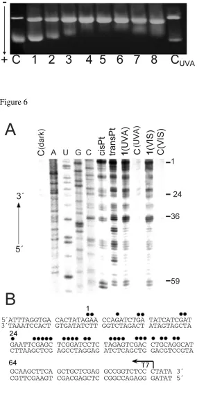
Transcription Mapping of DNA Adducts of Photoactivated 1.Cutting of pSP73KB DNA by NdeI
and HpaI restriction endonucleases yielded a 212-bp fragment (a substantial part of its nucleotide
sequence is shown in Figure 6B). This fragment contained T7 RNA polymerase promotor [in the upper
strand close to its 3'-end (Figure 6B)]. The first experiments were carried out using this linear DNA
fragment, randomly modified by transplatin or cisplatin in the dark and by 1 photoactivated by UVA or
visible light at rb = 0.01, for RNA synthesis by T7 RNA polymerase (Figure 6A, lanes transPt, cisPt,
1(UVA) and 1(VIS) respectively). RNA synthesis on the template modified by the platinum complexes
yielded fragments of defined sizes (Figure 6A), which indicates that RNA synthesis on these templates
was prematurely terminated. The sequence analysis revealed that the major bands resulting from
termination of RNA synthesis by the adducts of transplatin and photoactivated 1 were similar, appearing
mainly at G and C sites and to a considerably less extent also at adenine (A) sites (Figure 6B).
Importantly, the sequence dependence of the inhibition of RNA synthesis by the adducts of transplatin
and photoactivated 1 is considerably less regular than that by the adducts of cisplatin, indicating that 1
photoactivated by both UVA and visible light forms a greater variety of adducts with DNA and less
regularly than cisplatin does.
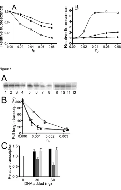
Characterization of DNA Adducts of Photoactivated 1 by EtBr Fluorescence. EtBr as a
fluorescent probe has been used to characterize perturbations induced in DNA by bifunctional adducts of
several platinum compounds (13, 40). Double-helical CT DNA was first modified by cisplatin,
transplatin, or 1 photoactivated by UVA or visible light. The levels of the modification corresponded to
the values of rb in the range between 0 and 0.08. Modification of DNA by all platinum complexes
resulted in a decrease of EtBr fluorescence (shown in Figure 7A for DNA modified by 1 photoactivated
by UVA). The decrease caused by the adducts of photoactivated 1 was markedly more pronounced than
that induced by the DNA adducts of cisplatin or transplatin at equivalent rb values. Identical experiment
carried out with CT DNA treated with 1 photoactivated by visible light (not shown) yielded identical
ACS Paragon Plus Environment
18 results. It was verified that irradiation of CT DNA by UVA or visible light in the absence of 1 for 5 h
had no effect on EtBr fluorescence.
Characterization of DNA Adducts of Photoactivated 1 by Terbium Fluorescence. Terbium ion
(Tb3+) fluorescence is used to investigate local perturbations of conformation induced in double-helical
DNA by various physical or chemical agents, including cisplatin (31, 32). This assay is based on the
observation that Tb3+ fluorescence is strongly enhanced when the ion is bound to the phosphate moieties
of G residues in distorted DNA regions (30-32). The modification of double-helical DNA by cisplatin
has been shown to result in substantially increased fluorescence of the lanthanide cation, which binds to
unplatinated G residues in distorted regions around the platination site (31, 32). In contrast, coordination
of complexes such as clinically ineffective transplatin or monofunctional [PtCl(dien)]Cl to
double-stranded DNA does not result in distortions of the helix structure that would affect terbium fluorescence
(31, 32).
CT DNA was modified by cisplatin, transplatin, or 1 photoactivated by UVA or visible light at rb
values in the range of 0.02 - 0.08, the resulting samples were treated with TbCl3 and the fluorescence
measured as described in the section Materials and Methods ((shown in Figure 7B for DNA modified by
1 photoactivated by UVA radiation)). In accord with previous results (31, 32), the modification by
cisplatin resulted in a significant enhancement of terbium fluorescence, while the modifications by
transplatin had only a negligible effect. The modification by photoactivated 1 also resulted in an
enhancement of the fluorescence, which was, however, markedly higher than that observed in the case
of DNA modified by cisplatin (shown for Figure 7B). These results confirm that DNA adducts of
photoactivated 1 induce local conformational alterations in double-stranded DNA that are markedly
more pronounced than those induced by cisplatin. This observation is in contrast to the modification of
DNA by transplatin, which does not induce in DNA conformational distortion detectable by terbium
fluorescence assay. Identical results were obtained when DNA was modified by 1 photoactivated by
visible light. It was verified that irradiation of CT DNA by UVA or visible light in the absence of 1 for 5
h had no effect on terbium fluorescence.
ACS Paragon Plus Environment
19
Transcription of DNA Modified by Photoactivated 1 by RNA Polymerase II In Vitro.One of the
key factors that is important for platinum drug-mediated cytotoxicity is the arrest of RNA synthesis by
Pt-DNA CLs (21). Therefore, the ability of 1 to affect transcription activity of human RNA polymerase
II (RNA pol II) was tested using HeLaScribe1 Nuclear Extract in vitro Transcription system kit. Using
an analogous procedure as described earlier (41), the RNA pol II transcription template pCMV-Gluc
either nonmodified or modified by 1 photoactivated by UVA or visible light was incubated with the
HeLa nuclear extract supplied with this kit. This extract can support accurate transcription initiation by
RNA pol II and exhibits both basal and regulated patterns of RNA transcription. This nuclear extract is
also the source for a variety of transcription factors, DNA binding proteins and the enzymatic machinery
involved in process of RNA synthesis (42). Specific transcription from the CMV promoter results in a
runoff transcript 688 nucleotides in length. The generated full length transcripts can be subsequently
detected by gel electrophoresis.
As seen in Figure 8A, in the absence of platination, a high level of full length transcript was
observed. In contrast, a significant decrease in the amount of full-length transcript was observed as a
result of increasing template modification by 1 photoactivated by both UVA and visible light (Figure
9A). The relative amount of full-length transcript generated from each reaction was quantitated and
plotted as a function of the level of the template platination (rb) (Figure 9B). The results obtained for
DNA modified by cisplatin in the dark under identical conditions were also included for comparative
purposes. Under the conditions employed, RNA pol II transcription was highly sensitive to even very
low levels of platinum damage on the template DNA, the damage by photoactivated 1 being more
effective in inhibiting RNA pol II transcription compared to cisplatin.
To investigate the possibility that RNA pol II catalytic activity was inhibited as a consequence of
hijacking factors essential for RNA pol II initiation by DNA adducts of photoactivated 1 (41), the
following competition experiments were performed. RNA pol II transcription of undamaged template
pCMV-Gluc was examined in the presence of increasing levels of a second, exogenous pUC19 plasmid
containing multiple lesions caused by either photoactivated 1 or cisplatin. As shown in Figure 8C, the
ACS Paragon Plus Environment
20 initial addition of control, undamaged exogenous plasmid resulted in an overall increase in the amount
of transcript generated by pCMV-Gluc which only slightly increased upon the further addition of
unplatinated plasmid. RNA pol II transcription of pCMV-Gluc template was significantly reduced by the
addition of increasing amounts of cisplatin modified exogenous plasmid. In contrast, a negligible
inhibition effect was seen if the transcription assay was performed in the presence of exogenous plasmid
containing adducts of 1 photoactivated by UV or visible light (shown in Figure 8C for DNA modified by
1 photoactivated by UVA).
Effect of the Single, Site-specific DNA Lesion of Photoactivated 1 on RNA Synthesis. The
previous results have shown that enhanced effects of photoactivated 1 on the inhibition of RNA
polymerization does not result from hijacking of transcription factors that are indispensable for the
transcription process. It is possible that platinum lesions formed by photoactivated 1 represent a stronger
block for RNA polymerase than cisplatin adducts do. Therefore, to investigate the effect of a single
lesion of 1 on the synthesis of RNA by RNA polymerase, 69 bp-long deoxyribo-oligonucleotide
duplexes were constructed so that they contained a single lesion of photoactivated 1 or cisplatin in the
top strand, approximately 20 or 16 bp downstream of the start site for T7 or SP6 polymerases,
respectively (Figure 9A). The constructs used in these studies contained either the 1,2-GG intrastrand
cross-link of cisplatin or the 1,3-GTG intrastrand CL of photoactivated 1, which is the major adduct
formed in DNA by this photoactivated complex (vide supra). The presence of the drug lesions and the
purity of the transcription templates were confirmed by gel electrophoresis, FAAS and DMS
footprinting (33).
Figure 9B shows that the GTG construct control template is efficiently transcribed by T7 RNA
polymerase to yield high levels of full length 54 nt transcript. In contrast, transcription of the template
containing a single 1,3-GTG intrastrand CL by T7 RNA polymerase (which transcribed the top,
platinated strand) resulted in a high level of shorter length transcripts arising from blockage of the T7
RNA polymerase by this lesion. Importantly, even after 1 h of reaction, no full-length transcript was
observed. Sequence analysis revealed that the T7 RNA polymerase was blocked at sites both one base
ACS Paragon Plus Environment
21 prior to the adduct site and at the first platinated guanine (Figure 9B). Transcription by T7 RNA
polymerase from the unplatinated GG construct templates yielded similar results to those of the
unplatinated GTG template (Figure 9B). The 1,2-GG intrastrand CL of cisplatin was also a block to the
progression to the T7 RNA polymerase, displaying a blockage pattern different from that of the GTG
adduct of photoactivated 1. T7 RNA polymerase was blocked mainly at the sites of the adducts (one half
prior to the first and second platinated guanine). In this case, the full length product was also observed,
although the intensity of this fraction was markedly reduced compared to the unmodified template. From
the intensity of the T7 RNA polymerase blockage sites relative to the intensity of the full-length
transcript, it can be calculated (taking into account the relative number of radiolabeled nucleotides
incorporated into the RNA) that there were 73% and 100% blockages by the major adducts formed by
cisplatin and photoactivated 1, respectively.
The effect of the GTG-adduct formed by photoactivated 1 (in the top strand) on transcription of the
bottom, unplatinated strand was also tested using the same constructs. SP6 RNA polymerase was
employed which transcribed the bottom, unplatinated strand of the constructs (Figure 9B, right panel).
The presence of adducts of both cisplatin and photoactivated 1 in the top strand of the construct had no
effect on the ability of SP6 RNA polymerase to transcribe the bottom, unplatinated strand and synthesize
full length products.
ACS Paragon Plus Environment
22 DISCUSSION
The cytotoxic effects of antitumor platinum drugs might arise from a number of mechanisms,
including tumor cell accumulation, protein interacions, DNA modifications and their cellular processing
(34, 43, 44). To support the view that DNA is a potential target for photoactivated 1, platinum levels on
nuclear DNA were determined after exposure of A2780 tumor cells to 1 photoactivated by UVA.
Measurements of DNA-bound platinum after exposure to the photoactivatable platinum drug under
irradiation revealed that the amount of platination by 1 was markedly (~16-fold) higher compared to that
determined after the treatment with the same dose of cisplatin in dark for the same time. Importantly, no
Pt bound to DNA was observed if the cells were treated with 1 in the dark. These results confirm that 1
when photoactivated accumulates in tumor cells, penetrates the nucleus and binds strongly to nuclear
DNA under conditions in which it exhibits cytotoxicity. This suggests that penetrating the nucleus and
binding to nuclear DNA may provide an important contribution to the mechanism of cytotoxicity of
photoactivated 1. DNA may therefore be a potential target for this cytotoxic photoactivatable platinum
complex, although we cannot rule out the possibility that nuclear DNA may not be the only target. In
other words, toxic effects of 1 in tumor cells may be associated with processes at the DNA level.
The finding that the photoactivated 1 is capable of delivering platinum to DNA in the cell nucleus
prompted us to examine the binding of 1 to DNA in a cell-free medium. The resulting DNA damage
triggers downstream effects including inhibition of replication and transcription, cell cycle arrest, and
apoptosis or necrosis (22, 45, 46). The results of these studies were compared with those previously
performed with conventional cisplatin and transplatin.
The CT DNA binding experiments carried out in the present work in a cell-free medium indicated
that modification reactions resulting in the irreversible coordination of photoactivated 1 were faster than
those of cisplatin or transplatin (Table 1). Platinum binding to CT DNA resulting from treatment of
DNA with 1 preirradiated for 2 h in the absence of DNA was significantly lower than that resulting from
treatment of DNA when 1 was preirradiated for 2 h in the presence of DNA (cf. Figures 2A and 2B).
ACS Paragon Plus Environment
23 This observation suggests that free 1 can be transformed into highly reactive species that can be trapped
by DNA if it is present in the same solution, whereas photoproducts formed in the absence of DNA
become less reactive towards it.
The transcription mapping experiments (Figure 6) indicate that 1 binds to DNA under irradiation
conditions at sites similar to those of transplatin, i.e., less regularly than cisplatin and mainly at single
guanines and cytosines (i.e., at the preferential DNA binding sites of transplatin and its antitumor
analogues (26, 38, 47)). Considerable evidence suggests that the antitumor efficacy of bifunctional
platinum compounds is the result of the formation of various types of inter- and intrastrand CLs;
however, their relative efficacy remains unknown. The results of this work are consistent with the view
that photoactivated 1 forms on DNA ca. only 12% interstrand (intramolecular) CLs (Figure 4, Table 1)
and ca. 37% of DNA adducts remain monofunctional even after 24 h (Figure 3, Table 1). The remaining
lesions are intrastrand adducts, presumably 1,3-CLs. Thus, it is reasonable to suggest that several aspects
of DNA binding mode of photoactivated 1 are similar to those of conventional transplatin (in the dark)
(Table 1). On the other hand, it cannot be excluded that identical types of DNA adducts of
photoactivated 1 and transplatin can distort DNA conformation differently and can be processed by
cellular components differently.
EtBr as a fluorescent probe can be used to characterize DNA binding of small molecules such as
platinum antitumor drugs (38, 48). The fluorescence of EtBr is markedly enhanced as a consequence of its
intercalation into DNA, but binding of EtBr to DNA by intercalation is blocked in a stoichiometric manner
by a wide spectrum of DNA-binding platinum drugs. Thus, for instance the modification of DNA by
cisplatin or transplatin results in a decrease of EtBr fluorescence intensity as compared with that for
non-platinated DNA. The molecules of photoactivated 1 bound to CT DNA sterically block approach of
molecules of EtBr to DNA and in this way hinder the intercalation of EtBr to as well, which lowers EtBr
fluorescence in comparison with the experiment in which unplatinated DNA was used (Figure 7A). The
decrease of EtBr fluorescence in case of DNA modifications by photoactivated 1 was markedly higher than
that observed in case of DNA modifications by convetional transplatin or cisplatin in the dark. Comparison
ACS Paragon Plus Environment
24 with cisplatin or transplatin suggests that the adducts of 1 photoactivated by UVA or visible light extend
over considerably more base pairs around the platination sites than in case of the adducts of transplatin or
cisplatin.
The results of experiments in which DNA modifications by photoactivated 1 were probed by EtBr
fluorescence (Figure 7A) also suggest that photoactivated 1 forms DNA adducts which cannot be
coclustered, from the viewpoint of their capability to inhibit EtBr fluorescence, with those formed by
'conventional' monofunctional platinum(II) complexes, such as cisplatin or transplatin. We also suggest
that the pyridine ligands in all, or in a significant fraction of, adducts (mono- and/or bifunctional) might
be well positioned to interact with the duplex. The extent of the observed decrease in EtBr fluorescence
indicates that the disturbance of the DNA helical structure by photoactivated 1 is not only an effect of
strong coordinative platinum binding but has to be explained by an additional interaction of the pyridine
ligand(s) of photoactivated 1 with the duplex. The suggestion that the pyridine ligands in the adducts of
photoactivated 1 interact with the duplex is further corroborated by the results of DNA unwinding
experiments.
Electrophoresis in native agarose gel was used to determine the unwinding induced in negatively
supercoiled plasmid by monitoring the degree of supercoiling (Figure 5). The unwinding angle
calculated in this way for 1 photoactivated by UVA or visible light was similar, 28 ± 3° or 27 ± 3°,
respectively. This unwinding angle is considerably greater than that found for cisplatin or transplatin
(13° and 9°, respectively, Table 1). It is reasonable to suggest that the large additional contribution to
unwinding is associated with interaction of yhe pyridine ligand(s) in photoactivated 1 with the duplex.
Adducts such as trans-[Pt(py)2(G)2] are likely to form. Thus, the large unwinding angle produced by
photoactivated 1 is good evidence that the pyridine ligand(s) substantially interacted with duplex DNA
upon coordinative binding of platinum. In other words, the unwinding angle observed for photoactivated
1 is consistent with DNA binding that involves a combined mode involving coordination of platinum to
a base residue and interaction of pyridine ligand(s) in photoactivated 1 with the duplex. DNA binding
that involves a similar combined DNA binding mode as that observed for some cationic platinum(II)
ACS Paragon Plus Environment
25 complexes that carry ethidium as a nonleaving group (ethidium is a well known DNA intercalator which
unwinds DNA by 26°) or quinoline, such as cis-[Pt(NH3)2Cl(N3/N8-ethidium)]+ or
trans-[PtCl2(NH3)(quinoline)] (13, 25). These results obtained with cationic platinum(II) complexes
revealed that the intercalating moiety needs to be cis to the Pt-N7 bond in order to interact effectively
with the DNA base stack. The analogy between the above-mentioned cationic cis-complex or
trans-[PtCl2(NH3)(quinoline)] and photoactivated 1 based on geometric considerations suggests that
DNA adducts of the latter may significantly contribute to the unwinding of supercoiled DNA.
The conclusion that the DNA binding mode of photoactivated 1 involves combined coordination of
platinum to a base residue and interaction of the pyridine ligand(s) in photoactivated 1 with the duplex
may also imply the following. Photoactivated 1 may be capable of forming DNA adducts which induce
conformational distortions in DNA which extend over more base pairs than in case of DNA adducts of
cisplatin or transplatin. The latter view that adducts of photoactivated 1 distort DNA conformation more
than adducts of cisplatin or transplatin is corroborated by the terbium fluorescence data (Figure 7B).
Enhancement of Tb3+ ion fluorescence is used to detect base residues in distorted DNA regions (
30-32). DNA modification by photoactivated 1 and by cisplatin resulted in an enhancement of the
fluorescence, whereas DNA modification by transplatin induced in DNA no conformational distortion
detectable by the terbium fluorescence assay (Figure 7B). Thus, the trend in efficiency of DNA adducts
of the platinum complexes tested in the present work to enhance Tb3+ ion fluorescence was
photoactivated 1 >> cisplatin > transplatin. These results along with those of DNA unwinding
experiments (Figure 5) support the thesis that DNA adducts of photoactivated 1 induce local
conformational alterations in double-stranded DNA that are markedly more extensive than those
induced by cisplatin or transplatin.
Studies on the early phases of molecular mechanisms underlying antitumor effects of platinum
antitumor drugs have focused on investigations of cellular responses to DNA damage by these
metallodrugs (46, 49, 50). Recently attention has been paid to the role of inhibition of transcription
elongation by RNA pol II by cisplatin adducts to further understand the mechanism of its biological
ACS Paragon Plus Environment
26 effects (20, 51-54). This important component of the mechanism underlying the antitumor effects of
platinum drugs has not been hitherto investigated in the case of photoactivatable platinum compounds.
We demonstrate in the present work for the first time (Figure 8) that RNA pol II transcription is highly
sensitive to even very low levels of modification on the template DNA by photoactivated 1. Thus,
already very low levels of DNA modification by photoactivated 1 may initiate transcription-coupled
subpathways leading to apoptosis or necrosis (55).
Importantly, adducts of photoactivated 1 are markedly more effective in inhibiting RNA pol II
transcription than those of conventional cisplatin (Figures 8B and 9). This different reduction in
transcript production may result from a different level of platination and/or conformational distortions at
the promoter, leading to promoter inactivation. The inhibition effects were observed, however, at levels
of platination by photoactivated 1 well below those expected to induce such damage [rb = 8 x 10⁻⁴, i.e.
only ~9 adducts per molecule of template DNA (5764 bp) on average]. Hence, the eventuality that the
observed different reduction in transcript production may result from a different level of platination
and/or conformational distortions at the promoter is unlikely. An alternative possibility is that platinum
lesions represent a steric blockage of RNA pol II inhibiting the prolongation of RNA transcription, or
that factors essential for RNA pol II initiation may also bind Pt-DNA adducts which do not represent
their natural binding sites. Therefore, in the presence of sufficient numbers of Pt-DNA adducts, these
factors may become limiting, resulting in an inhibition of transcription initiation due to the factors being
sequestered to platinated sites in DNA.
We examined in the present work whether some elements of the transcription complex might be
hijacked by DNA adducts of photoactivated 1 more or less than by those of cisplatin using a competition
assay (41) (Figure 8C). The initial addition of control, unplatinated exogenous pUC19 plasmid resulted
in an overall increase in the amount of transcript generated by pCMV-Gluc substrate, which increased
only slightly upon the further addition of unplatinated exogenous plasmid. Such an increase in
transcription efficiency has been already reported (41) and was attributed to an increase in
macromolecular crowding induced by the presence of higher amounts of DNA (41, 56). We observed in
ACS Paragon Plus Environment
27 the present work (Figure 8C), in accord with the previously published results (40, 41), that RNA pol II
transcription of the pCMV-Gluc template was markedly reduced by the addition of increasing amounts
of cisplatin-modified exogenous plasmid (Figure 8C). Thus, these results are consistent with and support
the 'hijacking' hypothesis raised earlier for mononuclear PtII drugs (41, 44, 57, 58) that RNA pol II
transcription of template modified by cisplatin is inhibited to a substantial extent due to the transfer of
some elements of the transcription complex away from their normal binding sites and in this way
interfer with transcription.
A markedly lower inhibition effect was seen if the transcription assay was performed in the presence
of exogenous plasmid modified by photoactivated 1 (Figure 8C). Thus, the stronger inhibition of
transcription by DNA adducts of photoactivated 1 appears to be due to pronouncedly more extensive
steric blockade of RNA pol II inhibiting the prolongation of RNA transcript and not due to sequestration
of elements of the transcription complex to platinated sites in DNA. Thus, this study has revealed that
the cause of the stalling of RNA pol II by DNA adducts of photoactivated 1 and cisplatin during
transcription is apparently distinctly different providing a basis for a different mechanism of action for
photoactivated 1 and cisplatin.
The observation in the present work that transcription elongation by RNA pol II was inhibited by
titrating some elements of transcription complex away from their normal binding sites much more by
DNA adducts of cisplatin than by those of photoactivated 1 (Figure 8C) deserves further discussion.
Many transcription factors are HMG-domain proteins (HMG = high mobility group). These proteins
have been shown to recognize and bind with a strong affinity to structural motifs in DNA that involve
directional rigid bends of the longitudinal axis of this double-helical nucleic acid. Major DNA adducts
of cisplatin (intrastrand CLs between neighboring purine residues which represent ~90% of all DNA
adducts of cisplatin (16)) produce in DNA a stable directional curvature (~40° toward major groove of
DNA) (44, 59). Therefore, this bending apparently represents an important structural motif responsible
for a high affinity of many transcription factors to DNA modified by cisplatin. In contrast, DNA adducts
of photoactivated 1 (presumably similar to those formed by conventional transplatin, i.e. mainly
1,3-ACS Paragon Plus Environment
28 intrastrand CLs, monofunctional adducts and to a much lesser extent interstrand CLs, Table 1) rather
produce in DNA flexible and relatively small nondirectional bends so that these adducts are not
recognized by HMG-domain proteins (60-62). Hence, DNA adducts of photoactivated 1 may lack, in
contrast to DNA adducts of cisplatin, a high-affinity structural motif which would attract transcription
factors.
In conclusion, the PtIV diazido complex trans,trans,trans-[Pt(N3)2(OH)2(pyridine)2] (1) is unreactive
in the dark but a potent anticancer drug when photoactivated, not only by UVA but also by visible light.
Complex 1 when photoactivated accumulates in tumor cells, penetrates the nucleus and binds strongly to
nuclear DNA under conditions in which it exhibits cytotoxicity. The nature of the DNA adducts,
including conformational alterations, induced by photoactivated 1 are distinctly different from those
produced in DNA by conventional cisplatin or transplatin. In addition, another critical difference
between photoactivated 1 and cisplatin emerged with respect to the ability of their DNA adducts to
inhibit transcription of DNA by stalling RNA pol II. Photoactivated 1 proves to be a significantly more
potent inhibitor of RNA synthesis than cisplatin (Figures 8 and 9, Table 1). Inhibition of DNA
transcription is considered to be a major mediator of the cell kill effect of cisplatin (20, 51, 63). The
observation that major adducts of photoactivated 1 are able to efficiently stall RNA pol II suggests that
transcription inhibition may contribute to the cytotoxicity levels observed for photoactivated 1 (3).
Hence, DNA adducts of 1 could trigger a number of downstream cellular effects different from those
triggered in cancer cells by DNA adducts of cisplatin. This might lead to the therapeutic effects that
could radically improve chemotherapy by platinum complexes. These findings do not rule out the
possibility that other photodecomposition products also contribute to the cytotoxicity of complex 1.
These might include for example the release of azide, reactions of azido radicals or nitrenes with
biomolecules. Such possibilities together with the present findings may explain the different
pharmacological effects of photoactivated 1 and conventional cisplatin and thereby provide new insights
into mechanisms associated with the antitumor effects of platinum complexes photoactivated by UVA
and/or visible light (3).
ACS Paragon Plus Environment
29 AUTHOR INFORMATION
Corresponding Author
*Tel: +420 541517174. Fax: +420-541240499. E-mail: [email protected].
Funding Sources
This work was supported by the Czech Science Foundation (Grants P301/10/0598 and 301/09/H004).
Research of JP was also supported by the student project of the Palacky University Olomouc (Grant PrF
2012 026). NF and PJS thank the EPSRC (EP/G006792/1) and the ERC (award no 247450) for funding.
ACKNOWLEDGEMENT
We thank members of EC COST Action D39 for stiumulating discussions.
ABBREVIATIONS
bp, base pair; cisplatin, cis-Pt(NH3)2Cl2; CL, cross-link; CT, calf-thymus; DTT, dithiothreitol ; EtBr,
ethidium bromide; FAAS, flameless atomic absorption spectroscopy; PAA, polyacrylamide; PDT,
photodynamic therapy; [PtCl(dien)]Cl, chloridodiethylenetriamineplatinum(II) chloride; rb, the number
of molecules of the platinum complex coordinatively bound per nucleotide residue; ri, the molar ratio of
free platinum complex to nucleotide phosphates at the onset of incubation with DNA; t50%, the time at
which the binding reached 50%; transplatin, trans-Pt(NH3)2Cl2; TU, thiourea.
ACS Paragon Plus Environment