A Thesis Submitted for the Degree of PhD at the University of Warwick
Permanent WRAP URL:
http://wrap.warwick.ac.uk/97302
Copyright and reuse:
This thesis is made available online and is protected by original copyright. Please scroll down to view the document itself.
Please refer to the repository record for this item for information to help you to cite it. Our policy information is available from the repository home page.
Post-Modification Strategies for Polymeric
Cyclic Peptide Nanotubes
Tammie Rose Barlow
A thesis submitted in partial fulfilment of the requirements for the degree of
Doctor of Philosophy in Chemistry
Department of Chemistry University of Warwick
The more you know, the more you know you don't know.
- Aristotle
It is finished.
Table of Contents ... i
List of Figures ... vii
List of Tables ... xv
List of Schemes ... xvii
Abbreviations ... xix
Acknowledgements ... xxii
Declaration ... xxv
List of Publications ... xxvi
Abstract ... xxvii
1 Introduction ... 1
1.1 Self-assembling Cyclic Peptides ... 3
1.1.1 Self-Assembly ... 3
1.1.2 Cyclic peptides comprising non-natural amino acids ... 7
1.1.3 External properties of the nanotubes ... 9
1.1.4 Controlling Assembly and Orientation ... 10
1.2 Cyclic Peptide-Polymer Conjugates ... 11
1.2.1 Complex Architectures and Functionality ... 15
1.3 Motivation for this work ... 18
1.4 References ... 19
2 RAFT Polymerisation and Post-modification of poly(bromo ethyl acrylate) .. 25
2.2 Results and Discussion ... 28
2.2.1 RAFT Homopolymerisation ... 28
2.2.2 RAFT block copolymerisation ... 30
2.2.3 Post-polymerisation Modification ... 33
2.3 Conclusion ... 38
2.4 Experimental ... 39
2.4.1 Materials ... 39
2.4.2 Instrumentation ... 40
2.4.3 (4-cyano pentanoic acid)yl ethyl trithiocarbonate (CPAETC) ... 40
2.4.4 Synthesis of 2-bromoethyl acrylate (BEA) ... 41
2.4.5 RAFT polymerisation of BEA ... 42
2.4.6 RAFT block copolymerisation of p(BEA)-b-p(BA) ... 43
2.4.7 RAFT polymerisation of BA ... 44
2.4.8 RAFT block copolymerisation of p(BA)-b-p(BEA) ... 45
2.4.9 Post-polymerisation substitutions of homopolymers ... 46
2.4.9.1 Substitution with Trimethyl amine ... 46
2.4.9.2 Substitution with Trimethyl phosphine ... 47
2.4.9.3 Substitution with Tetraethyl ammonium sulfite ... 48
2.4.9.4 Substitution with Sodium azide ... 49
2.4.9.5 Substitution with Thiophenol ... 50
2.4.9.6 Substitution with 1-β -D-Thioglucose ... 51
2.4.10 Post-polymerisation substitutions of block-copolymers ... 53
2.4.10.1 Substitution with Trimethyl amine ... 53
2.5 References ... 55
3 An Orthogonal Reaction Sequence for the Synthesis of Sugar-Coated Cyclic Peptide Nanotubes ... 59
3.1 Introduction ... 60
3.2 Results and Discussion ... 62
3.2.1 Synthetic Strategy ... 62
3.2.2 Post-polymerisation modification ... 67
3.2.3 Self-Assembly properties of CP-polymer conjugates ... 69
3.2.4 Assembly of pBEA-conjugates ... 70
3.2.5 Assembly of Sugar-Conjugates ... 73
3.2.6 Lectin binding studies ... 76
3.3 Conclusion ... 78
3.4 Experimental ... 79
3.4.1 Materials ... 79
3.4.2 Instrumentation ... 79
3.4.3 Synthesis of Linear Peptide ... 80
3.4.4 Cyclisation of Linear Peptide ... 82
3.4.5 Deprotection of Cyclic Peptide ... 83
3.4.6 Synthesis of N-hydroxysuccinimide-(4-cyano pentanoic acid)yl ethyl trithiocarbonate (NHS-CPAETC) ... 83
3.4.7 RAFT Polymerisation of BEA ... 84
3.4.8 Conjugation of Polymer to Cyclic Peptide ... 85
3.4.9 Substitution with 1-β-D-Thioglucose ... 85
3.4.11 Small Angle Neutron Scattering ... 86
3.4.12 SANS Sample Preparation ... 87
3.4.13 SANS Analysis ... 87
3.4.14 Static Light Scattering ... 87
3.4.15 SLS Sample Preparation ... 89
3.5 References ... 89
4 The Effect of Ionic Strength on the Self-Assembly of Polyelectrolyte-Cyclic Peptide Conjugates ... 93
4.1 Introduction ... 94
4.2 Results and Discussion ... 95
4.2.1 Synthetic Strategy ... 95
4.2.2 SANS Analysis of Polymer-Conjugates ... 97
4.2.3 Guinier-Porod analysis ... 97
4.2.4 Polyelectrolyte conjugates in D2O ... 99
4.2.5 The Effect of Salt on Self Assembly ... 100
4.2.6 Analysis of Anionic Polymer Conjugates in Solution ... 101
4.2.7 Analysis of Cationic Polymer Conjugates in Solution ... 103
4.3 Conclusion ... 104
4.4 Experimental ... 105
4.4.1 Materials ... 105
4.4.2 Instrumentation ... 105
4.4.3 RAFT polymerisation of BEA ... 106
4.4.4 Conjugation of Polymer to Cyclic Peptide ... 106
4.4.6 Substitution with Tetraethyl ammonium sulfite ... 107
4.4.7 Small Angle Neutron Scattering ... 107
4.4.7.1 SANS Sample Preparation ... 107
4.4.8 SANS Analysis ... 108
4.4.9 Debye Length ... 108
4.5 References ... 109
5 Effect of Self-Assembly versus Charge on the Cellular Uptake of Cyclic Peptide-Polymer Nanotubes ... 111
5.1 Introduction ... 112
5.2 Results and Discussion ... 113
5.2.1 Conjugate design ... 113
5.2.2 Synthetic strategy ... 114
5.3 Conclusion ... 122
5.4 Experimental ... 123
5.4.1 Materials ... 123
5.4.2 Methods & Instruments ... 123
5.4.3 Synthesis of Linear Peptide, Cyclic Peptide and Deprotection ... 124
5.4.4 Copolymerisation of PEGA and BEA ... 125
5.4.5 Conjugation of Polymer to Cyclic Peptide ... 127
5.4.6 Synthesis of 3',6'-Dihydroxy-N-(2-mercaptoethyl)-3-oxo-3H-spiro[isobenzofuran-1,9'- xanthene]-4-carboxamide (Cystamidefluorescein) ... 129
5.4.7 Post-modification of Polymers and Conjugates ... 130
5.4.7.1 Substitution with Cystamidefluorescein ... 130
5.4.7.1.1 Substitution with Trimethyl amine ... 131
5.4.7.3 Substitution with Thioglycerol ... 134
5.4.8 Static Light Scattering ... 135
5.5 References ... 135
Conclusion, Perspectives and Outlook ... 139
Appendix to Chapter 3 ... 144
A3.1 Calculation of Conjugation Efficiency ... 144
A3.2 Deconvolution of peptide-polymer conjugations ... 145
A3.3 pBEA Conjugate SANS Analysis ... 147
A3.4 Thioglucose Conjugate SANS Analysis ... 153
A3.5 SLS Analysis ... 155
A3.6 Details used in Igor for the Cylinder + Gaussian-Coil model1 ... 157
A3.7 References ... 157
Appendix to Chapter 4 ... 158
A4.1 SANS Data Analysis ... 158
Appendix to Chapter 5 ... 166
A5.1 HPLC analyses ... 166
A5.2 SLS Data ... 171
A5.3 Cell Experiments ... 176
A5.3.1Cell Lines ... 176
A5.3.2Cytotoxicity Assay ... 176
A5.3.3Cellular Uptake Experiments ... 176
List of Figures
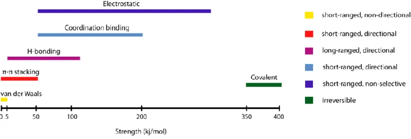
Figure 1.1: A comparison of the strength and properties of noncovalent interactions
involved in self-assembly. Adapted from Guler et al.17... 2
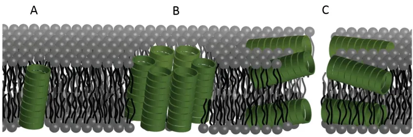
Figure 1.2: Illustration showing the different possible modes of bilayer channel
formation. Formation of cyclic peptide channels in a lipid bilayer can occur as (A) a unimeric pore, (B) a barrel stave or (C) through a bilayer or carpet-like disruption. Adapted from Danial et al.42 ... 9
Figure 1.3: Schematic of the LbL assembly approach used by Mizrahi et al. Blue/red represents positively/negatively charged CPs respectively. The grey ‘‘arm’’ represents the cysteine side chains used to tether CPs to the gold surface. Note: AFM tip not to scale. Adapted from Mizrahi et al.28 ... 11
Figure 1.4: Schematic showing the directed co-assembly CP-polymer conjugates.
(A) CP-polymer conjugates are first blended with block copolymers, (B) which results in formation of nanopores within microporous domains. Adapted from Xu et al.83 ... 15
Figure 1.5: (A) Synthesis of Janus conjugate [pBA71]-CP-[pSty68], (B) asymmetrical
conjugates with a “mixed” corona comprising two miscible polymers (PS and pCHA), (C) depiction of the “demixed” corona of a Janus nanotube arising from micro-phase separation of immiscible polymers (PS and pBA), and (D) schematic representation of the pore formed by Janus conjugates in a lipid bilayer. Adapted from Danial et al.70 ... 18
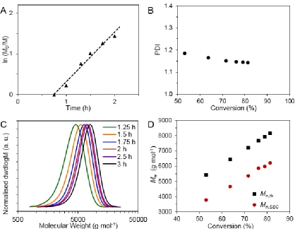
Figure 2.1: Kinetic data for pBEA targeting DP 50. (A) Kinetic plot for the RAFT
polymerisation of pBEA as determined by 1H NMR spectroscopy. Dashed line indicates
linearity. (B) Plot of polymer dispersity vs. conversion. (C) SEC traces of kinetic samples. (D) Theoretical Mn vs. Mn from SEC values. ... 29
Figure 2.2: Chain extension of: (A) pBEA50 macro-CTA (A1) with pBA70 and (B)
pBEA45 macro-CTA (A2) polymerised to 75% conversion, and subsequent chain
extension with pBA115. ... 31
Figure 2.4: (A) SEC trace of precursor pBEA50 (1), and post-substitution pTPEA50,
showing the similarity in distribution (B) 1H NMR in DMSO-d
6 indicating the shifts of
the pendant ethyl acrylate chain for protons α and β of pBEA50 (1) and protons α’ and β’
of pTPEA50. ... 34
Figure 2.5: (A) IR overlay showing the absorbance of the pBEA50 (1) vs. pAEA50 (B) 1H
NMR in DMSO-d6 indicating the shifts of the pendant ethyl acrylate chain for protons α
and β of pBEA50 (1) and protons α’ and β’ of pAEA50. ... 35
Figure 2.6: 1H NMR in DMSO-d
6 of tetraethyl ammonium sulfonate polymer from
pBEA50 ... 37
Figure 2.7:1H NMR in CDCl
3 of pBEA50. ... 43
Figure 2.8:1H NMR pBEA
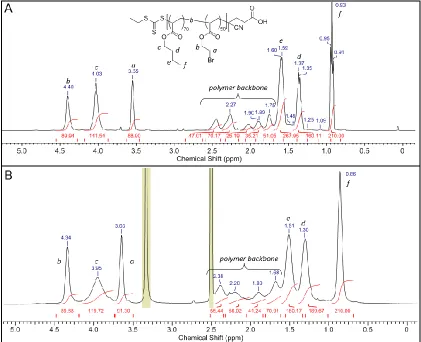
50-b-pBA70 (from macro-CTA A1) in (A) in CDCl3 and (B)
DMSO-d6. ... 44
Figure 2.9:1H NMR in CDCl
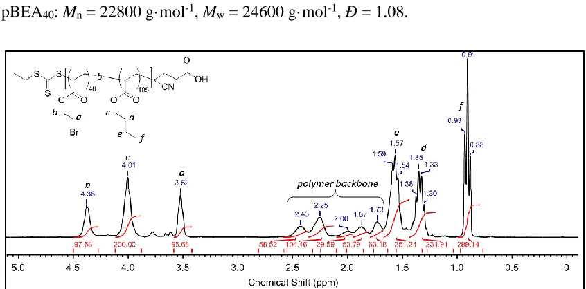
3 of pBA105-b-pBEA40 from macro-CTA A3. ... 45
Figure 2.10: 1H NMR in DMSO-d
6 of product from trimethyl amine substitution of
pBEA50. ... 47
Figure 2.11:1H NMR in DMSO-d
6 of product from trimethyl phosphine substitution of
pBEA50. ... 48
Figure 2.12: SEC trace of product of tetraethyl ammonium sulfite substitution of pBEA50
and pBEA150. ... 49
Figure 2.13: SEC trace of product of sodium azide substitution of pBEA50. ... 50
Figure 2.14: 1H NMR in CDCl
3 of thiophenol substitutions of pBEA50 conducted: (A)
without base and (B) in the presence of DIPEA. ... 51
Figure 2.15:1H NMR of pGluEA
50 in (A) DMSO-d6 and (B) D2O. ... 52
Figure 2.16: SEC trace of product of 1-β-D-thioglucose substitution of pBEA50. ... 53
Figure 2.17: 1H NMR in DMSO-d
6 of product from trimethyl amine substitution of
Figure 2.18:1H NMR in DMSO-d
6 of product from trimethyl phosphine substitution of
pBEA50-b-pBA70. ... 54
Figure 3.1: The principle of scattering. ... 61
Figure 3.2: Relationship between q-range and size of the window of observation showing
the complementarity of SLS and SANS. ... 62
Figure 3.3:1H NMR of CP-[pBEA
25]2 in DMSO-d6. ... 65
Figure 3.4: SEC chromatograms of conjugation reactions with varying polymer lengths:
(A) DP 10 and (B) DP 25. The black arrows highlight the decreasing polymer peaks and the green arrow to the increasing 2-arm conjugate peak. ... 66
Figure 3.5: Deconvolution number average molecular weight distributions of
CP-[pBEA25]2 conjugation at (A) 0.5 h, (B) 1 h, (C) 1.5 h, (D) 3.5 h. ... 67
Figure 3.6:1H NMR of CP-[pGluEA
10]2 in DMSO-d6. ... 69
Figure 3.7: SANS profiles of CP-[pBEAn]2 conjugates at 10 mg·mL-1 in THF-d8:
CP-[pBEA10]2 before (green circles) and after (green squares) filtration and
CP-[pBEA25]2 (blue diamonds). Data were fit to a Cylinder-Gaussian Coil model (red
lines). ... 72
Figure 3.8: SANS profiles of CP-[pGluEA10]2 (purple circles) and CP-[pGluEA25]2
(green circles) scattering profiles at 5 mg·mL-1 in D
2O. Data were fit to a
Cylinder-Gaussian Coil model (red lines). ... 74
Figure 3.9: Absorbance (λ = 420 nm) curves after adding 100 μL of 1 mg·mL-1 solution
of Con A in HBS buffer to the solution of the polymer CP-[pGluEA10]2 (50 μM per sugar
unit, in HBS). The linear fit of the steepest portions of the curves were used to calculate the clustering rate k = 1.31·10-3 s-1. ... 77
Figure 3.10: Absorbance (λ = 420 nm) curves after adding 100 μL of 1 mg·mL-1 solution
of Con A in HBS buffer to the solution of the polymer CP-[pGluEA25]2 (50 μM per sugar
Figure 3.11:1H NMR of CP-[pGluEA
25]2 in D2O. ... 86
Figure 4.1: SEC chromatograms of pBEA25 (green dashed), after CP-[pBEA25]2
conjugation is complete (red dotted) and CP-[pBEA25]2 after purification (black solid
line). ... 96
Figure 4.2: Results from Guinier-Porod Model for the Dimension Parameter, s (A) and
(B) the Radius of Gyration, Rg (Fits in Figure A4.3 and A4.6). ... 98
Figure 4.3: Scattering profiles of polyelectrolyte conjugates at a range of salt
concentrations (A) anionic CP-[pEASTEA25]2 and (B) cationic CP-[pTMAEA25]2 .... 100
Figure 4.4: Variation in length and radius of anionic CP-[pEASTEA25]2 conjugate
assemblies from cylinder model fitting (Fits in Figure A4.4 and A4.5). Lines between points are added to guide the eye. ... 102
Figure 4.5: Variation in length and radius of cationic CP-[pTMAEA25]2 conjugate
assemblies from Cylinder + Gaussian Coil model fitting. Lines between points are added to guide the eye. ... 104
Figure 5.1: Cellular toxicity against MDA-MB-231 cells after 24 h incubation time with
varying concentration of compound, as measured using XTT assay. ... 118
Figure 5.2: Intracellular fluorescence in MDA-MB-231 cells exposed to 50 μg·mL-1 of
polymers P1-9, and CP-polymer conjugates CP1-9 for 20 hours, as determined using flow cytometry and fluorescence correction factors (n = 4). ... 120
Figure 5.3: Intracellular fluorescence in MDA-MB-231 cells exposed to 50 µM of
conjugates with various amount of charge, introduced from heterogeneous mixing of neutral CP1 with (A) ammonium derivative CP5 and (B) phosphonium derivative CP9. ... 122
Figure 5.4:1H NMR of NHS-p(PEGA
75%-stat-BEA25%)28 in CDCl3. ... 126
Figure 5.5: SEC analyses of NHS-p(PEGA-stat-pBEA)n polymers (dotted line) and
Figure 5.6: Overlay of HPLC traces for 5(6)-carboxyfluorescein starting material (black line), cystamidefluorescein product in the disulfide form (blue line) and desired free-thiol cystamidefluorescein product following addition of TCEP (red line). The trio of peaks in the cystamidefluorescein traces are due to isomerisation of the dye. ... 130
Figure 5.7: 1H NMR of CP-[p(PEGA
75%-stat-pTMAEA25%)28] in DMSO-d6 following
substitution with trimethyl amine. ... 132
Figure 5.8: 1H NMR of CP-[p(PEGA
75%-stat-pTMAEA25%)28] in DMSO-d6 following
substitution with trimethyl phosphine. ... 133
Figure 5.9: 1H NMR of CP-[p(PEGA
98%-stat-pGlyEA2%)29] (CP1) in DMSO-d6
following substitution with thioglycerol. ... 135
A 3.1: Deconvolution of dwdlogM of CP-[pBEA10]2 conjugation before and after
addition of base. ... 145
A 3.2: Deconvolution of dwdlogM of CP-[pBEA25]2 conjugation. ... 145
A 3.3: Normalised Infrared transmittance spectra of CP-[pBEA10]2 (blue line) and
CP-[pBEA25]2 (red line). ... 146
A 3.4: SANS profile of CP-[pBEAn]2 conjugates at 10 mg·mL-1 in THF-d8:
CP-[pBEA10]2 before (green circles) and after (green squares) filtration and
CP-[pBEA25]2 (blue diamonds). ... 147
A 3.5: Guinier-Porod model fits of CP-[pBEAn]2 scattering profiles at 10 mg·mL-1 in
THF-d8 with different polymer arm DP: (A) DP10, (B) DP10 filtered, and (C) DP25.
... 148
A 3.6: Cylinder-Gaussian Coil model fits of CP-[pBEAn]2 scattering profiles at
10 mg·mL-1 in THF-d
8 with different polymer arm DP: (A) DP10, (B) DP10 filtered, and
(C) DP25. ... 149
A 3.7: Cylinder model fits of CP-[pBEAn]2 scattering profiles at 10 mg·mL-1 in THF-d8
A 3.8: Ellipsoid model fits of CP-[pBEAn]2 scattering profiles at 10 mg·mL-1 in THF-d8
with different polymer arm DP: (A) DP10, (B) DP10 filtered, and (C) DP25. ... 151
A 3.9: Sphere model fits of CP-[pBEAn]2 scattering profiles at 10 mg·mL-1 in THF-d8
with different polymer arm DP: (A) DP10, (B) DP10 filtered, and (C) DP25. ... 152
A 3.10: Model fits of CP-[pGluEA 10]2 (purple circles) and CP-[ pGluEA 25]2 (green
circles) scattering profiles at 5 mg·mL-1 in D
2O: (A) fits to a Cylinder-Gaussian Coil
model and (B) fits to a Cylinder model. ... 153
A 3.11: Model fits of CP-[pGluEA10]2 (purple circles) and CP-[pGluEA 25]2 (green
circles) scattering profiles at 5 mg·mL-1 in D
2O: (A) fits to an Ellipsoid model and (B) to
a Guinier-Porod model. ... 154
A 3.12: Concentration dependence of molecular weight (Ma) of (A) CP-[pBEA15]2 and
(B) CP-[pBEA29]2 in THF obtained by light scattering. ... 155
A 3.13: Evolution of KC/R as a function of q² obtained by light scattering for
CP-[pBEA15]2 in THF before (A) and after (B) filtration. ... 155
A 3.14: Evolution of KC/R as a function of q² obtained by light scattering for
CP-[pBEA29]2 in THF before (A) and after (B) filtration. ... 156
A 3.15: Evolution of KC/R as a function of q² obtained by light scattering for
(A) CP-[pBEA15]2 and (B) CP-[pBEA29]2 in THF 10 days after filtration. ... 156
A 3.16: Screenshot of Cylinder + Gaussian-Coil sum model selected. ... 157
A 4.1: BE_Polyelectrolyte fits of (A) CP-[pEASTEA25]2 at 8.6 mg·mL-1 and
5.9 mg·mL-1; and (B) CP-[pTMAEA
25]2 at 10 mg·mL-1 and 8.3 mg·mL-1 in D2O (†=held
parameter value). ... 158
A 4.2: Ellipsoid fits of (A) CP-[pEASTEA25]2 at 8.6 mg·mL-1 and 5.9 mg·mL-1; and
(B) CP-[pTMAEA25]2 at 10 mg·mL-1 and 8.3 mg·mL-1 in D2O (†=held parameter value).
A 4.3: Guinier-Porod model fits of CP-[pEASTEA25]2 scattering profiles in D2O with
(A) 0.18 M NaCl, (B) 1 M NaCl , (C) 2 M NaCl, (D) 3 M NaCl, (E) 4 M NaCl, and (F) 5 M NaCl. ... 160
A 4.4: Cylinder model fits of CP-[pEASTEA25]2 scattering profiles in D2O (A) at
different concentrations: 5.9 and 8.5 mg·mL-1 (B) with 0.18 M NaCl, (C) 1 M NaCl,
(D) 2 M NaCl (†=held parameter value). ... 161
A 4.5: Cylinder model fits of CP-[pEASTEA25]2 scattering profiles in D2O with (A) 3 M
NaCl, (B) 4 M NaCl,(C) 5 M NaCl, (D) 5 M NaCl re-run after 24 h (†=held parameter value). ... 162
A 4.6: Guinier-Porod model fits of CP-[pTMAEA25]2 scattering profiles in D2O with
(A) 0.18 M NaCl, (B) 1 M NaCl , (C) 2 M NaCl, (D) 3 M NaCl, (E) 4 M NaCl, and (F) 5 M NaCl. ... 163
A 4.7: Cylinder-Gaussian Coil model fits of CP-[pTMAEA25]2 scattering profiles in D2O
with (A) 0.18 M NaCl, (B) 1 M NaCl , (C) 2 M NaCl, (D) 3 M NaCl, (E) 4 M NaCl, and (F) 5 M NaCl (†=held parameter value). ... 164
A 4.8: Cylinder fit and model (red lines) and Gaussian-Coil fit and model (blue lines) of
CP-[pTMAEA25]2 in D2O with 5 M NaCl (†=held parameter value). ... 165
A 5.1: RP-HPLC traces of CP-polymer conjugates CP2-5: 15-100 % Methanol gradient
over 30 min. The strong UV absorbance is due to the Tryptophan residues on the CP. ... 166
A 5.2: RP-HPLC traces of CP-polymer conjugates CP6 - 9: 15-100 % Methanol gradient
over 30 min. The strong UV absorbance is due to the Tryptophan residues on the CP. ... 167
A 5.3: RP-HPLC traces of control polymers P2 - 5: 15-100 % Methanol gradient over 30
A 5.4: RP-HPLC traces of control polymers P6 - 9: 15 - 100 % Methanol gradient over 30 min. UV traces for the polymers are weak due to the absence of strongly UV active functionalities. ... 169
A 5.5: Zimm plots (KC/R) and concentration dependence of molecular weight for
(A) CP-[p(PEGA-stat-pTMAEA2%]29 and (B) CP-[p(PEGA-stat-pTMPEA2%]29. ... 172
A 5.6: Zimm plots (KC/R) and concentration dependence of molecular weight for
(A) CP-[p(PEGA-stat-pTMAEA5%]22 and (B) CP-[p(PEGA-stat-pTMPEA5%]22. ... 173
A 5.7: Zimm plots (KC/R) and concentration dependence of molecular weight for
(A) CP-[p(PEGA-stat-pTMAEA10%]26 and (B) CP-[p(PEGA-stat-pTMPEA10%]26. .... 174
A 5.8: Zimm plots (KC/R) and concentration dependence of molecular weight for
List of Tables
Table 2.1: Summary of BEA RAFT homopolymerisation ... 30
Table 2.2: Summary of pBEA substitutions and structural characteristics of polymers and derivatives. ... 38
Table 2.3: Summary of RAFT homopolymerisation for use as macro-CTAs. ... 46
Table 2.4: Summary of RAFT chain extensions. ... 46
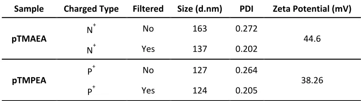
Table 2.5: DLS and Zeta Potential of block copolymers after substitution with trimethyl amine (pTMAEA) and trimethyl phosphine (pTMPEA). ... 55
Table 3.1: Details of NHS-pBEA polymers synthesised. ... 64
Table 3.2: Results of the Guinier-Porod analysis for Dimension Variable, s, and Radius of Gyration, Rg for CP-[pBEAn]2 conjugates. ... 71
Table 3.3: Parameters estimated from fitting to Cylinder-Gaussian Coil model. ... 72
Table 3.4:Nagg of CP-[pBEAn]2 conjugatesbefore and after filtration, and after 10 days equilibration time. ... 73
Table 3.5: Summary of Guinier-Porod model fits for CP-[pGluEAn]2 in D2O. ... 75
Table 3.6: Summary of Cylinder-Gaussian Coil model fits for CP-[pGluEAn]2 in D2O. ... 75
Table 4.1: Details of pBEA-NHS polymerisation. ... 106
Table 4.2: Quantities of NaCl added to give the desired ionic strength... 108
Table 4.3: Calculated values of the Debye length in nm and in Å. ... 109
Table 5.1: Details of NHS-p(PEGA-stat-BEA) polymers synthesised. ... 126
Table A 3.1: Conjugation efficiencies of pBEA conjugations using Eq. 2. ... 144
Table A 5.1: Zeta potential and electrophoretic mobility values for charged polymers and conjugates (P2 - 9 and CP2 - 9). ... 170
Table A 5.2: Summary of Nagg values obtrained by static light scattering. ... 171
Table A 5.3: Relative fluorescence of fluorescein-modified compounds in PBS and
fluorescence corrections of flow cytometry data. ... 177
Table A 5.4: Theoretical and experimental cellular fluorescence from mixtures of
List of Schemes
Scheme 1.1: Scheme highlighting the directionality of the amide bonds in the backbone
of CPs of alternating L/D α-amino acids in an octapeptide. Hydrogen bonding allows CP
units to self-assemble in an antiparallel β-sheet conformation. ... 4
Scheme 1.2: X-ray crystal structures of peptide nanotubes. (a) Antiparallel nanotubes formed by cyclo[(DlKl)2]. Two conformations of the peptide are present (pink and
yellow). Adjacent nanotubes are oriented head-to-tail. (b) Parallel nanotubes of
cyclo[(DaKa)2] formed by a single peptide conformer. Adjacent nanotubes are parallel to
one another and form sheets separated by HFIP molecules (orange). Adapted from Silk
et al.37 ... 5
Scheme 1.3: Schematic representation of the assembly of a cyclic octapeptide comprising
alternating L- and D- α-amino acids, displaying the dimensions of the internal pore. Red, green and blue protrusions from the circumference of the ring represent the amino acid side chains extending equatorially from the ring. The inset highlights the sequence-dependent ring distances including the hydrogen-bond length (≈ 2.85 Å) and inter-axial distance (≈ 4.73 Å). ... 6
Scheme 1.4: Schematic representation of the antiparallel β-sheet dimer composed of
D,L-α-octapeptides with strategically N-alkylated amino acids blocking the hydrogen
bonding from one face. ... 7
Scheme 1.5: Classes of cyclic peptides that assemble into nanotubes through β-sheet
interactions: (A) CPs comprising alternating D, L α- residues; (B) CPs incorporating
β-residues; (C) CPs with both α- and γ-residues and; (D) self-assembling heterocyclic peptides by incorporation of ε-amino acids. Adapted from Chapman et al.61 ... 8
Scheme 1.6: Convergent approach to cyclic peptide–polymer nanotubes via CuAAC.
Scheme 2.1: Scheme of entire synthesis, beginning with monomer synthesis, RAFT homo and block copolymerisation of BEA and nucleophilic substitution of pBEA precursors. ... 31
Scheme 2.2: Summary of the substitutions achieved with pBEA50 precursor. ... 33
Scheme 3.1: Reaction scheme of pBEA conjugations to a self-assembling cyclic peptide.
... 64
Scheme 5.1: Summary of compounds used in this study and their abbreviations. ... 117
Scheme 5.2: Conjugation of NHS-p(PEGA-stat-pBEA)n copolymers to cyclic D-, L-
α-octapeptide. ... 127
Scheme 5.3: Synthesis of cystamidefluorescein from cystamine.HCl and
Abbreviations
4,4′-Azobis(4-cyanovaleric acid) ACVA Atomic force microscopy AFM Atom transfer radical polymerisation ATRP
Butyl acrylate BA
Bromo ethyl acrylate BEA deuterated chloroform CDCl3
Cyclic peptide CP
cyclic peptide [poly(bromo ethyl acrylate)]2 CP-[pBEAn]2
cyclic peptide [poly(thioglucose ethyl acrylate)] 2 CP-[pGluEAn]2
cyclic peptide [poly(thioglycerol ethyl acrylate)] 2 CP-[pGluEAn]2
cyclic peptide [poly(trimethyl ammonium ethyl acrylate)] 2 CP-[pTMAEAn]2
cyclic peptide [poly(trimethyl phosphonium ethyl acrylate)] 2 CP-[pTMPEAn]2
(4-Cyano pentanoic acid)yl ethyl trithiocarbonate CPAETC Cyclic Peptide Nanotube CPNT Chain Transfer Agent CTA Copper Azide Alkyne Cycloaddition CuAAC
Dichloromethane DCM
N,N-Diisopropylethylamine DIPEA Dynamic Light Scattering DLS Dimethylamino pyridine DMAP
Dimethylformamide DMF
Dimethylsulfoxide DMSO
4-(4,6-Dimethoxy-1,3,5-triazin-2-yl)-4-methylmorpholinium DMTMM Degree of Polymerisation DP 1-Ethyl-3-(3-dimethylaminopropyl)carbodiimide EDC Electrospray Ionisation Time of Flight ESI ToF
Fourier Transform Infrared Spectroscopy FTIR
O-(Benzotriazole-1-yl)-N,N,N’,N’-tetramethyluronium HBTU Hexafluoro-2-propanol HFIP High-performance liquid chromatography HPLC
Infrared IR
Association constant Ka
Layer-by-layer LbL
Leucine Leu
Lysine Lys
Methanol MeOH
Megahertz MHz
Number Average Molecular Weight Mn
Molecular Weight Mw
Molecular Weight Cut Off MWCO Avogadro's Number Na Number of Aggregation Nagg N-acryloyl morpholine NAM NIST Centre for Neutron Research NCNR
N-hydroxysuccinimide NHS
N-hydroxysuccinimide-(4-cyano pentanoic acid)yl ethyl
Trithiocarbonate NHS-CPAETC
N-isopropyl acrylamide NiPAAm National Institute of Standards and Technology NIST
N-methyl morpholine NMM Nitroxide-mediated radical polymerisation NMP Nuclear Magnetic Resonance NMR
Nanotube NT
polyethylene glycol PEG poly(ethylene glycol) methyl ether acrylate PEGA poly(ethylene oxide) PEO poly(2-ethyl-2-oxazoline) pEtOx poly(thioglycerol ethyl acrylate) pGlyEA poly(methyl methacrylate) pMMA
polystyrene PS
poly(trimethyl ammonium ethyl acrylate) pTMAEA poly(trimethyl phosphonium ethyl acrylate) pTMPEA poly (thiophenol ethyl acrylate) pTPEA
Wave vector q
Reversible Addition Fragmentation chain-Transfer RAFT Reversible Deactivation Radical Polymerisations RDRP
Radius of gyration Rg
Hydrodynamic radius Rh
Refractive Index RI
Rayleigh ratio Rθ
Small Angle Neutron Spectroscopy SANS Size Exclusion Chromatography SEC Single-Electron Transfer Living Radical Polymerisation SET-LRP Static Light Scattering SLS Solid Phase Peptide Synthesis SPPS Transmission electron microscopy TEM Trifluoro acetic acid TFA
Tetrahydrofuran THF
Triisopropylsilane TIPS
Tryptophan Trp
Ultra Violet-Visible UV-Vis
Acknowledgements
I can manage to write a thesis, and yet I can’t seem to find the words to start thanking the many people who have helped me through these last few years! Hopefully I won’t forget anyone, but if you feel left out, then you know you’ve helped.
First of all, I’d like to thank my supervisor Professor Sébastien Perrier, for giving me the opportunity of a life-time to move across the world to study in England! Without this opportunity I would be stuck in Sydney, with stunning weather, surrounded by beaches, family and friends. Fortunately, I was sent to Coventry where I have made wonderful friends and colleagues, travelled the world, and under your guidance have become the scientist I am today. Thank you for your unfailing ability to sell me my own project (even when it’s going terribly wrong) and for patiently enduring the tearful breakdowns throughout my degree.
To my Chemistry teachers throughout the years: Ms Cheong and Mr Harris in high school, and at university, to Professor Kate Jolliffe, your enjoyable organic puzzles drew me to you as a supervisor. Thank you for giving me an opportunity all those years ago, without it, I would never be where I am today.
When I first began my degree in 2013, I had no friends on this side of the world. Fortunately, I landed in a wonderful group that became my friends and family over here. To my fellow soon to be doctors Liam and Sophie, thank you for always being there, for the copious amounts of beer and wine all over Australia and Europe and (to Junliang as well) thank you for being the best friends you could ever hope to have in your year with you!
fun times (you and your gorgeous fam too)! Your guidance and shenanigans are always appreciated. Ximo, you are a wonderful man, thank you for bringing sunshine and smiles into my life when you’re around. Ming, thank you for teaching me all about cyclic peptides and for your unwavering enthusiasm for the pub on a Thursday afternoon. Dr Guillaume “GG” Gody, a masssiiiiiifff thank you for teaching me so much about RAFT, even if you never understood a word I was saying. Matthias, it’s always a pleasure to be with you and your wonderful little family. Dr Ed, you are an incredible proof-reader and without you, this thesis wouldn’t be what it is; thank you so much! Dr Kempe, thanks for all the spicy soup and the QCM chips. Dr Jon Newland, while I know you’re not my post-doc, but thanks for teaching me how to keep it real as a PhD student.
To my loyal (to varying degrees) tea crew! May the summoning spoon ring on in your hearts. To the Perrier group, each and every one of you have been a pleasure to meet and work with these past years, thank you for your friendly faces each day in the corridor. To my football homies, thanks for all the laughs and bruises. To George, thank you for always smiling and cheering me up, even when it’s 1 am and we’re both still working. I also met many students here who have become some of my best friends. Liam and Sophie for helping me make a home with you guys. Sophie, thanks for reminding me to save the trees when I print, to try to eat less meat, and to never be greedy around the cupcakes. To Julia, for the insanely delicious baking, and providing such tempting cupcakes and lunch leftovers. To Cookie, thank you for always being such a cheerful, happy-go-lucky guy and for always keeping us on our toes in the office. To the chatty rays of sunshine that are Julia and Aggie, it is always a pleasure seeing your smiling faces wandering into the office, I love your enthusiasm for life, tea and talking.
To Anthony’s family, Tracey, Tina, Nanny, Ollie, Tash, Phil and co. thank you for welcoming me into your family and your lives. You have always made me feel completely welcomed and loved. The Sunday dinners were always great for a catch-up, and gave me a second family, while I’ve been so far away from my own.
was too stupid to be here, for convincing me I could do this when I felt like I couldn’t and for reminding me there’s so much more to life than a degree. I know it can’t have been easy putting up with me, but I am so, so grateful that you did.
Declaration
This thesis is submitted to the University of Warwick in support of my application for the degree of Doctor of Philosophy. It has been composed by myself and has not been submitted in any previous application for any degree. The experimental work contained in this thesis is original research carried out by the author, unless stated otherwise, in the Department of Chemistry at the University of Warwick.
The work presented was carried out by the author except in the following instances:
Chapter 5: XTT assays, cell uptake studies and fluorescence corrections were conducted
by Dr. Raoul Peltier (University of Warwick, School of Life Science).
List of Publications
Parts of this thesis have been published by the author:
1. Poly(bromoethyl acrylate): A Reactive Precursor for the Synthesis of Functional RAFT Materials. Barlow, T. R.; Brendel, J. C.; Perrier, S. Macromolecules 2016,
49, (17), 6203-6212.
2. Tunable Length of Cyclic Peptide–Polymer Conjugate Self-Assemblies in Water. Catrouillet, S.; Brendel, J. C.; Larnaudie, S.; Barlow, T.; Jolliffe, K. A.; Perrier,
S. ACS Macro Letters 2016, 5, (10), 1119-1123.
3. An Orthogonal Reaction Sequence for the Synthesis of Sugar-Coated Cyclic Peptide Nanotubes. Barlow, T. R.; Brendel, J. C.; Jolliffe, K. A.; Perrier, S.
Submitted for publication.
4. The Effect of Ionic Strength on the Self-Assembly of Highly Charged Polyelectrolyte-Cyclic Peptide Conjugates. Barlow, T. R.; Brendel, J. C.; Mansfield, E. D. H.; Rogers, S. E.; Jolliffe, K. A.; Perrier, S. Submitted for publication.
Abstract
Self-assembly is a powerful process by which complex architectures can be achieved from single monomer units for a diverse range in applications from drug delivery to nano-electronics, an excellent example of which is self-assembled cyclic peptide nanotubes. The addition of polymers greatly enhances the potential applications of cyclic peptide nanotubes, however, there lacks a fundamental understanding on how this affects assembly, and whether it can be controlled by environmental manipulation. Additionally, there are few synthetic methods by which it is possible to access more complex polymeric architectures without the need for complex conjugation strategies.
As such the present work aims to further develop a scope of synthetic possibilities for polymeric cyclic peptide nanotubes, generating materials by which it is possible to probe the self-assembly properties of these complex supramolecular systems. Initially the polymerisation of poly(bromo ethyl acrylate) (BEA) is explored via reversible addition-fragmentation chain transfer polymerisation (RAFT) and the post-modification possibilities this reactive polymeric precursor provides. From this, a library of polymer materials from a pBEA precursor via nucleophilic substitution with a range of nucleophiles can be generated.
Finally, the cell-penetration of charged copolymers of poly(poly(ethylene glycol) methyl ether acrylate) (pPEGA) conjugates were studied to determine whether charge or self-assembly has a greater effect on cellular uptake. It was identified that self-self-assembly results in far greater in vitro cell penetration than for charged analogues.
1
1
Introduction
Self-assembly is a powerful tool for the synthesis of complex materials. By carefully tailoring individual building blocks, supramolecular forces can be used to do the synthetic heavy-lifting for the difficult task of creating demanding and otherwise challenging structures. As such, a diverse range of morphologies can be formed including micelles, vesicles, nanotubes, and lamellar sheets. Nanotubular structures in particular have gained a lot of attention recently, for the interesting properties that arise from their shape.1-6 Since
their discovery in the 1990s, carbon nanotubes have championed the field of nanotube research,7-9 with a lot of attention paid to their unique structural, mechanical and
electronic properties. With the introduction of carbon nanotube synthesis in the early 1990s,10 the interest in nanotubes has vastly increased. Unfortunately, poor solubility,
toxicity, and lack of size uniformity is a major drawback for any specific biomedical applications. Many of the problems facing fullerene based nanotubular structures can fortunately be overcome by employing organic nanotubes that more closely mimic nature such as carbohydrates, lipids and polymer-based nanotubes. These nanotubes generally span the width region between 10 - 1000 nm; a scale that is inaccessible to top-down microfabrication techniques, nor carbon nanotube fabrication methods.11 Thus the study
virus for example, exists as a tubular structure comprising unimeric coat proteins that self-assemble into a rod-like helical structure around a single strand of RNA.11, 12
Nanotubular structures have advantages over other nanoparticle architectures in biological systems, due to their anisotropy resulting in increased circulation times and altered internalisation kinetics vs spherical and cubic nanoparticles.5, 6, 13, 14
There are a number of intermolecular interactions by which supramolecular self-assembly can be guided, including van der Waals, electrostatic, π-π, and hydrogen bonding to name but a few.15 Of these, hydrogen bonds are particularly noteworthy due to the directionality
of their interaction, enabling intricate supramolecular assemblies. Though hydrogen bonds are weak (Figure 1.1),16, 17 they are key to a number of biological interactions
including DNA and RNA strand recognition, and protein folding. The cumulative strength of multiple hydrogen bonds in concert with other weak attractive forces, can lead to strong and stable complex architectures.17, 18
Figure 1.1: A comparison of the strength and properties of noncovalent interactions
involved in self-assembly. Adapted from Guler et al.17
Numerous bio-molecules have been used in the preparation of nanotubes including DNA, lipids, carbohydrates, and peptides.11 Of these examples, peptides in particular are an
[image:33.595.118.535.389.530.2]alternating L- and D- amino acids, Gramicidin has been extensively studied for its
membrane transport properties.20 Studies have revealed that it is highly sensitive to
structural alterations, and varying a single amino acid in the peptide sequence (Gramicidin A, B, and C variants) results in a subtly different supramolecular conformation, leading to different membrane interactions.19
1.1
Self-assembling Cyclic Peptides
β-sheet forming cyclic peptides are a remarkable example of synthetic nanotube forming materials. By tailoring the chemical structure of the cyclic peptide, the properties of the supramolecular structure can be altered. In this way the building blocks can be adjusted to tailor the nanotube properties to meet the requirements for the desired application. Self-assembling cyclic peptides have thus become of particular interest as nanomaterial scaffolds in a range of fields from biomedical applications such as antimicrobials,21-23
biosensors,24 to nanoelectronics.25-28
1.1.1
Self-Assembly
The potential of cyclic peptides comprised of alternating L- and D- α-amino acids to
self-assemble was first realised by De Santis and co-workers in 1974.29 In their theoretical
study, they found that the backbone C=O and N-H bonds point in opposite directions and were nearly parallel to the symmetry of the ring (Scheme 1.1). They postulated that the direction of the backbone amide bonds would lead to inter-annular stabilisation that may result in a contiguous parallel or antiparallel β-sheet structure. It wasn’t until 1993 that the first self-assembling cyclic peptide was synthesised by Ghadiri et al;30 whereby they
assembled the cyclo[-D-Ala-L-Glu-D-Ala-L-Gln-]2 peptide into nanotubes by controlled
acidification. Their characterisation of the nanotubes by electron diffraction30, 31
suggested that the nanotubes formed an antiparallel β-sheet-like structure with an inter-unit spacing of 4.73 Å. Subsequent molecular dynamics32 and dimerisation studies33-36
Scheme 1.1: Scheme highlighting the directionality of the amide bonds in the backbone of
CPs of alternating L/D α-amino acids in an octapeptide. Hydrogen bonding allows CP units
to self-assemble in an antiparallel β-sheet conformation.
In recent work by Silk et al., the first crystal structure of any continuous nanotube, rather than a simple N-alkylated CP-dimer, was analysed.37 In their work, the authors
crystallised two cyclic octapeptides of alternating D- and L- α-amino acids comprising similar peptide sequences (Scheme 1.2). Remarkably, despite the similarity in the peptide sequence, the crystal structure revealed that the leucine bearing CPs formed nanotubes
Scheme 1.2: X-ray crystal structures of peptide nanotubes. (a) Antiparallel nanotubes
formed by cyclo[(DlKl)2]. Two conformations of the peptide are present (pink and yellow).
Adjacent nanotubes are oriented head-to-tail. (b) Parallel nanotubes of cyclo[(DaKa)2]
formed by a single peptide conformer. Adjacent nanotubes are parallel to one another and
form sheets separated by HFIP molecules (orange). Adapted from Silk et al.37
arise from the ability to manipulate the peptide sequence, thereby controlling the supramolecular properties. By substituting different amino acids, whether naturally occurring or synthetic, into the peptide chain the properties of the nanotube can be tailored to a desired application. The inclusion of hydrophobic amino acids, such as tryptophan for example, has been used to improve lipid membrane interaction,22, 32, 38-42 and charged
residues such as glutamic acid, aspartic acid, and lysine have been included to induce electrostatic charge that can control assembly.28, 30 These charged systems have also been
analysed for potential antimicrobial activities.21, 23, 43 Control of the peptide sequence is
pivotal to control the properties of the nanotube including the size of the pore, the internal and external functionality, as well as the extent of hydrogen bonding in the nanotube such as by N-alkylation of the peptide backbone.
Scheme 1.3: Schematic representation of the assembly of a cyclic octapeptide comprising
alternating L- and D- α-amino acids, displaying the dimensions of the internal pore. Red, green and blue protrusions from the circumference of the ring represent the amino acid side
chains extending equatorially from the ring. The inset highlights the sequence-dependent
inter-ring distances including the hydrogen-bond length (≈ 2.85 Å) and inter-axial distance
1.1.2
Cyclic peptides comprising non-natural amino acids
Numerous attempts have been made at controlling the extended hydrogen bonding network in self-assembling CPs. This has primarily been achieved using strategic
N-alkylation of the amide backbone.33, 35, 36, 44, 45 By incorporating N-methyl or N-alkyl
residues of the same stereochemistry, the hydrogen bonding on a single face of the CP is blocked and self-assembly is limited to dimerisation (Scheme 1.4). This model system improves solubility and provides an insight into the thermodynamics and association constants of different peptide sequences. The equilibrium dimerisation constant, Ka, of CP dimers can be readily studied by concentration dependent 1H NMR studies in
deuterated chloroform and is a far simpler task than the study of the association constant of non-alkylated analogues. Dimerisation constants measured for N-alkylated
D,L-α-octapeptides comprising Ala and Phe are among the highest reported, between
2,500 – 3,000 M-1.33, 34, 44 The low association constant suggests that the CPs interact via
a cooperative mechanism. While computational approaches have brought insight to the self-assembly process,46-48 understanding nanotube formation and controlling the length
and self-assembly of cyclic peptide nanotubes (CPNTs) remains challenging.
Scheme 1.4: Schematic representation of the antiparallel β-sheet dimer composed of
D,L-α-octapeptides with strategically N-alkylated amino acids blocking the hydrogen bonding from one face.
CPs comprising alternating D,L α-amino acids (cyclo[-D-Ala-L-Glu-D-Ala-L-Gln-]2) were
sequence lengths ranging from 6 amino acids,49, 50 through to 1051, 52 and 12 α-residues
have been reported.53 Additionally, sequences in which some or all of the α-amino acids
have been replaced with β-54, 55 and cyclic γ-residues56-59 have also been explored
(Scheme 1.5). These structures result in nanotubes with a larger internal pore diameter,
however successful nanotube formation is linked to ability of the CP to adopt a rigid ring conformation. Inclusion of γ-residues has been found to increase the strength of the assemblies formed despite decreasing the hydrogen bonding interaction,56, 60 with
association constants as high as 5.3 × 10⁵ M-1,33, 34 several orders of magnitude higher
than many α-peptides. In addition, the cyclic nature of these γ-residues lends additional structural rigidity to the backbone when compared to α-CPs. As such, the internal diameters available to this system are far greater than for α-CPs, due to the additional rigidity of the peptide and their ability to form favourable intermolecular hydrogen bonds. Alteration of the amino acid sequence modifies the internal pore size of the CPNT in addition to the amino acid side chains providing the external properties of the nanotube.
Scheme 1.5: Classes of cyclic peptides that assemble into nanotubes through β-sheet
interactions: (A) CPs comprising alternating D, L α- residues; (B) CPs incorporating β-residues; (C) CPs with both α- and γ-residues and; (D) self-assembling heterocyclic
1.1.3
External properties of the nanotubes
It is clear in that the ability to tailor the peptide sequence of CPs is fundamental to their practicality as nanomaterial scaffolds. Given that the side-chains extend equatorially from the ring of the peptide, it is possible to control the functionality of the periphery of the nanotube. Adaptation of the amino acid sequence has led to a range of studies of structure activity relationships for applications such as ion-channels38, 39, 45, 51, 52, 62 and
antimicrobials.21-23, 43, 50 The effect of peptide composition, including both natural and
non-natural amino acids, on lipid bilayer interactions was studied by Danial et al.42 They
found that CPs of different structures formed different pores within lipid bilayers (Figure 1.1). This research, along with other earlier studies, focused heavily on lipid bilayer interaction, with a view to understanding the mechanism by which these CPs disrupt membranes. As such, they could be used as an exciting new class of antimicrobial materials.21-23, 42, 43, 50
Figure 1.2: Illustration showing the different possible modes of bilayer channel formation.
Formation of cyclic peptide channels in a lipid bilayer can occur as (A) a unimeric pore,
(B) a barrel stave or (C) through a bilayer or carpet-like disruption. Adapted from Danial
et al.42
[image:40.595.114.537.382.523.2]focus of conjugating molecules to the exterior of the nanotubes has been to tailor the CPNT properties towards specific applications, such as drug delivery, ion channel mimics, and electron transport. Indeed, various macrocyclic and polycyclic conjugates have been synthesised for their electron transport properties including a 1,4,5,8-naphthalenetetracarboxylic diimide (NDI) conjugate.25 These CP-NDI conjugates
displayed strong fluorescence properties which facilitated the study of the peptide orientation and conformation upon assembly. The additional attractive forces of π-π stacking between the NDI units resulted in nanotubes hundreds of nanometres long with an extended electronically-delocalised state.25 This system provides a very good example
on how the addition of a molecule to the exterior of the nanotube can greatly enhance its physico-chemical properties, and therefore expand on the range of applications for these systems. In addition to controlling the peptide sequence, macromolecular conjugation is another primary focus of CPNT research, since the first instance of polymer-peptide nanotubes by Biesalski et al. in 2005 and is discussed at length in Section 1.2.
1.1.4
Controlling Assembly and Orientation
Controlling the self-assembly of CPNTs has been a key area of research, primarily focused on limitation of backbone hydrogen bonding, through strategic N-alkylation or electrostatic interactions introduced by charged amino acid residues,63 as in the seminal
work by Ghadiri et al. using controlled acidification.30 A good example of successful
control of both nanotube orientation and length was demonstrated by Mizrahi and co-workers; who exploited different peptide sequences as a means of controlling the orientation of the nanotube on a gold surface in order to assess electron transport properties.28 They designed a series of three CPs for layer by layer (LbL) assembly; one
for initial deposition on a gold surface, and a second and third (incorporating glutamic acid and lysine residues) to introduce negative and positive charges respectively (Figure 1.3). The first CP included diametrically opposing cysteines for attachment to the gold electrode, in addition to the lysine residues to provide a point for charge interactions for additional layers. Assembly was induced by interlayer electrostatic interaction, with subsequent layer deposition alternating between negative glutamic acid bearing CPs
LbL deposition is an interesting and successful approach to controlling NT length, however use of this system in solution severely limits the potential applications including cell-uptake and drug delivery.
Figure 1.3: Schematic of the LbL assembly approach used by Mizrahi et al. Blue/red
represents positively/negatively charged CPs respectively. The grey ‘‘arm’’ represents the
cysteine side chains used to tether CPs to the gold surface. Note: AFM tip not to scale.
Adapted from Mizrahi et al.28
The structure of the peptide can have a drastic effect on its ability to self-assemble into nanotubes. Various structures and conformations have been extensively studied in order to understand the driving forces behind this process, which has paved way for future research looking into functionalised nanotubes as a way of expanding potential applications.
1.2
Cyclic Peptide-Polymer Conjugates
Polymer-conjugates of these self-assembling CPs have garnered considerable interest of late, due to the wide range of functionalities that can be imparted by polymeric species. Biesalski et al. first synthesised polymer conjugates with self-assembling cyclic peptides
a successful synthetic approach, more recent advances have focused on convergent methods, which presents fewer limitations on reaction conditions and simpler characterisation methods.65, 66
Larnaudie et al. recently highlighted that both a convergent approach, grafting polymers to the CP core; and a divergent approach, polymerising from the CP core, results in similar materials, however convergent synthesis facilitates full characterisation of all structures including the peptide, polymer and resultant nanotube.66 Employing a
convergent approach to CPNT synthesis has led to many recent advances, including the creation of water-soluble conjugates,66, 67 stimuli responsive conjugates,41, 67-69 and more
complex asymmetrical Janus-like NTs (Section 1.2.1).70
The convergent approach to the synthesis of polymer conjugated CPNTs was first employed by Börner and co-workers,71 who used an
1-ethyl-3-(3-dimethylaminopropyl)carbodiimide (EDC) mediated condensation reaction to couple two poly(n-butyl acrylate) (pBA) arms with two lysine residues on a D, L-α-octapeptide core.
The resulting nanotubes showed similar dimensions to those obtained by the divergent approach mentioned previously, with lengths of up to 300 nm and widths of ~5 nm in the dry state by AFM.71 More recently, a highly efficient conjugation method for the
attachment of 2, 3, and 4 arms to cyclic octapeptides has been demonstrated by Chapman
et al. via copper catalysed azide–alkyne cycloaddition (CuAAC) chemistry (Scheme
1.6).72-74 Alkyne groups were installed on the α-chain end of polymers by reversible
Scheme 1.6: Convergent approach to cyclic peptide–polymer nanotubes via CuAAC.
Adapted from Chapman et al.72
CuAAC “click” reactions such as these are ideal as a peptide-polymer coupling strategy,75
as it uses functional groups that are orthogonal to both free radical polymerisation and peptide chemistries, in addition to being synthetically simple to introduce into both polymer and peptide. A library of peptide-polymer nanotubes has been synthesised by the CuAAC mediated convergent approach, including conjugates of poly(butyl acrylate),73, 76 poly(dimethylaminoethyl acrylate),72 poly(acrylic acid),67 poly(styrene) (PS),72 and
poly(hydroxyethyl acrylate).72, 74, 77 While the synthesis of CPNTs by CuAAC is
attractive, due to its highly efficient and orthogonal nature to the chemistries involved, the presence of copper has been highlighted as problematic for applications such as drug-delivery due to the toxicity of free metal ions, and some copper complexes,77 requiring
careful selection of the copper catalyst employed.78, 79 In addition to the potentially
cytotoxic nature of copper, it is also know to quench the fluorescence of dyes such as calcein,80 leading to potential problems in fluorescent labelled materials for cell-uptake
Attaching polymer arms to the CP core results in a number of changes to the properties of the nanotubes. For example, the steric bulk of the polymer arms limits the extent of hydrogen bonding, resulting in far shorter assemblies with no apparent lateral/inter-tube aggregation. In addition to altered nanotube dimensions, the CP-polymer nanotubes adopt enhanced solubility properties, more similar to those of the conjugated polymer arms. Functionalisation with PEG for example greatly enhances the aqueous solubility of the peptides, and provides a route for in vivo and in vitro applications. Another important factor is the length and graft density of the conjugated polymer chains on nanotube assembly. Biesalski et al. assessed the impact of polymer mass and graft density on the length and diameter of the nanotubes in the dry state.81 From AFM measurements they
determined that as polymer molar mass increased, nanotube length decreased. More recently this has been demonstrated in solution, using Small Angle Neutron Scattering (SANS) to highlight that polymer length and graft density can have a significant impact on nanotube length.76
While many strides have been taken towards understanding the effect of polymer length and steric interactions on nanotube assembly, the reality is that the effects on self-assembly are more complicated than simply the polymer size. Factors such as the steric bulk of the monomer in addition to the solvent necessary to solvate the nanotube add further layers of complexity to the study of self-assembly. In a study by Koh et al.,82 they
demonstrated that in highly hydrogen-bonding competitive solvents such as TFA, DMSO and DMF, the solvent competes with the backbone hydrogen-bonding necessary for self-assembly, resulting in essentially unimeric species with scattering barely above the background in SANS. Analysis of the same pBA30 conjugates in chloroform and THF
resulted in extended cylindrical structures with a length > 2100 Å. In 1:9 v/v mixtures of [D1]TFA/[D8]THF and [D1]TFA/CDCl3, 10 % TFA was sufficient to diminish NT length
1.2.1
Complex Architectures and Functionality
In addition to CuAAC, there are examples in the literature on the use of different synthetic strategies to create more complex systems and materials. For example, Xu et al. have also followed a convergent methodology for the synthesis of CP-polymer conjugates, conjugating low molecular weight polymers of poly(ethylene oxide) (PEO), PS and pMMA to cyclic D, L α-octapeptides (Figure 1.4).83, 84 They annealed the various
peptide-polymer conjugates with block-copolymers of pBA-b-pMMA that form hexagonally packed cylindrical micro domains which are normally orientated to the surface of the thin film. They found that CP-PEO conjugates in particular were selectively sequestered into the cylindrical micro domains, within which the conjugates self-assembled forming nano-channels in the free-standing block copolymer film (Figure 1.4). The nanoporous channels were tested for gas permeability with carbon dioxide and neopentane, and it was found that gas permeance was higher for the smaller CO2
molecules than the larger neopentane molecules.83
Figure 1.4: Schematic showing the directed co-assembly CP-polymer conjugates.
(A) CP-polymer conjugates are first blended with block copolymers, (B) which results
in formation of nanopores within microporous domains. Adapted from Xu et al.83
well-defined, uniform nanotubular materials. The initial paper by Biesalski and co-workers64 mention steric repulsion of the polymer arms as a potential mechanism by
which the nanotube length could be controlled. pH driven electrostatic repulsion is an attractive method for controlling nanotube assembly and has been used for unfunctionalised CP systems (vide supra). Incorporation of pH and temperature responsive polymer arms have also proven effective for controlling CPNT assembly.67-69
Chapman et al. studied thermoresponsive conjugates of CPs with poly(2-ethyl-2-oxazoline) (pEtOx).69 Poly(2-oxazoline)s with ethyl side chains are known to exhibit
molecular weight dependent LCST behaviours,85 and conjugates of CP-pEtOx were also
found to display a lower critical solution temperature LCST in water. Below the LCST transition, or cloud point temperature, the polymer is hydrophilic and water soluble. However due to the unfavourable entropy of mixing, as the temperature increases the polymer becomes hydrophobic and thus insoluble in water.69 The addition of the CP to
pEtOx resulted in different LCST behaviour to the native polymer, presumably due to the presence of a sterically hindered polymer environment around the CP core that modify the hydrogen bonding and hydrophobic interactions in the polymeric corona of the nanotube.
Addition of ionisable groups on conjugated polymer arms has also been identified as an effective way to manipulate polymeric CPNT assembly. Another study by Chapman et al. made use of poly (acrylic acid) conjugates (pAA),67 and Catrouillet et al. used
poly(dimethyl amino ethyl methacrylate) (pDMAEMA) conjugates,68 to control NT
assembly, by manipulating the pH of the solutions, in both cases assessing self-assembly by SANS. For CP-pAA conjugates, assembly into cylinders occurred when the pAA arms were in an uncharged state at pH 3, as observed by SANS and supported by DLS measurements, with assembly greatly diminished at pH 11. Catrouillet and co-workers demonstrated the reversibility of assembly of CP-pDMAEMA conjugates in their SANS study.68 At pH 9 the CP-pDMAEMA conjugates were uncharged and formed NTs with a
nanotubular assemblies with the accompanying increase in scattering intensity and comparable length of 59 Å and Nagg of 13.
Another example of exacting control of the polymer nanotube architecture is the orthogonal synthesis of asymmetrical Janus-like CPNTs by Danial et al.70 They employed
a two-step convergent strategy using consecutive orthogonal conjugation reactions that provided high-yielding conversions without the necessity of synthesising complex amino acids or linkers. They used a CuAAC/thiol-ene addition sequence, as well as a consecutive active-ester/thiol-ene sequence (Figure 1.5A) to synthesise Janus tubes both with and without copper. They discovered that the ligation relay employed had a significant impact on the conjugation efficiency of each technique. They found that CuAAC followed by thiol-ene resulted in quantitative conversion to the asymmetrical two arm product. However when the thiol-ene/CuAAC relay was employed, conjugation efficiencies were between 60-80% for thiol-ene addition, due to the limited solubility of the CP core in DMF. The Janus nature of the conjugate was assessed by 2D NMR, DSC and pore formation was observed in lipid bilayers. 2D NMR was used to observe the proton interactions between the asymmetrical polymer arms and DSC was employed to observe the changes in the Tg for conjugates of mixed corona (Figure 1.5B) and conjugates of Janus-like demixed corona (Figure 1.5C). The authors go on to assess the properties of the Janus NTs as nanopores following the well-established technique in which a fluorescent dye is encapsulated in large unilamellar vesicles (LUVs). As copper is known to quench fluorescence,80 the Janus NTs were recreated via an activated
Figure 1.5: (A) Synthesis of Janus conjugate [pBA71]-CP-[pSty68], (B) asymmetrical
conjugates with a “mixed” corona comprising two miscible polymers (PS and pCHA),
(C) depiction of the “demixed” corona of a Janus nanotube arising from micro-phase
separation of immiscible polymers (PS and pBA), and (D) schematic representation of the
pore formed by Janus conjugates in a lipid bilayer. Adapted from Danial et al.70
These studies highlight the interesting yet complex nature of hydrogen bond directed self-assembly of CPNTs, and while significant progress towards functionalising CP-polymer NTs has been made, there are few examples of fine control by manipulating the polymer corona.
1.3
Motivation for this work
[image:49.595.119.535.99.353.2]remains to be explored. From these studies, it is clear that a more comprehensive view on the self-assembly process is required. The addition of polymers greatly enhances the potential applications of CPNTs, however, as discussed, there lacks a fundamental understanding on how this affects assembly, and whether it can be controlled by environmental manipulation. Additionally, there are few synthetic methods by which it is possible to access more complex polymeric architectures.
As such, the aim of this thesis is to address these limitations, focusing on the synthesis and characterisation of polymeric CPNTs, and finally to demonstrate the bio-applications of these materials. Initially, a synthetic strategy by which complex functionalities can be introduced into the polymer arms of CPNTs will be developed. To this end the post-modification of an alkyl halide monomer will be explored, to determine the materials that can be made via this strategy. Following on from this, the viability of conjugating a reactive polymer to the CP will be explored, to determine the orthogonality of the reactive polymer to the peptide chemistry. Once a successful synthetic strategy is established, a library of materials polymeric materials of varying functionalities will be generated via
substitution of the precursor alkyl halide conjugate with different nucleophiles. The self-assembly properties of each of the functional materials will be studied by a combination of scattering techniques including SLS and SANS. Finally, based on this fundamental ground work, a series of polymer conjugates will be prepared to assess the cellular interactions and uptake of these materials as potential biomaterials.
1.4
References
1. Albanese, A.; Tang, P. S.; Chan, W. C. W., The Effect of Nanoparticle Size, Shape, and Surface Chemistry on Biological Systems. In Annual Review of Biomedical
Engineering, Vol 14, Yarmush, M. L., Ed. Annual Reviews: Palo Alto, 2012; Vol. 14, pp
1-16.
2. Elsabahy, M.; Wooley, K. L. Chemical Society Reviews 2012, 41, (7), 2545-2561. 3. Banerjee, A.; Qi, J.; Gogoi, R.; Wong, J.; Mitragotri, S. Journal of Controlled Release
2016, 238, 176-185.
4. Rathgeber, S.; Pakula, T.; Wilk, A.; Matyjaszewski, K.; Beers, K. L. The Journal of
Chemical Physics 2005, 122, (12), 124904.
![Figure 1.5: (A) Synthesis of Janus conjugate [pBA71]-CP-[pSty68], (B) asymmetrical](https://thumb-us.123doks.com/thumbv2/123dok_us/9453615.452501/49.595.119.535.99.353/figure-synthesis-janus-conjugate-pba-cp-psty-asymmetrical.webp)