Original Article
Endoplasmic reticulum stress plays an important role in
methotrexate-related cognitive impairment in adult rats
Li-Li Wu, Dan-Na Lin, Li-Hua Yu, Li-Hua Yang
Pediatric Center of Zhujiang Hospital, Southern Medical University, Guangzhou, China
Received May 18, 2017; Accepted August 1, 2017; Epub October 1, 2017; Published October 15, 2017
Abstract: The patients receiving methotrexate (MTX) treatment are inclined to suffer from cognition impairment, since MTX can induce apoptosis of neurons, while the underlying molecular mechanisms remain unknown. Thus we hypothesized that MTX-induced apoptosis of hippocampal neurons via activating endoplasmic reticulum stress (ERS) pathway, which leads to cognitive impairment in adult rats. In order to confirm our hypothesis, twenty male Sprague-Dawley rats weighting 180-220 g were divided into two groups: the control group (physiological saline) and the MTX60 group (MTX 60 mg/kg). Spatial memory was assayed by the Morris water maze test (MWM). In the mean time, another twenty-four rats were divided into four groups: the control group (physiological saline), MTX60 (MTX, 60 mg/kg), MTX100 (MTX, 100 mg/kg) and MTX250 (MTX, 250 mg/kg). Then, we observed the pathological changes of the hippocampus by hematoxylin-eosin stained. The expressions of C/EBP homologous protein (CHOP) and cas-pase-12 in the hippocampus were determined by Western blot and immunofluorescence. The apoptosis of neurons were assessed by TUNEL assay. The Morris water maze test showed that MTX induced spatial memory impairment in adult rats (P<0.05). The degenerated or apoptotic neurons were condensed and the number of neurons with nuclear pyknosis increased significantly in hippocampus CA1 area of rats in MTX groups. Additionally, both protein expressions of CHOP and caspase-12 and number of TUNEL positive cells were significantly increased in these MTX groups (P<0.05). The present results suggested that ERS mediated by apoptosis of hippocampal neurons might play an important role in the mechanism of MTX-induced cognitive impairment in adult rats.
Keywords: Methotrexate, apoptosis, cognitive impairment, endoplasmic reticulum stress, hippocampal neurons
Introduction
Methotrexate (MTX) is a well-known cytostatic agent used in combination chemotherapy for lymphatic system malignancies. It significantly improves the survival of patients with lympho-ma or acute lymphoblastic leukemia (ALL), and effectively prevents the relapse of central ner-vous system leukemia [1, 2]. However, intensi-fied treatment protocols brought about a paral-lel increase in neurotoxicity [3]. Particularly, deficits in neurological and cognitive function are distressing complications of therapy for survivors, as these can last for years after treatment and affect learning or occupational performance [4, 5].
Although some literatures had reported that potential mechanisms for cognitive impairment induced by MTX related to the apoptosis of hip-pocampal neurons [6, 7]. The precise
expres-sion of ER-resident proteins including calreticu-lin, protein disulphide isomerase A3 and A4, and GRP78 in a time-dependent manner [12]. Therefore, we propose that MTX-induced cogni-tive deficits may be related to neuronal apopto-sis and ERS in hippocampus.
Materials and methods
Animals
Adult male Sprague-Dawley rats weighting be- tween 180 g and 220 g were acquired from the Animal Center of the Southern Medical University (Guangzhou, China). The rat experi-ments were approved by the Animal Use and Protection Committee of Southern Medical University, and performed according to the National Institutes of Health Guide for the Care and Use of Laboratory Animals. Rats were indi-vidually housed under a 12/12 h light/dark cycle and given food, water with ad libitum fed. The rats were permitted to acclimate to their surroundings for 1 week to become accusto- med to the experimenter.
Drug treatment
Twenty of these rats received either MTX (Pfizer, USA) or physiological saline, and MTX was administered via intraperitoneal injection at a dose of 60 mg/kg. Cognitive performance was evaluated shortly on days 1 to 5 after drug treatment with the Morris water maze test. Another twenty-four rats were used to explore the effects of different does of MTX on neuro-nal apoptosis and CHOP and caspase-12 expression levels in hippocampus after admin-istration of an intraperitoneal injection of MTX (60 mg/kg, 100 mg/kg, 250 mg/kg respective-ly). The control group was injected intraperito-neally with 0.9% saline.
Sample preparation
On the third day after drug treatment, twenty-four rats were anesthetized by intraperitoneal injection of 10% chloral hydrate (0.35 ml/kg). The rats were perfused via left ventricle with approximately 300 ml normal saline. Then, the rat’s hippocampus was quickly separated on ice. Half of the hippocampus of the rats per group were removed and fixed in 4% parafor-maldehyde for over 24 h. After that they were routinely dehydrated, paraffin embedded and sectioned for hematoxylin and eosin (HE)
stain-ing, immunohistochemical stainstain-ing, and TUNEL assays. The other half of hippocampus per group were frozen immediately on dry ice, and stored at -80°C for protein analysis.
Morris water maze test (MWM)
After MTX treatment, the spatial memory abili-ties of rats were tested with MWM. The maze mainly consisted of a circular swimming pool (200 cm diameter and 60 cm height) which was filled with water to a depth of 30 cm and the water was made opaque by the addition of black non-toxic ink. The water temperature was maintained at 22°C-24°C. The maze was divid-ed into 4 quadrants at equal distance on the rim. An invisible platform (20 cm diameter) was submerged 1.5 cm below the surface of the water and fixed the location in the centre of the second quadrant of the pool.
During the four consecutive days, the rats were tested four times per day to learn the position of the platform. On each trial, the test began by releasing the rat from one of our predetermined starting positions on the rim of the pool. The starting position was varied on each trial in a quasi-random sequence. The rat faced the pool wall and allowed them to swim and find the hid-den platform in a maximum of two minutes. Rats who failed to swim to the platform within 120 seconds would be gently guided to the platform. After arriving at the platform, the rat was allowed to remain there for 15 seconds. The latency time to find the hidden platform was recorded and the average time of 4 trials represented as the daily result for the rat. On the 5th day, the hidden platform was removed, and each rat was allowed to swim freely for 60 seconds. The swimming time in the previous platform site was recorded during the probe trial.
Histological examination
The hippocampi were paraffin-embedded, sec-tioned into 4-μm serial sections, and stained with hematoxylin and eosin (H&E), as previous-ly described [13]. Then the HE-stained sections of the hippocampus were observed with an inverted microscope.
TUNEL assay
performed to determine the degree of DNA fragmentation using a commercial kit (prome-ga, United States). Briefly, after deparaffiniza-tion, the sections were treated with 20 ug/ml proteinase K for 30 min, fixed in 4% paraformal-dehyde for 10 min, and balanced with equilibra-tion Buffer for 10 min at room temperature. The sections were incubated with rTdT reaction liq-uid (contained Equilibration Buffer, Biotinylated Nucleotide Mix, and rTdT Enzyme) for 60 min at 37°C, and washed with 2X SSC solution for 15 min. After treatment with 3% H2O2 for 10 min, the sections were washed with PBS for 5 min×3, and incubated with Streptavidin HRP antibody at 37°C for 30 min. The sections were washed with PBS for 15 min at room tempera-ture, followed by hematoxylin counterstaining. Apoptotic nuclei were labelled with DAB stain-ing. The TUNEL positive cells and the total num-ber of cells in the hippocampus were counted under 400× magnification.
Western blot
Fresh frozen hippocampi were fully homoge-nized in RAPI lysis buffer (Beyotime Biotec- hnology, China) with Phenylmethanesulfonyl fluoride (PMSF). After centrifuging the tissue homogenate at 12,000 r/min for 20 min, se- parates the supernatant to measure the pro-tein concentration with BCA propro-tein assay kit (Beyotime Biotechnology, China). The samples were separated by 10% sodium dodecyl sul-fate-polyacrylamide gel electrophoresis (SDS-PAGE) and transferred onto a PVDF membrane. The membrane was blocked with 5% skim milk in Tris-buffered saline (TBS) for 2 h at room temperature, then they were incubated with primary antibody (CHOP, 1:800, Bioworld Te- chnology, Dublin, USA), caspase-12 (1:1000, Bioworld Technology, Dublin, USA) at 4°C over-night. Goat anti-rabbit IgG were used as sec-ondary antibodies (1:1000, Boster, china) and were coupled with the blots for another 2 h at room temperature. The blot developed with the enhanced Chemiluminescence(ECL; Millipore, Bedford, MA, USA). GADPH (1:10000) (Kang Chen Bio-tech, China) was used as a loading control. The immunoblot signal was visualized with an image analysis system using BIO-ID software (Vilber Lourmat, Marne La Vallec, France) and the integrated optical density of the protein band was determined using Image-J software.
Immunofluorescence staining
The mounted sections were washed in 0.01 M PBS, and endogenous peroxidase activity was blocked by incubation in 3% hydrogen peroxide for 10 min at room temperature. To facilitate antigen retrieval, the sections were heated to boiling at high temperature, using a 0.01 mol/L citrate buffer (pH 6.0) in microwave oven, and continued to boil at low temperature for 8 min. Then the sections were incubated in 10% normal horse serum in PBS for 30 min at 37°C, incubated at 4°C overnight in CHOP anti-body (1:100, ImmunoWay Biotechnology, USA) or Caspase-12 antibody (1:100, ImmunoWay Biotechnology, USA) in PBS, washed in PBS (3×5 min), incubated for 30 mins at room tem-perature with horseradish peroxidase (HRP) (ChemMateTM DAKO EnVisionTM Detection Kit, Dako, Glostrup, Denmark), and washed in PBS (3×5 min). The stained tissues were visualized by incubating the tissues sections in diamino-benzidine (DAB), and rinsing in PBS (3×5 min). After staining, the sections were counterstained with hematoxylin and dehydrated with ethanol and xylene before mounting on coverslips with Permount. To quantify the immunostaining, the optical density for CHOP and caspase-12 were calculated in the CA1 area of the hippocampal for all groups using Image-J software, after defining a threshold for background correction.
Statistical analysis
All statistical analysis was done with SPSS so- ftware, version 20.0 (SPSS Inc., Chicago, IL, USA). Group differences in the escape latency in the Morris water maze task were analyzed using one-way ANOVA with repeated measu- res. Data for the protein expression level, and the optical density of CHOP and Caspase-12-positive neurons, and the number of TUNEL positive neurons in hippocampus in different groups were analyzed by one-way ANOVA and Dunnett’s post-hoc test for the statistical analy-sis. Data are expressed as mean ± SD and a probability value less than 0.05 was consid-ered statistically significant.
Results
Effects of MTX on the memory and learning ability of the rats
the rats to MWM testing. As shown in Figure 1A, following four days of training, the latency to reach the platform in both of the groups sig-nificantly reduced during the learning phase, supported by a significant effect of training day
[image:4.612.90.372.73.230.2]We detected the apoptosis rate of hippocam-pus neurons in CA1 area with TUNEL stained. TUNEL positive cells were few in the control groups, and there were some TUNEL positive cells in the MTX60 group (Figure 3A). However, Figure 1. Rats treated with MTX performed more poorly in the Morris water
maze test. A. Effects of MTX on escape latency to find the hidden platform. There were no significant differences between MTX-treated rats and controls in escape latency on the first day. On day 4, MTX group rats spent more time to arrive the platform than control group (P<0.05). B. The time the rats spent in the target quadrant. During re-training on the fifth day post treatment, MTX rats spent significantly less time searching in the target quadrant com-pared with the control animals (P<0.05). Data are presented as mean ± SD. n=10, *P<0.05, vs control group.
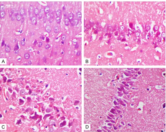
Figure 2. MTX induces neuronal pyknosis (×400). The neurons in control group displayed regular appearance with large and round nuclei but dense-ly stained, and distorted cells with pyknotic appearance were observed in MTX60, MTX100 and MTX250 group. A. control group; B. MTX60 group; C. MTX100 group; D. MTX250 group.
(P<0.01). There were no sig-nificant differences between MTX-treated and controls rats in escape latency on the first day. On the fourth day, MTX group rats spent more time to arrive the platform than con-trol group (P<0.05).
To observe memory retrieval, we analyzed the time that the rats spent in the quadrant fixed platform during the pro- be trial. As shown in Figure 1B, during re-training on the fifth day post treatment, rats treated with MTX appeared to perform worse than control rats, and MTX rats spent less time searching (27.3±3.98) the target quadrant as com-pared with the control animals (33.3±6.3) (P<0.05).
Effects of MTX on cellular morphology of hippocampal neurons in CA1 area
As compared with the control group, the number of neurons with nuclear pyknosis in MTX groups of the rats increased significantly in hippocampus CA1 area. Moreover, as co- mpared to the MTX60 group, the neuronal degeneration in MTX100 and MTX250 group was more severe. The neurons in control group displayed regu-lar appearance with regu-large and round nuclei but densely sta- ined, and distorted cells with pyknotic appearance were ob- served in MTX60, MTX100 and MTX250 groups (Figure 2).
[image:4.612.90.376.351.579.2]more TUNEL positive cells could be observed in the MTX100 and MTX250 group. The percentage of apoptotic hippocampal neurons in the con-trol, MTX60, MTX100 and MTX250 groups were 5.47±2.71%, 16.71±6.34%, 36.85±3.85% and 48.90±7.28%, respectively. The number of TUNEL-positive cells in MTX250 group increased nearly 9-fold more than the control group (Figure 3B). These findings indicate that MTX induced hippocampal neuronal death, as the dose of MTX increased, the apoptosis rate of hippocampus neurons increased (P<0.01).
Effects of MTX on the expression of CHOP and caspase-12 in hippocampal tissues
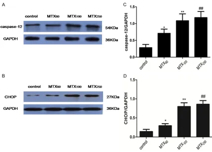
Western blot analysis showed that the expres-sion levels of CHOP and caspase-12 in MTX60, MTX100 and MTX250 groups significantly incr- eased as compared to the control group (P<0.05) (Figure 4A, 4C). Among the MTX60, MTX100 and MTX250 groups, as the dose of MTX increased, the expression levels of CHOP and caspase-12 increased (P<0.01 and P<0.01). However, there were no significant differences between MTX100 and MTX250 in the expression
levels of CHOP and caspase-12 (P>0.05) (Fig- ure 4B, 4D).
Immunofluorescence staining of caspase-12 and CHOP in hippocampal tissues
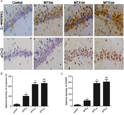
[image:5.612.87.520.76.413.2]230.37±19.51 in the MTX250 group as com-pared to the MTX group respectively. However, there were also no significant differences between MTX100 and MTX250 in the optical den-sity for CHOP and caspase-12 (Figure 5B, 5C). Discussion
According to some recent clinical researches, most of the ALL patients may present with dif-ferent degrees of impairments in attention, ver-bal and visual memories after chemotherapy [14]. Besides, several experimental studies have demonstrated that MTX may contribute to memory impairment by inducing apoptosis and inhibiting proliferation of hippocampal cell in the rat [15-17]. Furthermore, ERS-related neu-ronal apoptosis plays an important role in vari-ous kinds of neurodegenerative diseases, how-ever, there is few finding about the role of ERS played in MTX-induced neurotoxicity and cogni-tive dysfunction. Therefore, we hypothesized that ERS either mediated the apoptosis of
hip-pocampal neurons or induced memory impair-ment after the administration of high-dose MTX in adult rats.
In order to assess the effects of MTX on the memory and learning ability of rats, a widely used Morris water maze test was implemented in this study. In the Morris water maze test, we observed that MTX-treated rats took longer to find the escape platform location and shorter time to swim in the escape platform site com-pared to controls (Figure 1A, 1B). These results are similar to those of those of previous studies in which animals treated with MTX showed a defect in cognitive performance [6]. Therefore, we could conclude that MTX do induce im- pairment in spatial learning and recognition memory.
[image:6.612.91.517.68.374.2]bic system and associated with affective, cog-nitive and behavioral functions. The major result of the present study is that MTX treat-ment significantly increased the number of degenerated neuronal and the percentage of TUNEL positive neurons in hippocampus CA1 area (Figures 2 and 3). This is consistent with previous studies that discovered the number of neurons and neuroglial cells decreased in the CA3, CA4 of the hippocampus after intrathecal injection of MTX in rats [18]. Further, the pres-ent results also show that the hippocampal cell apoptosis increased as the dose of MTX increased. These findings indicated that meth-otrexate-related cognitive deficits may be
asso-ciated with the apoptosis of hippocampus ne- urons.
[image:7.612.93.524.75.473.2]ed, accompanied by an up-regulation of CHOP expression, cleavage of poly (ADP-ribose) poly-merase (PARP) and increase of cell apoptosis [23-26]. In the present study, we found that MTX exposure could up-regulate the protein expression of CHOP and caspase-12 in the hip-pocampus of adult rats, and there were no significant differences between MTX100 and MTX250 groups in the protein expression level (Figures 4 and 5). These results revealed that MTX-induced neuron apoptosis may be related to the ERS and, there is no obvious different in the protein expression of CHOP and caspase-12 between MTX100 and MTX250 group. However the protein expression of CHOP and caspase-12 was not in conformity with TUNEL statistical results. The percentage of TUNEL positive neu-rons of MTX250 group was more than the other three groups. It suggested that other apoptosis mechanism may involve in high-dose MTX-in- duced hippocampus apoptosis. Therefore, we need further studies for exploring it.
In summary, the present study demonstrated that MTX could induce hippocampal neuronal apoptosis and lead to cognitive impairment in adult rats through ERS pathway.
Acknowledgements
This study was supported by the Natural Science Foundation of Guangdong Province (2014A030313331).
Disclosure of conflict of interest
None.
Address correspondence to: Li-Hua Yang, Pediatric Center of Zhujiang Hospital, Southern Medical Uni- versity, No. 253, Middle Gongye Road, Haizhu Dis- trict, Guangzhou 510282, Guangdong Province, China. Fax: +02085143020; E-mail: dryanglihua@ 163.com
References
[1] Schrappe M, Reiter A, Riehm H. Prophylaxis and treatment of neoplastic meningeosis in childhood acute lymphoblastic leukemia. J Neurooncol 1998; 38: 159-165.
[2] Shuper A, Stark B, Kornreich L, Cohen IJ, Avin-er S, Steinmetz A, Stein J, Goshen Y, Yaniv I. Methotrexate treatment protocols and the cen-tral nervous system: significant cure with sig-nificant neurotoxicity. J Child Neurol 2000; 15: 573-580.
[3] Buizer AI, de Sonneville LM, van den Heuvel-Eibrink MM, Veerman AJ. Chemotherapy and attentional dysfunction in survivors of child-hood acute lymphoblastic leukemia: effect of treatment intensity. Pediatr Blood Cancer 2005; 45: 281-290.
[4] Buizer AI, de Sonneville LM, Veerman AJ. Ef-fects of chemotherapy on neurocognitive func-tion in children with acute lymphoblastic leuke-mia: a critical review of the literature. Pediatr Blood Cancer 2009; 52: 447-54.
[5] Cole PD, Kamen BA. Delayed neurotoxicity as-sociated with therapy for children with acute lymphoblastic leukemia. Ment Retard Dev Dis-abil Res Rev 2006; 12: 174-83.
[6] Yang M, Kim JS, Kim J, Kim SH, Kim JC, Kim J, Wang H, Shin T, Moon C. Neurotoxicity of meth-otrexate to hippocampal cells in vivo and in vi-tro. Biochem Pharmacol 2011; 82: 72-80. [7] Fardell JE, Vardy J, Logge W, Johnston I. Single
high dose treatment with methotrexate causes long-lasting cognitive dysfunction in laboratory rodents. Pharmacol Biochem Behav 2010; 97: 333-9.
[8] Rao RV, Ellerby HM, Bredesen DE. Coupling en-doplasmic reticulum stress to the cell death program. Cell Death Differ 2004; 11: 372-80. [9] Malhotra JD and Kaufman RJ. Endoplasmic re-ticulum stress and oxidative stress: a vicious cycle or a double-edged sword? Antioxid Redox Signal 2007; 9: 2277-93.
[10] Lindholm D, Wootz H, Korhonen L. ER stress and neurodegenerative diseases. Cell Death Differ 2006; 13: 385-92.
[11] Kuznetsov JN, Leclerc GJ, Leclerc GM, Barredo JC. AMPK and Akt determine apoptotic cell death following perturbations of one-carbon metabolism by regulating ER stress in Acute Lymphoblastic Leukemia. Mol Cancer Ther 2011; 10: 437-447.
[12] Kuznetsov JN, Leclerc GJ, Leclerc GM, Barredo JC. AMPK and Akt determine apoptotic cell death following perturbations of one-carbon metabolism by regulating ER stress in Acute Lymphoblastic Leukemia. Mol Cancer Ther 2011; 10: 437-47.
[13] Liu X, Wang M, Chen H, Guo Y, Ma F, Shi F, Bi Y, Li Y. Hypothermia protects the brain from tran-sient global ischemia/reperfusion by attenuat-ing endoplasmic reticulum response-induced apoptosis through CHOP. PLoS One 2013; 8: e53431.
[14] Espy KA, Moore IM, Kaufmann PM, Kramer JH, Matthay K, Hutter JJ. Chemotherapeutic CNS prophylaxis and neuropsychologic change in children with acute lymphoblastic leukemia: a prospective study. J Pediatr Psychol 2001; 26: 1-9.
Buwalda B. Long-lasting suppression of hippo-campal cell proliferation and impaired cogni-tive performance by methotrexated in the rat. Behav Brain Res 2008; 186: 168-75.
[16] Yang M, Kim JS, Kim J, Jang S, Kim SH, Kim JC, Shin T, Wang H, Moon C. Acute treatment with methotrexate induces hippocampal dysfunc-tion in a mouse model of breast cancer. Brain Res Bull 2012; 89: 50-6.
[17] Seigers R, Schagen SB, Coppens CM, van der Most PJ, van Dam FS, Koolhaas JM, Buwalda B. Methotrexate decreases hippocampal cell proliferation and induces memory deficits in rats. Behav Brain Res 2009; 201: 279-84. [18] Madhyastha S, Somayaji SN, Rao MS, Nalini K,
Bairy KL. Hippocampal brain amines in metho-trexate-induced learning and memory deficit. Can J Physiol Pharmacol 2002; 80: 1076-84. [19] Tabas I, Ron D. Integrating the mechanisms of
apoptosis induced by endoplasmic reticulum stress. Nat Cell Biol 2011; 13: 184-90. [20] Zhao L and Ackerman SL. Endoplasmic
reticu-lum stress in health and disease. Curr Opin Cell Biol 2006; 18: 444-52.
[21] Agarwal NK, Mueller GA, Mueller C, Streich JH, Asif AR, Dihazi H. Expression proteomics of acute promyelocytic leukaemia cells treat- ed with methotrexate. Biochim Biophys Acta 2010; 1804: 918-28.
[22] Herman S, Zurgil N, Deutsch M. Low dose methotrexate induces apoptosis with reactive oxygen species involvement in T lymphocytic cell lines to a greater extent than in monocytic lines. Inflamm Res 2005; 54: 273-80.
[23] Guan L, Han B, Li Z, Hua F, Huang F, Wei W, Yang Y, Xu C. Sodium selenite induces apopto-sis by ROS-mediated endoplasmic reticulum stress and mitochondrial dysfunction in hu-man acute promyelocytic leukemia NB4 cells. Apoptosis 2009; 14: 218-25.
[24] Hayashi T, Saito A, Okuno S, Ferrand-Drake M, Dodd RL, Nishi T, Maier CM, Kinouchi H, Chan PH. Oxidative damage to the endoplasmic re-ticulum is implicated in ischemic neuronal cell death. J Cereb Blood Flow Metab 2003; 23: 1117-28.
[25] Jiang X, Kanda T, Nakamoto S, Haga Y, Sasaki R, Nakamura M, Wu S, Mikata R, Yokosuka O. Knockdown of glucose-regulated protein 78 enhances poly (ADP-ribose) polymerase cleav-age in human pancreatic cancer cells exposed to endoplasmic reticulum stress. Oncol Rep 2014; 32: 2343-8.