Original Article
Hashimoto’s thyroiditis or/and thyroid cancer in
patients with diffuse large B-cell lymphoma
Yuanfang Yue1*, Jing Ma1*, Qian Li1, Tiantian She2, Han Li1, Su Liu1, Lin Chen1, Tinghui Yan1, Shuang Gao1, Zeng Cao1, Yong Yu1, Xiaofang Wang1, Hongliang Yang1, Haifeng Zhao1, Yizhuo Zhang1, Yafei Wang1
1Department of Hematology and Blood and Marrow Transplantation, Tianjin Medical University Cancer Institute
and Hospital. National Clinical Research Center for Cancer; Tianjin Key Laboratory of Cancer Prevention and Therapy, Tianjin, China; 2School of Medical Laboratory Science, Tianjin Medical University, Tianjin, China. *Equal
contributors and co-first authors.
Received April 7, 2016; Accepted June 13, 2016; Epub September 1, 2016; Published September 15, 2016
Abstract: Objectives Diffuse large B-cell lymphoma (DLBCL) is an aggressive and highly heterogeneous malignancy. However, its pathogenesis remains not so clear, since other diseases like thyroid problems have recently been re-ported to accompany with DLBCL. Therefore, we evaluated the association between thyroid diseases and DLBCL,
aiming to figure out their relationship and find valuable clues for the treatment of DLBCL. Method A total of 214 DLBCL patients who all had thyroid ultrasound examinations as part of the routine work-up were collected in this study from our hospital between 2010 and 2014. They were classified into three groups according to the results of thyroid ultrasound examination: Hashimoto’s thyroiditis (HT), suspected thyroid cancer (TC) and thyroid nodules/nor-mal groups. We comparatively analyzed the clinical characteristics, treatment response and prognosis among the three groups. Results Patients with HT, accounting for 18.7% (40/214) of all DLBCLs, were predominantly elderly females. They presented with the early-stage germinal center B cell-like (GCB) DLBCL and showed a better response to anti-DLBCL therapy than the other two groups. Patients with suspected TC accounted for 8.4% (18/214) of all DLBCLs, among which 4 patients were pathologically diagnosed with TC from the 6 patients who underwent thyroid biopsy. Additionally, there was no statistically significant difference in survival among the three groups, probably due to the short follow-up. Conclusion The high concurrency of DLBCL and thyroid diseases indicated an association between them. We thus propose that thyroid examination is necessary for DLBCL patients.
Keywords: Diffuse large B-cell lymphoma, Hashimoto’s thyroiditis, thyroid cancer, association, prognosis
Introduction
Diffuse large B-cell lymphoma (DLBCL), the most common subtype of lymphoma, is an aggressive and highly heterogeneous malig-nancy. With the development of intensive che-motherapy and novel targeted agents such as rituximab, anti-DLBCL therapy could lead to prolonged lifespan in patients of DLBCL. However, the pathogenesis of DLBCL remains unknown. It was reported to be associated with
immunodeficiency, autoimmune diseases or
viral infections [1].
Hashimoto’s thyroiditis (HT), an inflammatory
autoimmune disorder that has affected 4% of all females, is the most common cause for hypothyroidism [2]. HT is usually diagnosed by
MALT lymphoma arose from pre-existing HT, or, HT-indu-
ced lymphocytic infiltration
of thyroid gland. Additionally, salivary gland MALT lympho-ma was also found to co-exist with SS [9]. Furtherm- ore, Troch et al. demonstrat-ed that HT was not only associated with thyroidal MALT lymphoma, but also with non-thyroidal MALT ly- mphoma [10]. All these indi-cated a possible associa-tion between HT and lym-phoma. However, whether DLBCL co-exists with HT and how they are mutually associated has not been assessed so far.
In addition, we fortuitously found a few cases of DLBCL concomitant with thyroid cancer (TC). It’s also uncer-tain whether DLBCL is dire- ctly associated with TC, or HT acts as a bridge between them. Therefore, the objec-tive of this study is to ana-lyze the clinical characteris-tics and prognosis of all the cases of DLBCL concomi-tant with HT/TC and assess the relationship between DLBCL and HT/TC, thus pro-viding a better guidance for clinical practice.
Patients and methods
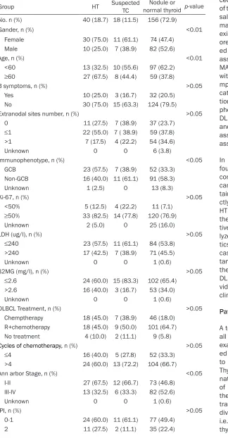
[image:2.612.92.416.100.722.2]A total of 214 DLBCL cases all having thyroid ultrasound examinations were collect-ed in this study from 2010 to 2014 at our institution. Thyroid ultrasound exami-nations were taken as part of routine work-up for all of them. According to their ul- trasound results they were divided into three groups, i.e. HT, suspected TC and thyroid nodules/normal gr- Table 1. Clinical characteristics of DLBCL patients among three
groups
Group HT Suspected TC normal thyroidNodule or p-value
No. n (%) 40 (18.7) 18 (11.5) 156 (72.9)
Gander, n (%) <0.01
Female 30 (75.0) 11 (61.1) 74 (47.4)
Male 10 (25.0) 7 (38.9) 82 (52.6)
Age, n (%) <0.01
<60 13 (32.5) 10 (55.6) 97 (62.2)
≥60 27 (67.5) 8 (44.4) 59 (37.8)
B symptoms, n (%) >0.05
Yes 10 (25.0) 3 (16.7) 32 (20.5)
No 30 (75.0) 15 (63.3) 124 (79.5)
Extranodal sites number, n (%) >0.05
0 11 (27.5) 7 (38.9) 37 (23.7)
≤1 22 (55.0) 7 ( 38.9) 59 (37.8)
>1 7 (17.5) 4 (22.2) 54 (34.6)
Unknown 0 0 6 (3.8)
Immunophenotype, n (%) <0.05
GCB 23 (57.5) 7 (38.9) 52 (33.3)
Non-GCB 16 (40.0) 11 (61.1) 91 (58.3)
Unknown 1 (2.5) 0 13 (8.3)
Ki-67, n (%) >0.05
<50% 5 (12.5) 4 (22.2) 11 (7.1)
≥50% 33 (82.5) 14 (77.8) 120 (76.9)
Unknown 2 (5.0) 0 25 (16.0)
LDH (ug/l), n (%) >0.05
≤240 23 (57.5) 11 (61.1) 84 (53.8)
>240 17 (42.5) 7 (38.9) 71 (45.5)
Unknown 0 0 1 (0.6)
β2MG (mg/l), n (%) >0.05
≤2.6 24 (60.0) 15 (83.3) 102 (65.4)
>2.6 16 (40.0) 3 (16.7) 53 (34.0)
Unknown 0 0 1 (0.6)
DLBCL Treatment, n (%) >0.05
Chemptherapy 18 (45.0) 7 (38.9) 46 (18.0)
R+chemotherapy 18 (45.0) 9 (50.0) 101 (64.7)
No treatment 4 (10.0) 2 (11.1) 9 (5.8)
Cycles of chemotherapy, n (%) >0.05
≤4 16 (40.0) 5 (27.8) 52 (33.3)
>4 24 (60.0) 13 (72.2) 104 (66.7)
Ann arbor Stage, n (%) <0.05
I-II 27 (67.5) 12 (66.7) 73 (46.8)
III-IV 13 (32.5) 6 (33.3) 82 (52.6)
Unknown 0 0 1 (0.6)
IPI, n (%) >0.05
0-1 24 (60.0) 11 (61.1) 77 (49.4)
oups. The clinical characteristics, treatment response and prognosis among the three gr- oups were comparatively analyzed. For detailed information, see Table 1.
The diagnostic criteria for DLBCL are based on
the 2008 WHO classification for DLBCL [1]. HT can be diagnosed (defined by Colin et al.) by
such indicators as markedly elevated levels of serum anti-thyroglobulin antibodies (anti-TG) or/and thyroid peroxidase (TPO) anti-bodies, a hypoechogenetic appearance on thy-roid ultrasound and histopathology of thythy-roid
fine-needle biopsies or surgical specimen [5].
As for TC, thyroid imaging reporting and data
system (TI-RADS) score ≥4 implied high risk of
malignancy and its diagnosis was based on the
2004 WHO classification for thyroid tumors
[11, 12]. The systemic examinations for all pa- tients included serological checkup and imag-ing examinations such as thyroid ultrasound, Computed Tomography (CT), X-radiation, Mag- netic Resonance Imaging (MRI), and Positron Emission Tomography-computed Tomography (PET-CT).
Statistical analysis
All data was processed using SPSS 19.0, and categorical variables were analyzed using Chi-square test.Over survival (OS) was defined as
the length of time from the date of diagnosis to the date of death or the end of follow-up. Kaplan-Meier method was used for survival analysis, with the differences between groups
transformed DLBCL (one derived from stage III follicular lymphoma (FL) and the other from MALT lymphoma). The remaining 22 cases, however, suffered from non-thyroidal lympho-ma, with only one case of transformed DLBCL (from stage III FL). There were 12 cases of nodal lymphoma and 10 cases of extra-nodal lymphomas, the latter comprised of 4 cases of HP-positive gastric lymphoma, 1 case of pulmo-nary lymphoma, 1 case of mediastinal lympho-ma, 1 case of intestinal lympholympho-ma, 1 case of omentum majus lymphoma, 1 case of parotid and submandibular glands lymphoma and 1 case of tonsil lymphoma.
4 cases (n=40, 10.0%) had a history of HT prior to the diagnosis of DLBCL while the other 36 cases were diagnosed with HT in addition dur-ing DLBCL stagdur-ing. 9 cases took levothyroxine orally due to low serum fF4 levels. 7 cases (n=40, 17.5%) had other immune-or infection-related diseases, 3 with a history of drug aller-gy, 1 with SS, 1 with a 10-year history of both SS and rheumatoid arthritis and 2 with chronic hepatitis B.
27 cases were clinically diagnosed as stage I-II DLBCL and 13 diagnosed as stage III-IV DLBCL. 18 cases were treated with rituximab in combi-nation with chemotherapy and another 18 cases were treated with traditional chemother-apy, altogether leading to 18 cases (n=40,
45%) of complete remission/uncertified com -plete remission (CR/CRu). 4 cases rejected treatment. At the end of follow-up (median
3 5 (12.5) 5 (27.8) 31 (19.9)
4-5 0 0 13 (8.3)
Response, n (%) <0.05
PD 6 (15.0) 6 (33.3) 50 (32.1)
SD 3 (7.5) 1 (5.6) 5 (3.2)
PR 3 (7.5) 0 24 (15.4)
CR/CRu 18 (45.0) 8 (44.4) 62 (39.7)
Relapse 10 (25.0) 3 (16.7) 7 (4.5)
Unknown 0 0 8 (5.1)
Follow-up, n (%)
Alive 21 (52.5) 7 (38.9) 79 (50.6)
Death 12 (30.0) 6 (33.3) 58 (37.2)
Unknown 7 (17.5) 5 (27.8) 20 (12.8)
Abbreviations: DLBCL, diffuse large B-cell lymphoma; HT, Hashimoto’s thyroiditis; TC, thyroid cancer; GCB, germinal center B-cell origin; IPI, International Prognostic Index; PD, progress disease; SD, steady disease; PR, partial response; CR, complete remis-sion; CRu, uncertified complete remission.
analyzed by the log-rank test. A P<0.05 was consid-ered to be statistically sig-
nificant.
Results
HT group
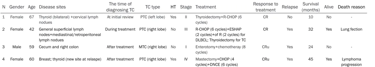
Table 2. Clinical characteristics of DLBCL patients with TC
N Gender Age Disease sites diagnosing TCThe time of TC type HT Stage Treatment Response to treatment Relapse (months) AliveSurvival Death reason
1 Female 67 Thyroid (bilateral) +cervical lymph nodues
At initial review PTC (left lobe) Yes II Thyroidectomy+R-CHOP (6 cycles)
CR No 10 No
-2 Female 42 General superficial lymph
nodes+mediastinal/retroperitoneal lymph nodues
During treatment PTC (right lobe) No III R-CHOP (6 cycles)+ESHAP (2 cycles)+of R (2 cycles) for DLBCL; Thyroidectomy for TC
CR Yes 32 Yes Lung fection
3 Male 59 Cecum and right colon After treatment MTC (right lobe) No I Enterotomy+chemotheray (8 cycles)
CRu Yes 24 No
-4 Female 60 Breast; thyroid (new site at relaspe) After treatment PTC (right lobe) Yes IV Mastectomy+CHOP (4 cycles)+DNCE (6 cycles)
CRu Yes 45 Yes Lymphoma progression
od: 12 months), there were 12 deaths (n=40, 30%) and the mean survival time was 38 months.
Suspected TC group
18 cases (n=214, 8.4%) of DLBCL were diag-nosed as TI-RADS 4, indicating high risk for TC.
Therefore, these 18 cases were classified as
suspected TC group. They were 11 females and 7 males, with the median age of 59 years (range 42-76 years). Only 6 cases (n=18, 33.3%) underwent thyroidal biopsy, among which 3 cases was pathologically diagnosed with papil-lary thyroid carcinoma (PTC) and 1 diagnosed with medullary thyroid carcinoma (MTC). One
case of confirmed TC was a 67-year-old female
who was initially diagnosed with PTC, HT and thyroid DLBCL. She achieved CR after thyroid-ectomy and 6 cycles of R-CHOP (rituximab plus cyclophosphamide, doxorubicin, vincristine and prednisone). However, she died of lung infec-tion eventually, with survival time up to 10
months. Another case of confirmed TC was a
42-year-old female diagnosed with nodal DLBCL. She suffered from a gradual enlarge-ment of thyroid gland after the lymphoma
treat-ment and later on was definitely diagnosed with
PTC. After the thyroidectomy she reached CR. A
third case of confirmed TC was a 59-year-old
male. He received an 8-month chemotherapy regimen due to the lymphoma of the cecum and right hemicolon, and reached CRu. Two months later, however, he was diagnosed with MTC. He died of lymphoma relapse ultimately, with the survival time up to 24 months. The last
case of confirmed TC was a 60-year-old female
with breast DLBCL. After the treatment of mas-tectomy and 4 cycles of CHOP regimen, she reached CR. Nonetheless, DLBCL relapsed in new locations (thyroid and systemic lymph nodes) within half a year. Results of histopatho-logical analysis after thyroidectomy showed a complicated pathological type: HT and MALT lymphoma in the left lobe of thyroid gland, with local lymphoma cells transforming into large cells, and PTC in the right lobe of thyroid gland. Following the treatment of 6 cycles of DECN (dexamethasone, navelbine, cisplatin and eto-poside), she returned to CR. Two above live
cases of confirmed TC kept alive throughout
the follow-up period, with no sign of disease progression. The detailed information can be seen in Table 2.
In the remaining 12 cases not undergoing thy-roid pathological examination, 6 had nodal DLBCL and 6 had extra-nodal DLBCL (1 at the skin of right lower limb, 1 at thyroid, 1 at medi-astinum, 1 at ilium and 2 at tonsil). In terms of other immune-or infection-related diseases, 1 case had suffered from systemic lupus erythe-matosus (SLE) for 20 years, with a history of allergy to antibiotics, 2 had both TC and HT and 1 had hepatitis B.
8 cases (n=18, 44.4%) were diagnosed at high risk for TC during the treatment of DLBCL and 3 out of them were eventually pathologically
con-firmed with TC through thyroid biopsy. For thera -peutic methods for DLBCL, 7 cases underwent R-CHOP chemotherapy, either alone or in combination with DNCE or RESHAP, and only 1 case received radiotherapy and 4 cycles of rituximab.
In respect to thyroid-related treatments, 4 cases underwent total thyroidectomy rather than chemotherapy or radiotherapy when they
had been pathologically confirmed with TC by
the frozen section technique. 12 cases received routine thyroid examination only. At the end of the follow-up (median period: 12 months), there were 6 deaths (n=18, 33%), with the mean sur-vival time up to 31 months.
Thyroid nodules/normal thyroid group
There were 74 females and 82 males in thyroid nodules/normal thyroid group, with the median age of 57 years (range 3-92 years). 109 cases (n=156, 69.8%) had thyroid nodules, 101 out of which were diagnosed with TI-RADS 1-2 and the rest diagnosed with TI-RADS 3. 14 cases had hepatitis B, 2 cases with family history of can-cer, 1 with a history of syphilis infection, 8 with a history of drug allergy, 1 with ankylosing spondylitis, 1 with rheumatoid arthritis and 1 having liver transplantation due to severe hepa-titis A. 2 cases also had bladder cancer and liver cancer while 1 case had suffered from breast cancer 5 years before the diagnosis of DLBCL.
By analyzing many clinical and prognostic parameters, we found that there was
statisti-cally significant difference in gender, age,
imm-unophenotype, staging and treatment response among the three groups. In contrast with the other two groups, DLBCL concomitant with HT were likely to affect elderly females. Additionally, they mostly presented with early-stage DLBCL and showed better response to treatment. In
spite of no significant difference in survival
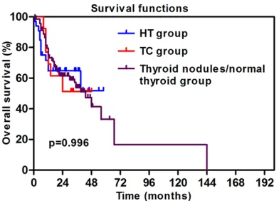
time among the three groups, thyroid nodules/ normal thyroid group was the best group while suspected TC group was the worst in terms of mean survival time (Figure 1).
Discussion
To date, there has been no report studying the link between HT and DLBCL with large samples, except 3 reports of rare cases [13-15]. Our data revealed that the incidence of HT in DLBCL patients was 18.7%, higher than those in gen-eral population (5-6%) [4] and MALT patients (16%)[10]. In the HT group, 18 cases had thy-roidal DLBCL and 22 had non-thythy-roidal DLBCL. Our data suggested that HT might be a high risk factor for DLBCL, no matter whether lymphoma was localized within the thyroid gland or not. Catarina Dias et al. summarized from large data that in patients with autoimmune rheu-matic diseases over-expression of regulatory cytokine BAFF (B-cell activating factor) could lead to B cell dysregulation and hyperactivity and Wang et at demonstrated that immune conditions were in coordination with immune-regulatory genes (TNF G308A and IL10 T3575A) polymorphisms which reported as risk factors
for non-Hodgkin lymphoma (NHL), both contrib-uting to the formation of lymphoma [16, 17]. Therefore, immune system dysfunction plays an important role in the development of DLBCL. However, whether DLBCL shares similar patho-genesis with HT requires further investigation. TC, the most common endocrine malignancy, was reported to be the fastest growing cancer in the United State in the last decade [18]. In China, the morbidity of TC increased gradually from year to year, especially in females [19]. Despite being a relatively indolent malignancy, TC relapsed frequently, thus leading to inc- reased mortality [18-20]. Most scholars insist-ed that thyroid nodules and HT were inclininsist-ed to develop into TC, but it was still controversial whether HT was a high risk factor for TC [21-23]. According to a Meta-analysis, 20% of thy-roid nodules were diagnosed as TC [24]. To improve the accuracy of thyroid ultrasound for TC diagnosis, thyroid imaging reporting and data system (TI-RADS) was introduced by Kwak et al. It had a sensitivity of 93% and a negative predictive value of 100% for TC diagnosis [11]. In our study, 18 cases were suspected as TC, 4 cases out of which were pathologically
con-firmed as TC through thyroid biopsy. However,
we didn’t get the accurate incidence of TC in DLBCL patients. There have been only two reports describing one mere case of thyroid MALT concomitant with both TC and HT so far, without elucidation of its pathogenesis howev-er [25, 26]. In this study, we reported 4 cases of DLBCL concomitant with both TC and HT (n=18, 22.2%). However, we still couldn’t deduce HT as the bridge linking DLBCL and TC due to a small sample. Katoh et al. observed an over-expres-sion of Forkhead-box (FOX) family which in- volved in transcription regulation and DNA repair in multiple cancers including TC and DLBCL [27] and maybe FOX overexpression possibly underlay the co-existence of TC and DLBCL.
[image:6.612.92.288.71.212.2]There were a few limitations in our study. Firstly, as a few DLBCL patients were diagnosed con-comitantly with TC during or after the treatme- nt of DLBCL, it was possible that treatment-related factors contributed to TC, which, how-ever, needed to be explored. Secondly, there was a lack of detailed and complete informa-tion regarding genetic aberrainforma-tions in DLBCL
Figure 1. Kaplan-Meier estimates for the OS rates
patients, such as BCL-2, C-MYC and EBV. As a result, this part of data was not shown.
In conclusion, DLBCL may be associated with HT/TC. It was necessary for clinicians to include thyroid examination in DLBCL diagnosis, in case of missed diagnosis of thyroid malignan-cy. Additionally, patients with HT should under-go thyroid examination regularly as well, in case of the development of DLBCL (thyroidal or non-thyroidal) or/and TC. On the other hand, we
should go on with this follow-up to figure out whether HT or TC influences the survival time of
DLBCL patients. Meanwhile, the common pathogenesis of the three diseases requires to be investigated, for it will provide new insights
for the identification of effective therapeutic
targets.
Acknowledgements
This work was supported in part by Tianjin Science and Technology Support Program
(13ZCZCSY20300); Planned Scientific
Rese-arch Program of Tianjin Municipal Education Commission (20140112).
Disclosure of conflict of interest
None.
Address correspondence to: Yafei Wang, Tianjin Me- dical University Cancer Institute and Hospital, 1 Huanhuxi Road, Hexi District, Tianjin 30060, China. Tel: 0086-18622221250; E-mail: Drwang2005@ 163.com
References
[1] Swerdlow SH, Campo E, Harris NL, et al. WHO classification of tumors of haematopoietic and lymphoid tissues. 4th edition. Lyon: IARC Press; 2008. pp. 441.
[2] Repplinger D, Bargren A, Zhang YW, Adler JT, Haymart M and Chen H. Is Hashimoto’s thy-roiditis a risk factor for papillary thyroid can-cer? J Surg Res 2008; 150: 49-52.
[3] Weissel M, Mayr N and Zeitlhofer J. Clinical sig-nificance of autoimmune thyroid disease in myasthenia gravis. Exp Clin Endocrinol Diabe-tes 2000; 108: 63-65.
[4] Sheth S. Role of ultrasonography in thyroid dis-ease. Otolaryngol Clin North Am 2010; 43: 239-255.
[5] Harris NL, Jaffe ES, Diebold J, Flandrin G, Muller-Hermelink HK, Vardiman J, Lister TA and Bloomfield CD. The World Health Organiza
-tion classifica-tion of neoplasms of the hemato -poietic and lymphoid tissues: report of the Clinical Advisory Committee meeting--Airlie House, Virginia, November, 1997. Hematol J 2000; 1: 53-66.
[6] Wundisch T, Kim TD, Thiede C, Morgner A, Al-pen B, Stolte M and Neubauer A. Etiology and therapy of Helicobacter pylori-associated gas-tric lymphomas. Ann Hematol 2003; 82: 535-545.
[7] Isaacson PG. Extranodal lymphomas: the MALT concept. Verh Dtsch Ges Pathol 1992; 76: 14-23.
[8] Wohrer S, Troch M, Streubel B, Zwerina J, Sk-rabs C, Formanek M, Hauff W, Hoffmann M, Mullauer L, Chott A and Raderer M. MALT lym-phoma in patients with autoimmune diseases: a comparative analysis of characteristics and clinical course. Leukemia 2007; 21: 1812-1818.
[9] Isaacson P, Norton A. Malignant lymphoma of the thyroid gland. In: Extranodal lymphomas. 1st edition. New York: Churchill Livingstone; 1994. pp. 103-115.
[10] Troch M, Woehrer S, Streubel B, Weissel M, Hoffmann M, Mullauer L, Chott A and Raderer M. Chronic autoimmune thyroiditis (Hashimo-to’s thyroiditis) in patients with MALT lympho-ma. Ann Oncol 2008; 19: 1336-1339.
[11] Kwak JY, Han KH, Yoon JH, Moon HJ, Son EJ, Park SH, Jung HK, Choi JS, Kim BM and Kim EK. Thyroid imaging reporting and data system for US features of nodules: a step in establish-ing better stratification of cancer risk. Radiolo -gy 2011; 260: 892-899.
[12] DeLellis RA, Lloyd RV, Heitz PU. World Health Organization Classification of Tumours. Pathol -ogy And Genetics Of Tumors Endocrine Organs. Lyon: IARC Press; 2014. pp. 136-166. [13] Ahmed T, Kayani N, Ahmad Z and Haque MN.
Non-Hodgkin’s thyroid lymphoma associated with Hashimoto’s thyroiditis. J Pak Med Assoc 2014; 64: 342-344.
[14] Agarwaf N, Wangnoo SK, Sidiqqi A and Gupt M. Primary thyroid lymphoma: a series of two cas-es and review of literature. J Assoc Physicians India 2013; 61: 496-498.
[15] Gulay K, Sukran E, Sumru Tanju, Ali S and Gul-sah K. Central nervous system lymphoma in a patient with Sjogren’s syndrome and autoim-mune thyroiditis (Hashimoto’s thyroiditis). Clin Rheumatol 2007; 26: 1377-1379.
[16] Dias C and Isenberg DA. Susceptibility of pa-tients with rheumatic diseases to B-cell non-Hodgkin lymphoma. Nat Rev Rheumatol 2011; 7: 360-368.
mecha-nisms in non-Hodgkin lymphoma: joint effects of the TNF G308A and IL10 T3575A polymor-phisms with non-Hodgkin lymphoma risk fac-tors. Cancer Res 2007; 67: 5042-5054. [18] Morris LG, Sikora AG, Tosteson TD and Davies
L. The increasing incidence of thyroid cancer: the influence of access to care. Thyroid 2013; 23: 885-891.
[19] Chen W, Zheng R, Zeng H, Zhang S and He J. Annual report on status of cancer in China, 2011. Chin J Cancer Res 2015; 27: 2-12. [20] Jemal A, Siegel R, Ward E, Hao Y, Xu J, Murray
T and Thun MJ. Cancer statistics, 2008. CA Cancer J Clin 2008; 58: 71-96.
[21] Larson SD, Jackson LN, Riall TS, Uchida T, Thomas RP, Qiu S and Evers BM. Increased in-cidence of well-differentiated thyroid cancer associated with Hashimoto thyroiditis and the role of the PI3k/Akt pathway. J Am Coll Surg 2007; 204: 764-773, 773-775.
[22] Salem IN, Ralph PT. Association of Hashimo-to’s thyroiditis and thyroid cancer. Curr Opin Oncol 2015; 27: 21-25.
[23] Chen YK, Lin CL, Cheng FT-F, Sung FC and Kao CH. Cancer risk in patients with Hashimoto’s thyroiditis: a nationwide cohort study. Br J Can-cer 2013; 109: 2496-2501.
[24] Brito JP, Gionfriddo MR, Al NA, Boehmer KR, Leppin AL, Reading C, Callstrom M, Elraiyah TA, Prokop LJ, Stan MN, Murad MH, Morris JC and Montori VM. The accuracy of thyroid nodule ul-trasound to predict thyroid cancer: systematic review and meta-analysis. J Clin Endocrinol Metab 2014; 99: 1253-1263.
[25] Melo GM, Sguilar DA, Petiti CM, Eichstaedt AG, Caiado RR and Souza RA. Concomitant thyroid Malt lymphoma and papillary thyroid carcino-ma. Arq Bras Endocrinol Metabol 2010; 54: 425-428.
[26] Jayaprakash K, Kishanprasad H, Hegde P and Chandrika R. Hashimotos Thyroiditis with Co-existent Papillary Carcinoma and Non-hodgkin Lymphoma-thyroid. Ann Med Health Sci Res 2014; 4: 268-270.