Toward dipolar recoupling in macroscopically ordered
samples of membrane proteins rotating at the magic angle
Clemens Glaubitz, Marina Carravetta, Mattias Edén,
Malcolm H. Levitt
Physical Chemistry Division, Arrhenius Laboratory Stockholm University, S-10691 Stockholm, Sweden
e-mail: [email protected]
Abstract
MAS NMR spectroscopy can be combined with the advantages of uniaxially ordered samples of membrane proteins as demonstrated in the so-called MAOSS (magic angle oriented sample spinning) experiment. Under these conditions, dipolar recoupling methods can be used to determine the orientation of internuclear vectors with respect to the MAS rotor frame. However, most approaches to measure dipolar couplings yield angle ambiguities even in the static, non-spinning case. Here, we present the possibility to overcome these problems by deriving a new homonuclear double-quantum radio frequency pulse sequence based on an eightfold symmetry. Only dipolar Hamiltonian terms with spatial components m=±2 are recoupled with high efficiency allowing unambiguous angle determinations. Preliminary data demonstrate the applicability to oriented samples.
Introduction
Magic angle spinning (MAS) is an essential NMR technique for studying disordered samples of biological materials such as biomembranes [1]. The large variety of applications involves for example investigations of diffusive ligands bound to transport proteins and receptors [2,3], the possibility to study locations of protein segments within the membrane[4] and structural studies of isotope labelled, immobilized proteins[5,6].
Especially the structural description of membrane proteins is technically challenging since data about secondary and tertiary conformation as well as about location and orientation in the anisotropic membrane environment have to be obtained. The latter question is best approached, by using macroscopically ordered samples, which is routinely done for membrane-bound peptides using static NMR techniques [7,8]. Macroscopically ordered samples with well oriented lipids and proteins can be prepared either by aligning them as bilayers on glass disks [9,10] or by using bicelles [11-13]. Since the signal frequency observed from second rank tensor interactions (such anisotropic chemical shift, quadrupolar and dipolar coupling) depends on its orientation with respect to the field of interaction, sharp resonance lines would be observed if all tensors would have the same orientation with respect to the magnetic field B0. However,
spread), which is common for large proteins such as rhodopsin or purple membrane, causes a significant contribution to the linewidth making this static NMR approach less straightforward[14].
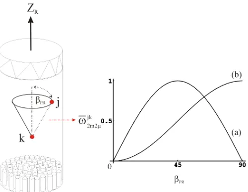
Fig. 1 Dipolar recoupling methods can be used to determine the orientation βPR of the
internuclear vectors between spins j and k with respect to the MAS rotor axis ZR in an ordered
sample (such as a macroscopically oriented membrane protein). However, most homo- as well as heteronuclear recoupling techniques used so far recouple terms of the dipolar Hamiltonian with spatial elements m=±1 which are scaled with sin2βPR leading to angle ambiguities (a).
Here it is shown that specific recoupling sequences can be designed which only use dipolar Hamiltonian terms with m=±2 (sin2β
PR) allowing an unambiguous determination of the
orientation of the dipolar coupling tensor (b)
[image:2.612.173.423.161.355.2]questions. Examples include the determination of the ligand orientation in bacteriorhodopsin [19] and bovine rhodopsin [20] as well as applications to peptides and lipids [15,21]. First experiments on oriented, multiple labelled samples of 15 N-Met-bacteriorhodopsin did show that in principle many angle constraints can be obtained simultaneously by deconvoluting 15N MAS sideband pattern (Glaubitz, C., Mason, J., Watts, A., unpublished results).
In all these cases – as in static NMR experiments using oriented samples - the anisotropies of deuterium quadrupolar or 13C and 15N chemical shift interactions were utilized. Analysing these data in terms of molecular structure however requires some knowledge about the tensor orientations within the molecular reference frame. Usually, the secondary structure has to be known in order to interpret especially the orientation of 15N CSA tensors[22,23]
A more direct approach would be possible by combining MAS on oriented samples with dipolar recoupling techniques. Selectively reintroducing dipolar coupling while maintaining the high spectral resolution achieved by MAS would allow determining directly the tilt angle βPR of the internuclear vector between spins j and k
with respect to the sample/rotor reference system (fig.1). However, most homo- and heteronuclear recoupling techniques used today feature a more or less complex orientational dependence on the Euler angles ΩPR. For example, the sequence C7 [24]
depends on sin2βPR while REDOR [25] and MELODRAMA [26] additionally depend
on cosγPR. The only exception described so far is rotational resonance at the n=2
condition [35]. However, this method generally requiring high spinning frequencies is limited to samples with appropriate differences in isotropic chemical shifts and is sensitive to interference from the chemical shift anisotropies.
Here, we demonstrate the design of specific recoupling schemes, which only recouple terms with sin2βPR dependence allowing unambiguous angle determination.
The method is broadband with respect to isotropic shifts, is robust with respect to chemical shift anisotropy and does not require very rapid sample spinning.
Theory
It has been shown in the past, that symmetry arguments can be exploited to design rf pulse sequences for tasks like homonuclear recoupling or heteronuclear decoupling in the presence of sample rotation [24,27-29]. Two symmetry classes, denoted as CNνn and
ν
n
RN , were discovered [27,29]. Sequences based on the symmetry class CNνn are
specified by three integer number N,n,ν with the following properties: (a) they consist of phase-shifted repetitions of a rf cycle C, each of which returns the irradiated spins to their initial state (in absence of other interactions), (b) each cycle has the duration
τC=(n τr/N) with τr=|2π/ωr|, i.e. N rf cycles span n rotor periods, and (c) the phase of the
qth cycle Cq is given by Φq=2πνq/N (q=0,1,2…N-1). The following selection rules for
0 ) 1 ( = λµ lm
H if mn−µν ≠ N×integer number (1)
0
) 2 (
:2 22 2 1 1 1
1mλµ lmλµ
=
lH
if × ≠ + − + × ≠ − × ≠ − number integer ) ( ) ( number integer number integer 1 2 1 2 2 2 1 1 N n m m N n m N n m ν µ µ ν µ ν µ (2)
A particular spin interaction is classified by index l referring to its spatial rotational rank (with m=-l,-l+1,…,0,…,+l) and λ denoting the rotational rank of spin polarization rotations by the resonant rf field (with µ=-λ,-λ+1,…,0,…,+λ). In this notation, isotropic and anisotropic chemical shifts have l=0, λ=1 and l=2, λ=1, respectively. Homonuclear dipolar couplings are described by l=λ=2. These selection rules have been used successfully to construct a number of pulse sequences e.g. to perform double -quantum filtered dipolar recoupling using the symmetry C712which found widespread application. The simplified average Hamiltonian for 1
2 7
C is given to [24]
∑
= + − − −=
λµω λµ λµ ω ω
lm jk jk jk jk jk jk
lm T T T
H 2122 22 212 2 2 2
) 1 (
(3)
where T2jk±2 are second rank spin operators for the interactions between spins j and k. Explicit expressions for the magnitude
ω
lmjkλµare given in references [24] and [27].These terms are proportional to the reduced Wigner function 0 ( PR)
l m
d β which becomes
PR
d 32sin2β
2 1
0± =± (4)
in equation (3) leading to angle ambiguities in ordered systems (fig.1). This problem could be solved by analysing expressions (1) and (2) for symmetries which result in similar properties as C712but with m=±2 with an average Hamiltonian of the form
jk jk jk jk T T
H 2222 22 2 222 2 2
) 1 ( − − − +
=ω ω (5)
Using m=±2 the magnitudes
ω
lmjkλµare scaled withPR
d 2β
8 3 2
2
0± = sin (6)
leading to a monotonic angle dependence for 0≤βPR≤90o (fig.1b). At the same time, for
optimal results, one has to ensure that chemical shift terms ω2m10 with m≠0 are
dipolar terms are suppressed. Each double quantum term should be associated with only a single spatial component m=2 or m=-2. This eliminates the orientation dependence on
γPR in the first order average Hamiltonian in favour of a maximum magnitude of the
effective dipolar Hamiltonian while creating a monotonic orientation dependence on βPR
(see fig.1).
A set of 23 possible symmetries in the range of 1≤N≤20, 1≤n≤5, 0≤ν≤10 are found analysing theorems (1) and (2). Here, we restrict ourselves to one particular solution using an eightfold symmetry with N=8, n=1, ν=3; denoted in the following as
3 1 8
C . The selection of particular space-spin components can be illustrated schematically using a
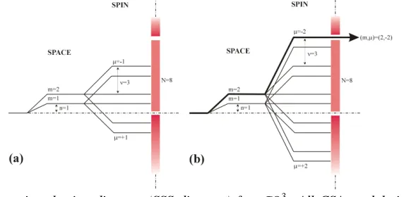
Fig. 2: Space-spin selection diagram (SSS diagram) for C813. All CSA modulation components are suppressed (a). One single 2Q dipole-dipole component with quantum numbers (m,µ )=(2,-2) is selected (b). The mirror images for m=-1 and m=-2 have been suppressed for simplicity.
space-spin selection diagram [28] as shown in Fig.2. Each level indicates a value of
mn-µν, while the barrier symbolises the inequality of theorem (1) [27]. As shown in fig.2a for CSA components (m={±1, ±2}, µ={0, ±1}) no pathway can be found to pass through the “selection wall”. This means that all CSA components are suppressed by
3 1 8
C at least in the first order average Hamiltonian. The fact that only homonuclear dipolar components with (m,µ)=(2,-2) and (-2,2) are allowed is shown in fig.2b. The terms with µ=±2 indicate double quantum coherence. Furthermore, each term is only associated with m=±2 rotational components which creates the desired monotonic orientation dependence required for studies on ordered systems.
Methods
[image:5.612.160.446.261.401.2]experiments. Usually, larger MAS rotors are necessary (6-7.5mm) for accommodating glass disks containing the protein sample. This limits power levels (up to 100kHz) and spinning rates (2-6kHz) to moderate levels, also due to sample stability aspects[18]. For
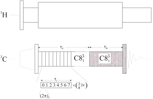
Fig. 3 Radio frequency pulse sequence for double quantum filtered measurements of dipolar couplings using C813. Eight rf cycles are timed to occupy one rotational period. Each cycle consi sts of one 2π pulse with a ¾ π phase increment. It has been shown, that distance data can be obtained by measuring the DQF efficiency as a function of the DQ excitation period τE while
keeping the reconversion time τR constant [30].
the demonstration here, we have chosen C0=(2π)0 which is stepwise phase incremented
by ¾ π as shown in fig.3. Eight C elements span one rotor period. The shaded sequence elements have variable phases according to standard procedures for selecting signals passing through double quantum coherence. The signal built-up can be measured for a variable double-quantum excitation time τE while keep the reconversion time τR
constant [28]. Oscillations in the observed built-up curve reveal the dipolar coupling between both spins. This approach has been shown to be especially useful in difficult situations, where the isotropic chemical shift of both spins are similar, such as in
[10,11-13C
2]-E-retinal[30].
The experimental situation encountered by performing MAS on oriented membrane proteins in MAOSS type of experiments (see fig.1) can be conveniently emulated by placing an isotope labelled single crystal in the MAS rotor. The carousel symmetry, i.e. the two-dimensional distribution of the protein about the MAS rotor axis can be mimicked by not synchronising the data acquisition with the sample rotation. In this case, a slow-speed 13C spectrum will only feature nearly absorptive sidebands in contrast to rotor -synchronized sampling [31,32]. A single crystal of [1,2-13C2]-glycine
[image:6.612.175.441.161.333.2]All experiments were performed using a Chemagnetics 200 Infinity spectrometer and a 4mm triple resonance probe.
Results and Discussion
The double-quantum filtering performance of C813 is shown in fig.4 for [1,2-13C2
]-glycine (a) and [10,11-13C2]-E-retinal (b). Conventional cross-polarized spectra at a
sample rotation rate of ωr/2π=5kHz are displaying both labelled sites in (a) and (b). The
sample of [10,11-13C2]-E-Ret was diluted to 10% in unlabelled material explaining the
additional intensities. The top spectra are the result of passing the cross-polarized signal through double-quantum coherence using the sequence in fig.3. Excitation and reconversion sequences were of the same length using q=24 elements. In both cases, ca 40% of the spin magnetization passed through the double -quantum filter. This shows, that C813 seems to be fairly robust with respect to differences in isotropic chemical shifts.
Fig. 4 MAS 13
C spectra of polycrystalline samples of [1,2-13
C2]-glycine (a) and [10,11-13
C2
]-E-retinal (b) with and without double quantum filtering using 3 1 8
C . Spectra were acquired at
ωr/2π=5kHz with τE=600µ, a CP contact time of 2ms and at 4.9T. Ca. 40% DQ efficiency is
achieved in both cases.
One possibility to determine dipolar couplings using these symmetries is to measure the observed double-quantum efficiency as a function of the excitation time τE
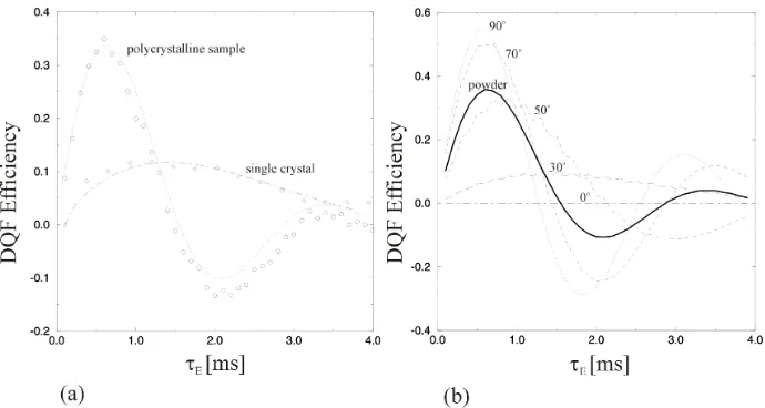
while keeping the reconversion sequences at constant length (Fig.3) [30]. The oscillations in the obtained built-up curves are a sensitive measure for the strength of the dipolar coupling. Fig.5a shows the signal built-up for a polycrystalline sample and for a single crystal of [1,2-13C2]-glycine. Both data sets were obtained at ωr/2π=5kHz.
[image:7.612.133.480.327.481.2]was not diluted with unlabelled glycine, and vibration effects. The same experiment performed at the single crystal shows a totally different signal built-up with a much longer periodicity indicating a smaller dipolar coupling. The tilt angle between the Cα and the carboxyl nuclei and the MAS rotor axis βPR was so determined to be 30o. The
unit cell of glycine as crystallized here in the monoclinic space group P21/n, contains
two pairs of molecules[33,34]. The molecules in each pair have a slightly different
Fig. 5 Comparison of DQF built-up in a polycrystalline and a single crystal sample of 1-2-13
C-glycine using the sequence shown in Fig.3 (a). Data were acquired at ωr/2π=5kHz with a fixed
reconversion period (τR= 600 and 1500 µs for poly and single crystal respectively). The best fit
for the single crystal built-up is obtained for βPR=30 o
. Simulated built-up curves demonstrate the monotonic dependence of the recoupled dipolar coupling from the tilt angle βPR which
allows unambiguous tilt angle measurements.
orientation, so that only a superposition of curves is observed. Also, the exact orientation of the crystal in the MAS rotor is not known, since its placement in the rotor was determined by its mechanical dimensions to ensure stable spinning, rather by its crystallographic axes. However, it was possible to verify the obtained results by observing dipolar splittings while spinning the crystal at the rotational resonance conditions n=1 and n=2 at ωr/2π=6.94 and 3.47kHz (data not shown). Interestingly, at
n=1 the average dipolar Hamiltonian depends on spatial components with m=±1 while at n=2 only m=±2 terms contribute to the observed dipolar coupling [35]. Therefore, an unambiguous additional angle determination was possible which lead to the same result. The fact, that only one dipolar splitting was observed at rotational resonance also indicates, that both inequivalent molecules in the unit cell differ only slightly in their orientation.
Various simulated built-up curves over the range 0≤ βPR ≤ 90o are shown in
[image:8.612.134.479.199.383.2]Conclusions
We have shown that the orientation of homonuclear dipolar couplings in ordered samples can be determined unambiguously. This is achieved by using an eightfold symmetry 3
1 8
C which selects only terms of the dipolar Hamiltonian which are scaled with sin2βPR . Angular constraints can so be obtained which are directly related to the
molecular structure rather than using CSA data. Similar information from oriented samples without sample rotation could only be obtained by acquiring data with different sample orientations as shown in early single crystal experiments [34]. This underlines the strength of the approach presented here with respect to the study of complex systems such as membrane proteins.
Further detailed studies and applications of these principles to a uniformly aligned, isotope labelled membrane protein are currently in progress and will be reported elsewhere.
Acknowledgements
Ole Johannesson, Henrik Luthmann, Andreas Brinkman, Xin Zhao, P.K. Madhu are acknowledged for technical help and discussions and Oleg Antzutkin for advice and preparing the single crystal sample.
References
[1] Smith, S.O., Aschheim, K., Groesbeck, M., Q.Rev.Biophys. 29 (1996) 395. [2] Watts, A., Burnett, I.J., Glaubitz, C., Gröbner, G., Middleton, D., Spooner, P.J.R.,
Williamson, P.T.F., Eur.Biophys.J. 28 (1998) 84.
[3] Glaubitz, C., Gröger, A., Gottschalk, K., Spooner, P., Watts, A., Schuldiner, S., Kessler, H., FEBS Lett. 480 (2000) 127.
[4] Gröbner, G., Glaubitz, C., Watts, A., J. Magn. Reson. 141 (1999) 335. [5] Griffin, R.G., Nat.Struct.Biol. 5 (1998) 508.
[6] Feng, X., Verdegem, P.J.E., Lee, Y.K., Sandstrom, D., Eden, M., BoveeGeurts, P., de Grip, W.J., Lugtenburg, J., deGroot, H.J.M., Levitt, M.H., J.Am.Chem.Soc. 119 (1997) 6853.
[7] Opella, S.J., Nat.Struct.Biol. 4 (1997) 854.
[8] Fu, R.Q., Cross, T.A., Ann.Rev.Biophys.Biomol.Struct. 28 (1999) 235. [9] Cross, T.A., Opella, S.J., Curr.Opin. Struct. Biol. 4 (1994) 574.
[10] Gröbner, G., Taylor A., Williamson, P.T.F., Choi, G., Glaubitz, C., Watts, J.A., de Grip, W.J., Watts, A., Anal. Biochem. 254 (1997) 132.
[11] Sanders, C.R., Hare B.J., Howerd, K., Prestegard, J.H., Progr.Nucl.Magn.Reson. 26 (1993) 421.
[12] Prosser, S.R., Hunt, S.A., Dinitale, J.A., Vold, R.R., J. Am. Chem. Soc. 118 (1996) 269. [13] Howard, K.P., Opella, S.J., J. Magn. Reson. 112 (1996) 91.
[14] Gröbner, G., Choi, G., Burnett, I., Glaubitz, C., Verdegem, P.J.E., Lugtenburg, J., Watts, A., FEBS Lett. 422 (1998) 201.
[16] Harbison, G.S., Vogt, V.D., Spiess, H.W., J.Chem.Phys. 86 (1987) 1206.
[17] Song, Z.Y., Antzutkin, O.N., Lee, Y.K., Shekar, S.C., .Rupprecht, A., Levitt,M.H., Biophys.J. 73 (1997) 1539.
[18] Glaubitz, C., Conc.Magn.Reson. 12 (2000) 137.
[19] Glaubitz, C., Burnett, I.J., Gröbner, G., Mason, A.J., Watts, A., J.Am.Chem.Soc. 121 (1999) 5787.
[20] Gröbner, G., Burnett, I., Glaubitz, C., Choi, G., Mason, J., Watts, A., Nature 405 (2000) 810-813.
[21] Glaubitz, C., Gröbner, G., Watts, A., Biochim. Biophys. Acta 1463 (2000).
[22] Hartzell, C.J., Whitfield, M., Oas, T.G., Drobny, G.P., J.Am.Chem.Soc. 109 (1987) 5966. [23] Walling, A.E., Pargas, R.E., deDios, A.C., J.Phys.Chem. 101 (1997) 7299.
[24] Lee, K.Y., Kurur, N.D., Helmle, M., Johannessen, O.G., Nielsen, N.C. and Levitt, M.H., Chem.Phys.Lett. 242 (1995) 242.
[25] Gullion, T. and Schaefer, J., Adv. Magn. Reson. 13 (1989) 57.
[26] Sun, B.Q., Rienstra, C.M., Costa, P.R., Williamson, J.R., Griffin, R.G., J.Am.Chem.Soc. 119 (1997) 8540.
[27] Eden, M. and Levitt, M.H., J. Chem. Phys. 111 (1999) 1511.
[28] Brinkmann, A., Eden, M. and Levitt, M.H., J. Chem. Phys. 112 (2000) 8539.
[29] Carravetta, M., Eden, M., Zhao, X., Brinkmann, A. and Levitt, M.H., Chem.Phys.Lett. 321 (2000), 205.
[30] Carravetta, M., Eden, E., Levitt, M.H., manuscript in preparation [31] Maricq, M.M. and Waugh, J.S., J.Chem.Phys. 70 (1979) 3300. [32] Antzutkin, O.N. and Levitt, M.H., J.Magn.Reson. 118 (1996) 295.
[33] Haberkorn, R.A., Stark, R.E, van Willigen, H., Griffin, R.G., J.Am.Chem.Soc. 103 (1981) 2534.
[34] Jönsson, P.G., Kvick, A., Acta.Cryst. B28 (1972) 1827.
![Fig. 4 MAS 13C spectra of polycrystalline samples of [1,2-13C2]-glycine (a) and [10,11-13C2]-E-retinal (b) with and without double quantum filtering using C831](https://thumb-us.123doks.com/thumbv2/123dok_us/1032428.618649/7.612.133.480.327.481/spectra-polycrystalline-samples-glycine-retinal-double-quantum-filtering.webp)