RESEARCH ARTICLE
HALLUX VALGUS – THE MODIFIED LAPIDUS PROCEDURE
*,1
Bäcker, H.,
2Magnilio, M.,
1Schwinghammer, A.,
1Attinger, M. C. and
1Krause, F.
1
University Hospital Bern, Inselspital, Department for Orthopedic and Traumatology, Freiburgstrasse 8,
3010 Bern, Switzerland
2
Department of Orthopaedic Surgery, Cantonal Hospital, Fribourg, Switzerland
ARTICLE INFO ABSTRACT
Hallux valgus is a frequent disease in the population. Several different surgical procedures exist. One of these techniques is the Lapidus procedure which is performed in 5 to 10% of all hallux surgeries. Indications are recurrent hallux valgus, lisfranc arthritis, intermetatarsal angle greater than 15° and hypermobility of the first ray. One of the disadvantages are high complication rate and a long postoperative rehabilitation period. Therefore the correct indication for this rather demanding procedure is important. We want to present information about the clinical examination of hallux valgus, indication for the Lapidus procedure and how it is performed at our center.
Copyright©2017, Bäcker et al. This is an open access article distributed under the Creative Commons Attribution License, which permits unrestricted use,
distribution, and reproduction in any medium, provided the original work is properly cited.
INTRODUCTION
Hallux valgus deformity is a frequent disease of the forefoot. The prevalence is described to be between 23 to 35.7%. (Nix et al., 2010) Based on the severity of hallux valgus deformity, a variety of different techniques exist, which underscores, that no single approach universally addresses the deformity. In 2007, Lin JS et al published a review of surgical treatment options concluding that no real consensus about the best treatment of choice exists (Lin and Bustillo, 2007). One method is the Lapidus procedure which was roughly described as fusion of the metatarsocuneiforme joint by Albrecht et al in 1911. (Albrecht, 1910). In the following years controversy discussion have been taken place on the sciences and impact in clinics. The etiology of hallux valgus based on the obliquity of the first tarsometatarsal (TMT) joint and a consequent adduction of the first metatarsal was published in 1932 by Kleinberg. Two years later Lapidus published this technique (Lapidus, 1934), stating an arthrodesis of the first cuneiforme-metatarsal joint and the first intercuneiforme-metatarsal joint. Later on this procedure was modified by Clark, using crossing screws without intermetatarsal fusion. (Clark et al., 1987).
*Corresponding author: Bäcker, H.
University Hospital Bern, Inselspital, Department for Orthopedic and Traumatology, Freiburgstrasse 8, 3010 Bern, Switzerland.
Indication versus contraindication
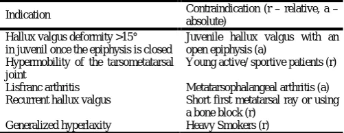
A simple or congenital hallux valgus is not being treated by the Lapidus procedure. It is only performed in about 5 to 10% of all hallux valgus corrections due to longer postoperative rehabilitation and higher complication rate like over-/ undercorrection, nonunion or broken screws. (Sangeorzan and Hansen, 1954). Indications are tarsometatarsal arthritis of the first joint due to hypermobility or anintermetatarsal angle greater than 15°, where distal bunion is thought to be insufficient (Schmid and Krause, 2014). In patients with metatarsophalangeal arthritis of the first ray (hallux rigidus) the Lapidus technique will not satisfy patients why this procedure should be avoided. In people with high activity such as professional athletes (i.e. sprinters or dancers), a relative contraindication exists. After TMT arthrodesis, only approximately 30% of patients will return to the same level of activity in sports prior surgery, caused by higher stiffness and less range of motion due to limitation in dorsiflexion. In sedentary patients the satisfaction rate is stated with 75% as good to excellent. (McInnes and Bouche, 2001) Additionally in heavy smokers a relative contraindication exists, as these patients are more likely to suffer from a nonunion or an infection after surgery up to 36,4% (Bettin et al., 2015). Following table summarizes the indication and contraindication of the Lapidus procedure.
ISSN: 0975-833X
International Journal of Current Research
Vol. 9, Issue, 07, pp.54657-54661, July, 2017
INTERNATIONAL JOURNAL OF CURRENT RESEARCH
Article History:
Received 19thApril, 2017
Received in revised form
28thMay, 2017
Accepted 04thJune, 2017
Published online 31stJuly, 2017
Citation: Bäcker, H., Magnilio, M., Schwinghammer, A., Attinger, M. C. and Krause, F. 2017.“Hallux valgus–The lapidus procedure”,International Journal of Current Research, 9, (07), 54657-54661.
Key words:
Hallux valgus, Modified, Lapidus procedure, Metatarsocuneiforme fusion, Arthritis.
RESEARCH ARTICLE
HALLUX VALGUS – THE MODIFIED LAPIDUS PROCEDURE
*,1
Bäcker, H.,
2Magnilio, M.,
1Schwinghammer, A.,
1Attinger, M. C. and
1Krause, F.
1
University Hospital Bern, Inselspital, Department for Orthopedic and Traumatology, Freiburgstrasse 8,
3010 Bern, Switzerland
2
Department of Orthopaedic Surgery, Cantonal Hospital, Fribourg, Switzerland
ARTICLE INFO ABSTRACT
Hallux valgus is a frequent disease in the population. Several different surgical procedures exist. One of these techniques is the Lapidus procedure which is performed in 5 to 10% of all hallux surgeries. Indications are recurrent hallux valgus, lisfranc arthritis, intermetatarsal angle greater than 15° and hypermobility of the first ray. One of the disadvantages are high complication rate and a long postoperative rehabilitation period. Therefore the correct indication for this rather demanding procedure is important. We want to present information about the clinical examination of hallux valgus, indication for the Lapidus procedure and how it is performed at our center.
Copyright©2017, Bäcker et al. This is an open access article distributed under the Creative Commons Attribution License, which permits unrestricted use,
distribution, and reproduction in any medium, provided the original work is properly cited.
INTRODUCTION
Hallux valgus deformity is a frequent disease of the forefoot. The prevalence is described to be between 23 to 35.7%. (Nix et al., 2010) Based on the severity of hallux valgus deformity, a variety of different techniques exist, which underscores, that no single approach universally addresses the deformity. In 2007, Lin JS et al published a review of surgical treatment options concluding that no real consensus about the best treatment of choice exists (Lin and Bustillo, 2007). One method is the Lapidus procedure which was roughly described as fusion of the metatarsocuneiforme joint by Albrecht et al in 1911. (Albrecht, 1910). In the following years controversy discussion have been taken place on the sciences and impact in clinics. The etiology of hallux valgus based on the obliquity of the first tarsometatarsal (TMT) joint and a consequent adduction of the first metatarsal was published in 1932 by Kleinberg. Two years later Lapidus published this technique (Lapidus, 1934), stating an arthrodesis of the first cuneiforme-metatarsal joint and the first intercuneiforme-metatarsal joint. Later on this procedure was modified by Clark, using crossing screws without intermetatarsal fusion. (Clark et al., 1987).
*Corresponding author: Bäcker, H.
University Hospital Bern, Inselspital, Department for Orthopedic and Traumatology, Freiburgstrasse 8, 3010 Bern, Switzerland.
Indication versus contraindication
A simple or congenital hallux valgus is not being treated by the Lapidus procedure. It is only performed in about 5 to 10% of all hallux valgus corrections due to longer postoperative rehabilitation and higher complication rate like over-/ undercorrection, nonunion or broken screws. (Sangeorzan and Hansen, 1954). Indications are tarsometatarsal arthritis of the first joint due to hypermobility or anintermetatarsal angle greater than 15°, where distal bunion is thought to be insufficient (Schmid and Krause, 2014). In patients with metatarsophalangeal arthritis of the first ray (hallux rigidus) the Lapidus technique will not satisfy patients why this procedure should be avoided. In people with high activity such as professional athletes (i.e. sprinters or dancers), a relative contraindication exists. After TMT arthrodesis, only approximately 30% of patients will return to the same level of activity in sports prior surgery, caused by higher stiffness and less range of motion due to limitation in dorsiflexion. In sedentary patients the satisfaction rate is stated with 75% as good to excellent. (McInnes and Bouche, 2001) Additionally in heavy smokers a relative contraindication exists, as these patients are more likely to suffer from a nonunion or an infection after surgery up to 36,4% (Bettin et al., 2015). Following table summarizes the indication and contraindication of the Lapidus procedure.
ISSN: 0975-833X
International Journal of Current Research
Vol. 9, Issue, 07, pp.54657-54661, July, 2017
INTERNATIONAL JOURNAL OF CURRENT RESEARCH
Article History:
Received 19thApril, 2017
Received in revised form
28thMay, 2017
Accepted 04thJune, 2017
Published online 31stJuly, 2017
Citation: Bäcker, H., Magnilio, M., Schwinghammer, A., Attinger, M. C. and Krause, F. 2017.“Hallux valgus–The lapidus procedure”,International Journal of Current Research, 9, (07), 54657-54661.
Key words:
Hallux valgus, Modified, Lapidus procedure, Metatarsocuneiforme fusion, Arthritis.
RESEARCH ARTICLE
HALLUX VALGUS – THE MODIFIED LAPIDUS PROCEDURE
*,1
Bäcker, H.,
2Magnilio, M.,
1Schwinghammer, A.,
1Attinger, M. C. and
1Krause, F.
1
University Hospital Bern, Inselspital, Department for Orthopedic and Traumatology, Freiburgstrasse 8,
3010 Bern, Switzerland
2
Department of Orthopaedic Surgery, Cantonal Hospital, Fribourg, Switzerland
ARTICLE INFO ABSTRACT
Hallux valgus is a frequent disease in the population. Several different surgical procedures exist. One of these techniques is the Lapidus procedure which is performed in 5 to 10% of all hallux surgeries. Indications are recurrent hallux valgus, lisfranc arthritis, intermetatarsal angle greater than 15° and hypermobility of the first ray. One of the disadvantages are high complication rate and a long postoperative rehabilitation period. Therefore the correct indication for this rather demanding procedure is important. We want to present information about the clinical examination of hallux valgus, indication for the Lapidus procedure and how it is performed at our center.
Copyright©2017, Bäcker et al. This is an open access article distributed under the Creative Commons Attribution License, which permits unrestricted use,
distribution, and reproduction in any medium, provided the original work is properly cited.
INTRODUCTION
Hallux valgus deformity is a frequent disease of the forefoot. The prevalence is described to be between 23 to 35.7%. (Nix et al., 2010) Based on the severity of hallux valgus deformity, a variety of different techniques exist, which underscores, that no single approach universally addresses the deformity. In 2007, Lin JS et al published a review of surgical treatment options concluding that no real consensus about the best treatment of choice exists (Lin and Bustillo, 2007). One method is the Lapidus procedure which was roughly described as fusion of the metatarsocuneiforme joint by Albrecht et al in 1911. (Albrecht, 1910). In the following years controversy discussion have been taken place on the sciences and impact in clinics. The etiology of hallux valgus based on the obliquity of the first tarsometatarsal (TMT) joint and a consequent adduction of the first metatarsal was published in 1932 by Kleinberg. Two years later Lapidus published this technique (Lapidus, 1934), stating an arthrodesis of the first cuneiforme-metatarsal joint and the first intercuneiforme-metatarsal joint. Later on this procedure was modified by Clark, using crossing screws without intermetatarsal fusion. (Clark et al., 1987).
*Corresponding author: Bäcker, H.
University Hospital Bern, Inselspital, Department for Orthopedic and Traumatology, Freiburgstrasse 8, 3010 Bern, Switzerland.
Indication versus contraindication
A simple or congenital hallux valgus is not being treated by the Lapidus procedure. It is only performed in about 5 to 10% of all hallux valgus corrections due to longer postoperative rehabilitation and higher complication rate like over-/ undercorrection, nonunion or broken screws. (Sangeorzan and Hansen, 1954). Indications are tarsometatarsal arthritis of the first joint due to hypermobility or anintermetatarsal angle greater than 15°, where distal bunion is thought to be insufficient (Schmid and Krause, 2014). In patients with metatarsophalangeal arthritis of the first ray (hallux rigidus) the Lapidus technique will not satisfy patients why this procedure should be avoided. In people with high activity such as professional athletes (i.e. sprinters or dancers), a relative contraindication exists. After TMT arthrodesis, only approximately 30% of patients will return to the same level of activity in sports prior surgery, caused by higher stiffness and less range of motion due to limitation in dorsiflexion. In sedentary patients the satisfaction rate is stated with 75% as good to excellent. (McInnes and Bouche, 2001) Additionally in heavy smokers a relative contraindication exists, as these patients are more likely to suffer from a nonunion or an infection after surgery up to 36,4% (Bettin et al., 2015). Following table summarizes the indication and contraindication of the Lapidus procedure.
ISSN: 0975-833X
International Journal of Current Research
Vol. 9, Issue, 07, pp.54657-54661, July, 2017
INTERNATIONAL JOURNAL OF CURRENT RESEARCH
Article History:
Received 19thApril, 2017
Received in revised form
28thMay, 2017
Accepted 04thJune, 2017
Published online 31stJuly, 2017
Citation: Bäcker, H., Magnilio, M., Schwinghammer, A., Attinger, M. C. and Krause, F. 2017.“Hallux valgus–The lapidus procedure”,International Journal of Current Research, 9, (07), 54657-54661.
Key words:
Table I. Indication vs contraindication
Indication Contraindication (r–relative, a–
absolute) Hallux valgus deformity >15°
in juvenil once the epiphysis is closed
Juvenile hallux valgus with an open epiphysis (a)
Hypermobility of the tarsometatarsal joint
Young active/ sportive patients (r)
Lisfranc arthritis Metatarsophalangeal arthritis (a)
Recurrent hallux valgus Short first metatarsal ray or using
a bone block (r)
Generalized hyperlaxity Heavy Smokers (r)
Diagnosis
For evaluation and diagnosis of a hallux valgus a clinical examination as well as radiographies are required.
Clinical examination: Primarily the foot shape and keratosis are examined. Plantar hyperkeratosis of the second distal metatarsus indicates a higher bearing of the second ray due to instable TMT I joint. For identifying symptomatic arthrosis patientsare asked for pain and if present, they shall localize it. When palpating the tenderness and passive motion of the tarsometatarsal joint and metatarsophalangeal joint, the different types of arthritis can be distinguished. When pain is localized in the metatarsophalangeal (MTP) joint at punctum maximum, a MTP arthritis is likely. This is an absolute contraindication as described earlier, as those patients will not become free of pain. However, in case pain is present in the cuneiformia medialis - metatarsal joint, it is likely that arthritis is already severe and the indication for the Lapidus procedure is set. Later on, the mobility of the first ray is examined. Therefore the patient shall sit and flex the knee in a neutral position of the ankle, stabilizing the foot - except ray one–with one hand by the examiner. The other hand moves the first ray from dorso-medial to plantar-lateral which is compared with the contralateral foot as published by Morton. (Morton, 1935)
To objectify mobility of the first ray, the Klaue device may be used, which consists of a modified ankle-foot orthosis with an attached micrometer. When comparing the control group with the hypermobility group, a difference in displacement of approximately 5mm can be observed (Klaue, 1994). All examinations must be performed on both feet to compare and identify individual differences.
Radiography: The dorso-plantar radiography of the weight-bearing foot allows to measure the hallux valgus angle as well as to identify arthritis, the epiphysis in juvenile patients or a shortage of the metatarsus I ray. In terms of an intermetatarsal angle more than 15°, the indication may be given for a Lapidus procedure. Hypermobility may be identified by bony hypertrophy of the second proximal metatarsus, (Morton, 1935) however these findings are insecure as they are infrequentaccording to Grebing et al. (2004). When a short first metatarsal ray is visible on dorso-plantar radiography, a bridging with a bone block interposition must be performed, as the Lapidus procedure leads to another shortage, (Catanzariti et al., 1999). Normally, on the lateral radiography, the metatarsocuneiform joint is slightly medial. A metatarsus primus varus deformity may be indicated by an excessive medial inclination of the metatarsocunei form joint (Berntsen, 1930; Haines, 1954). In few cases an os intermetatarseum is present which may reduce the range of motion due to a rigid joint–especially intermetatarsal I to II -. In about one fifth of all cases, a gap of the plantar aspect of the
first TMT joint may be seen, indicating instability. (Coughlin and Jones, 2007) (Figure I).
Authors preferred modified Lapidus procedure
Surgical procedure
The patient is placed in a supine position. For correct rotation of the foot a bolster may be placed under the buttock. To allow a safe and blood-free procedure, either a sterile Esmarch rubber bandage is wrapped around the ankle or a tourniquet which is located at the thigh is inflated to 350mmHg. To begin with, longitudinal incision of approximately 5cm length is performed, centered over the dorsomedial aspect of the tarsometatarsal joint. The soft tissue is dissected centering the first and second metatarsophalangeal joints longitudinally, as well as on the medial aspect of the first metatarsophalangeal joint. In the first web space a release of the adductor tendons and transverse metatarsal ligament is carried out, followed by an incision of the joint capsule from the medial side horizontally. The bony eminence–superior more than inferior - is resected of 2-4mm width afterwards, remaining the medial sesamoid groove. Now the extensor halluces longus tendon is retracted laterally and the joint capsule of the first TMT is cut dorsomedially. To perform a fusion of the first and second metatarsal ray, the approach is in the interval between the extensor halluces longus and brevis tendons. Special care has to be taken to the neurovascular bundle - dorsalis pedis artery and deep peroneal nerve -.
Using two Hohmann retractors, the joint can be exposed, moving the anterior tibial tendon on the medial and the neurovascular bundle on the lateral side. The articular cartilage of the TMT and plantar-laterally cartilage of the proximal metatarsal ray is completely removed with an osteotome and a small ring curette by exposing the plantar-lateral part with a K-wire distractor or spreader. For better healing a subchondral macrofracturing is carried out, using an osteotome and multiple 1.5mm drill holes on both sides of the arthrodesis. More precise correction of excessive valgus and mild plantar flexion can be achieved when resecting a small plantarly and laterally based wedge.
The present dorsomedial position of the first ray is moved to a slight plantar lateral position – according the biomechanical axis- and the intermetatarsal angle is closed by reducing the first metatarsal ray. It is important to keep in mind, not to perform an undercorrection of the metatarsal plantarflexion or an overcorrection. An excessive dorsiflexion may cause metatarsalgia of the second ray. The correct alignment of the plantar lateral aspect as well as residual cartilage or bony fragments have to be visualized by the surgeon as well as using radiography and must be corrected respectively resected. In case of any major osseous defect a local bone graft is obtained from the medial MTP joint eminence and placed into the arthrodesis site. The correct, modified alignment and rotation is temporarily fixated with a preliminary K-wire. Afterwards the alignment and rotation as well as the bone stock must be rechecked by the physician using radiography.
Once the TMT I joint is anatomically, two 3.5mm crossing lag screws are used for fixation. For more stability and to decrease risk of fracture, a notch at the entry point of the distal screw is performed using a triangle of six 2.5mm drill holes and resecting the surrounding cortices of the holes with a Ronguer.
Precise drilling to the medial border of the foot helps to direct the drill in axis of the os cuneiformia and to avoid any damage or iatrogenic fracture. A 3.5mm gliding hole is drilled, and for more lateral correction of the first metatarsal ray the 2.5mm drill can be placed slightly medially of the gliding hole in the os cuneiformia. Afterwards the second screw can be inserted from proximal. The direction of the screw should aim the lateral plantar first metatarsal cortex. Ideally the screws cross distally of the arthrodesis to allow further rotational stability. Our advice is to insert the screws when performing a dorsiflexion of the first metatarsophalangeal joint, to allow higher compression of the arthrodesis site by tensioning the plantar fascia.
When a fusion of the intermetatarsal joint (the articular surfaces – cartilage –) is required, the same method is performed described above, including resection of the intermetarsal cartilage. It is important to keep in mind to protect and preserve the dorsal pedis artery. In our center the modified Lapidus technique is preferred which includes a simple fusion of the first TMT joint. An arthrodesis is only performed in cases of a non-union or presence of an os intermetatarseum. For a good alignment of the hallux, the capsular tissue on the medial MTP joint are double-breasted. A fully correction is targeted hereby. Further corrections will lead to high recurrence rate. In residual deformity, the reason must be examined.The hallux phalangeus is most common which can be treated by performing an additional Akin osteotomy.
Finally all layers are closed of the incision and a compression dressing as well as a splitted Crus cast is applied.
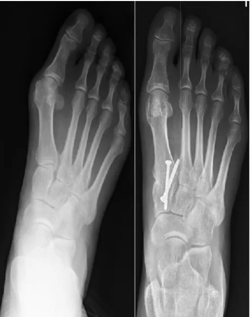
[image:3.595.310.558.48.315.2]In Figure I the pre- and postsurgical x-rays are shown, after Lapidus procedure using two crossing screws
Figure 1a
Figure 1b
Figure 1. a) dorsoplantar view b) lateral view before and after the Lapidus procedure. Prior surgery: Intermetatarsal angle of 16.8°,
Hallux valgus angle of 27.1°
Post-operative care
Depending on the implant used, the arthrodesis show high stability – locking plate vs. crossing screws. This enables a weight-bearing mobilization quite fast after surgery. Even full mobilization two weeks after osteosynthesis with locking plates showed no complications such as implant break or dislocation in alignment. (Basile et al., 2010; Cottom and Vora, 2013) Crossed screws arthrodesis is a slightly weaker fixation method, why a more careful post-operative care should be performed (Cohen et al., 2005; Egol et al., 2004). The first bandage is removed the first or second day after surgery. The lower leg cast is applied for 8 weeks without weight-bearing at all, for the first 4 weeks. The following 4 weeks a 10-20kg partial weight-bearing is enabled and weeks 8-12 a transition to fully weight-bearing is allowed. Ambulatory clinical and radiological follow-up examinations are performed 4and 8weeks after.
Complications
Because of only little subcutaneous tissue of the foot, the implanted material - two crossed screws may cause problems (i.e. irritation and pain) in up to 20%, (Coetzee et al., 2004). Locking plate osteosynthesis may cause even higher incidence of complications however are more stable directly post-operative. In case of earlier weight-bearing mobilization, the rate of non-union is higher in simultaneous bilateral Lapidus fusion. (Myerson et al., 1990; Sangeorzan and Hansen, 1989). In single Lapidus procedure the non-union rate is described from 2 to 10%, of which about half of the patients require a revision, (Thompson et al., 2005). Smoking has shown to be a major impact in healing according to Coetzee et al. (2004) Patients are informed about the risk of complications when smoking, however the surgical procedure is still performed at our department. Major symptoms of non-union is pain or radiological signs like missing ossification which requires a Precise drilling to the medial border of the foot helps to direct
the drill in axis of the os cuneiformia and to avoid any damage or iatrogenic fracture. A 3.5mm gliding hole is drilled, and for more lateral correction of the first metatarsal ray the 2.5mm drill can be placed slightly medially of the gliding hole in the os cuneiformia. Afterwards the second screw can be inserted from proximal. The direction of the screw should aim the lateral plantar first metatarsal cortex. Ideally the screws cross distally of the arthrodesis to allow further rotational stability. Our advice is to insert the screws when performing a dorsiflexion of the first metatarsophalangeal joint, to allow higher compression of the arthrodesis site by tensioning the plantar fascia.
When a fusion of the intermetatarsal joint (the articular surfaces – cartilage –) is required, the same method is performed described above, including resection of the intermetarsal cartilage. It is important to keep in mind to protect and preserve the dorsal pedis artery. In our center the modified Lapidus technique is preferred which includes a simple fusion of the first TMT joint. An arthrodesis is only performed in cases of a non-union or presence of an os intermetatarseum. For a good alignment of the hallux, the capsular tissue on the medial MTP joint are double-breasted. A fully correction is targeted hereby. Further corrections will lead to high recurrence rate. In residual deformity, the reason must be examined.The hallux phalangeus is most common which can be treated by performing an additional Akin osteotomy.
Finally all layers are closed of the incision and a compression dressing as well as a splitted Crus cast is applied.
[image:3.595.37.286.467.780.2]In Figure I the pre- and postsurgical x-rays are shown, after Lapidus procedure using two crossing screws
Figure 1a
Figure 1b
Figure 1. a) dorsoplantar view b) lateral view before and after the Lapidus procedure. Prior surgery: Intermetatarsal angle of 16.8°,
Hallux valgus angle of 27.1°
Post-operative care
Depending on the implant used, the arthrodesis show high stability – locking plate vs. crossing screws. This enables a weight-bearing mobilization quite fast after surgery. Even full mobilization two weeks after osteosynthesis with locking plates showed no complications such as implant break or dislocation in alignment. (Basile et al., 2010; Cottom and Vora, 2013) Crossed screws arthrodesis is a slightly weaker fixation method, why a more careful post-operative care should be performed (Cohen et al., 2005; Egol et al., 2004). The first bandage is removed the first or second day after surgery. The lower leg cast is applied for 8 weeks without weight-bearing at all, for the first 4 weeks. The following 4 weeks a 10-20kg partial weight-bearing is enabled and weeks 8-12 a transition to fully weight-bearing is allowed. Ambulatory clinical and radiological follow-up examinations are performed 4and 8weeks after.
Complications
Because of only little subcutaneous tissue of the foot, the implanted material - two crossed screws may cause problems (i.e. irritation and pain) in up to 20%, (Coetzee et al., 2004). Locking plate osteosynthesis may cause even higher incidence of complications however are more stable directly post-operative. In case of earlier weight-bearing mobilization, the rate of non-union is higher in simultaneous bilateral Lapidus fusion. (Myerson et al., 1990; Sangeorzan and Hansen, 1989). In single Lapidus procedure the non-union rate is described from 2 to 10%, of which about half of the patients require a revision, (Thompson et al., 2005). Smoking has shown to be a major impact in healing according to Coetzee et al. (2004) Patients are informed about the risk of complications when smoking, however the surgical procedure is still performed at our department. Major symptoms of non-union is pain or radiological signs like missing ossification which requires a Precise drilling to the medial border of the foot helps to direct
the drill in axis of the os cuneiformia and to avoid any damage or iatrogenic fracture. A 3.5mm gliding hole is drilled, and for more lateral correction of the first metatarsal ray the 2.5mm drill can be placed slightly medially of the gliding hole in the os cuneiformia. Afterwards the second screw can be inserted from proximal. The direction of the screw should aim the lateral plantar first metatarsal cortex. Ideally the screws cross distally of the arthrodesis to allow further rotational stability. Our advice is to insert the screws when performing a dorsiflexion of the first metatarsophalangeal joint, to allow higher compression of the arthrodesis site by tensioning the plantar fascia.
When a fusion of the intermetatarsal joint (the articular surfaces – cartilage –) is required, the same method is performed described above, including resection of the intermetarsal cartilage. It is important to keep in mind to protect and preserve the dorsal pedis artery. In our center the modified Lapidus technique is preferred which includes a simple fusion of the first TMT joint. An arthrodesis is only performed in cases of a non-union or presence of an os intermetatarseum. For a good alignment of the hallux, the capsular tissue on the medial MTP joint are double-breasted. A fully correction is targeted hereby. Further corrections will lead to high recurrence rate. In residual deformity, the reason must be examined.The hallux phalangeus is most common which can be treated by performing an additional Akin osteotomy.
Finally all layers are closed of the incision and a compression dressing as well as a splitted Crus cast is applied.
In Figure I the pre- and postsurgical x-rays are shown, after Lapidus procedure using two crossing screws
Figure 1a
Figure 1b
Figure 1. a) dorsoplantar view b) lateral view before and after the Lapidus procedure. Prior surgery: Intermetatarsal angle of 16.8°,
Hallux valgus angle of 27.1°
Post-operative care
Depending on the implant used, the arthrodesis show high stability – locking plate vs. crossing screws. This enables a weight-bearing mobilization quite fast after surgery. Even full mobilization two weeks after osteosynthesis with locking plates showed no complications such as implant break or dislocation in alignment. (Basile et al., 2010; Cottom and Vora, 2013) Crossed screws arthrodesis is a slightly weaker fixation method, why a more careful post-operative care should be performed (Cohen et al., 2005; Egol et al., 2004). The first bandage is removed the first or second day after surgery. The lower leg cast is applied for 8 weeks without weight-bearing at all, for the first 4 weeks. The following 4 weeks a 10-20kg partial weight-bearing is enabled and weeks 8-12 a transition to fully weight-bearing is allowed. Ambulatory clinical and radiological follow-up examinations are performed 4and 8weeks after.
Complications
surgical revision. In these cases, we use autologous cancellous bone graft interposition and performthe originally described Lapidus procedure - strong fixation including fusion of the basis of the first and second metatarsals.
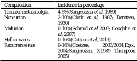
[image:4.595.49.274.349.450.2]Another specific complication is malalginment of the tarsometatarsal joint which can be identifiedin shortened and dorsiflexion of the first metatarsal ray. Typical symptoms are transfer metatarsalgia of the second or third ray, (Catanzariti et al., 1999; Bednarz and Manoli, 2nd) which occurs in approximately 10% (Myerson, 1990). For prevention an accurate joint preparation without removing too excessive wedges is essential. In this case a bridging with bone interposition should be performed to avoid a shortened ray (more than 5mm). A shortened ray between 5mmand 10mm a light plantarflexion of the first metatarsal suffices to prevent malalignment. When the metatarsal ray is shortened by more than 10mm or more a shortening metatarsal osteotomy (e.g. Weil osteotomy) of the second and third toe is indicated to prevent transfer metatarsalgia. However in cases of 20mm or more shortened first metatarsal, the reconstruction of the first ray using tricortical bone graft is required to restore the correct length. (Coetzee et al., 2004 and Mauldin et al., 1990).
Table 2. Complication rate
Complication Incidence in percentage
Transfer metatarsalgia 4-5%(Sangeorzan et al, 1989)
Non-union 2-10%(Clark et al, 1987; Berntsen,
1930)
Malunion 0-10%(Schmid et al 2007; Coughlin et
al, 2007)
Hallux varus 0-16%(Cottom et al, 2013)
Recurrence rate 0-16%(Coetzee, 2003/2004;Egol,
2004;Sangerozan, X1989 Thompson 2005)
Conclusion
The modified Lapidus procedure is a very effective and good procedure in Hallux valgus disease which is performed in 5-10%. Indications are primary and severe deformity, metatarsus primus varus, lisfranc arthritis and recurrent hallux valgus. Because of the most proximal correction method of the forefoot, it allows a variety of possibilities than other metatarsal osteotomies. The usage of crossing screws facilitates highest stability and less complications than locking plates. In post-operative care, the immobilizationtakes more time, about 6-8 weeks, however because of the difficultness of the procedure it is prone to complications in about one fifth to one fourth of patients. This rate also includes the usage in recurrence hallux or failure of primary hallux valgus correction.
Conflict of interest: None declared.
Information
This article is based on the coauthors paper “The modified Lapidus procedure”, originally published in Foot Ankle Clin. by Schmid T, Krause F in 2014.
REFERENCES
Albrecht, G.H. 2010. The pathology and treatment of hallux valgus. Russ Vrach. 10: 14.
Basile, P; Cook, EA; Cook, J.J. 2010. Immediate weight bearing following modified lapidus arthrodesis. The Journal of foot and ankle surgery: official publication of the American College of Foot and Ankle Surgeons. 49: 459-464. http://dx.doi.org/10.1053/j.jfas.2010.06.003
Bednarz, P.A. Manoli, A. 2000. 2nd: Modified lapidus procedure for the treatment of hypermobile hallux valgus. Foot & ankle international / American Orthopaedic Foot and Ankle Society [and] Swiss Foot and Ankle Society. 21: 816-821.
Berntsen, A. 1930. De l’hallux valgus: contribution a son etiologie et a son traitement. Rev Orthop. 3: 101-111 Bettin, C.C., Gower, K., McCormick, K., Wan, J.Y., Ishikawa,
S.N., Richardson, D.R., Murphy, G.A. 2015. Cigarette smoking increases complication rate in forefoot surgery; Foot Ankle Int. 2015 May;36(5):488-93. doi: 10.1177/1071100714565785. Epub 2015 Jan 12.
Catanzariti, A.R., Mendicino, R.W., Lee, M.S., Gallina, M.R. 1999. The modified Lapidus arthrodesis: a retrospective analysis. The Journal of foot and ankle surgery : official publication of the American College of Foot and Ankle Surgeons. 38: 322-332.
Clark, H.R., Veith, R.G. Hansen, S.T., Jr. 1987. Adolescent bunions treated by the modified Lapidus procedure. Bulletin of the Hospital for Joint Diseases Orthopaedic Institute. 47: 109-122.
Coetzee, J.C. and Wickum, D. 2004. The Lapidus procedure: a prospective cohort outcome study. Foot & ankle international / American Orthopaedic Foot and Ankle Society [and] Swiss Foot and Ankle Society. 25: 526-531. Coetzee, J.C., Resig, S.G. Kuskowski, M., Saleh, K.J. 2003.
The Lapidus procedure as salvage after failed surgical treatment of hallux valgus: a prospective cohort study. The Journal of bone and joint surgery American volume. 85-A: 60-65.
Coetzee, J.C., Resig, S.G., Kuskowski, M., Saleh, K.J. 2004. The Lapidus procedure as salvage after failed surgical treatment of hallux valgus. Surgical technique. The Journal of bone and joint surgery American volume. 86-A Suppl 1: 30-36.
Cohen, D.A., Parks, B.G. Schon, L.C. 2005. Screw fixation compared to H-locking plate fixation for first metatarsocuneiform arthrodesis: a biomechanical study. Foot & ankle international / American Orthopaedic Foot and Ankle Society [and] Swiss Foot and Ankle Society. 26: 984-989.
Cottom, J.M., Vora, A.M. 2013. Fixation of lapidus arthrodesis with a plantar interfragmentary screw and medial locking plate: a report of 88 cases. The Journal of foot and ankle surgery : official publication of the American College of Foot and Ankle Surgeons. 52: 465-469. http://dx.doi.org/ 10.1053/ j.jfas.2013.02.013
Coughlin, M.J., Jones, C.P. 2007. Hallux valgus: demographics, etiology, and radiographic assessment. Foot & ankle international / American Orthopaedic Foot and Ankle Society [and] Swiss Foot and Ankle Society. 28: 759-777. http://dx.doi.org/10.3113/FAI.2007.0759
Egol, K.A., Kubiak, E.N., Fulkerson, E., Kummer, F.J., Koval, K.J. 2004. Biomechanics of locked plates and screws. Journal of orthopaedic trauma. 18: 488-493. Grebing, B,R., Coughlin, M.J. 2004. Evaluation of Morton's
theory of second metatarsal hypertrophy. The Journal of bone and joint surgery American volume. 86-A: 1375-1386.
Haines, R.W. 1954. Mc, DA: The anatomy of hallux valgus. The Journal of bone and joint surgery British volume. 36-B: 272-293.
Klaue, K., Hansen, S.T., Masquelet, A.C. 1994. Clinical, quantitative assessment of first tarsometatarsal mobility in the sagittal plane and its relation to hallux valgus deformity. Foot & ankle international / American Orthopaedic Foot and Ankle Society [and] Swiss Foot and Ankle Society. 15: 9-13.
Lapidus, P.W. 1934. Operative correction of the metatarsus varus primus in hallux valgus. Surg Gynecol Obstet. 58: 16.
Lin, J.S., Bustillo, J., 2007. Surgical treatment of hallux valgus: a review. Curr. Opin. Orthop. 18, 112–117.
Mauldin, D.M., Sanders, M., Whitmer, W.W. 1990. Correction of hallux valgus with metatarsocuneiform stabilization. Foot & ankle. 11: 59-66.
McInnes, B.D. Bouche, R.T. 1992. Critical evaluation of the modified Lapidus procedure. The Journal of foot and ankle surgery : official publication of the American College of Foot and Ankle Surgeons. 40: 71-90.
Morton, DJ: The human foot, Columbia University Press, 1935.
Myerson, M. 1990. Metatarsocuneiform arthrodesis for treatment of hallux valgus and metatarsus primus varus. Orthopedics. 13: 1025-1031.
Myerson, M; Allon, S; McGarvey, W. 1992. Metatarsocunei form arthrodesis for management of hallux valgus and metatarsus primus varus. Foot & ankle. 13: 107-115. Nix, S., Smith, M., Vicenzino, B., 2010. 2010. Prevalence of
hallux valgus in the general population: a systematic review and meta-analysis. J. Foot Ankle Res. 3, 21.
Sangeorzan, B.J., Hansen, S.T., Jr. 1989. Modified Lapidus procedure for hallux valgus. Foot & ankle. 9: 262-266. Schmid, T., Krause, F. The modified Lapidus fusion; Foot
Ankle Clin. 2014 Jun; 19(2):223-33. doi: 10.1016/ j.fcl.2014.02. 005. Epub 2014 Mar 21. Review.
Thompson, IM; Bohay, DR; Anderson, JG: 2005. Fusion rate of first tarsometatarsal arthrodesis in the modified Lapidus procedure and flatfoot reconstruction. Foot & ankle international / American Orthopaedic Foot and Ankle Society [and] Swiss Foot and Ankle Society. 26: 698-703.