Electronic Thesis and Dissertation Repository
9-29-2016 12:00 AM
Development and Performance Evaluation of an Antibody-Based
Development and Performance Evaluation of an Antibody-Based
Technology for Detection of E. coli O157 in Meat Samples and Its
Technology for Detection of E. coli O157 in Meat Samples and Its
Potential Evolution Using Antibody Engineering
Potential Evolution Using Antibody Engineering
Yadira Tejeda SaldañaThe University of Western Ontario Supervisor
Dr. Michael J. Rieder
The University of Western Ontario Graduate Program in Pathology
A thesis submitted in partial fulfillment of the requirements for the degree in Doctor of Philosophy
© Yadira Tejeda Saldaña 2016
Follow this and additional works at: https://ir.lib.uwo.ca/etd
Part of the Food Microbiology Commons, Molecular Biology Commons, and the Pathogenic Microbiology Commons
Recommended Citation Recommended Citation
Tejeda Saldaña, Yadira, "Development and Performance Evaluation of an Antibody-Based Technology for Detection of E. coli O157 in Meat Samples and Its Potential Evolution Using Antibody Engineering" (2016). Electronic Thesis and Dissertation Repository. 4183.
https://ir.lib.uwo.ca/etd/4183
This Dissertation/Thesis is brought to you for free and open access by Scholarship@Western. It has been accepted for inclusion in Electronic Thesis and Dissertation Repository by an authorized administrator of
i Abstract
ii Keywords
Escherichia coli O157, pathogen detection, food safety, food microbiology, pre-collaborative study, rapid method, lateral flow immunoassay, monoclonal antibody,
iii
Acknowledgments
During the past five years, I had the opportunity and the pleasure to meet people who helped and encouraged me in this long journey. It has been a period of intense learning and challenges along the way, not only in the academic field, but also on a personal level. My whole PhD, including this thesis, would definitely not have been possible without all of this support in so many different ways.
Thanks to Dr. Rieder, my supervisor, for giving me the opportunity to participate in this project. His continuous trust in me and the academic freedom he afforded me throughout my studies allowed me to develop my own ideas. My deepest gratitude to my joint-supervisor Dr. McCormick. I was amazingly fortunate to have his guidance and support. I would like to thank him for always encouraging me, especially in those moments when I faltered. His motivation, patience and generosity have been truly inspirational, not to mention the countless skills that I was able to develop thanks to his mentorship.
I am also privileged to have Dr. Bend as part of my advisory committee. His insightful comments and constructive criticisms at different phases of my studies were always thought-stimulating. I am also grateful to him for giving me the opportunity to join Dr. Rieder’s project. In addition, I want to thank Dr. Chakraborty for his great support throughout my PhD not only as the Graduate Chair of the Pathology Department, but also as a member of my advisory committee.
I am also indebted to the members of Dr. McCormick’s lab, Katie, Will, and Kelcey, who were always willing to help me. Particularly, I would like to acknowledge Lorea, an amazing woman who was always there to encourage me. Her advice and positive energy were always game changers.
iv
in one way or another. I am really thankful to all of them that provided me with tools, knowledge, endless use of their lab equipment and reagents during my research that would otherwise not have been possible. A special thanks to Tracey Koning, from the Department of Pathology, for her endless patience and guidance, she always worked hard to understand all the perks of my Mexican scholarship.
My PhD would not have been possible without the funding from the Mexican National Council on Science and Technology (CONACyT), who financially supported my studies during these five years. In addition, thanks to the Western Graduate Research Scholarship (WGRS) for complementing my Mexican scholarship. Finally, thanks to MITACS for supporting part of the work done with our industrial partner IPOC.
v
Table of Contents
Abstract ... i
Acknowledgments ... iii
Table of Contents ... v
List of Tables ... xi
List of Figures ... xiii
List of Appendices ... xv
List of Abbreviation ... xvi
Chapter 1 Introduction ... 1
1.1. General Overview of Food Safety ... 2
Food Safety ... 2
1.1.1.1 Microbiological food safety ... 3
1.2. Escherichia coli O157 and Its Role in Food Safety ... 4
General Overview of Pathogenic E. coli ... 4
Main Reservoirs and Transmission ... 5
Mechanisms of Pathogenesis ... 7
Epidemiology and Economic Burden of E. coli O157 Infections ... 11
E. coli O157 Outbreaks, Recalls and Regulatory Aspects ... 12
1.3. Current State-of-the-Art in Detection of Food Pathogens ... 16
Trends in Rapid Point-of-Care (POC) Methods ... 17
1.3.1.1 Molecular methods ... 18
1.3.1.2 Optical methods ... 19
1.3.1.3 Nanotechnology ... 20
1.3.1.4 Microfluidics ... 20
1.3.1.5 Immunoassays ... 21
1.4. Overview of Lateral Flow Immunoassays (LFIAs) ... 22
vi
1.5. LFIAs for Detection of E. coli O157 in Food ... 27
Validation of Alternative Microbiological Methods ... 28
1.5.1.1 Health Canada’s procedure for the validation of alternative microbiological methods ... 32
1.6. Development of Single-Chain Variable Fragments (scFv) for Pathogen Detection ... 33
Structure of Immunoglobulins (Ig) ... 34
Antibody Engineering ... 38
1.6.2.1 Single-chain variable fragments (scFv) ... 41
1.6.2.2 Application of scFv to food pathogen detection ... 43
Chapter 2 Rationale, Hypotheses and Objectives ... 45
2.1. Rationale ... 46
2.2. Hypotheses ... 47
2.3. Objectives ... 47
Chapter 3 Materials and Method ... 48
3.1. Bacterial Strains, Sample Preparation and Culture Conditions ... 49
Inclusivity and Exclusivity Strains ... 49
Bacterial Culture Enumeration ... 50
Preparation of Stressed E. coli O157 Cells. ... 50
Artificial Inoculation of Food Samples ... 51
Determination of Aerobic Plate Count (APC) of Food Samples ... 52
Evaluation of Enrichment Conditions ... 52
Cloning and Expression Bacterial Strains ... 52
3.2. Development of a Lateral Flow Immunoassay (LFIA) for Detection of E. coli O157 ... 54
Assembling of the LFIA ... 54
In-Tube Sandwich Immunoassay ... 54
Optimization of Reagents and Blocking Conditions ... 59
vii
Determination of the Limit of Detection (LOD) ... 61
3.3. Relative Validation of a LFIA Test Kit for Detection of E. coli O157 in Raw Meat Products ... 64
Inclusivity and Exclusivity Study ... 67
Relative Validation of the LFIA Test Kit Using a Protocol for Unpaired Samples ... 67
3.3.2.1 Sample preparation ... 67
3.3.2.2 Preparation of inocula for processed and unprocessed food samples ... 68
3.3.2.3 Food sample inoculation ... 71
3.3.2.4 Most Probable Number (MPN) determination of inoculated bulk samples . 71 3.3.2.5 LFIA Test Kit alternative method ... 72
3.3.2.6 Reference method MFHPB-10 ... 73
3.3.2.7 LOD ... 73
Evaluation of Probability of Detection (POD) and Performance Parameters ... 73
3.4. Development of a Humanized Single-Chain Variable Fragment (scFv) Against the O-antigen of E. coli O157 ... 79
Hybridoma Cell Line Growth Conditions, Screening and Propagation ... 79
Sodium Dodecyl Sulfate Polyacrylamide Gel Electrophoresis (SDS-PAGE) and Western Blotting (WB) for Protein Visualization ... 81
Enzyme-Linked Immunosorbent Assay (ELISA) and Fluorescent-Antibody Microscopy for Antibody Functionality Assessment ... 81
Murine mAb Characterization and Purification ... 83
Hybridoma RNA Isolation ... 84
mAb Variable Regions Reverse Transcription (RT) and Polymerase Chain Reaction(PCR) ... 85
Plasmid DNA Isolation ... 87
DNA Visualization and Gel Extraction ... 87
DNA Sequencing ... 87
Construction of the Humanized scFvO157 ... 88
viii
3.4.11.1 Restriction digestion ... 92
3.4.11.2 DNA ligation ... 92
3.4.11.3 Transformation of chemical competent E. coli cells ... 93
3.4.11.4 E. coli clone selection ... 93
Expression of scFvO157 ... 93
Purification and Refolding of scFvO157 ... 95
3.4.13.1 Native conditions ... 95
3.4.13.2 Denaturing conditions ... 95
3.4.13.3 Hybrid conditions ... 96
Chapter 4 Results ... 97
4.1. Development of a LFIA for Detection of E. coli O157 ... 98
Preparation and Evaluation of Bacterial Cultures ... 98
4.1.1.1 Preparation of healthy bacterial cultures ... 98
4.1.1.2 Preparation of stressed E. coli O157 cells ... 98
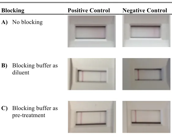
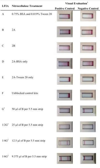
Optimization of the LFIA Device Blocking Conditions ... 99
4.1.2.1 Assessment of different nitrocellulose blocking solutions ... 102
Optimization of the In-Tube Sandwich Immunoassay ... 106
4.1.3.1 Optimization of the pH ... 107
4.1.3.2 Optimization of the colloidal gold conjugated secondary antibody (CGC) 107 4.1.3.3 Optimization of the antibodies ... 110
Development of a Tandem LFIA Test Kit ... 115
Pairing the Enrichment Procedure with the LFIA Test Kit ... 119
4.1.5.1 Assessment of the RapidCult™ enrichment medium ... 119
4.1.5.2 Assessment of mTSBN and mTSB enrichment media ... 120
4.1.5.3 Assessment of TSBN enrichment medium ... 123
Assessing the LOD Using Meat Samples ... 126
Estimating E. coli O157 Content in Artificially Inoculated Meat Samples Using the Tandem LFIA Test Kit ... 126
ix
Inclusivity and Exclusivity Study ... 133
Sample and Inocula Preparation ... 134
Artificial Inoculation and Experimental Layout Using an Unpaired Samples Protocol ... 134
Evaluation of Probability of Detection (POD) ... 136
Evaluation of Performance Parameters ... 136
Determination of the LOD ... 141
4.3. Development of the scFvO157 ... 141
Stability of the Hybridoma Cell Line ... 141
Anti-O157 mAb Characterization ... 143
4.3.2.1 Anti-O157 mAb purification and ELISA ... 143
4.3.2.2 Determination of the anti-O157 mAb isoelectric point (pI) ... 148
RT-PCR and Sequencing of the Variable Heavy and Light (VH and VL) Chains of the Anti-O157 mAb ... 148
Characterization of the Anti-O157 mAb VH and VL Chains ... 151
Construction of the Humanized scFvO157 ... 155
Molecular Cloning of the Humanized scFvO157 ... 161
Expression of the Humanized scFvO157 ... 166
Re-Design of the scFvO157 Structure ... 171
Chapter 5 Discussion ... 175
Chapter 6 Conclusions, Limitations and Future Directions ... 188
6.1. Overall Findings and Implications ... 189
6.2. Research Limitations ... 191
6.3. Future Directions ... 193
Extending the Application of the LFIA Test Kit ... 193
Collaborative Study ... 194
Tandem LFIA Test Kit as a Semi-Quantitative Assay ... 194
Alternative Strategies for Improving the Synthesis of Soluble scFvO157 ... 195
x
Re-Evaluation of the scFvO157 Using Bioinformatics ... 197
References ... 199
Appendices ... 224
xi
List of Tables
Table 1. Major E. coli O157 foodborne outbreaks in the USA and Canada, 2006-2016. 14
Table 2. Commercial LFIAs for detection of E. coli O157 in food samples (as of June,
2016). ... 29
Table 3. Summary of applications of scFv as immunodiagnostics reagents (as of June, 2016). ... 42
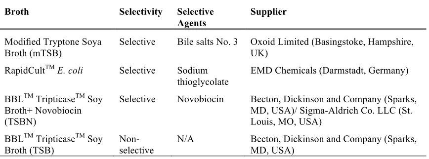
Table 4. Enrichment broths and their selective agents. ... 53
Table 5. Cloning and expression E. coli competent cells. ... 55
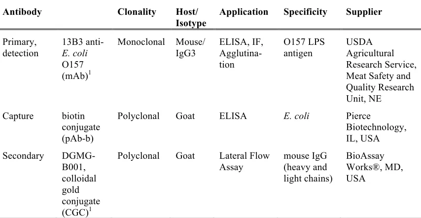
Table 6. Antibodies used for the development of the LFIA Test Kit. ... 58
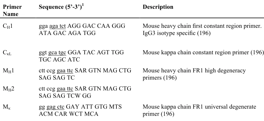
Table 7. Sequences of primers for mAb VH and VL chains RT-PCR. ... 86
Table 8. Sequences of standard primers provided by the Sequencing Facility at Robarts Research Institute, Western University. ... 89
Table 9. Plasmids used in this study. ... 94
Table 10. Initial assessment of nitrocellulose blocking conditions. ... 103
Table 11. Screening of blocking conditions for the LFIA devices. ... 104
Table 12. Visual interpretation of an E. coli O157 concentration curve using the tandem LFIA device. ... 118
Table 13. E. coli O157 concentrations and intensity scores using the tandem LFIA Test Kit with meat samples. ... 130
Table 14. Estimation of TVC and inoculation levels for each food item. ... 135
xii
Table 16. Summary of the alternative method (AM) and reference method (RM) unpaired samples results for the processed raw meat food type. ... 138
Table 17. POD analysis for unprocessed and processed raw meat food types. ... 139
Table 18. Performance parameters for the Alternative Method. ... 140
Table 19. Determination of the LOD for the raw meat products category using MPN. . 142
Table 20. anti-O157 mAb VH and VL chains BLASTP results. ... 154
xiii
List of Figures
Figure 1. Overview of the routes of transmission and human disease due to E. coli O157.
... 8
Figure 2. Overview of the main components of a typical LFIA. ... 24
Figure 3. Overview of the structure of an IgG antibody. ... 36
Figure 4. Examples of antibody engineering. ... 39
Figure 5. Schematic representation of the LFIA. ... 56
Figure 6. LFIA standard results. ... 62
Figure 7. Workflow diagram for the relative validation of unpaired samples for the LFIA Test Kit. ... 65
Figure 8. Scheme of the distribution of samples for the unpaired validation study. ... 69
Figure 9. Flow chart of the LFIA Test Kit alternative method. ... 74
Figure 10. LOD sample preparation scheme. ... 76
Figure 11. Humanized scFvO157 construct. ... 90
Figure 12. Preparation and assessment of stressed E. coli O157 cells. ... 100
Figure 13. Optimization of the in-tube sandwich immunoassay pH conditions. ... 108
Figure 14. Optimization of the colloidal gold conjugate secondary antibody concentration. ... 111
Figure 15. Optimization of the mAb and pAb-b concentrations. ... 113
Figure 16. E. coli O157 concentration curve using the tandem LFIA device. ... 116
xiv
Figure 18. TSBN enrichment medium time-course combined with the tandem LFIA Test
Kit using artificial inoculated ground beef samples. ... 124
Figure 19. Assessment of the LOD of the LFIA Test Kit using food samples. ... 127
Figure 20. Relationship between the tandem LFIA device control and test line intensities with E. coli O157 concentrations in meat samples. ... 131
Figure 21. Anti-O157 mAb isotyping results. ... 144
Figure 22. Overview of the anti-O157 mAb purification steps and ELISA results. ... 146
Figure 23. pI determination of the anti-O157 mAb using IEF. ... 149
Figure 24. Gel electrophoresis of VH and VL chains PCR products. ... 152
Figure 25. anti-O157 mAb VH and VL chain amino acid sequences and COBALT analysis results. ... 156
Figure 26. Construction of the humanized scFvO157 based on 3D modeling. ... 159
Figure 27. Deduced amino acid and DNA sequences used for scFvO157 expression. .. 162
Figure 28. Cloning of the scFvO157 construct into pET32a(+) expression plasmid. .... 164
Figure 29. Overview of the purification and cleavage of the scFvO157. ... 167
Figure 30. Effect of temperature and induction time in the expression of soluble scFvO157. ... 169
xv
List of Appendices
Appendix A: Characterization of inclusivity and exclusivity strains ... 224
Appendix B: MFHPB-10 Isolation of Escherichia coli O157:H7/NM from foods and environmental surface samples ... 233
Appendix C: LFIA Test Kit package insert ... 239
Appendix D: MFHPB-33 Enumeration of total aerobic bacteria in food products and food ingredients using 3M™ Petrifilmt™ aerobic count plates ... 243
Appendix E: Buffer and reagents prepared for this study. ... 246
xvi
List of Abbreviation
°C degrees Celsius
µg microgram
µl microliter
µm micrometer
µM micromolar
µTAS micro total analysis systems
×g times gravity
AC alternative confirmation result A/E attachment and effacing AF alternative final result
AIDS acquired immunodeficiency syndrome
AM alternative method
AOAC Association of Analytical Communities AP alternative presumptive result
APC aerobic plate count
ATCC American Type Culture Collection BAM Bacteriological Analytical Manual, FDA BHI brain heart infusion
bp base pair
BPW buffered peptone water BSA bovine serum albumin
CH constant region of the heavy chain
CL constant region of the light kappa chain
CDC Centers for Disease Control and Prevention, USA cDNA complementary DNA
CDR complementarity determining region CFIA Canadian Food Inspection Agency CFU colony forming unit
CGC colloidal gold conjugate secondary antibody
xvii
CO2 carbon dioxide
COBALT constraint-based multiple alignment tool CR-SMAC cefixime rhamnose sorbitol MacConkey CT-SMAC cefixime tellurite sorbitol MacConkey
d day
Da dalton
DAEC diffusely adherent Escherichia coli DALY disability-adjusted life year
DMSO dimethyl sulfoxide DNA deoxyribonucleic acid
dNTP deoxyribonucleotide triphosphates eae E. coli attaching and effacing gene EAEC enteroaggregative Escherichia coli EHEC enterohemorrhagic Escherichia coli EIEC enteroinvasive Escherichia coli ELISA enzyme-linked immunosorbent assay EPEC enteropathogenic Escherichia coli EtBr ethidium bromide
ETEC enterotoxigenic Escherichia coli Fab antigen binding site
FBS fetal bovine serum Fc crystallizable fragment
FDA Food and Drug Administration, USA
FERG Foodborne Disease Burden Epidemiology Reference Group FITC fluorescein isothiocyanate
FN false negative
FP false positive
FR framework region
FSIS Food Safety and Inspection Service
g gram
xviii
GMQE global model quality estimation
h hour
HACCP Hazard Analysis and Critical Control Point HCl chlorhydric acid
HIV human immunodeficiency virus HRP horseradish peroxidase
HUS hemolytic uremic syndrome IEF isoelectric focusing
Ig immunoglobulin
IgG immunoglobulin G
IMS immunomagnetic separation IPTG isopropyl-D-thiogalactopyranoside
ISO International Standardization Organization
kb kilo-base
kDa kilodalton
kg kilogram
L liter
LAMP loop-mediated isothermal amplification
LB luria broth
LCL lower confidence limit
LEE locus of enterocyte effacement LFIA lateral flow immunoassay LOD limit of detection
LPS lipopolysaccharide
M molar
MFHPB Microbiology Food Health Protection Branch
MFLP Laboratory Procedure for the microbiological analysis of food
mg milligram
MgCl2 magnesium chloride
min minute
xix
MLG Microbiology Laboratory Guidebook, USDA FSIS
mm millimeter
mm2 square millimeter
mM millimolar
mAb monoclonal antibody
MMC Microbiological Methods Committee MPN most probable number
MSA multiple sequence alignment mTSB modified tryptic soy broth
mTSBN modified tryptic soy broth with novobiocin MWCO molecular weight cut-off
N novobiocin
NaCl sodium chloride
NAAT nucleic acid amplification test
NALF(IA) nucleic acid lateral flow (immunoassay) NASBA nucleic acid sequence based amplification Ni-NTA nickel-nitrilotriacetic acid
nm nanometer
NMWL nominal molecular weight limit
OD optical density
O/N overnight
pAb polyclonal antibody
PHAC Public Health Agency of Canada PBS phosphate-buffered saline
PBST phosphate-buffered saline with Tween 20 PCR polymerase chain reaction
PDB Protein Database PEG polyethylene glycol
pI isoelectric point
xx
POD probability of detection PVA polyvinyl acetate PVP polyvinylpyrrolidone
qPCR quantitative polymerase chain reaction RACE rapid amplification of cDNA ends RBSS rehydration buffer stock solution
RM reference method
RMSD root-mean-square deviations RNA ribonucleic acid
rpm revolutions per minute
RT room temperature
RT-PCR reverse transcription polymerase chain reaction
RT-qPCR quantitative reverse transcription polymerase chain reaction
RU reflective units
s second
scFv single-chain variable fragment
SDS-PAGE sodium dodecyl sulfate polyacrylamide gel electrophoresis
SE standard error
SERS surface enhanced Raman spectroscopy SFM serum free medium
SIB Swiss Institute of Bioinformatics SPR surface plasmon resonance
Stx Shiga toxin
STEC Shiga toxin-producing Escherichia coli TBE tris/borate/EDTA buffer
TBS tris-buffered saline
TBST tris-buffered saline with Tween 20 TCA trichloroacetic acid
TE Tris-EDTA
TEV tobacco etch virus
xxi
TN true negative
TP true positive
TrxA thioredoxin TSA tryptic soy agar
TSAYE tryptic soy agar with yeast extract TSB tryptic soy broth
TSBYE tryptic soy broth with yeast extract TSBN tryptic soy broth with novobiocin TVC total viable count
U units
UCL upper confidence limit
USDA United States Department of Agriculture
UV ultraviolet light
V volts
VH variable heavy chain
VL variable light chain
Vh volt hours
VTEC verotoxigenic Escherichia coli
v/v volume/volume
WB western blotting
WHO World Health Organization
1.1.General Overview of Food Safety
Food Safety
The World Health Organization (WHO) defines food safety as a set of actions necessary to ensure that all food is as safe as possible throughout the production chain (1). Moreover, it is considered a multidisciplinary activity that requires full integration of a broad spectrum of disciplines, from technological to legal, while requiring the engagement of the different stakeholders within the food supply chain in order to create a successful food safety management system. Borchers et al. defined food safety as a “reasonable certainty of no harm” because it is impossible, from a feasible and economic perspective, to ensure with absolute certainty that all food will be safe (2). In addition, food safety can be considered as an intrinsic attribute that refers to the absence of hazards with an acceptable risk (3).
In the last few decades, food safety has been gaining more attention as a global health issue due to the huge impact that foodborne illness is having on public health and socio-economic development (4). Main concerns involve the emergence and/or redistribution of microbial and chemical hazards (e.g. mycotoxins), especially due to extreme weather conditions (5–7) and the increase in global food trade (7,8). Safe food and water supplies are relevant components of a healthy environment; therefore any new threats caused by the world’s evolution and dynamics can alter the agro-food production chain. The latest estimations from WHO indicate that >200 different diseases, including diarrhea and cancer, are linked to consumption of unsafe food (9), causing approximately 1 in 10 people worldwide to become ill and 420,000 to die annually, representing 33 million disability-adjusted life years (DALYs)1 lost due to consumption of unsafe food (10). Specifically within Canada, approximately 3,000 food safety investigations are carried out each year, resulting in almost 250 recalls with an estimate of 4 million cases of food-related illness reported annually (11).
1
In fact, in 2000 food safety was recognized as an essential public health function by the Member States of the WHO (1). More recently, in April 2015, the WHO official World Health Day was dedicated to raising awareness about food safety and to highlight the impact of food safety on public health (9). At the national level, governments have implemented new programs and initiatives with the aim of increasing food safety awareness among all parties involved in food production, from farm-to-table and from governments-to-consumers. As an example, in 2010 the Healthy People 2020 initiative was launched in the USA. Included as one of its main goals is the reduction of foodborne diseases by improving food safety measures (12). Meanwhile in Canada, the Safe Food for Canadians Action Plan came into force in 2015. Among the activities included in this Action Plan are strengthening and developing food safety rules to update the Canadian food safety system and to better protect Canadians from food safety risks as a result (13).
1.1.1.1 Microbiological food safety
These differ from those considered to be major contributors to the number of hospitalizations (nontyphoidal Salmonella spp., Campylobacter spp. and verotoxigenic E. coli O157 (VTEC O157), in order) and deaths (L. monocytogenes, nontyphoidal Salmonella spp., and VTEC O157, in order), suggesting that the latter tend to have more severe outcomes (15). The statistics presented showed that foodborne pathogens result in considerable morbidity and mortality. Unlike toxic agents, foodborne pathogens can enter at almost any stage during the food production chain. In addition, bacterial pathogens can easily reach a new host using food as a vehicle because they can adapt, survive and/or grow within the food chain (8,16). Moreover, foodborne pathogens are dynamic and their effects are difficult to predict; they are constantly evolving into new, resistant strains and emerging in unusual food commodities (8). For this reason, microbiological food safety requires different approaches and strategies than food toxicity, in order to counteract the challenges that microorganisms represent to food safety (16). Specifically within Canada, the Safe Food for Canadians Framework is expected to establish effective prevention and control measures targeting the pathogens responsible for the greatest burden of disease and most severe illness. Focusing on microbial food safety, such measures include more stringent controls and testing requirements for pathogens like E. coli and Listeria, better tracing systems and compliance verification (11).
1.2.Escherichia coli O157 and Its Role in Food Safety
General Overview of Pathogenic E. coli
which causes dysentery and bloody diarrhea, is an intracellular pathotype that invades and replicates within colonic epithelial cells (20,23). In fact, EIEC is the only pathotype that does not adhere to the epithelial cells using their fimbriae or pili, as the rest of the pathogenic E. coli do (22). EAEC and ETEC are known to be major causes of traveler’s diarrhea. In addition, the EAEC mechanism of pathogenicity relies on bacterial cell stacking attachment to enterocytes from either the large or the small bowel, forming “bricked wall” biofilms on the cell surface (20,22,25). DAEC is a relatively new pathotype, which requires attachment to eukaryotic cells, but through the formation of finger-like cellular projections that engulf the bacterial cell (20,23,26). All five of these pathotypes are relevant to public health due to their potential to cause disease through consumption of contaminated food and water. Much research has focused on understanding the EHEC pathotype that has been in the spotlight since the first serotype, E. coli O157:H7 was discovered in 1982 during an outbreak related to contaminated ground beef hamburgers (21,27,28). Since then, E. coli O157 has been persistently linked to food and waterborne outbreaks with severe health consequences including hemorrhagic colitis and hemolytic-uremic syndrome (HUS). In addition, several studies have reported that E. coli O157 has an extremely low infectious dose (<100 cells) (20,26,29,30), thermal resistance above normal ground beef cooking temperature (71°C) (29,31,32) and acid resistance that allows survival in environments with low pH (33–36). These features, together with the production of one or both of the potent Shiga toxins, Stx1 and Stx2, (18,37,38) have made E. coli O157 a major foodborne pathogen that requires sensitive and precise surveillance coupled with control measures to counteract its public health and economic effects.
Main Reservoirs and Transmission
their feces (43–45) at a typical range of 10 to 100 CFU/g of feces (44). Normally, E. coli O157 is found at the terminal end of the colon, although colonization has been shown to be more prominent at the recto-anal junction (44,46). This has led to the hypothesis that some cattle, called “supershedders”, can excrete higher levels, >104 CFU/g of feces, of E. coli O157 (18,44,46,47). Although they represent less than 10% of the total cattle, studies have shown that “supershedders” might be responsible for 99% of the E. coli O157 environmental contamination (44). This represents a major risk factor for humans because E. coli O157 is known to remain viable in feces, soil, and water for extended periods of time (40,46). For example, contamination of produce fields directly with feces or indirectly through contaminated water has been traced as the potential cause of the increasing number of E. coli O157 outbreaks linked to leafy green vegetables (45,48).
Focusing on North America, two recent studies estimated that 65-68% of E. coli O157 infections in the USA are transmitted by food products (27,43). In fact, many of the recent outbreaks are linked to leafy vegetables, which, together with beef, caused >25% of the E. coli O157 outbreaks and >40% of the illnesses reported in the 2003-2012 period in the USA (27). In agreement with USA findings, the most recent source attribution of enteric illness data in Canada estimated that foodborne transmission is still considered the main route (49). However, these estimations were obtained through an expert elicitation rather than an outbreak data analysis and were based on the analysis of a broad range of transmission routes, including water, person-to-person and animal contact in addition to food. Concerning this, E. coli O157 outbreaks are also linked to additional minor transmission routes besides contaminated food (2,27,43,44), such as water (2,27,43,44), direct contact with infected animals (27,43,44) or their environment (27,43) (Figure 1A). Moreover, person-to-person contact and fomite are also possible sources (27,43), although from 1982-2002 it only accounted for 14% of E. coli O157 outbreaks, mainly in child daycare centers (43); while from 2003-2012 it represented only 10% of the outbreaks identified in the USA (27).
(50). This study provided the first extensive analysis of illness attributions from Canadian foodborne outbreaks however, it did not reflect the emerging impact of fresh produce. In light of the high consumption rate of fresh fruits and vegetables in Canada, a recent report gathered data from 27 produce-related outbreaks occurring from 2001 through 2009 and estimated that 66% of these were caused by bacteria, of which 33% were attributed to E. coli O157 (51). These data show that, together with beef, fresh produce is becoming a major source of E. coli O157 infections (51).
Mechanisms of Pathogenesis
BOVINE RESERVOIR OF E. coli O157
Shedding
HUMANS
Milk (other meat)Ground beef Environment Feces/ Manure
Water Soil
Other food e.g. produce
Large bowel
A/E lesion
Epithelial cell InMmin Stx
Blood vessel
Kidney Stomach
Small bowel Mouth
A)
B)
E. coli O157
Stxs are the other major virulence factor that E. coli O157 possesses. They are classified into two main groups, Stx1 and Stx2, based on their antigenicity (53). E. coli O157 can produce both or only one type of Stx (21,42,54). Although Stx1 is structurally identical to the toxin produced by S. dysenteriae type 1, some studies have shown that Stx2 is 1,000 times more toxic for renal cells thus Stx2 is commonly associated with the pathogenesis of HUS (18,21,42,54). Once the A/E lesion is formed, Stxs are released and bound to their receptor within epithelial cells (18), triggering the uptake of the toxin by endocytosis (18,21). It is after the onset of hemorrhagic colitis that Stxs enter the blood, leading to renal failure associated with HUS (53) (Figure 1B). Stxs will produce damage to the vascular endothelial cells in the glomeruli and arterioles of the kidney, triggering clotting and clogging resulting in accumulation of waste in the blood (20,21), Meanwhile in the intestine, Stx is also involved in the development of bloody diarrhea, hemorrhagic colitis, and induction of apoptosis and necrosis of the epithelial intestinal cells resulting in perforation (20,22).
Signs and symptoms may appear within an incubation period of 3-8 days (2,21), progressing from stomach cramps (2,27,54), watery diarrhea (52,54,55), bloody diarrhea (2,27,52,54) and vomit (2,27) to hemorrhagic colitis (52), renal failure (56) and/or HUS (27,52,56). However, the severity of the disease depends on complex mechanisms that interplay between regulation of the expression of virulence features and host factors (57). In fact, the role of other virulence factors can be as relevant as the LEE island, because atypical EHEC strains lacking the LEE have occasionally been shown to produce HUS (20,42,54). Additional virulence factors include hemolysin, a toxin that can contribute to the disruption of erythrocytes (18,21,57), O157 lipopolysaccharide that has a proinflammatory effect (53), and secreted proteins that aid in the formation of the A/E lesion (21,42), among many others.
of 3-5% (2,21). Thus far, there is no treatment for E. coli O157 infections, other than monitoring the illness, providing comfort, and preventing dehydration through proper hydration and nutrition. In addition, patients who develop complications may need further treatment, such as dialysis, to treat kidney failure (54). Although treatments have been developed, it has been shown that some E. coli O157 strains can increase the production of Stxs when exposed to antibiotics such as ampicillin, tetracycline or norfloxacin (55). On the other hand, some approaches have focused on preventing the release of Stxs during the diarrheal phase to decrease further damage, however results lacked efficacy (37). Therefore, current research is focused on improving the prevention of infections by adopting measures throughout the food chain to reduce the risk of transmission of E. coli O157 to humans.
Epidemiology and Economic Burden of E. coli O157 Infections
approximately 37,800 on-going cases, while new primary cases had a cost of illness of $26.7 million CAD (22,300 cases annually), adding up to a total annual cost of illness of $403 million CAD (62). Despite these estimates being dramatic, they do not represent the true total cost of E. coli O157 illness because they mainly focus on medical and productivity losses, which ignore industrial and/or government costs (i.e. cost due to recalls) (61,62).
E. coli O157 Outbreaks, Recalls and Regulatory Aspects
Meanwhile in Canada, in 1990, STEC (including E. coli O157) became a notifiable disease, requiring the Public Health Agency of Canada (PHAC) to report any cases (66). Recently, the Canadian “Guidance Document on Escherichia coli O157:H7 and E. coli O157:NM in Raw Beef” was released. This document provides better recommendations, focusing on Good Agricultural Practices (GMPs) and Hazard Analysis and Critical Control Point (HACCP) programs, aiming to enhance the verification and control to minimize the prevalence of E. coli O157 in raw beef products (70). Due to the nature of raw beef, it possesses a high risk for contamination with E. coli O157 either during slaughtering or further processing and/or packaging. In addition, according to Statistics Canada and the Canadian Meat Council, beef continues to be the meat with the highest per capita consumption of 12 kg (boneless weight) annually (71). Therefore, in pursuit of minimizing the risk that this highly-consumed product represents to Canadians, Health Canada established that both precursor materials and finished raw ground beef products and beef products processed for raw consumption should not contain detectable levels of E. coli O157 (70).
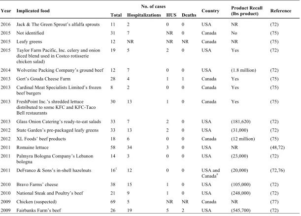
Table 1. Major E. coli O157 foodborne outbreaks in the USA and Canada, 2006-2016.
Year Implicated food No. of cases Country Product Recall
(lbs product) Reference Total Hospitalizations HUS Deaths
2016 Jack & The Green Sprout’s alfalfa sprouts 11 2 0 0 USA NR (72)
2015 Not identified 31 7 NR 0 Canada No (75)
2015 Leafy greens 12 NR NR NR Canada NR (75)
2015 Taylor Farm Pacific, Inc. celery and onion diced blend used in Costco rotisserie chicken salad)
19 5 2 0 USA Yes (72)
2014 Wolverine Packing Company’s ground beef 12 7 0 0 USA (1.8 million) (72)
2013 Gort’s Gouda Cheese Farm 28 4 1 1 Canada Yes (75)
2013 Cardinal Meat Specialists Limited’s frozen beef burgers
8 2 0 0 Canada Yes (75)
2013 FreshPoint Inc.’s shredded lettuce distributed to some KFC and KFC-Taco Bell restaurants
30 13 1 0 Canada Yes (75)
2013 Glass Onion Catering’s ready-to-eat salads 33 7 2 0 USA (181,620) (72) 2012 State Garden’s pre-packaged leafy greens 33 13 2 0 USA (31,000) (72)
2012 XL Foods’ beef products 18 6 0 0 Canada (12 million) (75)
2011 Romaine lettuce 58 34 3 0 USA NR (48,72)
2011 Palmyra Bologna Company’s Lebanon bologna
14 3 0 0 USA (23,000) (72)
2011 DeFranco & Sons’s in-shell hazelnuts 161 12 0 0 USA and
Canada# (20,000) (72,76)
2010 Bravo Farms’ cheese 38 15 1 0 USA (105,000) (72)
2010 National Steak and Poultry’s beef 21 9 1 0 USA (248,000) (72)
2009 Chicken (suspected) 69 5 NR NR Canada NR (77)
Year Implicated food No. of cases Country Product Recall (lbs product) Reference Total Hospitalizations HUS Deaths
2009 JBS Swift Beef Company’s beef 23 12 2 0 USA (421,280) (72)
2009 Nestle Toll House raw refrigerated cookie dough
72 34 10 0 USA 3.6 million
packages
(72,78)
2008 Aunt Mid’s Produce Company’s iceberg lettuce
742 21 NR 0 USA and
Canada
No (51,77)
2008 Romaine lettuce 29 NR 1 NR Canada No (51,77)
2008 Harvey’s Restaurant (Spanish onions (suspect))
235 26 NR 0 Canada No (51,77)
2008 Kroger/Nebraska Ltd. ground beef 49 27 1 0 USA (5.3 million) (72) 2007 Totino’s and Jeno’s frozen pizza (pepperoni) 21 8 4 0 USA 5 million pizzas (72,79)
2007 Topp’s ground beef patties 40 21 2 0 USA (21.7 million) (72)
2006 Lettuce (suspect) 7 NR NR NR Canada NR (51)
2006 Natural Selection Foods, LLC’s fresh bagged spinach
1993 102 31 3 USA and
Canada# NR (51,72,80,81)
2006 Taco Bell 71 53 8 NR USA NR (72)
NR: not reported; Yes: there was a product recall but no information regarding quantities; No: there was no product recall linked to the outbreak. 1In Canada, 8 cases were found, all of them hospitalized
2Three cases were in Canada
increasing number of E. coli O157 outbreaks in Canada and the USA (43,48,51,80). In fact, after the 2006 outbreaks caused by fresh spinach in the USA, the FDA announced the implementation of the “Leafy Greens” initiative with the aim of identifying the potential public health concerns inherent to these products (82). Finally, outbreaks and recalls represent a relevant source of information and provide an opportunity to learn while prompting governments to improve guidelines and regulations to strengthen the food safety systems throughout the whole food chain.
1.3. Current State-of-the-Art in Detection of Food Pathogens
It is evident that food pathogen detection is an important environmental health milestone towards reducing the burden of foodborne diseases and economic losses due to contaminated food. The major objective of testing plans is to verify the adequacy of control manufacturing processes during food production (83). Focusing on E. coli O157, the impact of testing strategies on reducing risk has been proved since 2002, when the USDA-FSIS reassessed the HACCP plans and extended the testing programs, after determining that E. coli O157 is likely to occur at all stages of raw beef production (69). Further implementation of these measures, resulted in a reduction of the prevalence of E. coli O157 in ground beef from 0.73% in 2002 to 0.17% in 2006 (70), while the number of recalls also decreased from 21 in 2002 to 5 in 2005 (84). In Canada, data from 2009 have been encouraging, showing that testing of beef trim, potentially used for the production of ground beef, has prevented contaminated product to be further processed (70). Currently, as part of the new guidance document on E. coli O157, it is recommended that all precursor material used for the production of raw ground beef and beef products should be tested and only lots below detectable levels should be accepted (70). Therefore, testing programs, combined with proper sampling protocols and process controls, have played a key role not only in reducing the risk of E. coli O157 contamination, but also in determining the efficacy of the control measures established to prevent E. coli O157 contamination throughout the manufacturing process.
easy-to-use for early identification of potential hazards. Conversely, however, the “gold standard” conventional testing methods are known to be time-consuming and lengthy because they rely on culture, isolation, and biochemical identification. Food pathogens are commonly found in lower concentrations than food microbiota (85); thus a selective enrichment step is required to enable the recovery of the target microorganism while suppressing non-target organisms (86,87). The enrichment step involves transferring the food sample into a selective nutrient medium and incubation to allow the multiplication of the target pathogen to a detectable level (86,88,89). Furthermore, an isolation step using selective and differential agar plating helps to identify presumptive colonies of the target pathogen. Lastly, typical colonies are screened using biochemical and/or serological analysis. Presumptive results from this procedure can take up to four days, while confirmation may require up to one week (86,90,91). Therefore, one of the main challenges that food producers normally face when implementing testing protocols is that lengthy methods can delay the release of minimally processed products with short shelf life such as raw meat until they are screened and considered to be microbiologically safe (88,92). Consequently, a major research field has focused on the refinement of current methods and the development of more efficient technologies designed improve testing programs. Based on the needs of the food sector, three main characteristics of such tests are crucial: speed, sensitivity, and ease-of-use (16,91,92). Interestingly, the food pathogen testing market was valued at 7.42 billion USD as of 2015 and is expected to continue growing due to the establishment of ever more stringent regulations (93). Moreover, in a report from 2003, it was estimated that the beef and poultry industry in the USA performed approximately 369 tests per processing plant per week, representing 22% of the total microbial tests within the USA food industry (91). Therefore, due to the establishment of new testing plans and recent enhancement of regulations applicable to the meat industry, it is expected that the food pathogen rapid testing market will continue growing.
Trends in Rapid Point-of-Care (POC) Methods
diseases such as dengue, hepatitis B, HIV/AIDS, malaria and syphilis in patients from developing countries (94–97). The success achieved by on-site disease diagnosis and the rise in the market availability of POC tests helped to establish the optimal characteristics that a POC test should have. Indeed, in 2004 WHO’s Sexually Transmitted Diseases Diagnostics Initiative determined that a POC test should comply with the “ASSURED” principle, which means: Affordable, Sensitive, Specific, User-friendly, Rapid, Equipment-free and Delivered (95–98). Based on this, a POC test can be defined as a simple and affordable assay for food producers that is performed at the location where the sample is found, and will provide a rapid outcome, which is crucial for taking appropriate immediate action (95,98,99).
Even though most of the progress has been achieved in developing medical diagnostic tools, affordable and rapid technologies are also necessary to improve other fields such as environmental and food safety (99–101). As noted above, there is a dire need in food safety diagnostics for more rapid and sensitive methods that can replace traditional techniques that require more than two days to determine the presence of pathogens in food samples (91). However, due to recent advances in biotechnology, chemistry, and molecular biology, it has been possible to address some of the challenges that conventional pathogen detection possesses. In fact, significant advances towards developing rapid state-of-the-art microbiological methods that do not require laboratory facilities or special training so that they can be used throughout the food chain, but maintain high specificity and sensitivity, have occurred (101). The upcoming sections will provide an overview of the most relevant methodologies that are influencing the development of rapid POC tests for food safety.
1.3.1.1Molecular methods
loop-mediated isothermal amplification (LAMP), with the advantage that it does not require thermocycling instruments or purified DNA (90,102,106), an important advantage for a POC test model. LAMP is known to be more specific and sensitive than standard PCR methods (106). Conversely, multiplex quantitative PCR (qPCR) has focused on the detection of several pathogens using only one sample, markedly reducing the labor intensive limitation of culture methods and traditional single PCR techniques (86,90,106,107).
Regardless of the advantages that molecular diagnostic methods have such as specificity and sensitivity (95), one of their major shortcomings is they can detect DNA/RNA from dead microorganisms, resulting in misinterpretation of results in complex samples such as food (107). Moreover, most of these methods still require complex equipment, special training, several steps for sample preparation, and are considered to be expensive processes for routine food analysis when compared to a conventional culture method (95,107,110).
1.3.1.2Optical methods
Although some optical techniques have been used for more than a decade, the main challenge has been to transfer them into portable POC tests. The best example is surface plasmon resonance (SPR), which has the advantage of detecting molecular interactions without the need for labeled reagents (95,96). Thus, it can be used with nucleic acids or immunoassays (96). SPR has been used to detect C. jejuni, S. enterica ser. Typhimurium,
1.3.1.3Nanotechnology
Some of the advances in nanotechnology in the diagnostics field are focused on labeling with metal or magnetic nanoparticles for immunoassays (96,111,113). The range of reagents for labeling goes from antibodies (106,114–116) to nucleic acids (99), the basis for biosensors (nanosensors) for real-time detection (108,117). Quantum dots are also a novel alternative for fluorescent labeling because they possess higher brightness, photostability and are more resistant to chemical degradation that traditional fluorescent dyes (96,99,118). In the food safety field, quantum dots have been used for detection of
E. coli O157, Salmonella and L. monocytogenes (115). Working with nanomaterials has the advantage of increasing the number of reactive sites, resulting in higher sensitivity and specificity (94). The development of nanosensors represents a portable alternative with a shorter time-to-result at a low cost (117). Nanocantilevers, which are made of silicon-based materials and can detect biological binding interactions with high sensitivity and short time-to-result, exemplify this novel class of label-free nanosensors (101,117).
1.3.1.4Microfluidics
mechanisms of microbial drug resistance (95). It is evident that microfluidics represents one of the most promising research areas in POC test development, with the potential to optimize sensitivity and specificity without requiring laboratory equipment.
1.3.1.5Immunoassays
Enzyme immunoassays are one of the first described immunochemical techniques still widely used for diagnostics due to its ability to produce a colorimetric reaction that can be quantitated and visualized at a macroscopic scale (95,123). The most well-known immunoassay is the enzyme-linked immunosorbent assay (ELISA), which has become a notable tool for in vitro diagnostics, regulatory and quality assessments. Immunoassays are a versatile method that has been combined with several of the technologies described above. Recently, Weidemaier et al. reported a nanotechnology-based immunoassay using antibody-conjugated magnetic SERS nanoparticles added directly into the food sample, which capture the target pathogens as they grow and produces a real-time detectable signal read through the sample vessel (111). Moreover, Ho et al. developed a colorimetric immunoassay based POC test using immunoliposomes with an encapsulated visible dye to detect E. coli O157 (124). However, in light of developing POC tests, lateral flow immunoassays (LFIA) or immunochromatographic test strips are considered the most popular POC technology in clinical diagnostics (95,96). In fact, they are defined as an ASSURED technology (125,126). Thus, it is not surprising that it has also gained popularity within other fields such as food safety and has become a target technology for research (127). LFIAs are similar to an ELISA because they rely on the principle of antigen-antibody binding (128). Therefore, their sensitivity and specificity will be completely dependent on the concentration and accessibility of the target analyte in the sample, as well as of the binding strength and affinity between the antibody and the antigen (106). Contrary to ELISA, LFIAs have the advantage that results can be obtained relatively fast and do not require intensive training (95,96). Similar to other immunoassays, LFIAs have been combined with other techniques such as the prototype developed by Mondesire
commonly known as NALF or NALFIA (nucleic acid lateral flow or nucleic acid lateral flow immunoassay). The former does not require antibodies because it uses amplicons or probes as capture and detection reagents, while the latter uses antibody recognition against a labeled amplicon (128). By combining LFIA not only with DNA/RNA techniques and also with nanoparticles labeling or miniaturized thin-layer chromatography, the sensitivity and selectivity of the assays has been improved (130).
1.4. Overview of Lateral Flow Immunoassays (LFIAs)
The first lateral flow or dipstick assay was developed in the 1960s for quantifying glucose in urine (131). Its principle relied on an enzymatic reaction that caused a color change, which was compared to a color chart to obtain a semi-quantitative result (131). However, it was two decades later, in the mid-1980s, when LFIAs were introduced to the clinical diagnostic field with the development of the pregnancy test (100,127). Shortly after, their application gradually expanded to other fields, where on/off signals were sufficient, such as drug screening (100), detection of cardiac markers (121), food (130,132,133) and environmental applications (100,130,133), particularly in resource-limited countries.
of LFIAs has mainly focused on presence/absence formats for rapid and easy screening of food samples (90,106,133). Indeed, progress has been such that Dzantiev et al. compiled the recent advances on immunochromatographic assays developed for food analysis between 2007 and 2013 (132). These authors estimated that 14% of the scientific publications on immunochromatographic test systems were focused on the development of LFIAs for pathogen detection whereas 30% focused on mycotoxins detection. Furthermore, low development costs and facile production (127) give LFIAs an extra advantage over other technologies. From an economic perspective, a publication from 2009 estimated that the food and beverage sector are the largest producer of rapid tests while being the third highest consumer of LFIA-based tests with $30 million USD generated in 2007, just below the clinical and veterinary sectors (139). LFIAs combine the selectivity and sensitivity of immunoassays, such as ELISA, with the simplicity and speed of operation of a POC device. LFIAs are designed to provide results in less than 15 min after loading the sample; thus they represent an excellent alternative for the routine testing of food products.
LFIAs Principle and Main Components
LFIAs rely on the movement, through capillarity, of a liquid sample that is initially loaded onto a sample pad found at one extreme of the nitrocellulose membrane, towards the other extreme where an absorbent pad will capture the remainder of the liquid. Capillary movement through the membrane allows the sample to pass through different zones, where immobilized recognition reagents will interact with the target analyte to form complexes that will attach within the test zone, while unreacted reagents will continue flowing to the control line, where they will attach (130,140). The target analyte complexes captured in the test zone will produce a visible line, similar to the one produced by unreacted reagents captured within the control line. However, the control line will develop whether or not the sample contains antigen, thereby ensuring that the test system is functioning properly (Figure 2).
Sample
Sample pad
Nitrocellulose membrane
Absorbent pad FLOW
Conjugate pad Test line
formed by the primary detection antibody or a secondary antibody against the primary, which are conjugated to a label or marker (141). Although new alternatives for preparing recognition reagents, such as quantum dots (141,142) and liposomes with colored dyes (124,141,143), are being introduced, colloidal gold nanoparticles remain the most frequently used label alternative for LFIAs mainly due to their availability and low cost for large scale production (114,130,140,141). In addition, two main types of reactions are commonly used, which are based on the analyte to be determined. The direct assay normally is selected when the target has more than one epitope. In this case, the recognition reagent will bind to one epitope, while the capture reagent, normally attached to the test line, will bind an alternative epitope (130,133,140). In this case, the development of a test line is directly linked to the presence of the target analyte. On the other hand, the competitive assay is frequently selected when the target analyte is a small molecule, which is the case for mycotoxins (134,144). Two alternatives can be used, either the capture reagent is attached to the test line and labeled analyte competes with the sample analyte, or a protein-analyte is attached to the test line while the labeled antibody is initially mixed with the sample. In both cases, a colored test line will be indirectly linked to the presence of the target analyte in the sample (114,130,133). Therefore, because bacteria possess several surface antigens, the sandwich assay is preferred for development of LFIA for pathogen detection.
reagents being stored in a tube, to which the sample is added, acting as a reconstituent, before inserting the LFIA test strip (145). Although both capture and detection antibodies are responsible for the analytical characteristics of the LFIA based on their interactions with the target analyte (146), mAbs are key in determining the specificity of the method. Therefore, efforts are continuously being made to maintain and improve the affinity of the mAbs. This includes using labeled secondary antibodies instead of direct labeling of the mAb because it has been shown that conjugation can decrease the affinity of the antibody while interfering in the antibody-antigen binding due to steric hindrance (130). In addition, research has focused on developing more specific antibodies with the adequate binding characteristics for their application in immunoassays. This has included optimizing hybridoma technologies as well as the development of antibody derivatives through genetic engineering (147). As a result of these approaches, LFIAs have evolved to comply with the needs demanded by the recent diagnostics markets.
1.5. LFIAs for Detection of E. coli O157 in Food
LFIAs, similar to most of the novel technologies that have been developed for pathogen detection, rely on biorecognition of surface antigens in order to detect whole bacterial cells (101). Some of the surface antigens found in E. coli O157 are commonly used as detection biomarkers, while also serving to classify E. coli isolates by serotyping. The latter is a technique based on three main antigens: “O”, “H” and “K”, which are frequently identified by serology (52,148). The former is the outermost variable part of the polysaccharide in the outer membrane (somatic antigen), the second is a flagellar protein, and the third is a capsular antigen (18,52,149,150). However, the K-antigen is rarely used by laboratories; only the O:H combination is considered the standard for classifying or serotyping E. coli
immunogenic, it is considered a target for both immune cells and bacteriophages (149). Furthermore, contrary to other surface antigens, it is heat-stable (149). Therefore, the O-antigen is of particular interest for epidemiological studies, classification during outbreak investigations and as a diagnostic target (150). Indeed, through surveillance and epidemiological data, different O-serogroups have been associated more often with outbreaks than others, despite their H-antigen. This has been the case of E. coli O157:non-H7 strains, which have been isolated from humans and patients with diarrhea including
E. coli O157:H45, O157:H39 and O157:H16, among others (154). In light of these findings and considering E. coli O157 as one of the most implicated serotypes involved in human illness, antibodies against the O157-antigen have been constantly produced and used in the development of LFIAs for routine screening of E. coli O157 in food (155,156) or clinical samples (157,158).
While there have been numerous LFIAs described for detecting E. coli O157 in food samples, only a few of them have been marketed. Nine gold nanoparticle-based LFIAs and one using magnetic nanoparticles, were commercially available at the time of writing (Table 2). Six of these were previously reported by Farrokh et al. and Jasson et al. (42,159) as validated methods for the detection of E. coli O157 in different types of food. Contrary to this, the remaining four methods, MaxSignal®, Quicking, SAS™, and SMART™-II, did not possess supporting information regarding their validation status (160–163). Besides commercialized LFIAs, Singh et al. summarized the information of six non-commercial LFIAs for the detection of E. coli O157, which have been developed and published by different research groups (133). Overall, the LFIAs described thus far, require an enrichment step in order to achieve detectable levels that range between 104 and 105 CFU/ml (Table 2) (133,159). Despite the fact that the enrichment step can be as short as 6 h, it is still necessary and a major feature to be considered when selecting a rapid POC method.
Validation of Alternative Microbiological Methods
Table 2. Commercial LFIAs for detection of E. coli O157 in food samples (as of June, 2016).
Brand Manufacturer Food Type
Enrichment (h) Enrichment broth
Sensitivity Validation Reference
Dupont™ Lateral Flow System
DuPont Ground beef, boneless beef trim
Yes (8-18) P/E
1 CFU/ 25g AOAC-RI PTM (42,164)
FoodChek™* FoodChek™ Systems Inc.
Raw ground beef, beef trim
Yes (6-8) mTSB
1 CFU/ 25g AOAC-RI PTM (42,165,166)
MaxSignal®E. coli
O157 Strip Test Kit
Bioo Scientific Corporation
Meat and meat products, dairy products
Yes (18-24) mEC (meat)/ mTSB-n (dairy)
1×10⁴ CFU/ml
post-enrichment
N/S (160)
Quicking Quicking Biotech Co., Ltd.
N/S Yes 1×10⁵ CFU/ml
post-enrichment
N/S (161)
RapidChek®E. coli
O157 (incl. H7)
Romer Labs Raw ground beef, raw boneless beef, apple cider
Yes (8-18) P/E, mEC or EEB
N/S AOAC-RI PTM USDA FSIS MLG
(42,167)
Reveal 2.0 NEOGEN Corporation
Raw ground beef, raw beef trim
Yes (8-20) P/E
1 CFU/ 25 or 375g
104 CFU/ml post
enrichment
AOAC-RI PTM (42,168)
SAS™ E. coli O157 and O157:H7 Test
SA Scientific N/S Yes (16-24) mEC
N/S N/S (162)
Singlepath®E. coli
O157
EMD Millipore Corporation
Raw ground beef, pasteurized milk
Yes (24) mTSB-n and/or EEB
1 CFU/ml or 25g AOAC-RI PTM Health Canada
Brand Manufacturer Food Type
Enrichment (h) Enrichment broth
Sensitivity Validation Reference
SMART™-II Rapid E. coli O157 Strip Test
New Horizon Diagnostics
N/S Yes 1 CFU/25g
3.3×10⁴ CFU/ml
post-enrichment
N/S (133,163)
VIP® Gold- EHEC BioControl
Systems, Inc.
N/S Yes N/S AOAC OMA (42,170,171)
The information provided in this table is based on the latest version of the manufacturer’s web pages, product data sheets and/or validation certificates available at the time of writing.
*FoodChek™ test is a magnetic nanoparticle LFIA.
AOAC OMA: AOAC INTERNATIONAL Official Method of AnalysisSM AOAC-RI PTM: AOAC Research Institute Performance Tested MethodSM N/S: not specified
Consequently, the development and commercialization of rapid methods have advanced because they represent an alternative to maintaining processing efficiency while complying with screening requirements. However, due to the critical role rapid methods play in ensuring the safety of food, evidence regarding their performance and fit for purpose is required before they can be considered reliable alternatives for pathogen screening (159,172,173). This process is known as validation and normally involves two main phases: 1) comparison of the alternative rapid method against a reference method, and 2) an interlaboratory study (105,159,174). The latter requires the participation of different laboratories in order to assess the reproducibility and repeatability of the results obtained with the alternative method (105). During the validation process, the alternative method is represented by the method that has been designed and is intended to be used instead of a gold standard, also known as the reference method (172). Most often an alternative method will be a system that intends to reduce the time necessary for getting a reliable result (159). Therefore, a rapid alternative method will be any new technique or system that can be used instead of a traditional culture method and provide results in a shorter time with a high degree of reliability. On the other hand, a reference method will comprise of any widely accepted method, normally internationally recognized with a well-established protocol, such as traditional culture methods (159,172).
specifically designed to comply with the needs of their particular market. They may not be fully recognized worldwide or by all stakeholders. In addition, validation by any of these bodies is not considered mandatory, however, the use of alternative methods may be restricted by the needs of certain stakeholders that require a specific validation scheme (e.g.
official control by government agencies) (159). For this reason, method developers need to select carefully the type of validation protocol they will follow.
1.5.1.1 Health Canada’s procedure for the validation of alternative microbiological methods
In Canada, the Microbiological Methods Committee (MMC) is responsible for supplying the appropriate methods for ensuring food safety throughout the supply chain. To do that, the MMC reviews all submitted methodologies to guarantee that they are fit for purpose and that sound science was used along the validation procedures (174). Once a method is approved, it is included in the Compendium of Analytical Methods, which contains all methodologies that are used by Health Canada, the Canadian Food Inspection Agency (CFIA), and other organizations, to determine compliance with standards and regulations (174).