Prediction of poor outcome within the
first 3 days of postanoxic coma
E.G.J. Zandbergen, MD; A. Hijdra, PhD; J.H.T.M. Koelman, PhD; A.A.M. Hart, PhD; P.E. Vos, PhD;
M.M. Verbeek, PhD; and R.J. de Haan, PhD, for the PROPAC Study Group*
Abstract—Objective: To determine the optimal timing of somatosensory evoked potential (SSEP) recordings and the additional value of clinical and biochemical variables for the prediction of poor outcome in patients who remain comatose after cardiopulmonary resuscitation (CPR).Methods:A prospective cohort study was conducted in 32 intensive care units including adult patients still unconscious 24 hours after CPR. Clinical, neurophysiologic, and biochemical variables were recorded 24, 48, and 72 hours after CPR and related to death or persisting unconsciousness after 1 month.Results:Of 407 included patients, 356 (87%) had a poor outcome. In 301 of 305 patients unconscious at 72 hours, at least one SSEP was recorded, and in 136 (45%), at least one recording showed bilateral absence of N20. All these patients had a poor outcome (95% CI of false positive rate 0 to 3%), irrespective of the timing of SSEP. In the same 305 patients, neuron-specific enolase (NSE) was determined at least once in 231, and all 138 (60%) with a value ⬎33 g/L at any time had a poor outcome (95% CI of false positive rate 0 to 3%). The test results of SSEP and NSE overlapped only partially. The performance of all clinical tests was inferior to SSEP and NSE testing, with lower prevalences of abnormal test results and wider 95% CI of false positive rates. Conclusion: Poor outcome in postanoxic coma can be reliably predicted with somatosensory evoked potentials and neuron-specific enolase as early as 24 hours after cardiopulmonary resuscitation in a substantial number of patients.
NEUROLOGY 2006;66:62–68
The prediction of poor outcome in patients who
re-main unconscious after cardiopulmonary
resuscita-tion (CPR) has recently been addressed in a number
of systematic reviews. A positive predictive value of
100% has been demonstrated for the absence of early
cortical responses (N20) of the somatosensory evoked
potentials (SSEPs) in the first week after CPR,
1-3and
similar values were found for the absence of
pupil-lary and corneal reflexes and of any motor response
3 days after the hypoxic–ischemic insult.
1,4The
use-fulness of biochemical markers of brain damage in
serum or CSF remained uncertain, because of the
small number of patients in most studies and
meth-odologic flaws in some.
5With these reviews, a
num-ber of questions could not be answered. 1) What is
the earliest time when SSEP results may be
consid-ered reliable? 2) Are the predictive values of separate
variables additive? 3) Can biochemical markers of
anoxic–ischemic brain damage contribute to the
pre-diction of poor outcome? We designed our study
(Prognosis in Postanoxic Coma) to address these
questions.
Methods. From January 2000 to May 2003, we performed a multicenter prospective cohort study to correlate early clinical, neurophysiologic, and biochemical findings with clinical outcome. To study prediction in regular clinical practice, we chose a prag-matic design, with various types of hospitals and without central-ized assessment of the neurophysiologic tests. CSF studies were not feasible with this design. Patients admitted to the intensive care units of 32 Dutch hospitals (13 teaching hospitals and 19 nonteaching hospitals) were included. The study was approved by the ethical review boards of all participating hospitals.
Consecutive patients with CPR for primary or secondary circu-latory arrest, persisting coma 24 hours after CPR, age 18 years or older, and informed consent from a legal representative were in-cluded. We defined coma as no eye opening to external stimuli, motor response to pain flexion or worse, and no speech. Exclusion criteria were a life expectancy of no more than several months caused by pre-existent disease, death by brain criteria after 24 hours, and concomitant head injury.
Timing of assessments. Baseline characteristics registered were gender, age, medical history, prearrest level of functioning, cause of the arrest, location of arrest, cardiac rhythm before CPR, time between arrest and initiation of CPR, and duration of CPR. All patients underwent standard assessments 24, 48, and 72 hours (⫾4 hours) after CPR, including neurologic examination, median nerve SSEP, and blood sampling at all time points and EEG at 72 hours. For practical reasons, SSEP recording was not
Additional material related to this article can be found on theNeurology
Web site. Go to www.neurology.org and scroll down the Table of Con-tents for the January 10 issue to find the title link for this article.
*See the Appendix for a complete list of Group members.
From the Departments of Neurology and Clinical Neurophysiology (E.G.J.Z., A.H., J.H.T.M.K., R.J.D.H.) and Clinical Epidemiology and Biostatistics (A.A.M.H.), Academic Medical Centre, University of Amsterdam, and Department of Neurology (P.E.V.) and Laboratory of Pediatrics and Neurology (M.M.V.), University Medical Centre Sint Radboud, Nijmegen, the Netherlands.
Supported by grants from the Dutch Heart Foundation (Nederlandse Hartstichting) and the Dutch Brain Foundation (Hersenstichting Nederland). Disclosure: The authors report no conflicts of interest.
Received April 27, 2005. Accepted in final form October 5, 2005.
always possible on weekends. If the 72-hour SSEP was due on a weekend day, the recording was postponed to Monday.
Clinical assessment. Within the first 24 hours post CPR, sed-atives and muscle relaxants were stopped. When this was not possible, propofol was used as the standard sedative, and this medication was stopped shortly before clinical assessment and EEG recording. Morphinomimetics and benzodiazepines were avoided as much as possible. Clinical features registered included cardiovascular stability (spontaneously stable, stable with medica-tion, unstable), Glasgow Coma Scale, pupillary and corneal re-flexes of each eye (present or absent), eye movements (spontaneously roving, on cervico-ocular testing, absent), and sei-zure or myoclonus (absent, sporadic, status). We also recorded the Acute Physiology and Chronic Health Evaluation-II (APACHE-II) score over the first 24 hours. Outcome was registered after 1 month using the Glasgow Outcome Scale. Poor outcome was de-fined as either death or persisting unconsciousness after 1 month. In patients who died, the probable cause of death was registered. In surviving patients, 1-year outcome was assessed by telephone contact with the patient, a family member, or the general practi-tioner of the patient.
SSEPs. We recorded SSEPs with standard procedures.6,7
Lo-cal cliniLo-cal neurophysiologists in each center assessed the record-ings. The results for the N20 were documented for each side separately as absent (only in the presence of a cervical potential) or present or the recording was judged to be technically insuffi-cient. For our primary analysis, we defined “SSEP absent” as N20 absent on both sides and “SSEP not absent” as all remaining combinations of left and right recordings.
EEG. Local clinical neurophysiologists assessed the EEG re-cordings. They coded their findings using the classification of Hockaday et al.8In addition, they registered the presence or
ab-sence of a burst-suppression pattern and of epileptiform activity (absent, sporadic, frequent, status).
Determination of S-100B and neuron-specific enolase. Blood sampling and measurements were done using standard proce-dures.9For our primary analysis, we defined abnormal values as
⬎0.7 g/L for S-100B and⬎33 g/L for neuron-specific enolase (NSE), based on our meta-analysis in which these values resulted in the lowest false positive rates.5
Blinding. Treating physicians were blinded for the results of the first and second day SSEP and for all blood tests. Absence of SSEP at 72 hours was considered a sufficiently reliable predictor of poor outcome to allow its use for treatment decisions,1-3and the
result of 72-hour SSEP testing was therefore made available to the treating physicians.
Treatment and treatment restrictions. The protocol prescribed no specific treatments. Standard care in Dutch intensive care units generally consists of standard supportive care, valproate or phenytoin for epilepsy, and valproate or clonazepam for myoclo-nus. During the last phase of the study, induced hypothermia (32 to 34 °C) was gradually introduced as a treatment of anoxic-ischemic brain damage.10,11These patients were included. From a
methodologic point of view, it was desirable that all patients re-ceive maximal treatment during a certain period, but this could not be realized on ethical and practical grounds. Treating physi-cians could decide to stop or forego further treatment of patients with established predictors of poor outcome. With regard to neuro-logic prognosis, they were offered the following guidelines: 1) Chances for survival or recovery of consciousness are virtually nil when the N20 of the SSEP or the pupillary reflexes or motor responses are absent or when the EEG is isoelectric at 72 hours or later; 2) chances for survival without severe disability are virtu-ally nil with a burst-suppression EEG at 72 hours or later or with motor response flexion or worse at 7 days or later.1,12Timing and
motivation of any change in treatment level were documented. Treatment levels were no treatment restrictions; treatment re-stricted to standard supportive care, such as can be given on regular wards (no-resuscitation order, weaning from the ventila-tor and transfer to the regular ward, no readmission to the inten-sive care unit; all other treatment given when necessary); and no treatment other than palliation.13
Analyses. We designed the study to test two hypotheses: 1) The bilateral absence of N20 of the SSEP 24 and 48 hours after CPR predicts poor outcome as accurate as the absence of such responses 72 hours after CPR; 2) in a number of patients in whom N20 of the SSEP is present in the first 72 hours after CPR, poor
prognosis can be accurately predicted with clinical, EEG, or bio-chemical variables. Hypothesis 1 was tested with descriptive sta-tistics on the basis of 2⫻2 tables with “bilateral absence of N20” and “poor outcome” as present or absent. We calculated false pos-itive rates (patients with abnormal test result and favorable out-come/all patients with abnormal test result) and positive likelihood ratios with their 95% CIs. To calculate likelihood ratios when one of the cells of the 2⫻2 table contained no observations, we added a value of 0.5 to each cell. For the second hypothesis, we performed univariate analysis with similar descriptive statistics, using all remaining variables (table 1). We used predefined cut-off levels for the primary analysis of NSE and S100b levels (see above). The performance of these tests was further evaluated with receiver operating characteristic curves. Sample size calculation was based on 5% as the upper limit of the CI of a false positive rate for a prognostic variable, which means that we would need 80 patients to identify such a variable. With a conservative estima-tion of the prevalence of useful predictors of 20%, 400 patients were required to identify such variables reliably.
Results. We included 407 patients in the study (see table E-1 on theNeurologyWeb site at www.neurology.org). Six centers included more than 20 patients, 8 centers between 20 and 10, and 18 centers fewer than 10. Poor outcome occurred in 356 (87%) of the patients, 349 of whom had died. Of the 51 patients who were conscious after 1 month, 34 were severely disabled, 10 moderately disabled, and 7 had made a good recovery. Of the 34 patients who were severely disabled after 1 month, 11 had died after 1 year, 13 remained severely disabled, and 10 had become inde-pendent (moderate disability or good recovery).
Characteristics of neurophysiologic, biochemical, and clinical tests are presented in tables 2 to 4. The absence of SSEP, EEG Hockaday grade 5, and serum levels of NSE of ⬎33g/L were superior to all other variables with regard to the percentage of abnormal test results (high), false
Table 1Variables and values used in analyses
Clinical characteristics
●APACHE-II score during first 24 h (⬎25 vsⱕ25)*
●Circulation (unstable vs spontaneously stable)
●Epilepsy or myoclonus (present vs absent)
●Pupillary reflexes (bilaterally absent vs. present)
●Corneal reflexes (bilaterally absent vs present)
●Eye movements (absent vs spontaneous or on cervico-ocular testing)
●Motor response (absent vs present spontaneously or with painful stimuli)
Neurophysiologic variables
●SSEP N20 (bilaterally absent vs not bilaterally absent)
●EEG at 72 h (Hockaday grade 5 vs others)*
●EEG at 72 h (burst suppression vs no burst suppression)*
●EEG at 72 h (epileptiform activity vs. no epileptiform activity)* Biochemical variables
●Serum NSE (⬎33 vsⱕ33g/L)
●Serum S-100B (⬎0.7 vsⱕ0.7g/L)
All variables except (*) tested at 24, 48, and 72 (⫾4) h after car-diopulmonary resuscitation.
positive rates (low), and likelihood ratios (high). No false positive predictions were made with any of these variables, with the upper limit of the 95% CI of their false positive rates below 5%.
In 256 patients, at least two SSEP recordings hade been done. In nine of these, N20 was present in the first and absent in a later recording. In five other patients, N20 was absent at 24 hours, but the response recurred in a later recording. All these 14 patients had a poor outcome. In 167 patients, serum NSE had been determined at all three time intervals, and all 88 (53%) patients with at least one abnormal NSE value had a poor outcome. The results of SSEP recording and NSE determination only partially overlapped (see table E-2 on theNeurology Web site). All 192 patients in whom one of the tests was abnormal had a
poor outcome (95% CI of the false positive rate 0 to 2%). Most of the abnormal test results were obtained 24 hours after CPR (74% of abnormal results for SSEP, 65% for NSE, and 78% for one of these tests). Overlap between SSEP and EEG results was more extensive, but still in-complete: Of all patients without absent SSEP, only 13% had a low voltage EEG (Hockaday grade 5; all voltages ⬍20 V) or a burst-suppression pattern. Of the 305 pa-tients who were still unconscious after 72 hours, no abnor-mal test result for SSEP, NSE, or EEG was found in 105, 81 (77%) of whom had a poor outcome.
Receiver operating characteristic curves for the two bio-chemical tests validated the cut-off value for NSE we se-lected from our structured review,5but not that for S100b, which proved more time dependent than that of NSE
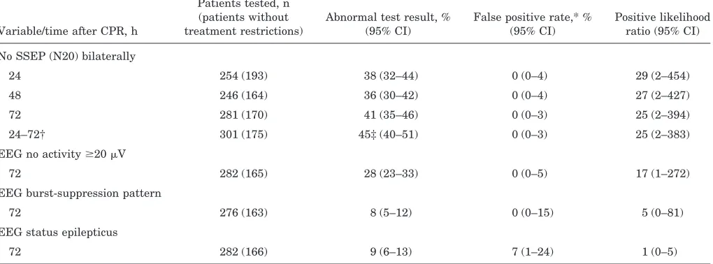
(fig-Table 2Prediction of poor outcome with neurophysiologic variables
Variable/time after CPR, h
Patients tested, n (patients without treatment restrictions)
Abnormal test result, % (95% CI)
False positive rate,* % (95% CI)
Positive likelihood ratio (95% CI)
No SSEP (N20) bilaterally
24 254 (193) 38 (32–44) 0 (0–4) 29 (2–454)
48 246 (164) 36 (30–42) 0 (0–4) 27 (2–427)
72 281 (170) 41 (35–46) 0 (0–3) 25 (2–394)
24–72† 301 (175) 45‡ (40–51) 0 (0–3) 25 (2–383)
EEG no activityⱖ20V
72 282 (165) 28 (23–33) 0 (0–5) 17 (1–272)
EEG burst-suppression pattern
72 276 (163) 8 (5–12) 0 (0–15) 5 (0–81)
EEG status epilepticus
72 282 (166) 9 (6–13) 7 (1–24) 1 (0–5)
* Patients with abnormal test result and favorable outcome/all patients with abnormal test result (1⫺positive predictive value). † Refers to 305 patients who were still comatose after 72 h and in whom SSEP testing had been performed at least once. ‡ At least one abnormal test result.
CPR⫽cardiopulmonary resuscitation; SSEP⫽somatosensory evoked potential.
Table 3Prediction of poor outcome with biochemical variables
Variable/time after CPR, h
Patients tested, n (patients without treatment restrictions)
Abnormal test result, % (95% CI)
False positive rate,* % (95% CI)
Positive likelihood ratio (95% CI)
NSE⬎33g/L
24 272 (206) 42 (36–48) 0 (0–3) 36 (2–563)
48 241 (157) 52 (46–59) 0 (0–3) 45 (3–715)
72 209 (108) 46 (40–53) 0 (0–4) 39 (3–610)
24–72† 231 (110) 60‡ (53–66) 0 (0–3) 23 (2–357)
S100b⬎0.7g/L
24 273 (207) 45 (40–51) 3 (1–8) 5 (2–12)
48 238 (155) 44 (38–50) 2 (0–7) 9 (2–36)
72 207 (108) 35 (29–42) 0 (0–5) 30 (2–466)
24–72† 230 (110) 53‡ (46–59) 2 (1–7) 3 (1–9)
* Patients with abnormal test result and favorable outcome/all patients with abnormal test result (1⫺positive predictive value). † Refers to 305 patients who were still comatose after 72 h and in whom NSE or S100b testing had been performed at least once. ‡ At least one abnormal test result.
ure). With the mandatory high specificity, sensitivity of NSE testing is clearly superior to that of S100b testing.
Treatment had been restricted in 23% of patients at 24 hours and in 28% at 48 hours. In all but two patients, these restrictions were of category B. Despite blinding of the treating physicians for the 24- and 48-hour SSEP and all NSE values, treatment was more often restricted in patients with absent SSEP and abnormal NSE values (see table E-3 on theNeurologyWeb site).
Only 10 patients were treated with induced hypother-mia, 8 of whom had a poor outcome (not significantly dif-ferent from untreated patients). These 10 patients included 4 in whom at least one SSEP had been absent and 3 in whom serum NSE had been above the cut-off value at least once; all these patients had a poor outcome.
Discussion.
We have demonstrated that the
bilat-eral absence of the N20 of the SSEP in patients with
postanoxic coma of at least 24 hours’ duration is
invariably associated with poor outcome. In 45% of
patients, SSEP were absent within the first 3 days
after CPR, in about three-fourths of these already
after 24 hours. Absent SSEP were found more often
than any other neurophysiologic or clinical predictor
with 100% predictive value. Serum NSE above 33
g/L proved to be equally accurate, with a
preva-lence of 60%. An important additional finding,
con-firming earlier reports,
14,15was the incomplete
overlap of the results of SSEP and NSE. The
preva-lence of at least one abnormal test result derived
from patients in whom both tests were performed
was 66%. Finally, in patients with no absent SSEP
and NSE
ⱕ
33
g/L, a small number with poor
out-come could be identified with EEG (burst
suppres-sion or no voltage
⬎
20
V). Of all 356 patients with
poor outcome, this outcome could be reliably
pre-dicted with these three variables in the first 3 days
after CPR in 252 (71%).
We tested a large number of variables (see table
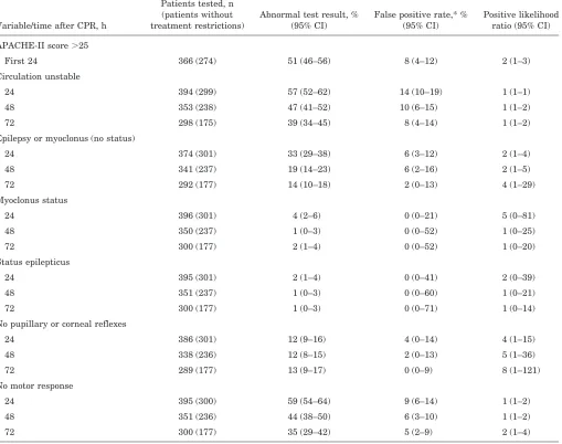
Table 4Prediction of poor outcome with clinical variablesVariable/time after CPR, h
Patients tested, n (patients without treatment restrictions)
Abnormal test result, % (95% CI)
False positive rate,* % (95% CI)
Positive likelihood ratio (95% CI)
APACHE-II score⬎25
First 24 366 (274) 51 (46–56) 8 (4–12) 2 (1–3)
Circulation unstable
24 394 (299) 57 (52–62) 14 (10–19) 1 (1–1)
48 353 (238) 47 (41–52) 10 (6–15) 1 (1–2)
72 298 (175) 39 (34–45) 8 (4–14) 1 (1–2)
Epilepsy or myoclonus (no status)
24 374 (301) 33 (29–38) 6 (3–12) 2 (1–4)
48 341 (237) 19 (14–23) 6 (2–16) 2 (1–5)
72 292 (177) 14 (10–18) 2 (0–13) 4 (1–29)
Myoclonus status
24 396 (301) 4 (2–6) 0 (0–21) 5 (0–81)
48 350 (237) 1 (0–3) 0 (0–52) 1 (0–25)
72 300 (177) 2 (1–4) 0 (0–52) 1 (0–20)
Status epilepticus
24 395 (301) 2 (1–4) 0 (0–41) 2 (0–39)
48 351 (237) 1 (0–3) 0 (0–60) 1 (0–21)
72 300 (177) 1 (0–3) 0 (0–71) 1 (0–14)
No pupillary or corneal reflexes
24 386 (301) 12 (9–16) 4 (0–14) 4 (1–15)
48 338 (236) 12 (8–15) 2 (0–13) 5 (1–36)
72 289 (177) 13 (9–17) 0 (0–9) 8 (1–121)
No motor response
24 395 (300) 59 (54–64) 9 (6–14) 1 (1–2)
48 351 (236) 44 (38–50) 6 (3–10) 1 (1–2)
72 300 (177) 35 (29–42) 5 (2–9) 2 (1–4)
* Patients with abnormal test result and favorable outcome/all patients with abnormal test result (1⫺positive predictive value).
1), and the 100% predictive value of some of these
could well have arisen by chance. This is borne out
by the fact that some patients (2/57) have been
re-ported with NSE values
⬎
33
g/L who survived.
5,15On the other hand, the low false positive rates for
SSEP and serum NSE confirm our previous
find-ings
1,5and are supported by subsequently reported
data.
2,3,14-16Our results must therefore be regarded
sufficiently robust to warrant application in clinical
practice.
The patient characteristics at inclusion do not
dif-fer from previously described cohorts with regard to
age and sex distribution, initial rhythm disturbance,
proportion of out-of-hospital arrests, and
comorbid-ity. The proportion of patients with poor outcome
(87%) is at the upper end of the wide range reported
for both retrospectively and prospectively studied
co-horts, with mortality rates varying from 28 to 92%.
1The main difference between these cohorts and the
one we studied was the time of inclusion: We
in-cluded only patients still unconscious 24 hours after
CPR, whereas all other studies included patients
with much shorter periods of unconsciousness,
vary-ing from the time of return of spontaneous
circula-tion to a few hours thereafter. Because most patients
who recover consciousness do so within the first day,
inclusion of patients at a later stage explains the
high proportion of patients with poor outcome in our
cohort.
We have defined death or persisting
unconscious-ness after 1 month as “poor outcome.” This was
based on our previous argumentation that the
chance of recovery of consciousness in patients who
are still unconscious 1 month after CPR is virtually
nil, especially when the SSEPs are absent.
1Patients
who were conscious but severely disabled after 1
month were not included in the “poor outcome”
cate-gory, because further improvement in these patients
may occur. In our cohort, one-third of such patients
lived independently 1 year after the cardiac arrest.
One could argue that prediction of poor outcome
should be aimed at the clinical condition after a
longer period, say 1 year, and should then include
“severe disability,” a condition many patients would
like to avoid. We could not find useful differences
between the small numbers of patients who were
severely disabled or living independently after 1
year. Obviously, a major effort would be required to
find reliable rules for such a prediction.
Regular clinical practice, with its more or less
tacit knowledge about predictors of poor outcome, is
incompatible with a strict experimental study
de-sign. The tendency to restrict treatment selectively
in patients with characteristics recognized or
pre-sumed to be predictive of poor outcome may then
result in the finding that such characteristics are
indeed good predictors of poor outcome (the fallacy of
a “self-fulfilling prophecy”). We could not (and did
not wish to) prevent physicians to restrict treatment.
In our protocol, we provided guidelines for such
deci-sions, with as most important characteristic the
postponement of such decisions to at least 72 hours
after CPR. Despite this, some restriction had been
implemented in 23% of patients after 24 hours and
in 28% after 48 hours and significantly more often in
patients with absent SSEP or high values of serum
NSE at these time points, test results of which the
treating physicians were not aware. This could be
explained by the higher prevalence in these patients
of clinical variables generally recognized to be
associ-ated with poor outcome (see table E-3 on the
Neurol-ogy
Web site). An alternative or complementary
explanation of insufficient blinding would only apply
to the SSEP results and therefore seems unlikely. It
is apparent that selective treatment restrictions are
difficult to prevent. The main conclusions of our
study, however, are not invalidated by this finding,
because two-thirds of the patients with absent SSEP
at 24 and more than half at 48 hours were treated
without restrictions. Despite this maximal
treat-ment, all these patients had a poor outcome.
During the last phase of our study, induced
hypo-thermia (32 to 34 °C) became gradually accepted as a
treatment of anoxic–ischemic brain damage.
10,11Al-though we have included some patients who were
treated with hypothermia, our results are essentially
derived from patients in whom no such treatment
has been given. The question then is whether our
results could be applied to patients treated with
hy-pothermia. The need for predicting outcome and
changing treatment options accordingly only arises,
of course, after the treatment, that is, when the
pa-tient is normothermic again. Because the effects of
low body temperature on SSEP are immediately
re-versed with rewarming, SSEP can then be used
safely. Furthermore, cooling to 30 °C influences the
Figure. Receiver operating characteristic curves for thelatencies of the cortical responses, but not the
re-sponses themselves.
17,18All this has recently been
substantiated in a study in which all patients with
postanoxic coma who were treated with hypothermia
and had absent SSEP while hypothermic had a poor
outcome.
19In another study, serum NSE levels at 24,
36, and 48 hours associated with poor outcome were
significantly higher in patients treated with induced
hypothermia compared with those of untreated
pa-tients.
16However, in no patient in this series,
irre-spective of treatment and outcome, values greater
than 33
g/L were found, so that no false prediction
of poor outcome would have been made when this
cut-off had been applied. We conclude that SSEP
recording and serum NSE determination can be used
in patients who are treated with hypothermia.
The mainstay of outcome prediction has always
been the clinical neurologic examination, and the
al-gorithms of Levy et al.
12based on eye opening, motor
response, and brainstem reflexes have been widely
used for nontreatment decisions. Direct comparison
of clinical and laboratory tests has been rare, and
structured reviews could only summarize evidence
for separate categories of tests.
1-5With our study, we
could make such a comparison, and it is evident that
the predictive value of some laboratory tests (SSEP
and serum NSE) is superior to that of clinical tests
in terms of low false positive rates and high
preva-lence of abnormal test results. Based on all available
evidence,
1-5,14-16confirmed and extended by the
cur-rent results, we therefore propose the following
strategy: Patients who are still unconscious at least
24 hours after an anoxic–ischemic insult undergo
SSEP recordings and determination of serum NSE.
When N20 is bilaterally absent or serum NSE is
⬎
33
g/L, further treatment will be withheld. When
equivocal SSEP recordings are obtained and serum
NSE is
ⱕ
33
g/L, repeat SSEP testing is indicated.
Repeat testing in patients in whom both tests were
initially normal may identify additional patients
with poor prognosis within the first 72 hours. In
patients with normal SSEP and NSE results after 72
hours, additional findings that are sufficient to
forego further treatment are absence of corneal or
pupillary reflexes after 72 hours, or an EEG with
burst suppression, or minimal or absent cortical
ac-tivity. With this proposed testing strategy, most
pa-tients who will not recover consciousness can be
reliably identified within the first 3 days after CPR.
Finally, we emphasize that no tests are available
that can reliably predict the recovery of
conscious-ness or the quality of life in surviving patients.
Appendix
PROPAC (Prognosis in Postanoxic Coma) Study Group. Principal investi-gators. A. Hijdra, E.G.J. Zandbergen, J.H.T.M. Koelman, R.J. de Haan, (Department of Neurology and Clinical Neurophysiology, Academic Medical Centre and University of Amsterdam, Amsterdam, the Netherlands); P.E. Vos (Department of Neurology, University Medical Centre Sint Radboud, Nijmegen, the Netherlands).
Laboratory. M.M. Verbeek (Laboratory of Pediatrics and Neurology, University Medical Centre Sint Radboud, the Netherlands)
Statistics. A.A.M. Hart (Department of Clinical Epidemiology and Bio-statistics, Academic Medical Centre, University of Amsterdam, Amsterdam, the Netherlands)
PROPAC Collaborators. Alkmaar–Medisch Centrum Alkmaar (M.M. Veering, B.M. van Geel, A.J.M. Soomers); Amsterdam–Academisch Medisch Centrum (A. Hijdra, J. Kesecioglu, J.H.T.M. Koelman); Amsterdam–Sint Lucas-Andreas Ziekenhuis (E.J. Wouda, C.P. Vos, D.M. Laman); Amster-dam–Slotervaart Ziekenhuis (I.H. Kwa, H.L. Hamburger); Amsterdam– Vrije Universiteit Medisch Centrum (M.A. Visser, R.J.M. Strack van Schijndel, E. Vriens); Arnhem–Ziekenhuis Rijnstate (R.H. Boerman, F.H. Bosch, H.A. Bosker); Breda–Ignatius Ziekenhuis (E.A.C.M. Sanders, G.J. Scheffer); Delft–Reinier de Graaf Gasthuis (W.M.J.H. Grosveld, E.F. Salm); Den Haag–Leyenburg Ziekenhuis (R.W.M. Keunen, G.R. de Ruiter, J.W.J. van Wezemael, D.L.J. Tavy); Dordrecht–Albert Schweitzer Ziekenhuis (L.I. Hertzberger, V.M.H. Nanninga-van den Neste); Geldrop–Sint Anna Zieken-huis (A. Boon, P.E. Polak); Gorinchem–Beatrix ZiekenZieken-huis (M.H. Dijkman); Gouda–Groene Hart Ziekenhuis (G.A.M. Verheul, W.P. Kingma, M.J.W. van Hessen, E. Siebenga); Heerlen–Atrium Medisch Centrum (C.A.M. Roz-eman, W. Roosendaal); Hilversum–Ziekenhuis Hilversum (D. Herderscheeˆ, J. Beute, P.A.R. de Milliano); Leiden–Leids Universitair Medisch Centrum (G.W. Lammers, H.I.J. Harinck, J.G. van Dijk); Leiderdorp–Rijnland Ziek-enhuis (E.P. Vries, M.J.F.M. Jansen); Maastricht–Academisch ZiekZiek-enhuis Maastricht (M.C.T.F.M. de Krom, G. Ramsay, C. de Zwaan, F. Spaans, V. van Kranen-Mastenbroek); Nieuwegein–Sint Antonius Ziekenhuis (H.P. Siegers, L.J. Bras, E.H.J.F. Boezeman); Nijmegen–Universitair Medisch Centrum Sint Radboud (P.E. Vos, C. Zimmerman, H. Gehlmann, M.J. Zwarts); Roosendaal–Franciscus Ziekenhuis (H.N.A.M. Wouters, G. Buunk, R.J. Bos); Rotterdam–Medisch Centrum Rijnmond (H.J. van den Brand, J. Ligthart, A. Corsten, T.A. Rijpstra, D.C.A. van Hoogenhuyze); Rotterdam– Sint Franciscus Gasthuis (C. Bulens, A.P. Rietveld); Tilburg–Sint Elisabeth Ziekenhuis (C.C. Tijssen, B. Speelberg, R.L.L.A. Schellens); Utrecht– Diakonessenhuis (W. Weststrate, W.N.M. Hustinx); Utrecht–Universitair Medisch Centrum (G.W. van Dijk, A.C. van Huffelen, H. Franssen); Veld-hoven–Sint Joseph Ziekenhuis (B. J. van Kasteren, J.A.P. Hiel, J. de Kon-ing, R.F. de Visser); Venlo–Sint Maartens Gasthuis (G.M.J. Lassouw, N. Foudraine, F. van Rey, S. Aertz); Woerden–Hofpoort Ziekenhuis (E.J. Wier-inga, H.J. Blom, E.B. Brinkman); Zwolle–Isala Klinieken De Weezenlanden (S. Mellema, F.T.F. Snellen, A.J.W. van ’t Hof) and Sophia Ziekenhuis (A. Otten)
References
1. Zandbergen EGJ, De Haan RJ, Stoutenbeek CP, Koelman JHTM, Hij-dra A. Systematic review of early prediction of poor outcome in anoxic– ischaemic coma. Lancet 1998;352:1808–1812.
2. Robinson LR, Micklesen PJ, Tirschwell DL, Lew HL. Predictive value of somatosensory evoked potentials for awakening from coma. Crit Care Med 2003;31:960–967.
3. Carter BG, Butt W. Review of the use of somatosensory evoked poten-tials in the prediction of outcome after severe brain injury. Crit Care Med 2001;29:178–186.
4. Booth CM, Boone RH, Tomlinson G, Detsky AS. Is this patient dead, vegetative, or severely neurologically impaired? JAMA 2004;291:870– 879.
5. Zandbergen EGJ, De Haan RJ, Hijdra A. Systematic review of predic-tion of poor outcome in anoxic-ischaemic coma with biochemical mark-ers of brain damage. Intensive Care Med 2001;27:1661–1667. 6. Madl C, Kramer L, Yeganehfar W, et al. Detection of nontraumatic
patients with no benefit of intensive care treatment by recording of sensory evoked potentials. Arch Neurol 1996;53:512–516.
7. Sherman AL, Tirschwell DL, Micklesen PJ, Longstreth JRWT, Robin-son LR. Somatosensory potentials, CSF creatine kinase BB activity, and awakening after cardiac arrest. Neurology 2000;54:889–894. 8. Hockaday JM, Potts F, Epstein E, Bonazzi A, Schwab RS.
Electroen-cephalographic changes in acute cerebral anoxia from cardiac or respi-ratory arrest. Electroencephalogr Clin Neurophysiol 1965;18:575–586. 9. Vos PE, Lamers KJ, Hendriks JC, et al. Glial and neuronal proteins in
serum predict outcome after severe traumatic brain injury. Neurology 2004;62:1303–1310.
10. Bernard SA, Gray TW, Buist MD, et al. Treatment of comatose survi-vors of out-of-hospital cardiac arrest with induced hypothermia. N Engl J Med 2002;346:557–563.
11. The hypothermia after cardiac arrest study group. Mild therapeutic hypothermia to improve the neurologic outcome after cardiac arrest. N Engl J Med 2002;346:549–556.
12. Levy DE, Caronna JJ, Singer BH, Lapinski RH, Frydman H, Plum F. Predicting outcome from hypoxic-ischemic coma. JAMA 1985;253:1420– 1426.
13. Clinical Care Committee of the Massachusetts General Hospital. Opti-mum care for hopelessly ill patients. N Engl J Med 1976;295:362–364. 14. Meynaar IA, Oudemans-van Straaten HM, Wetering J, et al. Serum
neuron-specific enolase predicts outcome in post-anoxic coma: a pro-spective cohort study. Intensive Care Med 2003;29:189–195.
electro-physiological investigations may provide high prognostic certainty in patients after cardiac arrest. Eur Neurol 2003;49:79–84.
16. Tiainen M, Roine RO, Pettila¨ V, Takkunen OS. Serum neuron-specific enolase and S-100B protein in cardiac patients treated with hypother-mia. Stroke 2003;34:2881–2886.
17. Stecker MM, Cheung AT, Pochettino A, et al. Deep hypothermic circu-latory arrest: I. Effects of cooling on electroencephalogram and evoked potentials. Ann Thorac Surg 2001;71:14–21.
18. Kottenberg-Assenmacher E, Armbruster W, Bornfeld N, Peters J. Hy-pothermia does not alter somatosensory evoked potential amplitude and global cerebral oxygen extraction during marked sodium nitroprusside-induced arterial hypotension. Anesthesiology 2003;98: 1112–1118.
19. Tiainen M, Kovala TT, Takkunen OS, Roine RO. Somatosensory and brainstem auditory evoked potentials in cardiac arrest patients treated with hypothermia. Crit Care Med 2005;33:1736–1740.
Neuro
Images
Cavernoma of cavernous sinus
Narayanam R.S. Surendrababu, DNB; and Ankamma Rao, DNB, Tamilnadu, India
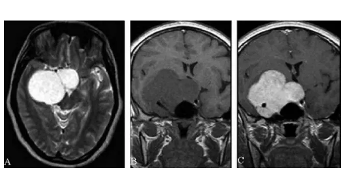
A 40-year-old woman presented with right-sided ophthalmople-gia, ptosis, and retro-orbital pain. MRI showed a large, lobulated, hyperintense lesion in the right cavernous sinus on T2-weighted image (figure, A). The lesion was hypointense on T1-weighted
image (figure, B), with intense enhancement (figure, C). The imag-ing differentials are cavernoma, menimag-ingioma, and schwannoma. Cavernous sinus cavernoma is a rare vascular malformation, which represents 3% of all benign cavernous sinus tumors.1
Marked hyperintensity on T2-weighted images with intense and homogenous enhancement are characteristic. Red cell– labeled blood pool scintigram is more specific for diagnosis.2
Preoperative diagnosis is crucial because of intraoperative pro-fuse hemorrhage.
Copyright © 2006 by AAN Enterprises, Inc.
1. Linskey ME, Sekhar LN. Cavernous sinus hemangiomas: a series, a review, and an hypothesis. Neurosurgery 1992;30:101–108.
2. Salanitri GC, Stuckey SL, Murphy M. Extracerebral cavernous hemangi-oma of the cavernous sinus: diagnosis with MR imaging and labeled red cell blood pool scintigraphy. Am J Neuroradiol 2004;25:280–284.
Figure. (A) Axial T2-weighted MR im-age reveals a well-circumscribed, large, lobulated, markedly hyperintense lesion in the right cavernous sinus with mass effect on the midbrain. (B) Coronal T1-weighted MR image shows a well-circumscribed, large, lobulated, hypoin-tense lesion in the right parasellar region with encasement of the right internal carotid artery and sellar exten-sion. (C) Coronal T1-weighted postga-dolinium MR image shows an intense and homogenous enhancement of the lobulated right cavernous sinus lesion.
The authors report no conflicts of interest.