O R I G I N A L R E S E A R C H
Pre-mRNA Processing Factor 8 Accelerates the
Progression of Hepatocellular Carcinoma by
Regulating the PI3K/Akt Pathway
This article was published in the following Dove Press journal: OncoTargets and Therapy
Shouhan Wang1 Min Wang2 Bin Wang1 Jiaqi Chen1 Xianbin Cheng1 Xiaodan Sun 3
1Department of Hepatopancreatobiliary
Surgery, Cancer Hospital of Jilin Province,
Changchun, People’s Republic of China;
2Department of Pathology, Cancer
Hospital of Jilin Province, Changchun,
People’s Republic of China;3Department
of 2nd Gynecologic Oncology Surgery, Cancer Hospital of Jilin Province,
Changchun, People’s Republic of China
Background: The specific function of pre-mRNA processing factors (Prps) in human
malignancies has not been yet investigated. The aim of the present study was to determine the impacts of Prp8 in a common human malignancy, hepatocellular carcinoma (HCC).
Materials and Methods: RT-qPCR and Western blotting were performed to measure the
expression levels of Prp8 in various HCC cell lines and HCC tissues. A hepatic astrocyte line was transfected with a eukaryotic expression plasmid to overexpress Prp8. In addition, the endogenous expression level of Prp8 in HCC cells was silenced using a short hairpin RNA method, and the role of Prp8 on cell proliferation and migration was examined by Cell Counting Kit-8, wound healing assay and Transwell assays following knockdown in HCC cells, and overexpression in astrocytes.
Results: Upregulation of Prp8 expression was found to be associated with poor clinical
outcomes in patients with HCC. The upregulation of Prp8 promoted cell viability, metastasis and the activity of the PI3K/Akt pathway in hepatic astrocytes cells and HCC cells. Interestingly, loss of Prp8 had no obvious impact on cell viability and migration in hepatic astrocytes, but significantly inhibit the cell malignancy of HCC cells. Functionally, the inhibition of the PI3K/Akt pathway reversed the increased cell viability and migration of HCC cells induced by Prp8 via inhibiting EMT process.
Conclusion: Collectively, the present results suggested that Prp8 served as a tumor
pro-moter in HCC by targeting and regulating the PI3K/Akt pathway.
Keywords:pre-mRNA processing factor 8, phosphatidylinositol 3-kinase, protein kinase B,
hepatocellular carcinoma
Introduction
Pre-mRNA splicing is essential for gene expression in all eukaryotes.1In higher
eukaryotes, such as mammals, ~95% of the nucleotides in the primary transcript
(pre-mRNA) of a protein-encoding gene are introns.2 These introns need to be
removed precisely by splicing before the mRNA can be transported from the nucleus into the cytoplasm, where it can be translated.3Alternative splicing greatly expands the gene coding capacity and >60% of human genes are alternatively
spliced.4 It is also becoming increasingly clear that alternative splicing is
a fundamental component of eukaryotic gene regulation, influencing cell
differen-tiation, development and many processes in the nervous system.5A typical intron
contains a conserved 5ʹsplice site (5ʹss), a branch point sequence (BPS) followed
by a polypyrimidine tract (PYT), and a 3ʹ ss.6 Introns are removed through two
Correspondence: Xiaodan Sun Department of 2nd Gynecologic Oncology Surgery,Cancer Hospital of Jilin Province,No. 1018, Huguang Road,
Changchun, People’s Republic of China
Email [email protected]
OncoTargets and Therapy
Dove
press
open access to scientific and medical research
Open Access Full Text Article
OncoTargets and Therapy downloaded from https://www.dovepress.com/ by 118.70.13.36 on 25-Aug-2020
transesterification reactions catalyzed by the spliceosome.5
The spliceosome contains five smalls nuclear RNAs
(snRNAs), such as U1, U2, U4, U5 and U6 snRNAs,
which form five small nuclear ribonucleoproteins
(snRNPs) with their associated proteins, in addition to
numerous other protein splicing factors.7 Notably, the
total number of proteins in the spliceosome is more than
100.8The formation of the E-complex involves the initial
recognition of an intron by the spliceosome.5The 5ʹss is recognized by U1 snRNP, whereas the BPS and PYT interact with other splicing factors. Subsequently, the U2 snRNP joins the spliceosome to form the a complex, which is followed by the recruitment of the U4/U6.U5
triple snRNP (tri-snRNP), forming the B complex.9
Extensive structural rearrangements occur at this stage to form the catalytically active B complex that mediated the
first splicing step.10 After thefirst step reaction, the spli-ceosome repositions the substrate, allowing the second
catalytic reaction and forming the C complex.11
The second reaction is followed by post-catalytic rearran-gements to release the mature mRNA for the nuclear export, releasing the lariat intron, which will be degraded,
and the snRNPs, which will be recycled.12
Errors in splicing contribute to >30% of human genetic disorders, including retinitis pigmentosa (RP), spinal
muscu-lar atrophy and myotonic dystrophy.13 RP is an autosomal
dominant genetic disorder that leads to photoreceptor
degen-eration and vision impairment.14 Mutations or deletions of
a number of splicing factors, including pre-mRNA proces-sing factor 8 (Prp8), small nuclear ribonucleoprotein U5 subunit 200 (Brr2), Prp3 and Prp31, have been found to cause various subtypes of RP.15These proteins are all com-ponents of the U4/U6.U5 tri-snRNP complex and are ubiqui-tously expressed in all tissues.16Intriguingly, mutations or heterozygous deletion of these splicing factors affect primar-ily photoreceptors, which are one of the most metabolically active cell types in the body, and have no obvious effect on
any other organs.17 Furthermore, a 90% reduction in the
protein level of splicing factor 3b subunit 1 (SF3b1), a key component of the U2 snRNP complex, leads to developmen-tal defects in very specific organs instead of lethality or widespread defect in many organs, highlighting the cell type-specific effects of inhibiting the basal splicing machinery.18 Therefore, the present study hypothesized that inhibiting the spliceosomal activity may selectively inhibit cancer cell growth or survival with limited effect on normal cells.16
Approaches aimed to control the metastasis and recur-rence of hepatocellular carcinoma (HCC) has been found
to be insufficient in the treatment of this disease, and no
currently available treatments have been identified to be
efficient against metastatic HCC.19Our present study
iden-tified that Prp8 was upregulated in HCC; however, to the
best of our knowledge, there are no studies investigating the impact of Prp8 on the tumorigenesis of malignant HCC. Therefore, the aim of the present study was to investigate the expression pattern of Prp8, its role and its underlying mechanisms in HCC malignancy. Importantly, the regulatory mechanism of the Prp8/PI3K/Akt axis in HCC remains unclear. Therefore, the dysregulation of Prp8 and its regulatory mechanism in HCC were examined in the present study. The present results may facilitate the
development of a novel and efficient HCC treatment.
Materials and Methods
Experimental Sample
In the present study, HCC and normal hepatic specimens were obtained from 206 patients in Cancer Hospital of Jilin Province between May 2006 and September 2013. Before the experi-ment, written informed consent was collected from all the patients. The participants did not receive any treatment except for surgery. The present study was approved by The Institutional Ethics Committee of Cancer Hospital of Jilin Province.
Clinical Tissue Sample Collection
We collected 130 primary gastric cancer tissue samples and 108 paired adjacent normal tissues from patients who under-went surgery at the Hospital of Chengdu University of TCM (Chengdu, China). None of the patients received any antic-ancer therapy before tumor resection and diagnosed with any additional malignancies. Pathological staging was based on
the UICC/AJCC TNM Classification (8th edition of 2016).
Immunohistochemistry Analysis
Routine hematoxylin and eosin staining were performed prior
to immunohistochemistry analysis. Briefly, paraffi
n-embedded samples were cut into 3-μm sections, and then
dewaxed with xylene and rehydrated for peroxidase (DAB) immunohistochemistry staining. For antigen retrieval, sections were heated at 97 ° C for 20 min. Following a brief proteolytic digestion and a peroxidase blocking, the sections were incu-bated with corresponding primary antibody Prp8 (cat. no. # ab79237, Abcam) overnight at 4 ° C, and HRP/Fab polymer
conjugate (Cat. PV-6000-D, Zhongshan Goldenbridge
Biotechnology Co. Ltd., China) was applied as secondary
OncoTargets and Therapy downloaded from https://www.dovepress.com/ by 118.70.13.36 on 25-Aug-2020
antibody. Finally, the sections were stained with diaminoben-zidine substrate and counter-stained with hematoxylin. Two independent investigators semi quantitatively evaluated PRP8 positivity without prior knowledge of clinicopathologic data.
The final immunoreactivity scores (IRS) were assessed
according to scores of the percentage of positive stained cells
(0 point, 0–5% positive cells; 1 point, 6–25%; 2 point,
26–50%; 3 point, 51–75%; 4 point, 76–100%) plus staining intensity scores (0 point, no staining; 1 point, weak staining; 2 point, moderate staining; 3 point, strong staining). The final IRS more than 4 were accepted as strong positivity, while, others were weak positivity.
Cell Lines and Transfection
Human hepatic astrocyte line LX-2, and MHCC97H Hep3B, Huh1 HCC cell lines were obtained from American Type Culture Collection (ATCC; Rockville, Md.) and seeded in RPMI-1640 medium containing 10% FBS. All cells were cultured at 37°C in 5% CO2. Prp8 vector and control vector were bought from Shanghai Genechem Co., Ltd. Prp8 vectors were transfected into HCC cells and using Lipofectamine 2000 (Invitrogen;
Thermo Fisher Scientific, Inc.) following the
manufac-turer’s instructions and G418 (Sigma-Aldrich; Merck
KGaA) was used to expand G418-resistant clones in cul-ture as a monoclonal population.
PI3K Inhibitor Treatment
The PI3K inhibitor LY294002 was diluted to afinal
concen-tration of 40 µM in DMSO and stored at−20°C, cells were
subsequently treated for 24 h at 20 nM in order to efficiently inhibit PI3K. Cells treated with the same volume of DMSO served as the control group.
RNA Extraction and Reverse
Transcription-Quantitative PCR (RT-qPCR)
Total RNA was extracted using TRIzol reagent (Invitrogen; Thermo Fisher Scientific, Inc.) as previously described.20The cDNA was synthesized by PrimeScript RT reagent (Takara Bio, Inc.). RT-qPCR was performed using SYBR Green Master Mix II (Takara Bio, Inc.) according to the manufac-turer’s instructions. The expression levels of Prp8 and Prp31 were normalized to U6 or GAPDH. The expression levels ofthe genes investigated were calculated using the 2−ΔΔCq
method. The primers used in the present work were as follows:
Prp8 forward, 5ʹ- ATGAAGAGCAATCCATTCTGGTGG
AC-3ʹ and reverse, 5ʹ- TTGGCACGGTTGATGGTAGGTG
ACCACCA-3ʹ; Prp31 forward, 5ʹ- GGATCCATGTCTCTGG
CAGATGAGCTCTTA-3ʹ and reverse, 5ʹ- CCGCGGTCAG
GTGGACATAAGGCCACTCTT-3ʹ; GAPDH forward, 5ʹ
-ACATCGCTCAGACACCATG-3ʹand reverse, 5ʹ-TGTAGT
TGAGGTCAATGAAGGG-3ʹ.
Western Blot Analysis
Cells were lysed using RIPA buffer (Beyotime Institute of Biotechnology). Then, the supernatant containing the total
protein was collected as previously described.21 The
pro-tein was separated by 10% SDS-PAGE. The propro-tein was blocked using 5% non-fat milk for 1 h. The membranes were incubated with the following primary antibodies: Prp8 (cat. no. # ab79237, Abcam.), Prp31 (1:1000
dilu-tion; cat. no. #ab188577, Abcam), PI3 Kinase p110α(cat.
no.#4255, Cell Signaling Technology, Inc.), Akt (cat. no.
#2920, Cell Signaling Technology, Inc.), anti-pAkt
(Thr308) (cat. no. #13038, Cell Signaling Technology, Inc.), N-Cadherin (cat. no. #13116, Cell Signaling Technology, Inc.), E-Cadherin (cat. no. #14472, Cell Signaling Technology, Inc.), Vimentin (cat. no. #5741, Cell Signaling Technology, Inc.), andβ-actin (1:2000 dilu-tion; cat. no. #ab107061, Abcam). Primary antibodies were incubated with the membranes overnight at 4°C. The diluted secondary antibodies were added to the mem-branes for 1 h. Finally, the protein was examined using an ECL reagent (EMD Millipore).
Immuno
fl
uorescence
The cells were washed 3 times with PBS, fixed with 4%
paraformaldehyde for 10 min at room temperature, per-meabilized with 0.1% Triton X-100, and blocked in PBS with 2% bovine serum albumin for 1 h. The staining was performed with a rabbit anti-human Prp8 antibody. Images were obtained using an Olympus IX81 microscope with an MT20/20 illumination system.
Short Hairpin RNA (shRNA) Method
The packaging construct (pHelper 1.0), the (vesicular sto-matitis virus G, VSV-G) VSVG-expressing construct (pHelper 2.0), pGCSIL-EGFP plasmid, pGCSIL-scramble vector and pGCSIL Prp8-shRNA construct were pur-chased from Genechem Biotech Co., Ltd. TheshRNA-mediated knockdown was performed as previously
described.22 HEK 293T cells (at 70–80% confluence)
maintained in 6-well dishes were transfected with the aforementioned constructs using Lipofectamine (cat. no.
11668027; Thermo Fisher Scientific, Inc.), according to
OncoTargets and Therapy downloaded from https://www.dovepress.com/ by 118.70.13.36 on 25-Aug-2020
the manufacturer’s protocol. The viral stocks were
con-centrated via ultracentrifugation and dissolved in Hanks’
balanced salt solution. The HCC cells and hepatic
astro-cytes were transfected with the viral stocks at
a multiplicity of infection of 200.
Cell Counting Kit-8 (CCK8) Assay
Transfected cells (4x10³cells/well) were seeded in
RPMI-1640 with 10% FBS for 24, 48, 72 or 96 h. Next, the suspen-sion of cells was incubated with 20μL of CCK8 for 4 h. Then,
150μL DMSO was added into the medium. After 10 min, cell
viability was assessed using a microplate reader (Olympus Corporation) to determine the optical density at 490 nm.
Colony Formation Assay
Briefly, stably transfected LX2 or Hep3B cells were plated
in 6-well plates at 40 cells per well at 37°C for 10 days.
the cell colonies were washed with PBS twice, and fixed
with methanol for 30 min, and dyed with 0.1% crystal violet diluted in PBS for 15 min. The colonies which contained more than 120 cells were counted.
Transwell Chamber Assay
Transwell assay was used to assess cell invasion. Upper chambers were coated with Matrigel (BD Biosciences) to detect Hep3B or LX-2 cell invasion. The transfected cells
(5x10³cells/well) were seeded in the upper chamber, and
the lower chamber wasfilled with 10% FBS. The invasive
cells werefixed and stained for 30 min. Finally, the
inva-sive cells werefixed and stained for 30 min, and examined
under a light microscope (Olympus Corporation).
Wound Healing Assay
A cell monolayer scratch assay was performed as
described previously.23Briefly, LX2 or Hep3B cells were
seeded in 6 well plates at 3.5x10⁵cells per well, grown to
A
B
C
Normal HCCPrp8
Prp8 Prp31
0 50 100 150
Rel
at
ive
p
ro
te
in
expr
ess
io
n
of
P
rp
s
LX-2 Huh1 Hep3B MHCC97H
** ** **
LX2 Huh1 Hep3B MHCC97H
Prp31
β-actin Prp8
20um 20um
D
E
β-actin Prp8
Normal HCC
F
Nonneop last
ic
HC Ctis
sues
0 20 40 60 80
R
e
la
ti
ve
pr
ot
ei
n
e
xpr
e
ss
io
n
of
P
rp
8
Nonneoplastic HCC tissues **
0 20 40 60 80
0 50 100
Months elapsed
Pe
rce
nt
s
ur
v
iv
a
l
Prp8 negative Prp8 positive
Prp8 Prp31 0
10 20 30 40 50
Rel
at
ive
m
R
N
A
quan
ti
ty
of
P
rp
s
LX-2 Huh1 Hep3B MHCC97H ** **
**
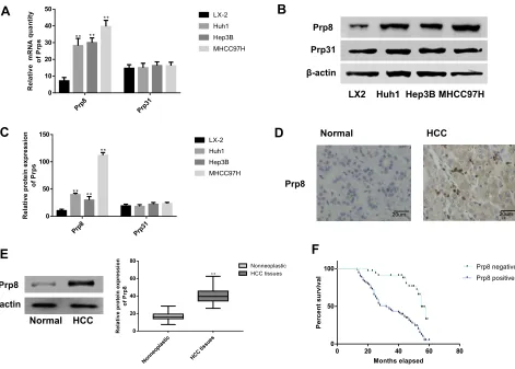
Figure 1The expression of Prp8 is increased in HCC cells and tissues. (A) Prp8 mRNA expression in HCC cells compared with hepatic astrocytes. **P<0.01, compared with the hepatic astrocytes. (B) Prp8 protein expression in HCC cells compared with hepatic astrocytes. (C) Relative protein expression of Prp8 in HCC cells versus hepatic astrocytes. **P<0.01, compared with the hepatic astrocytes. (D) Prp8 protein expression in HCC tissues was explored via IHC. (E) Prp8 protein expression in HCC tissues was examined via western-blotting. **P<0.01, compared with the hepatic tissues. (F) High Prp8 expression is associated with poor prognosis in HCC patients.
OncoTargets and Therapy downloaded from https://www.dovepress.com/ by 118.70.13.36 on 25-Aug-2020
~100% confluence and pretreated with mitomycin C (10
μg/mL) for 2 h to inhibit cell proliferation before
scratch-ing. A scratch wound was created using a 20μL pipette tip
and was imaged at the same position at 0, 12 and 24 h.
Statistical Analysis
Data are presented as the mean ± SD, which were analyzed using SPSS 17.0 or Graphpad Prism 6.
Chi-squared test, one-way ANOVA with Tukey’s post hoc
test and Univariate Kaplan-Meier method with Log rank test were used to calculate differences between groups. P<0.05 was considered to indicate a statistically signifi -cant difference.
Results
The Expression of Prp8 Is Increased in
HCC Tissues
The alternation of Prp8 and Prp31 expression was detected in HCC cells. RT-qPCR and Western blotting showed that the expression of Prp8 was increased in Hep3B, MHCC97H and Huh1 cells compared with LX-2 cells
(P<0.01; Figure 1A–C), although there were no
differences in Prp31 expression levels among LX-2, Hep3B, MHCC97H and Huh1 cells. Similarly, Prp8 expression was found to be higher in HCC tissues
com-pared with normal hepatic tissues (P<0.01; Figure 1D
and E). In addition, the correlation between abnormal
Prp8 expression and clinical features in HCC patients
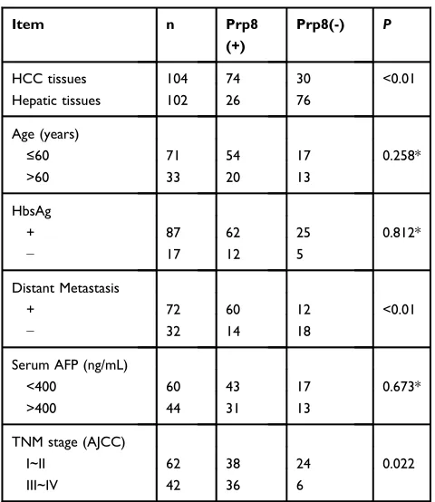
was investigated. As shown inTable 1, the dysregulation
of Prp8 was associated with tumor node and metastasis (TNM) stage (P<0.05) and distant metastasis (P<0.01). Furthermore, patients with HCC presenting high Prp8 expression showed a shorter overall survival time, indicat-ing that upregulation of Prp8 predicted poor prognosis in
patients with HCC (P<0.01, Figure 1F). The present
results indicated that Prp8 may function as an important regulator of the pathogenesis of HCC.
Overexpression of Prp8 Promotes Cell
Viability and Migration of Human Hepatic
Astrocyte Line
Prp8 was overexpressed in human hepatic astrocyte line LX-2 to perform a gain-of-function experiment. Prp8 expression was upregulated after overexpression in LX-2 cells (P<0.01,
Figure 2AandB). The effect of Prp8 on the PI3K/Akt
path-way was investigated to further examine its role in HCC. Prp8 overexpression was found to promote the expression level of PI3K-p110α (P<0.01) and phosphorylated Akt (P<0.01) in
LX-2 cells (Figure 2AandB). Besides, immunofluorescence
data discovered that Prp8 was mainly expressed in the cell nucleus (Figure 2C). CCK8 and colony formation assay revealed that overexpression of Prp8 promoted cell prolifera-tion and viability in LX-2 cells (P<0.01,Figure 2DandE). Transwell and wound healing assay showed that cell migra-tion and invasion were increased following Prp8 overexpres-sion in LX-2 cells (P<0.01,Figure 2F–H). Collectively, Prp8 promoted the proliferative, migratory and invasive abilities of hepatic astrocytes.
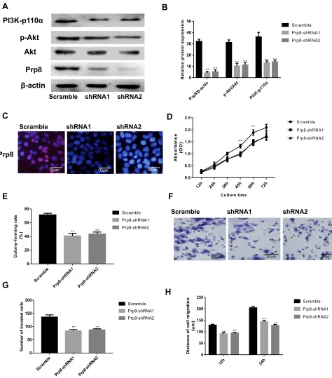
Loss of Prp8 Has an Inhibitory Effect on
Cell Viability, Migration and Invasion in
HCC Cells
Hep3B cells were selected for further functional assay due to the high expression level of Prp8 in this cell type. Prp8 was silenced in Hep3B cells to perform a loss-of-function
experi-ment. Western-blot and immunofluorescence assay revealed
that Prp8 expression was decreased following Prp8
knock-down in Hep3B cells (P<0.01, Figure 3A–C). Prp8
knockdown was found to inhibit the expression level of
Table 1 Expression of Prp8 and the Clinicopathological
Characteristics in HCC Patients
Item n Prp8
(+)
Prp8(-) P
HCC tissues 104 74 30 <0.01
Hepatic tissues 102 26 76
Age (years)
≤60 71 54 17 0.258*
>60 33 20 13
HbsAg
+ 87 62 25 0.812*
− 17 12 5
Distant Metastasis
+ 72 60 12 <0.01
− 32 14 18
Serum AFP (ng/mL)
<400 60 43 17 0.673*
>400 44 31 13
TNM stage (AJCC)
I~II 62 38 24 0.022
III~IV 42 36 6
Note:*No statistical significance was found with the Chi-square test/Chi-Square Goodness-of-Fit Test.
Abbreviations:AFP, alpha fetoprotein; TNM, tumor node and metastasis; AJCC, American Joint Committee on Cancer.
OncoTargets and Therapy downloaded from https://www.dovepress.com/ by 118.70.13.36 on 25-Aug-2020
PI3K-p110α(P<0.01) and phosphorylated Akt (P<0.01) in
Hep3B cells (Figure 3AandB). CCK8 and colony formation
assay revealed that loss of Prp8 reduced cell proliferation and
viability in Hep3B cells (P<0.01, Figure 3D and E).
Similarly, knockdown of Prp8 reduced cell invasion cells
(P<0.01, Figure 3Fand G). Wound healing assay showed
that cell migration was inhibited following Prp8 knockdown
in Hep3B cells (P<0.01, Figure 3H). Taken together, Prp8
knockdown reduced the proliferative, migratory and invasive abilities of HCC cells.
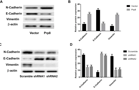
Prp8 Activates Epithelial-Mesenchymal
Transition (EMT) in HCC
The effect of Prp8 on the EMT process was investigated to further examine its role in HCC. Prp8 overexpression was found to promote the expression level of N-cadherin and Vimentin, and to inhibit the expression of E-cadherin in LX-2 cells, suggesting that Prp8 may be involved in EMT
in hepatic astrocytes (Figure 4AandB). Conversely, Prp8
knockdown reduced the expression levels of N-cadherin and Vimentin, and increased the expression of E-cadherin
in HCC Hep3B cells (Figure 4CandD).
Overexpression of Prp8 Has a Positive
Effect on Cell Viability, Migration and
Invasion in HCC Cells
Prp8 was overexpressed in human HCC cell line Huh1 to perform a gain-of-function experiment. Prp8 expression was upregulated after overexpression in Huh1 cells (P<0.01,
Figure 5A and B). The effects of Prp8 on the PI3K/Akt
pathway and EMT process were investigated to further examine its role in HCC. Prp8 overexpression was found to promote the expression level of PI3K-p110α, phosphorylated Akt, N-cadherin and Vimentin, but inhibit the expression of
E-cadherin in Huh1 cells (P<0.01;Figure 5AandB). CCK8
and colony formation assay revealed that overexpression of
A
B
D
C
Vector Prp8
PI3K-p110α Akt β-actin Prp8 p-Akt 20um 20um Prp8 /β-actin p-Akt/Akt PI3 K-p110α 0 10 20 30 40 50 Re la ti ve p ro te in e xp re s s io n Vector Prp8 ** ** **
12h 24h 36h 48h 60h 72h
0.0 0.5 1.0 1.5 2.0 2.5 Culture time A bs o rb an c e (O D ) Vector Prp8 ** ** ** ** Vec
tor Prp8
0 20 40 60 80 Col ony-f or mi ng ra te (% ) Vector Prp8 ** Vect or Prp8 0 50 100 150 N u mb e r o f in v ad e d c e ll s Vector Prp8 **
12h 24h
0 50 100 150 200 250 Culture time D is tan ce of c el l m ig rat io n (u m ) Vector Prp8 ** **
F
G
H
LX2-Vector LX2-Prp8
E
LX2-Vector LX2-Prp8
Prp8
10um 10um
Figure 2Upregulation of Prp8 promoted cell viability and migration in a human hepatic astrocyte line. (A) The effect of Prp8 on the PI3K/Akt pathway was investigated. (B) The relative protein expression of Prp8, PI3K-p110αand phosphorylated Akt. **P<0.01, compared with the empty vector groups. (C) The expression location of Prp8 was explored via immunofluorescence (D) CCK8 assay revealed that impact of Prp8 on cell proliferation. **P<0.01, compared with the empty vector groups. (E) Colony formation assay revealed that overexpression of Prp8 promoted cell viability. **P<0.01, compared with the empty vector groups. (F) Transwell assay was used to explore the impact of Prp8 on cell invasion. (G) The analyze of the invaded cell number. **P<0.01, compared with the empty vector groups. (H) The ability of cell migration was explored via wound healing assay. **P<0.01, compared with the empty vector groups.
OncoTargets and Therapy downloaded from https://www.dovepress.com/ by 118.70.13.36 on 25-Aug-2020
Prp8 promoted cell proliferation and viability in Huh1 cells
(P<0.01, Figure 5C and D). Transwell and wound healing
assay showed that cell migration and invasion were increased
following Prp8 overexpression in Huh1 cells (P<0.01,
Figure 5E–G). Collectively, Prp8 promoted the proliferative,
migratory and invasive abilities of HCC cells.
A
B
D
C
E
PI3K-p110α
Akt
β-actin Prp8 p-Akt
20um
Prp8
F
G
H
Scramble shRNA1 shRNA20 50 100 150 20
Nu
mb
er
of
in
v
ade
d
c
el
ls Scramble
Prp8-shRNA1 Prp8-shRNA2
** **
Scramble shRNA1 shRNA2
Scramble shRNA1 shRNA2
0 10 20 30 40 50
R
e
la
ti
ve
pr
ot
ei
n
expr
e
ss
io
n
Scramble
Prp8-shRNA1 Prp8-shRNA2
* * * *
* * * * * * * *
0.0 0.5 1.0 1.5 2.0 2.5
C ulture tim e
Abs
o
rba
n
c
e
(O
D
)
Scram ble
Prp8-shRNA1
Prp8-shRNA2 * *
* *
0 20 40 60 80
C
ol
ony-for
m
ing
rat
e
(%
)
Scramble Prp8-shRNA1 Prp8-shRNA2
** **
0 50 100 150 200 250
D
ist
a
nc
e
o
f
c
e
ll
m
ig
ra
ti
on
(u
m
)
Scramble
Prp8-shRNA1 Prp8-shRNA2
** **
** ** 10um
20um 20um
10um 10um 10um
Figure 3Loss of Prp8 has an inhibitory effect on cell viability and metastasis in HCC cells. (A) Prp8 expression and the PI3K/Akt pathway were inhibited following Prp8 knockdown in Hep3B cells. (B) The relative protein expression of Prp8, PI3K-p110αand phosphorylated Akt. **P<0.01, compared with the scramble group. (C) The expression location of Prp8 was explored via immunofluorescence (D) CCK8 assay revealed that loss of Prp8 reduced cell proliferation. **P<0.01, compared with the scramble group. (E) Colony formation assay revealed that the loss of Prp8 inhibited the ability of colony formation. **P<0.01, compared with the scramble group. (F) Transwell assay showed that cell invasion was inhibited by Prp8 knockdown in Hep3B cells. (G) The analyze of the invaded cell number. **P<0.01, compared with the scramble group. (H) The ability of cell migration was explored via wound healing assay. **P<0.01, compared with the scramble group.
OncoTargets and Therapy downloaded from https://www.dovepress.com/ by 118.70.13.36 on 25-Aug-2020
LY294002 Treatment Reversed the
Carcinogenesis Effect of Prp8 in Hepatic
Astrocytes
Functionally, the effect of Prp8 on PI3K/Akt pathway in hepatic astrocytes was abolished following treatment with 20 nM PI3K tyrosinase inhibitor LY294002 for 24 h. In addition, the effect of LY294002 on the PI3K/Akt pathway
was also identified in LX-2 cells (Figure 6A and B).
LY294002 treatment was found to inhibit the expression level of N-cadherin and Vimentin, and to promote the expression of E-cadherin in LX-2 cells with Prp8
over-expressing (P<0.01, Figure 6A and B). The LY294002
treatment suppressed the effects of Prp8 on cell
prolifera-tion and viability in LX-2 cells (Figure 6C and D).
Similarly, the effect of Prp8 on LX-2 cell invasion
(P<0.01, Figure 6E and F) and migration (P<0.01,
Figure 6G) were also inhibited following treatment with LY294002. Collectively, LY294002 treatment reversed the carcinogenic effects of Prp8 in hepatic astrocytes. In
sum-mary, Prp8 was identified to serve a carcinogenic role via
PI3K/Akt pathway in HCC progression.
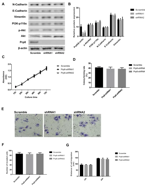
Loss of Prp8 Has No Obvious Impact on
Cell Viability in Hepatic Astrocytes
Prp8 was silenced in LX-2 cells to perform a loss-of-function experiment. Prp8 expression was reduced
follow-ing Prp8 knockdown in LX-2 cells (P>0.05, Figure 7A
and B). The loss of Prp8 has no obvious effect on the
expression level of N-cadherin, Vimentin, E-cadherin,
PI3K-p110α, and phosphorylated Akt in LX-2 cells
(P>0.05, Figure 7A and B). CCK8 assay revealed that
loss of Prp8 had no obvious impact on cell proliferation in LX-2 cells (P>0.05,Figure 7C). Colony formation assay revealed that the Prp8 knockdown have no obvious impact on the ability of colony formation in LX-2 cells (P>0.05,
Figure 7D). Transwell assay (P>0.05, Figure 7E and F)
and wound healing assay (P>0.05,Figure 7G) showed that
cell migration and invasion were not altered fo llowing
N-Cadherin
Vimentin
β-actin E-Cadherin
Scramble shRNA1 shRNA2
A
Vector Prp8 N-Cadherin
Vimentin
β-actin E-Cadherin
N-Cadh erin
E-C adher
in
Vim entin 0
10 20 30 40
R
e
la
ti
ve
pr
ot
ei
n
expr
ess
io
n
Scramble shRNA1 shRNA2 **
**
** **
** ** N-C
adhe rin
E-Cadhe rin
Vim entin
0 10 20 30 40
R
el
at
ive
pr
ot
ei
n
exp
ress
io
n
Vector Prp8 **
**
**
C
B
D
Figure 4Prp8 activates EMT in HCC. (A) Prp8 regulated the expression levels of E-cadherin, N-cadherin and Vimentin in LX-2 cells. (B) The relative protein expression of E-cadherin, N-cadherin and Vimentin in LX-2 cells. **P<0.01, compared with the empty vector groups. (C) Prp8 regulated the expression levels of E-cadherin, N-cadherin and Vimentin in Hep3B cells. (D) The relative protein expression of E-cadherin, N-cadherin and Vimentin in Hep3B cells. **P<0.01, compared with the scramble group.
OncoTargets and Therapy downloaded from https://www.dovepress.com/ by 118.70.13.36 on 25-Aug-2020
A
B
D
C
20um
Vect or
Prp8 0
50 100 150
Nu
m
be
r
of
in
v
a
de
d
c
el
ls Vector
Prp8 **
G
0 1 2 3Culture time
A
b
sor
ba
nce
(O
D
)
Vector Prp8
** **
**
0 100 200 300
Culture time
D
is
tan
ce
of
c
e
ll
m
ig
rat
io
n
(u
m
)
Vector Prp8 **
** 20um
E
PI3K-p110α
Akt
β-actin Prp8 p-Akt
Vector Prp8
Prp8 /β-a
ctin
p-Akt /Akt
PI3K -p110α
N-Cad her
in
E-Cadheri n
Vime ntin 0
20 40 60
Re
la
ti
ve
p
rot
ei
n
e
xpr
e
ss
io
n
Vector Prp8
** **
**
**
** **
N-Cadherin
Vimentin E-Cadherin
Vector Prp8
Vect or
Prp8 0
20 40 60 80 100
C
ol
ony-for
m
ing
ra
te
(%
)
Vector Prp8 **
F
Figure 5Upregulation of Prp8 promoted cell viability and migration in HCC cells. (A) The effects of Prp8 on the PI3K/Akt pathway and EMT process were investigated. (B) The relative protein expression of Prp8, PI3K-p110α, phosphorylated Akt, E-cadherin, N-cadherin and Vimentin. **P<0.01, compared with the empty vector groups. (C) CCK8 assay revealed that impact of Prp8 on cell proliferation. **P<0.01, compared with the empty vector groups. (D) Colony formation assay revealed that overexpression of Prp8 promoted cell viability. **P<0.01, compared with the empty vector groups. (E) Transwell assay was used to explore the impact of Prp8 on cell invasion. (F) The analyze of the invaded cell number. **P<0.01, compared with the empty vector groups. (G) The ability of cell migration was explored via wound healing assay. **P<0.01, compared with the empty vector groups.
OncoTargets and Therapy downloaded from https://www.dovepress.com/ by 118.70.13.36 on 25-Aug-2020
20um
DMSO LY294002 β-actin
Prp8
0 10 20 30 40 50
Re
la
tiv
e
p
ro
te
in
ex
p
re
ssi
o
n DMSO
LY294002
* * * * * * * *
* *
0.0 0.5 1.0 1.5 2.0 2.5
Culture time
Ab
s
o
rb
a
n
c
e
(OD
)
DMSO LY294002
** *
**
DMSO
LY29 400
2 0
20 40 60 80
Co
lo
n
y
-f
o
rm
in
g
ra
te
(%
)
DMSO LY294002
**
20um 0
20 40 60 80 100
Num
b
e
r
of
in
v
a
d
e
d
c
e
lls DMSO
LY294002
* *
0 50 100 150 200 250
Di
s
tan
c
e
o
fc
el
l
mi
g
rat
io
n
(um
)
DMSO LY294002
**
**
A
B
C
D
E
Akt p-Akt
PI3K-p110α
DMSO LY294002
F
G
N-CadherinVimentin E-Cadherin
Figure 6LY294002 treatment reverses the carcinogenic-inducing effects of Prp8 in hepatic astrocytes. (A) LY294002 treatment reversed the effects of Prp8 on PI3K/Akt pathway and EMT process in hepatic astrocytes. (B) The relative protein expression of Prp8, PI3K-p110α, phosphorylated Akt, E-cadherin, N-cadherin and Vimentin. **P<0.01, compared with the DMSO groups. (C) The LY294002 treatment suppressed the promoted effects of Prp8 on cell proliferation. **P<0.01, compared with the DMSO groups. (D) Colony formation assay revealed that the LY294002 treatment inhibited the ability of colony formation. **P<0.01, compared with the DMSO groups. (E) Promoted effect of Prp8 on cell invasion were inhibited by treatment with LY294002. (F) The analyze of the invaded cell number. **P<0.01, compared with the DMSO groups. (G) The ability of cell migration was explored via wound healing assay. **P<0.01, compared with the DMSO groups.
OncoTargets and Therapy downloaded from https://www.dovepress.com/ by 118.70.13.36 on 25-Aug-2020
A
B
D
C
β-actin Prp8
F
Scramble shRNA1 shRNA2
0 20 40 60 80 100
N
um
be
r
o
f
in
v
a
d
ed
c
el
ls Scramble
Prp8-shRNA1 Prp8-shRNA2
Scramble shRNA1 shRNA2
0.0 0.5 1.0 1.5 2.0
Culture time
A
bs
o
rba
n
ce
(O
D
)
Scramble Prp8-shRNA1 Prp8-shRNA2
0 10 20 30 40 50
Col
ony-f
or
mi
n
g
ra
te
(%
)
Scramble Prp8-shRNA1 Prp8-shRNA2
0 50 100 150 200
D
is
tan
ce
of
c
e
ll
mi
gr
a
ti
on
(u
m
)
Scramble
Prp8-shRNA1 Prp8-shRNA2
10um
E
G
PI3K-p110αAkt p-Akt N-Cadherin
Vimentin E-Cadherin
0 10 20 30
R
e
la
ti
ve
pr
ot
ei
n
e
xpr
e
s
s
io
n
Scramble shRNA1 shRNA2
* ** *
10um 10um
Figure 7Loss of Prp8 has no obvious impact on cell viability in hepatic astrocytes. (A) The loss of Prp8 has no obvious effect on the expression level of N-cadherin, Vimentin, E-cadherin, PI3K-p110α, and phosphorylated Akt in LX-2 cells. (B) The relative expression of Prp8, PI3K-p110α, phosphorylated Akt, E-cadherin, N-cadherin and Vimentin in LX-2 cells. **P<0.01, compared with the scramble group. (C) CCK8 assay revealed that loss of Prp8 had no obvious impact on cell viability in LX-2 cells. (D) The impact on the ability of colony formation was explored via colony formation assay. (E) Transwell assay showed that cell invasion was not obviously affected by Prp8 knockdown in LX-2 cells. (F) The analyze of the invaded cell number. (G) Wound healing assay showed that cell migration was altered by Prp8 knockdown in LX-2 cells.
OncoTargets and Therapy downloaded from https://www.dovepress.com/ by 118.70.13.36 on 25-Aug-2020
Prp8 knockdown in LX-2 cells. Collectively, Prp8 knock-down had no obvious impact on the proliferative, migra-tory and invasive abilities of hepatic astrocytes.
Discussion
Inhibition of basal spliceosomal activity may have limited effect on splicing in all cells, but normal cells may tolerate the slightly lowered spliceosomal activity.24However, can-cer cells may be much more susceptible to a reduction in spliceosomal activity due to the rapid proliferation and high metabolic demand of cancer cells. This effect was identified for proteasome inhibitors, such as velcade, which have been successfully used for cancer therapy.25In this present study, it was hypothesized that inhibition of multiple spliceosomal components in addition to SF3b can selectively inhibit cancer cell growth and survival with limited side effects on normal cells.26The present results on Prp8 knockdown, a component of the tri-snRNP, in HCC Hep3B cells support the hypothesis of the present study. The present results revealed that the loss of Prp8 had no significant impact on cell viability of hepatic astrocytes, but significantly inhibited the viability and metastatic potential of HCC cells. To date, suppression of Prp8 function has never been clinically targeted in HCC. However, the expression patterns of Prps in patients with HCC requires further study. The present observations sug-gested that Prp8 was upregulated in HCC tissues and was associated with distant metastasis. In addition, the influence of Prp8 on the PI3K/Akt signaling pathway in hepatic astro-cytes and HCC cells were examined, and the present results suggested that Prp8 promoted the viability and metastatic potential of hepatic astrocytes and HCC cells via the PI3K/ Akt signaling pathway. Moreover, the present observations suggested that the inhibition of PI3K/Akt signaling pathway induced a reduction of the metastatic potential of HCC cells that stably expressed Prp8. Furthermore, knockdown of Prp8 in an HCC cell line (Hep3B) leads to a suppression of the PI3K/Akt signaling pathway and reduced the metastatic potential of Hep3B cells via suppressing EMT process. Nevertheless, the detailed molecular mechanisms of the sig-nal transduced from nuclear Prp8 to PI3K require further investigation.
Recent studies also demonstrated that the splicing apparatus is a limiting factor and various pre-mRNAs may compete with each other when the availability of the splicing apparatus is limited. Inhibition of the basal spliceosomal components may lead to increased competi-tion for a limited amount of funccompeti-tional spliceosome and selectively affect alternative splicing events that contain
sub-optimal splicing sites. Therefore, inhibition of the basal splicing machinery can adversely affect cancer cells via changes in alternative splicing events that are critical to cancer cells. A number of natural products isolated from the fermentation broths of Pseudomonas spp. and Streptomyces spp. that have potent anti-tumor
properties support this hypothesis.27The spliceosome
con-tains multiple enzymes, including eight RNA helicases,
one GTPase, and various prolylisomerases and kinases.28
These enzymes and many protein interactions in the spli-ceosome may be inhibited by small molecules, and
target-ing these spliceosomal components may represent
a unique approach for cancer therapy. The potential side effect of these splicing-targeted therapies on
photorecep-tors is not a significant concern due to the existence of
blood-retinal barrier, which has a similar structure as the blood-brain barrier and can prevent most small molecules
from penetrating the barrier.29These compounds werefirst
identified due to their potent cytotoxic and cell cycle
arresting effect in multiple tumor cell lines, as these mole-cules exhibit an in vitro IC50 in the low nM range, and significant anti-tumor activity in animal models.30Recent mechanistic studies found that these compounds bind most
tightly to the SF3b complex, which contains five protein
components, of the spliceosome in cellular extracts.7
Although the exact binding partner of these compounds in SF3b remains to be determined, accumulating evidence suggested that they bind to the interface between the
sub-units of SF3b proteins.31 These compounds may have
more potent growth arresting and cytotoxic effects on cancer cells, with no apparent general toxic effects due to extensive inhibition of general splicing and gene expres-sion. One analog of these compounds (E7107) is currently in Phase I clinical trial for treating solid tumors.32
Conclusion
In summary, the present study identified an upregulation in Prp8 in HCC, which was associated with poor prognosis in HCC patients. Functionally, the loss of Prp8 may specifically inhibit cell viability and metastasis and activated EMT and PI3K/Akt in HCC cells. Although the present study has preliminarily investigated the regulatory mechanism of Prp8, further studies on Prp8 in HCC are required.
Abbreviations
Prp8, pre-mRNA processing factor 8; IHC, immunohisto-chemical analysis; CCK-8, Cell Counting Kit-8; Akt, pro-tein kinase B; HCC, hepatocellular carcinoma.
OncoTargets and Therapy downloaded from https://www.dovepress.com/ by 118.70.13.36 on 25-Aug-2020
Data Sharing Statement
The datasets used and/or analyzed during the present study are available from the corresponding author on reasonable request.
Acknowledgments
We would like to thank American Journal Experts (AJE) and Spandidos Publications English Language Editing Service for help with this manuscript.
Disclosure
The authors declare that they have no competing interests.
References
1. Li C, Xu X. Biological functions and clinical applications of exoso-mal non-coding RNAs in hepatocellular carcinoma.Cell Mol Life Sci. 2019;76(21):4203–4219. doi:10.1007/s00018-019-03215-0
2. Naftelberg S, Schor IE, Ast G, Kornblihtt AR. Regulation of alter-native splicing through coupling with transcription and chromatin structure. Annu Rev Biochem. 2015;84:165–198. doi:10.1146/ annurev-biochem-060614-034242
3. Yoshimoto R, Kaida D, Furuno M, et al. Global analysis of pre-mRNA subcellular localization following splicing inhibition by spliceostatin A.Rna.2017;23(1):47–57. doi:10.1261/rna.058065.116 4. Park E, Pan Z, Zhang Z, Lin L, Xing Y. The expanding landscape of alternative splicing variation in human populations.Am J Hum Genet. 2018;102(1):11–26. doi:10.1016/j.ajhg.2017.11.002
5. Shi Y. Mechanistic insights into precursor messenger RNA splicing by the spliceosome.Nat Rev Mol Cell Biol.2017;18(11):655–670. doi:10.1038/nrm.2017.86
6. Finci LI, Zhang X, Huang X, et al. The cryo-EM structure of the SF3b spliceosome complex bound to a splicing modulator reveals a pre-mRNA substrate competitive mechanism of action.Genes Dev. 2018;32(3–4):309–320. doi:10.1101/gad.311043.117
7. Lee SC, Abdel-Wahab O. Therapeutic targeting of splicing in cancer. Nat Med.2016;22(9):976–986. doi:10.1038/nm.4165
8. Effenberger KA, Urabe VK, Jurica MS. Modulating splicing with small molecular inhibitors of the spliceosome.Wiley Interdiscip Rev RNA.2017;8(2):e1381. doi:10.1002/wrna.1381
9. Yan C, Hang J, Wan R, Huang M, Wong CC, Shi Y. Structure of a yeast spliceosome at 3.6-angstrom resolution.Science.2015;349 (6253):1182–1191. doi:10.1126/science.aac7629
10. Chen Z, Gui B, Zhang Y, et al. Identification of a 35S U4/U6.U5 tri-small nuclear ribonucleoprotein (tri-snRNP) complex intermediate in spliceosome assembly.J Biol Chem.2017;292(44):18113–18128. doi:10.1074/jbc.M117.797357
11. Bao P, Boon KL, Will CL, Hartmuth K, Luhrmann R. Multiple RNA-RNA tertiary interactions are dispensable for formation of a functional U2/U6 RNA catalytic core in the spliceosome.Nucleic Acids Res.2018;46(22):12126–12138.
12. Wilkinson ME, Lin PC, Plaschka C, Nagai K. Cryo-EM studies of Pre-mRNA splicing: from sample preparation to model visualization. Annu Rev Biophys.2018;47:175–199. doi:10.1146/annurev-biophys -070317-033410
13. Scotti MM, Swanson MS. RNA mis-splicing in disease. Nat Rev Genet.2016;17(1):19–32.
14. Diakatou M, Manes G, Bocquet B, Meunier I, Kalatzis V. Genome editing as a treatment for the most prevalent causative genes of autosomal dominant retinitis pigmentosa.Int J Mol Sci.2019;20(10). 15. Krausova M, Stanek D. snRNP proteins in health and disease.Semin Cell Dev Biol.2018;79:92–102. doi:10.1016/j.semcdb.2017.10.011 16. Carey KT, Wickramasinghe VO. Regulatory potential of the RNA
processing machinery: implications for human disease.Trends Genet. 2018;34(4):279–290. doi:10.1016/j.tig.2017.12.012
17. Stegeman R, Hall H, Escobedo SE, Chang HC, Weake VM. Proper splicing contributes to visual function in the aging Drosophila eye. Aging Cell.2018;17(5):e12817. doi:10.1111/acel.12817
18. Sveen A, Kilpinen S, Ruusulehto A, Lothe RA, Skotheim RI. Aberrant RNA splicing in cancer; expression changes and driver mutations of splicing factor genes. Oncogene. 2016;35 (19):2413–2427. doi:10.1038/onc.2015.318
19. Colloca G, Venturino A. Trial-level analysis of progression-free survival and response rate as end points of trials offirst-line che-motherapy in advanced ovarian cancer.Med Oncol.2017;34(5):87. doi:10.1007/s12032-017-0939-9
20. Zhang X, Ruan Y, Li Y, Lin D, Quan C. Tight junction protein claudin-6 inhibits growth and induces the apoptosis of cervical carci-noma cells in vitro and in vivo. Med Oncol. 2015;32(5):148. doi:10.1007/s12032-015-0600-4
21. Zhang X, Wang H, Li Q, Li T. CLDN2 inhibits the metastasis of osteosarcoma cells via down-regulating the afadin/ERK signaling pathway. Cancer Cell Int. 2018;18:160. doi:10.1186/s12935-018-0662-4
22. Zhang X, Wang X, Wang A, Li Q, Zhou M, Li T. CLDN10 promotes a malignant phenotype of osteosarcoma cells via JAK1/Stat1 signaling.J Cell Commun Signal.2019;13(3):395–405. doi:10.1007/ s12079-019-00509-7
23. Niu G, Ye T, Qin L, et al. Orphan nuclear receptor TR3/Nur77 improves wound healing by upregulating the expression of integrin beta4.FASEB J.2015;29(1):131–140. doi:10.1096/fj.14-257550 24. Taylor J, Lee SC. Mutations in spliceosome genes and therapeutic
opportunities in myeloid malignancies.Genes Chromosomes Cancer. 2019;58(12):889–902. doi:10.1002/gcc.22784
25. Li T, Ho L, Piperdi B, et al. Phase II study of the proteasome inhibitor bortezomib (PS-341, Velcade) in chemotherapy-naive patients with advanced stage non-small cell lung cancer (NSCLC). Lung Cancer.2010;68(1):89–93. doi:10.1016/j.lungcan.2009.05.009 26. Brierley CK, Steensma DP. Targeting splicing in the treatment of
myelodysplastic syndromes and other myeloid neoplasms. Curr Hematol Malig Rep. 2016;11(6):408–415. doi:10.1007/s11899-016-0344-z
27. Harvey AL, Edrada-Ebel R, Quinn RJ. The re-emergence of natural products for drug discovery in the genomics era. Nat Rev Drug Discov.2015;14(2):111–129. doi:10.1038/nrd4510
28. Kastner B, Will CL, Stark H, Luhrmann R. Structural insights into nuclear pre-mRNA splicing in higher eukaryotes.Cold Spring Harb Perspect Biol.2019;11(11). doi:10.1101/cshperspect.a032417 29. Diaz-Coranguez M, Ramos C, Antonetti DA. The inner blood-retinal
barrier: cellular basis and development. Vision Res. 2017;139:123–137. doi:10.1016/j.visres.2017.05.009
30. Agrawal AA, Yu L, Smith PG, Buonamici S. Targeting splicing abnormalities in cancer. Curr Opin Genet Dev. 2018;48:67–74. doi:10.1016/j.gde.2017.10.010
31. Lin JC. Therapeutic applications of targeted alternative splicing to cancer treatment. Int J Mol Sci. 2017;19(1). doi:10.3390/ ijms19010075
32. Kim YJ, Abdel-Wahab O. Therapeutic targeting of RNA splicing in myelodysplasia.Semin Hematol.2017;54(3):167–173. doi:10.1053/j. seminhematol.2017.06.007
OncoTargets and Therapy downloaded from https://www.dovepress.com/ by 118.70.13.36 on 25-Aug-2020
OncoTargets and Therapy
Dove
press
Publish your work in this journal
OncoTargets and Therapy is an international, peer-reviewed, open access journal focusing on the pathological basis of all cancers, potential targets for therapy and treatment protocols employed to improve the management of cancer patients. The journal also focuses on the impact of management programs and new therapeutic
agents and protocols on patient perspectives such as quality of life, adherence and satisfaction. The manuscript management system is completely online and includes a very quick and fair peer-review system, which is all easy to use. Visit http://www.dovepress.com/ testimonials.php to read real quotes from published authors.
Submit your manuscript here:https://www.dovepress.com/oncotargets-and-therapy-journal
OncoTargets and Therapy downloaded from https://www.dovepress.com/ by 118.70.13.36 on 25-Aug-2020