R E V I E W
Mitochondrial Dynamic Dysfunction as a Main
Triggering Factor for In
fl
ammation Associated
Chronic Non-Communicable Diseases
This article was published in the following Dove Press journal: Journal of Inflammation Research
Zeleke Geto 1
Meseret Derbew Molla2 Feyissa Challa 1
Yohannes Belay3
Tigist Getahun 1
1National Reference Laboratory for
Clinical Chemistry, Ethiopian Public Health Institute, Addis Ababa, Ethiopia;
2Department of Biochemistry, School of
Medicine, College of Medicine and Health Sciences, University of Gondar, Gondar, Ethiopia;3National Reference Laboratory
for Hematology and Immunology, Ethiopian Public Health Institute, Addis Ababa, Ethiopia
Abstract:Mitochondria are organelles with highly dynamic ultrastructure maintained by
flexible fusion and fission rates governed by Guanosine Triphosphatases (GTPases) dependent proteins. Balanced control of mitochondrial quality control is crucial for maintaining cellular energy and metabolic homeostasis; however, dysfunction of the dynamics of fusion andfission causes loss of integrity and functions with the accumula-tion of damaged mitochondria and mitochondrial deoxyribose nucleic acid (mtDNA) that can halt energy production and induce oxidative stress. Mitochondrial derived reactive oxygen species (ROS) can mediate redox signaling or, in excess, causing activation of inflammatory proteins and further exacerbate mitochondrial deterioration and oxidative stress. ROS have a deleterious effect on many cellular components, including lipids, proteins, both nuclear and mtDNA and cell membrane lipids producing the net result of the accumulation of damage associated molecular pattern (DAMPs) capable of activating pathogen recognition receptors (PRRs) on the surface and in the cytoplasm of immune cells. Chronic inflammation due to oxidative damage is thought to trigger numerous chronic diseases including cardiac, liver and kidney disorders, neurodegenerative diseases (Parkinson’s disease and Alzheimer’s disease), cardiovascular diseases/atherosclerosis, obesity, insulin resistance, and type 2 diabetes mellitus.
Keywords:mitochondria, dynamics, inflammation, non-communicable diseases
Introduction
Mitochondria are one of the cell organelles that are characterized as round, bean-like,
seen as an oval shape under the electron microscope.1 They contain a dynamic
branched network that constantly fuses and divide under the control of specific fusion
andfission machinery2which is consistent with the endosymbiotic theory of bacterial
ancestor evolution.3By nature, mitochondria are a highlyflexible ultrastructure
orga-nelle designed to regulate the bioenergetics flux of key molecular elements.2,4
Mitochondrial proteomics depict that around 1200 proteins are encoded in the nuclear genome with only 13 of them being coded in the maternally inherited mitochondrial
genome.5 The overall dynamic nature of mitochondria is governed by Guanosine
Triphosphatases (GTPases) dependent antagonist activities called fusion andfission.
Bidirectional crosstalk between mitochondria and the nucleus is strictly controlled by
different signaling pathways and with the dynamic fusion and fission nature of
mitochondria.6Fusion proteins can be found in outer membrane mitofusins (Mfn1 &
Mfn2) and inner membrane optic atrophy 1 (Opa 1). Fission proteins (Dynamin related
Correspondence: Zeleke Geto Email [email protected]
Journal of In
fl
ammation Research
Dove
press
open access to scientific and medical research
Open Access Full Text Article
Journal of Inflammation Research downloaded from https://www.dovepress.com/ by 118.70.13.36 on 24-Aug-2020
protein 1 (Drp1)) with other proteins mediate the mitochon-drial ultrastructure process.7,8So, balanced control of mito-chondrial dynamics is very important which, if not balanced, can lead to mitochondrial dysfunction. Mitochondrial dys-function is a condition characterized by loss of membrane potential to decrease Adenosine Triphosphate (ATP) produc-tion, decrease respiration or oxidative phosphorylation lead-ing to a metabolic shift to the glycolysis dependent ATP generation that takes place outside mitochondria which increases the formation of mitochondrial reactive oxygen
species (ROS).4,9,10 Uncontrolled production of ROS can
further damage/distract the mitochondrial membrane and its
major constitutes like DNA, lipids, and proteins.11 These
fragments can initiate mitophagy to promote cell survival or can induce the initiation of the intrinsic pathway of
apoptosis.12,13 Initially, this condition can be regulated by
mitochondrial fusion/fission activities. Fusion delays the
onset of apoptosis by inhibiting mitochondrial fragmentation
whilefission has a positive role in apoptosis.14,15However,
the failure of such quality control can contribute to the development of degenerative diseases like type 2 diabetes, cancer, cardiovascular disorders, neuropathies such as
Parkinson’s and Alzheimer’s disease and age-related
disorders.12,16–20 Mitochondrial dysfunctions play a central
role in chronic inflammation through activation of signaling
pathways, including mitochondrial calcium handling ROS production and activation of nuclear factor kappa B
(NF-kB).21 Damaged mitochondria and degraded mtDNA
pro-duce an accumulation of Danger Associated Molecular Patterns (DAMPs) which can bind and activate membrane or cytoplasmic pathogen recognition receptors (PRRs) to
stimulate inflammatory responses.22,23Mitochondrial quality
control failure with the downregulation of mitophagy results
in spontaneous inflammasomes activation as a consequence
of mitochondrial ROS burst.24Oxidative stress due to ROS
burst also damages endothelial cells, which are recognized factors for atherosclerosis; decreased nitric oxide (NO) synthesis contributes to hypertension, upregulates the
secre-tion of adhesion molecules and inflammatory cytokines, and
is responsible for the oxidation of low-density lipoproteins.25
Muscle cell mitochondrial dysfunctions lead to a reduction in fatty acid oxidation and inhibition of glucose transport, which is an indication of insulin resistance, and further
results in obesity.26Obesity increases the likelihood of
var-ious diseases, particularly atheromatous heart disease, type 2
diabetes, breathing difficulties during sleep, certain types
of cancer, osteoarthritis and chronic periodontitis.27,28
However, the exact molecular mechanism of mitochondrial
dysfunction and its association with this chronic non-communicable disease is not fully addressed. Therefore, this review aims to describe mitochondrial dynamic
dysfunc-tions as the main determinant factors for infl
ammatory-related non-communicable diseases.
Mitochondrial Dynamics and
Functions
Mitochondria are vital to life. They generate Adenosine Triphosphate (ATP) by the breakdown of fuels (i.e., glucose, amino acids, and fatty acids) through a series of redox
reac-tions performed by a set offive electron transport chain (ETC)
enzyme complexes of the mammalian OXPHOS system.29,30
To control the required maintenance of mitochondrial mor-phology in a dynamic environment, mitochondria continu-ously undergo tightly regulated and opposite remodeling
process called fusion and fission activities.31–33 Fusion and
fission activity of the mitochondria is regulated by the
coordi-nated action of the series well-conserved GTPases proteins. These are mitofusins(Mfn1 & Mfn2) transmembrane GTPases embedded in the mitochondrial outer membrane, Optic atro-phy1 (OPA) is a dynamin-related GTPases associated with the
mitochondrial inner membrane or intermembrane space.34–36
Proper balance of the antagonist activities of fusion andfission is crucial for fundamental mitochondrial integrity and func-tioning including energy metabolism, ROS generation, and
apoptosis regulation.35,37 Fission promotes the removal of
damaged mitochondria by mitophagy to maintain proper assembly of electron transport chain complexes. This can allow mitochondria to exchange lipid membranes, and intra-mitochondrial contents while fusion escapes autophagy-mediated destruction to maintain proper mitochondrial ultrastructure and elongation (Figure 1).23,35,38Fusion allows for mitochondrial interconnection, favoring mtDNA mixing, signal transmission and exchange of metabolites with
in-network. On the other hand,fission ensures equal organelle
segregation between daughter cells and target defective
mito-chondria for their subsequent removal through mitophagy.39,40
The dynamic process controls mitochondrial morphology, biogenesis, transportation and localization, quality control and degradation, and apoptotic cell deaths.35,41,42
Mitochondria are integral to normal cellular function as they are responsible for energy production through oxidative phosphorylation; they synthesize key molecules including the phospholipids and Heme, calcium homeostasis, apoptotic acti-vation, and cell death.43–45Mitochondria have a unique feature of semi-autonomous in which exactly 13 proteins used in ATP
Journal of Inflammation Research downloaded from https://www.dovepress.com/ by 118.70.13.36 on 24-Aug-2020
production through oxidative phosphorylation are coded by its
mtDNA. The remaining 1200–1500 mitochondrial proteins
are nuclear gene products that are imported into the
organelle.5,46,47The maintenance of mtDNA is important for
normal and efficient functioning as it codes proteins for
oxi-dative phosphorylation of ATP production from oxidation of sugar, fats, and proteins, as well as tRNAs and ribosomal RNAs that are needed for their translation in the mitochondrial
matrix.48 As the principal functions of mitochondria are to
synthesize ATP from the oxidation of sugar, fat, and proteins through the process of OXPHOS via endosymbiotic principle. Electrons from reduced equivalents are transported along the respiratory chain protein complex to generate electrochemical
proton gradient or membrane potential (ΔΨm) across the inner
mitochondrial membrane producing ATP.10,27,48Under normal
conditions, 1–2% of electrons can leak from electron transport
chain and reduced to superoxide radical there by producing
reactive oxygen species (ROS), which will be detoxified by
the action of antioxidant enzymes such as superoxide dismu-tase, catalase, and glutathione peroxidase.49,50However, when the production of ROS overrides the capability of antioxidants,
oxidative stress will damage cellular macromolecules (i.e. DNA, lipids and proteins). This is linked to multiple patholo-gical conditions such as: neurodegenerative diseases; diabetes;
cancer; and premature aging.29,51The damaged mitochondria
with cellular stress are removed by selective mitochondrial autophagy called mitophagy otherwise damaged mitochondria
accumulated and induce mitochondrion mediated cell death.52
Fusion andfission regulate mitochondrial damage and repair
antagonistically; fusion enriches damaged mitochondria with normal genome and proteins to escape mitochondria from
damage while fission, in contrast, destined damaged
mito-chondria for destruction by mitophagy.31,53,54 Alteration of normal mitochondrial function results in signaling distur-bance, energy-dependent disturdistur-bance, and genetic defects of the mitochondrial genome.
Mitochondrial Dysfunction and
In
fl
ammation
Imbalanced activities of fusion and oppositefission lead to
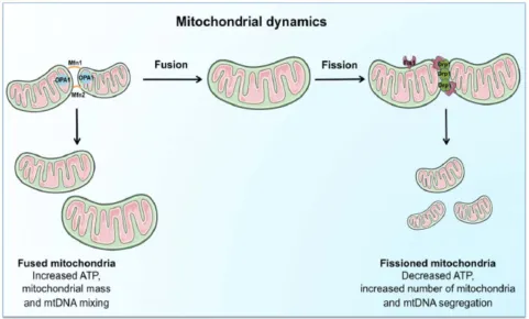
mitochondrial dysfunction. These further results in; mitochon-drial fragmentation, loss of oxidative phosphorylation Figure 1Schematic representation of the regulation and coordination of mitochondrial dynamics.
Notes: The coordination of fusion andfission machinery, involving mitofusins (Mfn) 1 and 2, optic atrophy protein 1 (OPA1), dynamin-related protein 1 (Drp1). Mitochondrial fusion, by interconnecting organelles, promotes mtDNA mixing and enhances bioenergetics efficiency whilefission removes damaged mitochondria via mitophagy. Reproduced from Picca A, Lezza AMS, Leeuwenburgh C, et al. Fueling inflamm-aging through mitochondrial dysfunction: mechanisms and molecular targets.Int J Mol Sci. 2017;18(5):933.23
Journal of Inflammation Research downloaded from https://www.dovepress.com/ by 118.70.13.36 on 24-Aug-2020
(OXPHOS), mtDNA depletion and ROS production. These
are associated with metabolic dysfunction or disease.55The
impairment of mitochondrial quality control to remove damaged and dysfunctional mitochondria leads to the accu-mulation of damage associated with membrane patterns (DAMPs) released from injured cells, cell-free mtDNA,
N-formyl peptides and Cardiolipin.56Mitochondrial DAMPs
can bind and activate membrane or cytosolic pathogen recog-nition receptors (PRRs) such as toll-like receptors (TLR), nod like receptors (NLRs), like those recognized by
pathogen-associated molecular pattern (PAMPs).57 Thus, it activates
different early-phase inflammatory mediators like tumor
necrosis factor α (TNF-α), interleukins, interferon-gamma
(IFN-γ) and ROS/RNS.23 These coexistences of cellular
responses to danger of oxidative stress and accumulation of
mitochondrial DNA leads to chronic inflammation.23
Mitochondrial respiratory chain, NADPH oxidases, and 5-lipoxygenase are the major cellular sources of ROS
production.58 The byproducts of oxidative stress: ROS; and
RNS can also further generated because of inflammatory cell
activity.59,60 Surprisingly, oxidative stress activates several transcription factors (NF-kB and activated protein 1) leading
to the production and activation of pro-inflammatory
cyto-kines, chemocyto-kines, and lymphocytes which in turn leads to the production of more ROS and RNS, principally in the form of superoxide, nitric oxide (NO), and peroxynitrite.61–63This complicated bidirectional amplifying and the self-sustaining relationship between the development of chronic
oxidative stress and chronic systemic inflammations outcomes
to modification of DNA, tertiary protein structure, and lipid
peroxidation of the cell membrane. This further results in a net accumulation of DAMPs that can further exacerbate the conditions.64–66Thus, in turn, it will have a deleterious effect of damaging mitochondria synergistically leading to the deple-tion of ATP producdeple-tion and promoting a switch to anaerobic
glycolysis.67 Therefore, mitochondrial dysfunction with fi
s-sion upregulation prevents the elimination of damaged mito-chondria producing ROS and RNS that exceed the antioxidant activity is likely initiating factors in inflammation, aging, and age-related diseases (Figure 2).44
Mitochondrial Dysfunction and
Signaling Pathway in In
fl
ammation
Mitochondria are multifunctional organelles that contain different proteins used for biosynthesis, metabolism and
cell death or survival functions.68 The vast majority of
proteins are encoded in the nucleus and imported to
mitochondria after translated. Translated proteins were shared with end-to-end collision fashion of mitochondrial
fusion movement along the cytoskeleton.2,34 Each fusion
movement of mitochondria is mediated by outer mem-brane proteins Mfn 1 and 2 and inner memmem-brane with
OPA1 catalyzed by dedicated GTPases.69–71 Since this
continuous fusion of individual mitochondria may produce
abnormally elongated mitochondria that were
non-functional, an opposing process called fission becomes
available.32Thefission process prevents abnormal
elonga-tion of mitochondria by splitting a single mitochondrion
into two separate organelles.72 If the balance favors fi
s-sion, mitochondria become fragmented, small spherical organelle while if fusion is favored, elongated and banded
together mitochondria will be formed.35 Mitochondrial
dynamics imbalance affects energy production, apoptosis, mitophagy, mitochondrial movement, mtDNA stability
and the tolerance of cells to mtDNA mutations.73–75 The
abnormalities of dynamics dysfunction and bidirectional signaling pathway between mitochondria and nucleus are exposed to, constantly alter physiological, environmental
and pathological stimuli.2,6As the center for many cellular
functions, especially in energy supply with oxidative meta-bolism, maintaining mitochondrial quality control is
cru-cial for tissue homeostasis.23,39Mitochondrial dysfunction
with the release of ROS and mitochondrial-derived
DAMPs contribute to initiating an inflammatory response
by invoking pro-inflammatory cytokines that interact with
receptors like those involved in the pathogen-associated
response.76 Mitochondria play a central role in sterile
inflammation through the activation of several pathways.21
Mitochondrial Ca2+ Handling and
ROS Derived In
fl
ammation
Mitochondrial energy generation in the inner membrane through the electron transport chain by using oxygen as an
electron acceptor is the major source of ROS generation.77
ROS are produced by one-electron transfer from redox donor NADH and FADH2 to molecular oxygen in mitochondrial
ETC especially from complex I and complex III.78A
redox-sensitive inflammatory signaling pathway engages
mitochon-drial calcium handling, ROS production, and nuclear factorκB
(NF-κB) activation.79The activation of NF-kB provokes the
transcription of pro-inflammatory cytokines such as TNF-α,
IL1, IL6, and IL8.77Under calcium overloads, such as burn
injury or sepsis, the electron transport chain (ETC) becomes dysfunctional and elevated ROS generation occurs. Such
Journal of Inflammation Research downloaded from https://www.dovepress.com/ by 118.70.13.36 on 24-Aug-2020
a ROS burst represents a major pro-inflammatory stimulus through the modulation of the expression and activity of NF-κB (Figure 2).23,77 Antioxidants produced by specific nitric oxide synthase in the biological system eliminate ROS
that results in overwhelming the cellular repair system.80
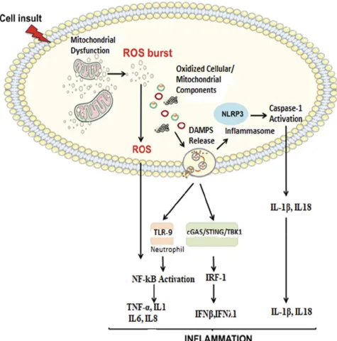
Imbalance of two opposite and antagonist forces, production of ROS and antioxidants called oxidative stress are damaging to mitochondrial cellular components like DNA, protein, lipids and other molecules. Oxidative stress in cellular milieu causes mitochondrial dysfunction and accumulation of damaged Figure 2Proposed signaling pathways through which damaged-associated molecular patterns (DAMPs) can trigger inflammation.
Notes:The impairment of the mitochondrial quality control process may lead to an accumulation of intracellular oxidized components and their release as DAMPs. Damaged mtDNA molecules, either TFAM-bound (green circles) or unbound (red circles) may be released as DAMPs. These, in turn, can activate an inflammatory response via three distinct signaling pathways by interacting with by interacting with 1) Toll-like receptors (TLRs), 2) nucleotide-binding oligomerization domain (NOD)-like receptor family pyrin domain containing 3 (NLRP3) inflammasome, and 3) cytosolic cyclic GMP-AMP synthase (cGAS)-stimulator of interferon genes (STING) DNA-sensing system. Reproduced from Picca A, Lezza AMS, Leeuwenburgh C, et al. Fueling inflamm-aging through mitochondrial dysfunction: mechanisms and molecular targets.Int J Mol Sci. 2017;18(5):933.23
Abbreviations:IFN, interferon; IL, interleukin; IRF-1, interferon regulatory factor 1; mtDNA, mitochondrial DNA; NF-kB, nuclear factor-kB; ROS, reactive oxygen species; TBK1, TANK-binding kinase 1; TNF-α, tumor necrosis factor-alpha.
Journal of Inflammation Research downloaded from https://www.dovepress.com/ by 118.70.13.36 on 24-Aug-2020
products which are recognized by pattern recognition recep-tors initiating sterile inflammations.81The coexistence of oxi-dative stress and mitochondrial dysfunction giving a role for the accumulation of DAMPs which proposed to initiate sterile inflammation.23
Mitochondrial- DAMPs Derived
In
fl
ammation
Due to the“bacterial ancestor”theory of mitochondria, the
releasing of contents due to damage or dysfunction to cytoplasm provokes an immunological response. In addi-tion to mitochondrial dysfuncaddi-tion with oxidative stress,
several conditions characterized by an inflammatory
response (e.g., trauma, HIV, cancer) are associated with increased levels of circulating mitochondrial DAMPs. DAMPs, in turn, induce caspase-1 activation and the
release of pro-inflammatory cytokines like IL1B and
IL18 whichfinally leads to sterile inflammation.58In
par-ticular, mitochondrial DNA activates different infl
amma-tory responses through different receptors, (Figure 2)
triggering transcription and translation of inflammatory
cytokines. Uncontrolled and excessive release of mito-chondrial DAMPs associated with severity and contributes
to the dysregulated process observed in numerous infl
am-matory and autoimmune conditions.44
Mitochondrial Dysfunctions
Associated Non-Communicable
Diseases
Non-communicable diseases (NCDs) are increasingly becoming the leading public causes of morbidity and
mor-tality globally.82The leading NCDs encompass a cluster of
illness including cardiovascular diseases (CVDs), diabetes mellitus type 2, chronic obstructive lung disease, and
cancer. These NCDs have common and key modifiable
behavioral risk factors like unhealthy diet, lack of physical activity, the harmful use of alcohol, tobacco use and which in turn leads to overweight and obesity, raised blood
pressure, and raised cholesterol, and ultimately disease.83
Cardiovascular diseases accounted for most NCD deaths (17.5 million NCD deaths), followed by cancers (8.2mil-lion NCD deaths), respiratory diseases (4.0 mil(8.2mil-lion NCD
deaths) and diabetes mellitus (1.5million NCD deaths).84
Globally, CVDs being the largest contributor to global mortality, accounting for nearly half of 36 million annual NCDs deaths and produces immense health and economic
burdens.85
A common feature of all NCDs is excessive fatigue which results from mitochondrial function impairment; the crucial
organelle responsible for cellular energy production.86 As
stated above, defects in mitochondrial gene results mutation of proteins that involve mitochondrial dynamic activities then
proceed to its physiological activity impairment.34,35 The
mitochondrial genome is susceptible to oxidative damage as a result of its residence close to the electron transport system, mtDNA lacks histone protein (protects from damage), and repair mechanism rather it Induces to apoptosis once it
becomes damaged.87,88 Thus, lack of repair mechanism of
damaged mtDNA means cells cannot copy mtDNA accurately resulting in errors of transcription, deletions and mutations and also oxidation from ROS results in a series of cellular abuse; loss of membranes integrity, diminishing proton gradient caus-ing less ATP production, unfoldcaus-ing and loss of cellular protein
affinity for their respective enzymes, and releasing
cyto-chromes C into the cytosol stimulating apoptosis, all in a continuous feed-forward cycle of cellular, tissue and organ
dysfunction to cause chronic diseases.87 Additionally, high
ROS concentration permits histone acetylation to predomi-nate, which accelerates faulty nuclear transcription and thus replication. This initiates the release of NF-kB into the nucleus
(a significant pro-inflammatory cytokine which also damages
nDNA). Simultaneously, cell differentiation and apoptosis signals are silenced with histone acetylation, eventually
result-ing in over-replication favorresult-ing tumorigenesis.89,90 As the
mitochondrial inner membrane is the main site of electron transport chain, dwelling different enzymes and proteins which are also becomes the main source of oxidative stress. Enzymes and proteins (i.e. cytochrome c), and low-molecular-weight redox intermediates (i.e. coenzymes such as ubiqui-none or coenzyme Q) that transport reducing equivalents, in the form of hydrogen atoms or just their electrons, down the redox potential from respiratory substrates to oxygen: an oxidative pathway composed of four multiple-subunit
com-plexes in the mitochondria which leaks 0.2–2% electrons to
molecular oxygen producing super oxide or hydrogen
peroxide.27These enzymes and proteins are the primary
tar-gets of oxidative stress products like ROS and RNS, and it is not surprising that disruption of function and structure of mitochondria are now thought to trigger numerous diseases, including cardiac, liver and kidney disorders, and
neurodegen-erative diseases (e.g. Parkinson’s disease and Alzheimer’s
disease), aging processes and multiple organ failure in septic shock. Cardiovascular disease/atherosclerosis, obesity and
type 2 diabetes are closely associated with chronic infl
amma-tion characterized by abnormal cytokine producamma-tion, increased
Journal of Inflammation Research downloaded from https://www.dovepress.com/ by 118.70.13.36 on 24-Aug-2020
levels of acute-phase reactants and activation of a network of
inflammatory signaling pathways.91 Mitochondrial
dysfunc-tion because of unbalanced fusion andfission, genetic defects
of mtDNA or oxidative damage with ROS and RNS can
impair energy metabolism, the central alteration in obesity.8
Cells can manage nutrient supply by increasing mitochondrial contents. However, persistent nutrient leftover devastates the mitochondrial system and causes its dysfunction leading to the accumulation of incompletely oxidized lipid products to cause fat accumulation and oxidative stress which damage endothe-lial cells.92,93Obesity is key in the development of major non-communicable diseases (NCDs) including diabetes mellitus, hypertension, metabolic syndrome, non-alcoholic fatty liver disease, cardiovascular disease and several classes of cancer including colorectal, liver, breast, pancreatic endometrial,
renal, prostate, lymphoma and myoma.8,27 Dysfunctional
mitochondria create a vicious cycle of impaired beta-oxidation giving rise to the production of ceramides, free
radicals and inflammatory cytokines which damages the
mito-chondrial membrane (as shown inFigure 3) and DNA further
jeopardizes mitochondrial respiratory capacity.94Oxygen-free
radicals may themselves initiate a chronic inflammatory
pro-cess in which the pathology is characteristics of obesity and linked to insulin resistance and type 2 diabetes.95
Therefore, preventing chronic sterile inflammation
derived with mitochondrial dysfunction resulting in
oxida-tive stress and DAMPs activated inflammasome can signifi
-cantly diminish non-communicable diseases.97,98Reducing
cellular and mitochondrial membrane and DNA damage and loss of membrane integrity is important in preventing loss of
cellular energy and regulating cellular life span.99 Since
oxidative stress is the consequence of imbalance between pro- and antioxidants species, supplements containing diet-ary antioxidants and some accessory molecules, such as zinc and certain vitamins, are important in maintaining free-radical scavenging systems, biosynthetic capacity,
mem-branes, enzymes and DNA.100Utility of oral mitochondrial
replacement supplements, such as replacement glycerol phospholipids, l-carnitine, alpha-Lipoic acid, coenzyme
Q10, NADH, pyrroloquinoline quinone and other
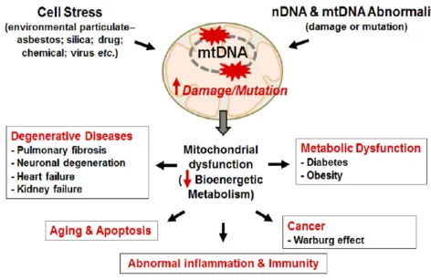
Figure 3Mitochondrial damage induces diverse chronic and degenerative diseases.
Notes:Mitochondrial damage and mutation can be caused by stress from environment particulate and/or DNA abnormality. DNA mutation/damage causes mitochondrial dysfunction reducing bioenergetics metabolism promoting chronic inflammation and chronic diseases, like cancer, CVD, Diabetes, Obesity, and aging. Kim SJ, Cheresh P, Jablonski RP, Williams DB, Kamp DW. The Role of Mitochondrial DNA in Mediating Alveolar Epithelial Cell Apoptosis and Pulmonary Fibrosis.Int J Mol Sci. 2015;16 (9):21486-21519.96
Journal of Inflammation Research downloaded from https://www.dovepress.com/ by 118.70.13.36 on 24-Aug-2020
mitochondrial supplements also improves mitochondrial
function.86 Anaerobic and resistance training intervention
induced in a mouse model showed an increase in the activa-tion of the signaling pathway involved in mitochondrial
biogenesis.101Preclinical evidence from animal and in vitro
studies suggested that regular endurance-based exercise intervention is known to be a potent stimulus for muscle
mitochondrial biogenesis.102It is also shown that
endurance-based exercise increases muscle oxidation capacity of mito-chondria by increasing the activity of citrate synthase and respiratory chain complexes.103–105
Conclusion
Mitochondria are central to the regulation of energy metabo-lism and cellular homeostasis due to their principal role in bioenergetics, ROS production, ion homeostasis, apoptosis and signal transduction. This organelle is highly dynamic and can re-program itself depending on various environmental and intracellular signals important for multiple mitochondrial functions, including mtDNA stability, respiratory function, apoptosis, response to cellular stress, and mitochondrial degradation. The dynamic process of mitochondria may not be balanced as a result of proteins required for fusion and
fission that decreases the crucial role of mitochondria
bioe-nergetics and the accumulation of damaged mitochondria producing ROS.ROS generated by dysfunctional mitochon-dria further damage mitochonmitochon-drial of different organs and tissues that cannot function properly to result in chronic and age-related disorders. Non-communicable diseases that are increasing worldwide in recent years are the consequences of unhealthy diets and physical inactivity, which shares basic
mechanisms of mitochondrial defects, systemic infl
amma-tion, and oxidative stress. Consequent oxidative stress caus-ing endoplasmic reticulum (ER) and mitochondrial stress leads to excess accumulation of food energy favoring obesity and the development of other metabolic syndrome and related complications. Maintaining the health of mitochon-dria is very valuable and could aid in the prevention of age-related disorders.
Disclosure
The authors declare that there is no conflict of interest
regarding the publication of this paper.
References
1. Rongvaux A. Innate immunity and tolerance toward mitochondria. Mitochondrion.2018;41:14–20. doi:10.1016/j.mito.2017.10.007
2. Mishra P, Chan DC. Mitochondrial dynamics and inheritance dur-ing cell division, development and disease.Nat Rev Mol Cell Biol. 2014;15:634–646. doi:10.1038/nrm3877
3. Zorov DB, Plotnikov EY, Silachev DN, et al. Microbiota and mitobiota. Putting an equal sign between mitochondria and bacteria.Biochemistry (Moscow).2014;79:1017–1031. doi:10.1134/S0006297914100046 4. Willems PHGM, Rossignol R, Dieteren CEJ, Murphy MP,
Koopman WJH. Redox homeostasis and mitochondrial dynamics. Cell Metab.2015;22:207–218. doi:10.1016/j.cmet.2015.06.006 5. Calvo SE, Mootha VK. The mitochondrial proteome and human
disease. Annu Rev Genomics Hum Genet. 2010;11:25–44. doi:10.1146/annurev-genom-082509-141720
6. Shadel GS, Horvath TL. Mitochondrial ROS signaling in organis-mal homeostasis. Cell. 2015;163(3):560–569. doi:10.1016/j. cell.2015.10.001
7. Cao YL, Meng S, Chen Y, et al. MFN1 structures reveal nucleotide-triggered dimerization critical for mitochondrial fusion. Nature.2017;542:372–376. doi:10.1038/nature21077
8. Cerdá C, Sánchez C, Climent B, et al. Oxidative stress and DNA damage in obesity-related tumorigenesis.Adv Exp Med Biol.2014. 9. Nagdas S, Kashatus DF. The interplay between oncogenic signaling networks and mitochondrial dynamics.Antioxidants.2017;6(2):33. 10. Ježek J, Cooper KF, Strich R. Reactive oxygen species and mito-chondrial dynamics: the yin and yang of mitomito-chondrial dysfunction and cancer progression. Antioxidants. 2018;7:13. doi:10.3390/ antiox7010013
11. Glowacki S, Synowiec E, Blasiak J. The role of mitochondrial DNA damage and repair in the resistance of BCR/ABL-expressing cells to tyrosine kinase inhibitors. Int J Mol Sci. 2013;14:16348–16364. doi:10.3390/ijms140816348
12. Bordi M, Nazio F, Campello S. The close interconnection between mitochondrial dynamics and mitophagy in cancer. Front Oncol. 2017;7. doi:10.3389/fonc.2017.00081
13. Martinez-Carreres L, Nasrallah A, Fajas L. Cancer: linking power-houses to suicidal bags. Front Oncol. 2017;7. doi:10.3389/fonc. 2017.00204
14. Kalkavan H, Green DR. MOMP, cell suicide as a BCL-2 family business.Cell Death Differ.2018;25:46–55. doi:10.1038/cdd.2017.179 15. Suárez-Rivero J, Villanueva-Paz M, de la Cruz-ojeda P, et al. Mitochondrial dynamics in mitochondrial diseases. Diseases. 2016;5:1. doi:10.3390/diseases5010001
16. Simula L, Nazio F, Campello S. The mitochondrial dynamics in cancer and immune-surveillance. Semin Cancer Biol. 2017;47:29–42. doi:10.1016/j.semcancer.2017.06.007
17. Wada J, Nakatsuka A. Mitochondrial dynamics and mitochondrial dysfunction in diabetes.Acta Med Okayama.2016;70(3):151–158. 18. Marín-García J, Akhmedov AT. Mitochondrial dynamics and cell
death in heart failure.Heart Fail Rev.2016;21(2):123–136. 19. Wu Q, Luo CL, Tao LY. Dynamin-related protein 1 (Drp1) mediating
mitophagy contributes to the pathophysiology of nervous system dis-eases and brain injury.Histol Histopathol.2017;32(6):551–559. 20. López-Lluch G. Mitochondrial activity and dynamics changes
regarding metabolism in ageing and obesity.Mech Ageing Dev. 2017;162:108–121. doi:10.1016/j.mad.2016.12.005
21. López-Armada MJ, Riveiro-Naveira RR, Vaamonde-García C, Valcárcel-Ares MN. Mitochondrial dysfunction and the infl amma-tory response.Mitochondrion.2013;13(2):106–118. doi:10.1016/j. mito.2013.01.003
22. Mathew A, Lindsley TA, Sheridan A, et al. Degraded mitochon-drial dna is a newly identified subtype of the damage associated molecular pattern (DAMP) family and possible trigger of neurodegeneration.J Alzheimer’s Dis.2012;30:617–627. doi:10. 3233/JAD-2012-120145
23. Picca A, Lezza AMS, Leeuwenburgh C, et al. Fueling inflamm-aging through mitochondrial dysfunction: mechanisms and molecular targets. Int J Mol Sci.2017;18(5):933. doi:10.3390/ijms18050933
Journal of Inflammation Research downloaded from https://www.dovepress.com/ by 118.70.13.36 on 24-Aug-2020
24. Nakahira K, Haspel JA, Rathinam VAK, et al. Autophagy proteins regulate innate immune responses by inhibiting the release of mitochondrial DNA mediated by the NALP3 inflammasome.Nat Immunol.2011;12:222–230. doi:10.1038/ni.1980
25. Liu TF, Brown CM, El Gazzar M, et al. Fueling theflame: bioe-nergy couples metabolism and inflammation. J Leukoc Biol. 2012;92:499–507. doi:10.1189/jlb.0212078
26. Cannon B, Nedergaard J. Neither brown nor white. Nature. 2012;488:286–287. doi:10.1038/488286a
27. Bullon P, Newman HN, Battino M. Obesity, diabetes mellitus, atherosclerosis and chronic periodontitis: a shared pathology via oxidative stress and mitochondrial dysfunction? Periodontol. 2000;64(1):139–153.
28. Chaffee BW, Weston SJ. Association between chronic periodontal disease and obesity: a systematic review and meta-analysis. J Periodontol.2010;81:1708–1724. doi:10.1902/jop.2010.100321 29. DiMauro S, Schon EA. Mitochondrial respiratory-chain diseases.
N Engl J Med.2003;348:2656–2668. doi:10.1056/NEJMra022567 30. DiMauro S. Mitochondrial diseases. Biochim Biophys Acta
Bioenerg.2004;1658(1–2):80–88.
31. Westermann B. Mitochondrial fusion andfission in cell life and death. Nat Rev Mol Cell Biol.2010;11:872–884. doi:10.1038/nrm3013 32. Bleazard W, McCaffery JM, King EJ, et al. The dynamin-related
GTPase Dnm1 regulates mitochondrial fission in yeast. Nat Cell Biol.1999;1:298–304. doi:10.1038/13014
33. Westrate LM, Drocco JA, Martin KR, Hlavacek WS, MacKeigan JP. Mitochondrial morphological features are associated withfission and fusion events. PLoS One. 2014;9:e95265. doi:10.1371/journal. pone.0095265
34. Chan DC. Fusion andfission: interlinked processes critical for mito-chondrial health. Annu Rev Genet. 2012;46:265–287. doi:10.1146/ annurev-genet-110410-132529
35. Detmer SA, Chan DC. Functions and dysfunctions of mitochondrial dynamics. Nat Rev Mol Cell Biol. 2007;8:870–879. doi:10.1038/ nrm2275
36. Pernas L, Scorrano L. Mito-morphosis: mitochondrial fusion,fi s-sion, and cristae remodeling as key mediators of cellular function. Annu Rev Physiol.2016;78:505–531. doi:10.1146/annurev-physiol -021115-105011
37. Zhu X, Perry G, Smitha MA, Wang X. Abnormal mitochondrial dynamics in the pathogenesis of Alzheimer’s disease. J Alzheimer’s Dis.2013.
38. García-Escudero V, Martín-Maestro P, Perry G, Avila J. Deconstructing mitochondrial dysfunction in alzheimer disease. Oxid Med Cell Longev.2013;2013:1–13. doi:10.1155/2013/162152 39. Twig G, Elorza A, Molina AJA, et al. Fission and selective fusion govern mitochondrial segregation and elimination by autophagy. EMBO J.2008;27(2):433–446. doi:10.1038/sj.emboj.7601963 40. Ono T, Isobe K, Nakada K, Hayashi J-I. Human cells are protected from
mitochondrial dysfunction by complementation of DNA products in fused mitochondria.Nat Genet.2001;28:272–275. doi:10.1038/90116 41. Zorzano A, Hernández-Alvarez MI, Sebastián D, Muñoz JP. Mitofusin 2
as a driver that controls energy metabolism and insulin signaling. Antioxid Redox Signal.2015;22:1020–1031. doi:10.1089/ars.2014.6208 42. Yin F, Cadenas E. Mitochondria: the cellular hub of the dynamic coordinated network. Antioxid Redox Signal. 2015;22:961–964. doi:10.1089/ars.2015.6313
43. Duchen MR. Mitochondria and calcium: from cell signalling to cell death.J Physiol.2000;529:57–68. doi:10.1111/tjp.2000.529.issue-1 44. Hernández-Aguilera A, Rull A, Rodríguez-Gallego E, et al. Mitochondrial dysfunction: a basic mechanism in infl ammation-related non-communicable diseases and therapeutic opportunities. Mediators Inflamm.2013;2013:1–13. doi:10.1155/2013/135698 45. McInnes J. Mitochondrial-associated metabolic disorders:
founda-tions, pathologies and recent progress. Nutr Metab. 2013;10:63. doi:10.1186/1743-7075-10-63
46. Bonawitz ND, Clayton DA, Shadel GS. Initiation and beyond: multiple functions of the human mitochondrial transcription machinery.Mol Cell. 2006;24:813–825. doi:10.1016/j.molcel.2006.11.024
47. Ryan MT, Hoogenraad NJ. Mitochondrial-nuclear communications. Annu Rev Biochem. 2007;76:701–722. doi:10.1146/annurev. biochem.76.052305.091720
48. Osellame LD, Blacker TS, Duchen MR. Cellular and molecular mechanisms of mitochondrial function. Best Pract Res. 2012;26:711–723. doi:10.1016/j.beem.2012.05.003
49. Lenaz G, Baracca A, Barbero G, et al. Mitochondrial respiratory chain super-complex I-III in physiology and pathology. Biochim Biophys Acta Bioenerg. 2010;1797:633–640. doi:10.1016/j. bbabio.2010.01.025
50. Srivastava S. Emerging therapeutic roles for NAD+ metabolism in mitochondrial and age-related disorders.Clin Transl Med.2016;5. doi:10.1186/s40169-016-0104-7
51. Schieber M, Chandel NS. ROS function in redox signaling and oxidative stress. Current Biology. 2014;24:R453–R462. doi:10.1016/j.cub. 2014.03.034
52. McCarthy CM, Kenny LC. Immunostimulatory role of mitochon-drial DAMPs: alarming for pre-eclampsia?Am J Reprod Immunol. 2016;76(5):341–347. doi:10.1111/aji.2016.76.issue-5
53. Chauhan A, Vera J, Wolkenhauer O. The systems biology of mitochondrialfission and fusion and implications for disease and aging. Biogerontology. 2014;15:1–12. doi:10.1007/s10522-013-9474-z
54. Westermann B. Bioenergetic role of mitochondrial fusion andfission. Biochim Biophys Acta Bioenerg.2012;1817(10):1833–1838. 55. Liesa M, Shirihai OS. Mitochondrial dynamics in the regulation of
nutrient utilization and energy expenditure. Cell Metab. 2013;17:491–506. doi:10.1016/j.cmet.2013.03.002
56. Zhang Q, Raoof M, Chen Y, et al. Circulating mitochondrial DAMPs cause inflammatory responses to injury. Nature. 2010;464:104–107. doi:10.1038/nature08780
57. Collins LV, Hajizadeh S, Holme E, Jonsson I-M, Tarkowski A. Endogenously oxidized mitochondrial DNA induces in vivo and in vitro inflammatory responses.J Leukoc Biol.2004;75:995–1000. doi:10.1189/jlb.0703328
58. Salminen A, Ojala J, Kaarniranta K, Kauppinen A. Mitochondrial dysfunction and oxidative stress activate inflammasomes: impact on the aging process and age-related diseases.Cell Mol Life Sci. 2012;69:2999–3013. doi:10.1007/s00018-012-0962-0
59. Fischer MT, Sharma R, Lim JL, et al. NADPH oxidase expression in active multiple sclerosis lesions in relation to oxidative tissue damage and mitochondrial injury. Brain. 2012;135:886–899. doi:10.1093/brain/aws012
60. Morris G, Anderson G, Dean O, et al. The glutathione system: a new drug target in neuroimmune disorders.Mol Neurobiol.2014. 61. Reuter S, Gupta SC, Chaturvedi MM, Aggarwal BB. Oxidative stress, inflammation, and cancer: how are they linked? Free
Rad Biol Med. 2010;49:1603–1616. doi:10.1016/j.
freeradbiomed.2010.09.006
62. Prolo C, Álvarez MN, Radi R. Peroxynitrite, a potent
macrophage-derived oxidizing cytotoxin to combat invading pathogens.BioFactors.2014;40:215–225. doi:10.1002/biof.1150 63. Vaamonde-García C, Riveiro-Naveira RR, Valcárcel-Ares MN,
Hermida-Carballo L, Blanco FJ, Lõpez-Armada MJ. Mitochondrial dysfunction increases inflammatory responsiveness to cytokines in nor-mal human chondrocytes. Arthritis Rheum. 2012;64:2927–2936. doi:10.1002/art.34508
64. Galley HF. Bench-to-bedside review: targeting antioxidants to mitochondria in sepsis.Critical Care.2010;14. doi:10.1186/cc9098 65. Ortiz GG, Pacheco-Moisés FP, Bitzer-Quintero OK, et al. Immunology and oxidative stress in multiple sclerosis: clinical and basic approach. Clin Devel Immunol. 2013;2013:1–14. doi:10.1155/2013/708659
Journal of Inflammation Research downloaded from https://www.dovepress.com/ by 118.70.13.36 on 24-Aug-2020
66. Lucas K, Maes M. Role of the toll like receptor (TLR) radical cycle in chronic inflammation: possible treatments targeting the TLR4 pathway. Mol Neurobiol. 2013;48:190–204. doi:10.1007/s12035-013-8425-7
67. Morris G, Berk M. The many roads to mitochondrial dysfunction in neuroimmune and neuropsychiatric disorders. BMC Med. 2015;13 (1):68.
68. Auld D, Lea W, Davis MI, et al..Proteomic mapping of mitochon-dria in living cells via spatially restricted enzymatic tagging. Science.2013;339:1328–1331. doi:10.1126/science.1230593 69. Santel A, Fuller MT. Control of mitochondrial morphology by
a human mitofusin.J Cell Sci.2001.
70. Rojo M, Legros F, Chateau D, Lombès A. Membrane topology and mitochondrial targeting of mitofusins, ubiquitous mammalian homologs of the transmembrane GTPase Fzo. J Cell Sci. 2002;115(8):1663–1674.
71. Alexander C, Votruba M, Pesch UEA, et al. OPA1, encoding a dynamin-related GTPase, is mutated in autosomal dominant optic atrophy linked to chromosome 3q28.Nat Genet.2000;26:211–215. doi:10.1038/79944
72. Richter V, Singh AP, Kvansakul M, Ryan MT, Osellame LD. Splitting up the powerhouse: structural insights into the mechanism of mitochondrialfission.Cell Mol Life Sci.2015;72:3695–3707. doi:10.1007/s00018-015-1950-y
73. Chen H, Vermulst M, Wang YE, et al. Mitochondrial fusion is required for mtdna stability in skeletal muscle and tolerance of mtDNA mutations. Cell. 2010;141:280–289. doi:10.1016/j. cell.2010.02.026
74. Tait SWG, Green DR. Mitochondria and cell death: outer mem-brane permeabilization and beyond. Nat Rev Mol Cell Biol. 2010;11:621–632. doi:10.1038/nrm2952
75. Chan NC, Salazar AM, Pham AH, et al. Broad activation of the ubiquitin-proteasome system by Parkin is critical for mitophagy.Hum Mol Genet. 2011;20:1726–1737. doi:10.1093/ hmg/ddr048
76. Quirós PM, Langer T, López-Otín C. New roles for mitochondrial proteases in health, ageing and disease.Nat Rev Mol Cell Biol. 2015;16:345–359. doi:10.1038/nrm3984
77. Schreck R, Rieber P, Baeuerle PA. Reactive oxygen intermediates as apparently widely used messengers in the activation of the
NF-kappa B transcription factor and HIV-1. EMBO J.
1991;10:2247–2258. doi:10.1002/embj.1991.10.issue-8
78. Huang LS, Cobessi D, Tung EY, Berry EA. Binding of the respira-tory chain inhibitor antimycin to the mitochondrial bc1 complex: a new crystal structure reveals an altered intramolecular hydrogen-bonding pattern. J Mol Biol. 2005;351:573–597. doi:10.1016/j.jmb.2005.05.053
79. Maass DL, White J, Sanders B, Horton JW. Role of cytosolic vs. mitochondrial Ca2+ accumulation in burn injury-related myocar-dial inflammation and function.Am J Physiol - Hear Circ Physiol. 2005;288:H744–H751. doi:10.1152/ajpheart.00367.2004
80. Fiers W, Beyaert R, Declercq W, Vandenabeele P. More than one way to die: apoptosis, necrosis and reactive oxygen damage. Oncogene.1999;18:7719–7730. doi:10.1038/sj.onc.1203249 81. Hu F, Liu F. Mitochondrial stress: a bridge between mitochondrial
dysfunction and metabolic diseases?Cell Signal.2011;23:1528–1533. doi:10.1016/j.cellsig.2011.05.008
82. Ekpenyong CE, Udokang NE, Akpan EE, Samson T. Double bur-den, non-communicable diseases and risk factors evaluation in Sub-Saharan Africa: the Nigerian experience. Eur J Sustain Devel.2012;1:249. doi:10.14207/ejsd.2012.v1n2p249
83. Riley L, Gouda H, Cowan M.Noncumminicable Diseases Progress Monitor 2017. World Health Organization;2017:2017.
84. Mendis S, Armstrong T, Bettcher D, et al.Global Status Report on Noncommunicable Diseases 2014. World Health Organisation; 2014.
85. Benziger CP, Roth GA, Moran AE. The global burden of disease
study and the preventable burden of NCD. Glob Heart.
2016;11:393–397. doi:10.1016/j.gheart.2016.10.024
86. Nicolson GL. Mitochondrial dysfunction and chronic disease: treat-ment with natural suppletreat-ments.Integr Med.2014.
87. Zeviar DD, Gonzalez MJ, Massari JRM, Duconge J, Mikirova N. The role of mitochondria in cancer and other chronic diseases. J Orthomol Med.2014;29(4):157.
88. Mason PA. Mismatch repair activity in mammalian mitochondria. Nucleic Acids Res.2003;31:1052–1058. doi:10.1093/nar/gkg167 89. González MJ, Rosario-Pérez G, Guzmán AM, et al. Mitochondria,
energy and cancer: the relationship with ascorbic acid.J Orthomol Med.2010.
90. Cui H, Kong Y, Zhang H. Oxidative stress, mitochondrial dysfunc-tion, and aging.J Signal Transduct.2012;2012:1–13. doi:10.1155/ 2012/646354
91. Lim S, Cho YM, Park KS, Lee HK. Persistent organic pollutants, mitochondrial dysfunction, and metabolic syndrome.Ann N Y Acad Sci.2010;1201:166–176. doi:10.1111/j.1749-6632.2010.05622.x 92. Camps J, Rodríguez-Gallego E, García-Heredia A, et al.
Paraoxonases and chemokine (C-C motif) ligand-2 in noncommu-nicable diseases.Adv Clin Chem.2014.
93. Chang J-C. Regulatory role of mitochondria in oxidative stress and atherosclerosis. World J Cardiol. 2010;2:150. doi:10.4330/wjc.v2. i6.150
94. Kelley DE, He J, Menshikova EV, Ritov VB. Dysfunction of mitochondria in human skeletal muscle in type 2 diabetes. Diabetes.2002;51:2944–2950. doi:10.2337/diabetes.51.10.2944 95. Rogge MM. The role of impaired mitochondrial lipid oxidation in
obesity. Biol Res Nurs. 2009;10:356–373. doi:10.1177/109980 0408329408
96. Kim S-J., Cheresh P, Jablonski RP, Williams DB, Kamp DW. The Role of Mitochondrial DNA in Mediating Alveolar Epithelial Cell Apoptosis and Pulmonary Fibrosis. International Journal of Molecular Sciences. 2015;16(9):21486-21519 doi:10.3390/ijms160921486
97. Xu D, Finkel T. A role for mitochondria as potential regulators of cellular life span.Biochem Biophys Res Commun.2002;294:245–248. doi:10.1016/S0006-291X(02)00464-3
98. Conti V, Izzo V, Corbi G, et al. Antioxidant supplementation in the treatment of aging-associated diseases.Front Pharmacol.2016;7. doi:10.3389/fphar.2016.00024
99. Sztretye M, Dienes B, Gönczi M, et al. Review article astaxanthin: a potential mitochondrial-targeted antioxidant treatment in diseases and with aging. Oxid Med Cell Longevity.2019;2019.
100. Nicolson GL, Ash ME. Lipid replacement therapy: a natural medicine approach to replacing damaged lipids in cellular membranes and orga-nelles and restoring function. Biochim Biophys Acta Biomembr. 2014;1838:1657–1679. doi:10.1016/j.bbamem.2013.11.010
101. Fiuza-Luces C, Valenzuela PL, Laine-Menéndez S, et al. Physical exercise and mitochondrial disease: insights from a mouse model. Front Neurol.2019;10. doi:10.3389/fneur.2019.00790
102. Irrcher I, Adhihetty PJ, Joseph AM, Ljubicic V, Hood DA. Regulation of mitochondrial biogenesis in muscle by endurance exercise. Sports Med. 2003;33:783–793. doi:10.2165/00007256-200333110-00001
103. Jeppesen TD, Schwartz M, Olsen DB, et al. Aerobic training is safe and improves exercise capacity in patients with mitochondrial myopathy.Brain.2006;129:3402–3412. doi:10.1093/brain/awl149 104. Jeppesen TD, Dunø M, Schwartz M, et al. Short- and long-term
effects of endurance training in patients with mitochondrial myopathy.Eur J Neurol.2009;16:1336–1339. doi:10.1111/j.1468-1331.2009.02660.x
105. Taivassalo T, Shoubridge EA, Chen J, et al. Aerobic conditioning in patients with mitochondrial myopathies: physiological, biochemical, and genetic effects. Ann Neurol. 2001;50:133–141. doi:10.1002/ (ISSN)1531-8249
Journal of Inflammation Research downloaded from https://www.dovepress.com/ by 118.70.13.36 on 24-Aug-2020
Journal of Inflammation Research
Dove
press
Publish your work in this journal
The Journal of Inflammation Research is an international, peer-reviewed open-access journal that welcomes laboratory and clinical findings on the molecular basis, cell biology and pharmacology of inflammation including original research, reviews, symposium reports, hypothesis formation and commentaries on: acute/chronic inflammation; mediators of inflammation; cellular processes; molecular
mechanisms; pharmacology and novel anti-inflammatory drugs; clin-ical conditions involving inflammation. The manuscript management system is completely online and includes a very quick and fair peer-review system. Visit http://www.dovepress.com/testimonials.php to read real quotes from published authors.
Submit your manuscript here:https://www.dovepress.com/journal-of-inflammation-research-journal
Journal of Inflammation Research downloaded from https://www.dovepress.com/ by 118.70.13.36 on 24-Aug-2020