Abstract
The dopamine D3 receptor is an important CNS target for the treatment of a variety of neurological diseases. Selective dopamine D3 receptor antagonists modulate the improvement of psychostimulant addiction and relapse. In this study, five and six featured pharmacophore models of D3R antagonists were generated and evaluated with the post-hoc score combining two survival scores of active and inactive. Among Top 10 models, APRRR215 and AHPRRR104 were chosen based on the coefficient of determination (APRRR215: R2training = 0.80; AHPRRR104: R2training = 0.82) and predictability (APRRR215: Q2test = 0.73, R2predictive = 0.82; AHPRRR104: Q2test = 0.86, R2predictive = 0.74) of their 3D-quantitative structure–activity relationship models. Pharmacophore-based virtual screening of a large compound library from eMolecules (> 3 million compounds) using two optimal models expedited the search process 100-fold speed increase compared to the docking-based screening (HTVS scoring function in Glide) and identified a series of hit compounds having promising novel scaffolds. After the screening, docking scores, as an adjuvant predictor, were added to two fitness scores (from the pharmacophore models) and predicted Ki (from PLSs of the QSAR models) to improve accuracy. Final selection of the most promising hit compounds were also evaluated for CNS-like properties as well as expected D3R antagonism.
Keywords: Pharmacophore, 3D-QSAR, virtual screening, D3R selective antagonist, molecular
docking, CNS-like
Introduction
Dopamine receptors are a class of G protein-coupled receptors in the central nervous system (CNS). There are at least five subtypes of dopamine receptors: D1R, D2R, D3R, D4R, and D5R. The D1R and D5R are members of the D1-like family of dopamine receptors, whereas the D2R, D3R, and D4R are members of the D2-like family [1]. The D3R is found within a key neuronal network involved in motivation and cognition. In contrast to the D2R, the D3R does not appear to play a role in the regulation of movements [2, 3]. Furthermore, the D3R subtype is considered to represent an important target for the treatment of a variety of neurological diseases, such as schizophrenia and Parkinson’s disease [4]. D3R-selective antagonists can be potential therapeutic agents for the treatment of psychostimulant addiction and relapse [5-9]. However, many clinically approved drugs targeting the D3R do not show significant selectivity over the D2R and other receptors [9]. Thus, it is necessary to develop selective D3R antagonists.
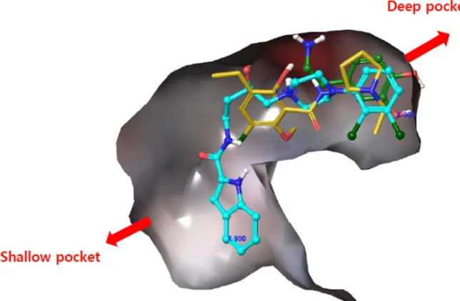
our docking approach identified a common binding site, a deep pocket common to all three compounds, and a shallow pocket occupied by some antagonists (Figure 1). R-22, a selective D3R antagonist, occupied the deep pocket and was found bound to the outer binding pocket to a greater extent [12]. The residue information derived from the docking study was used to develop novel molecules [13], and models including pharmacophore features were built from the dataset containing selected D3R antagonists with high efficacy, selectivity, and metabolic stability.
Figure 1: Superimposed 3PBL protein-ligand docking of dopamine (green; endogenous D3R agonist), eticlopride (yellow; non-selective D3R antagonist), and R-22 (sky blue; selective D3R antagonist).
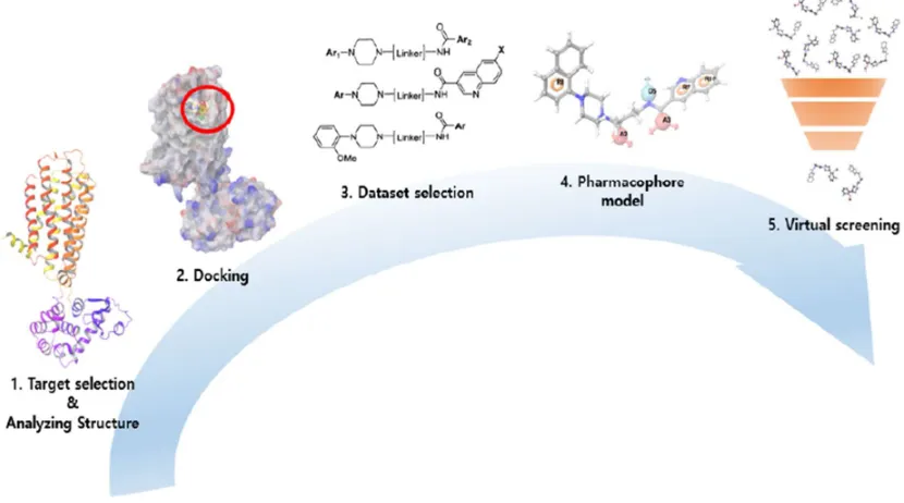
Figure 2: Workflows of this study.
Materials and methods
Data collection and molecular docking.
Identification of pharmacophore hypotheses.
Pharmacophore models were built using Pharmacophore Alignment and Scoring Engine (PHASE) running on Maestro 10.4 (Schrödinger). The best docking pose was considered to be a bioactive conformer of each conformer and used for the development of the pharmacophore model. The activities of compounds were scaled from a minimum value of 1.01 to a maximum value of 3.93 (to be consistent with ones in the table), with an activity threshold of 1.35; this meant that compounds with a Ki of < 44 nM were considered to be antagonists (actives). The pharmacophore features used for hypothesis generation were hydrogen bond acceptor (A), hydrogen bond donor (D), hydrophobic group (H), positively ionisable (P), negatively ionisable (N), and aromatic rings (R) defined by a set of chemical structure patterns. For the current dataset of 50 conformers, five or six features were chosen for model construction. The pharmacophore feature of active ligands that contain identical sets of features with very similar spatial arrangements were grouped together to give rise to a common pharmacophore hypothesis. In the present study, pharmacophore-based QSAR modeling was conducted by dividing the dataset into a 35-member training set (70%) and a 15-member test set (30%) in a random manner. The chemical features associated with ligand-protein binding and the training set data are used to create a model, and the test set is used to evaluate this model [19].
Internal and external QSAR model validation.
The success of a virtual screening campaign using a QSAR model depends heavily on the quality of the model. Several statistical parameters in PHASE, such as R2 for the training set and Q2 for the test set, the standard deviation, root mean square error, and variance ratio (F), can be used to evaluate the robustness of a QSAR model [20-23]. To test the reliability of QSAR models, compounds that were not used for model development must be used: (1) test set (15 compounds), (2) 3rd set (out of initial dataset). The external validation of the 3rd set was conducted using D3R antagonists extracted from the ChEMBL database. ChEMBL compounds with D3R Ki data were imported and filtered using a cut-off of pKi < 0 uM. The selected compounds were then docked to 3PBL. This external validation was followed by advanced screening using ligands that could generate conformers, and that matched a minimum of 4 out of 5, or 5 out of 6 site points. The predictive values derived from this screening process were then compared with the experimental values.
-2 and 6, the number of hydrogen bond donors is < 4, and the number of hydrogen bond acceptors is < 8. The ROC curve analysis describes the sensitivity (true positive rate, Se) for any possible change in the number of selected compounds as 1 – Sp (specificity, which is defined as the true negative rate) [24].
Se = 𝑛𝑢𝑚𝑏𝑒𝑟 𝑜𝑓 𝑠𝑒𝑙𝑒𝑐𝑡𝑒𝑑 𝑎𝑐𝑡𝑖𝑣𝑒𝑠 𝑡𝑜𝑡𝑎𝑙 𝑛𝑢𝑚𝑏𝑒𝑟 𝑜𝑓 𝑎𝑐𝑡𝑖𝑣𝑒𝑠 =
𝑇𝑃 𝑇𝑃 + 𝐹𝑁
Sp = 𝑛𝑢𝑚𝑏𝑒𝑟 𝑜𝑓 𝑑𝑖𝑠𝑐𝑎𝑟𝑑𝑒𝑑 𝑖𝑛𝑎𝑐𝑡𝑖𝑣𝑒𝑠 𝑡𝑜𝑡𝑎𝑙 𝑛𝑢𝑚𝑏𝑒𝑟 𝑜𝑓 𝑖𝑛𝑎𝑐𝑡𝑖𝑣𝑒𝑠 =
𝑇𝑁 𝑇𝑁 + 𝐹𝑃
In the ROC analysis, true positive (TP) means that the active compound is measured as active in the test. Likewise, true negative (TN) means to infer that inactive compound is measured as inactive. In contrast, false positive (FP) means that inactive is measured to active and false negative (FN) means that active is measured to inactive. A model with fewer FP and FN errors is superior model, and selectivity and specificity are the ratios of these. R software (version 3.3.2 probably need a reference) was used to plot ROC curves, calculate the area under the curve (AUC), and generate box plots, to compare the range of experimental and predictive values.
Pharmacophore-based virtual screening
The validated pharmacophore models were chosen for queries in our pharmacophore-based virtual screening [15]. Screening was conducted to identify hit compounds with chemical features corresponding to those of the template [25]. If a molecule can be fitted inside pharmacophore features, it could be considered a hit molecule based on the fitness score [26]. Existing conformers of all ligands in the database were included and screened for matches on at least 4 out of 5 or 5 out of 6 site points using the advanced pharmacophore method in PHASE (Schrödinger) running on a Linux-x86_64. The output of this QSAR model represented a maximum of 100,000 hits (≤ 1 hit per molecule) and considered atom types when computing volume scores. The commercially available compound library from eMolecules was used as the source of screening compounds to find novel hit compounds.
Hit selection
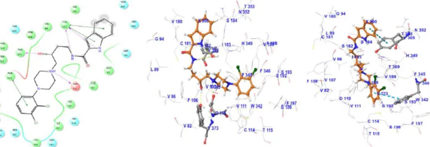
Figure 3: Docking pose showing the interaction between R-22 and 3PBL residues. In the left-hand picture, the blue line shows Pi-Pi stacking, the purple line shows hydrogen bond interaction, and the yellow-green line shows hydrophobic interaction.
A summary of the hit selection workflow is summarized in Figure 3. Hit compounds identified by screening using two models were sorted by fitness score in descending order; Then, they were filtered using the following drug-like criteria based on Lipinski’s rule: AlogP < 5; molecular refractivity of 40 ~ 130; nitrogen-containing; Molecular weight (MW) of 180 ~ 500 and preferably around 400; Polar surface area (PSA) < 90; Hydrogen bonding donor (HBD) < 5; and Hydrogen bonding acceptor (HBA) < 10 [27]. Filtered hits were processed through Ligprep for conversion to 3D, docked to 3PBL to investigate their interaction with the target protein, and filtered using the secondary criteria of a docking score < -7, a ligand efficiency < -0.3 [28], and a charge range from -1 to 1. Qikprop in Maestro is used to apply CNS-like property filtering criteria of QPlogBB > -0.523, BB > 0.3 [29-31], and CNS ≥ 1; the score value of -2 is CNS inactive, and the score value of 2 is CNS active [32].
Results and Discussion
Docking analysis of D3R selective antagonists
Pharmacophore modeling and 3D-QSAR models
The knowledge from docking analysis made us consider models containing five or six features to describe the interactions with two pockets. After building models to explore every plausible hypotheses from training set, the next scoring technique was applied to evaluate the models according to site, vector, volume, selectivity, conformational energy and activity.
Survival score = WsiteSsite + Wvec Svec + Wvol Svol + Wsel Ssel + W m rew – WEΔE + Wact A
The selection of superior hypothesis was committed based on outstanding post-hoc survival score combining two survival scores of active and inactive. Top 10 pharmacophore models showing superior post-hoc scores were used to generate 3D-QSAR models. Internal validation of the chosen PLS models produced reliable coefficient of determination (R squared of each hypothesis > 0.7, Max. 0.88) and their high calibrated values (Q squared of each hypothesis > 0.6, Max. 0.73) in Table2. In addition, Pearson correlation constants also showed more than 0.8 in all of our chosen models. Finally, two models (APRRR215 and AHPRRR104) were selected for virtual screening based on these statistical analyses and the appropriate location of features (Figure 4). In the simple regression analysis of the experimental activities with predicted activities, the best model, APRRR215, presented Act (pred.) = 0.80 Act (exp.) + 0.49 (R predictive squared = 0.80) in the training set and Act (pred.) = 0.62 Act (exp.) + 1.10 (R predictive squared = 0.78) in the test set (Figure 5). Another chosen model, AHPRRR104, indicated that Act (pred.) = 0.82 Act (exp.) + 0.45 (R predictive squared = 0.82) in the training set and Act (pred.) = 0.66 Act (exp.) + 0.98 (R predictive squared = 0.74) in the test set (Figure 6).
Figure 4: Representative 3D-QSAR models: APRRR215 (upper) and AHPRRR104 (lower).
Figure 5: APRRR215 regression lines for the training (blue) and test (red) sets.
Figure 6: AHPRRR104 regression lines for the training (blue) and test (red) sets.
Visualization of the validation was presented in ROC analysis to show how effectively the pharmacophore models distinguished between active and inactive compounds. Sensitivity (in other words, true positive rate, recall, hit rate) and specificity (in other words, true negative rate) are general indices to show the predictive power of a validated model. The ROC curve is a graphical representation of the false positive rate, (1 – specificity) of x-axis and the true positive rate, sensitivity of y-axis for each of the possible cut-offs [36]. The accuracy of this test is measured by the AUC of the ROC curve. According to the general judgement on an AUC value of ROC curve (AUC of poor model = 0.5, AUC of a moderate accurate model = 0.5 to 0.7, and very accurate if the AUC > 0.9) [37], the AUC of APRRR215 was 0.696 showing moderate usefulness in global scaffolds (Figure 7). In addition, the distribution of the experimental and predictive values was visually compared through box-and-whisker plot in Figure 8. Even though the model presented the limit to residual between experimental value and predicted value of active compounds, median values of two distributions could be well-matched and two distributions were identical after normalization.
novel D3R-selective antagonists. Our pharmacophore-based virtual screening played the role of high-throughput filter against compounds that lack the features essential for binding. The validated two best pharmacophore models allowed a researcher to efficiently screen a large commercial compound library from eMolecule (more than 3 millions), and the results were filtered to identify compounds with CNS drug-like properties (Figure9).
The current limitation of this study & future possibility
3D-consistent with proposed binding conformer of R-22 and each pharmacophore feature could describe non-covalent bonding interaction between R-22 and corresponding residues. Secondly, in the view of screening efficiency, the pharmacophore based screening was 100-fold faster than high-throughput docking without any pre-processing in one sub job. Under the condition of Intel Xeon E5-2650v2 (20M Cache, 2.60 GHz, 8.00 GT/s, 8x4 cores), HTVS docking of 100,000 chemicals from eMolecules (> 3 million compounds) consumed total 57959 seconds (16 hours) CPU time in 16 parallel sub jobs. Under the same resource, the pharmacophore based screening of whole eMolecule chemicals could completed within 72 hours in one job. Because parallel (thread) processing of both screenings does not have dependency between sub jobs, the pharmacophore based screening was also superior to docking in the speed of total job. Finally, double filtering through two screening and data fusion of scored values (e.g., the fitness score of pharmacophore model, predicted pKi of QSAR, and docking score) can improve predictive power [43, 44].
Conclusion
In this study, we have developed pharmacophore models of D3R selective antagonists to efficiently identify a series of compounds with novel scaffolds, having both predicted D3R antagonism and CNS drug-like properties. The models were built from docking conformers, and evaluated by combined survival scores of active and inactive of the models. And then two best models (APRRR215 and AHPRRR104) were chosen by predictive power among their 3D-QSAR models generated from Top10 among five/six-featured models. In the view of screening speed and performance, the pharmacophore models were efficient at finding 2D-novel and 3D-similar scaffolds with known D3R antagonists. Our docking model, as a second predictor, could compensate the limited accuracy of fitness score or predicted pKi through the data combination of the scored values. Moreover, CNS-like property was used as an additional criterion with the two models for choosing promising hit compounds. In conclusion, our current work shows that (1) efficient screening of a large compound database (more than 300 millions) is possible, and (2) the interpretation of models is well matched in pharmacophore features and docking poses.
Acknowledgment
This study was supported by the Basic Science Research Program of the National Research Foundation of Korea (NRF) funded by the Ministry of Education, Science, and Technology (No.: 2017R1E1A1A01076642).
Competing interests
The authors declare no competing financial interests.
Author contributions
wrote the manuscript. S. J. C. revised the manuscript. All the authors read and approved the final manuscript.
References:
1. Contreras F, F.C., Bolívar A, Simonovis N, H and A.-H.M. ernández-Hernández R, Velasco M Dopamine, hypertension and obesity. J Hum Hypertens, 2002. 16(1): p. 13-17. 2. Heidbreder, C.A. and A.H. Newman, Current perspectives on selective dopamine D(3) receptor antagonists as pharmacotherapeutics for addictions and related disorders. Annals of the New York Academy of Sciences, 2010. 1187: p. 4-34.
3. Gurevich, E.V. and J.N. Joyce, Distribution of Dopamine D3 Receptor Expressing Neurons in the Human Forebrain: Comparison with D2 Receptor Expressing Neurons. Neuropsychopharmacology, 1999. 20(1): p. 60-80.
4. Maramai, S., et al., Dopamine D3 Receptor Antagonists as Potential Therapeutics for the Treatment of Neurological Diseases. Frontiers in Neuroscience, 2016. 10: p. 451.
5. Joyce, J.N., Dopamine D3 receptor as a therapeutic target for antipsychotic and antiparkinsonian drugs. Pharmacology & Therapeutics, 2001. 90(2): p. 231-259.
6. Pilla, M., et al., Selective inhibition of cocaine-seeking behaviour by a partial dopamine D3 receptor agonist. Nature, 1999. 400(6742): p. 371-375.
7. Koob, G.F. and S.B. Caine, Cocaine addiction therapy[mdash]Are we partially there? Nat Med, 1999. 5(9): p. 993-995.
8. Levant, B., The D3 Dopamine Receptor: Neurobiology and Potential Clinical Relevance. Pharmacological Reviews, 1997. 49(3): p. 231.
9. Newman, A.H., P. Grundt, and M.A. Nader, Dopamine D3 Receptor Partial Agonists and Antagonists as Potential Drug Abuse Therapeutic Agents. Journal of Medicinal Chemistry, 2005. 48(11): p. 3663-3679.
10. Feng, Z., T. Hou, and Y. Li, Selectivity and activation of dopamine D3R from molecular dynamics. Journal of molecular modeling, 2012. 18(12): p. 5051-5063.
11. Griffon, N., et al., Antipsychotics with inverse agonist activity at the dopamine D3 receptor. J Neural Transm (Vienna), 1996. 103(10): p. 1163-75.
12. Chien, E.Y.T., et al., Structure of the human dopamine D3 receptor in complex with a D2/D3 selective antagonist. Science (New York, N.Y.), 2010. 330(6007): p. 1091-1095. 13. Doman, T.N., et al., Molecular Docking and High-Throughput Screening for Novel Inhibitors of Protein Tyrosine Phosphatase-1B. Journal of Medicinal Chemistry, 2002. 45(11): p. 2213-2221.
14. Marriott, D.P., et al., Lead Generation Using Pharmacophore Mapping and Three-Dimensional Database Searching: Application to Muscarinic M3 Receptor Antagonists. Journal of Medicinal Chemistry, 1999. 42(17): p. 3210-3216.
15. Olson, E.C. and R.E. Christoffersen, Computer-assisted drug design. 1979: ACS Publications.
16. Boateng, C.A., et al., High Affinity Dopamine D(3) Receptor (D(3)R)-Selective Antagonists Attenuate Heroin Self-Administration in Wild-Type but not D(3)R Knockout Mice. Journal of Medicinal Chemistry, 2015. 58(15): p. 6195-6213.
derivatives as ERK kinase inhibitors utilizing double tools of 3D-QSAR and side-chain hopping, Bioorg. Med. Chem. Lett. 2011, 21, 4900.
22. Kim, M. H., Ryu, J. S. & Hah, J. M. 3D-QSAR studies of 1,2-diaryl-1H-benzimidazole derivatives as JNK3 inhibitors with protective effects in neuronal cells. Bioorganic & medicinal chemistry letters 23, 1639-1642, doi:10.1016/j.bmcl.2013.01.082 (2013).
23. Gadhe, C. G., Lee, E. & Kim, M. H. Finding new scaffolds of JAK3 inhibitors in public database: 3D-QSAR models & shape-based screening. Archives of pharmacal research 38, 2008-2019, doi:10.1007/s12272-015-0607-6 (2015).
24. Taha, M.O., et al., Pharmacophore and QSAR modeling of estrogen receptor β ligands and subsequent validation and in silico search for new hits. Journal of Molecular Graphics and Modelling, 2010. 28(5): p. 383-400.
25. Yang, S.-Y., Pharmacophore modeling and applications in drug discovery: challenges and recent advances. Drug discovery today, 2010. 15(11): p. 444-450.
26. Seidel, T., et al., Strategies for 3D pharmacophore-based virtual screening. Drug Discovery Today: Technologies, 2011. 7(4): p. e221-e228.
27. Lipinski, C.A., et al., Experimental and computational approaches to estimate solubility and permeability in drug discovery and development settings1PII of original article: S0169-409X(96)00423-1. The article was originally published in Advanced Drug Delivery Reviews 23 (1997) 3–25.1. Advanced Drug Delivery Reviews, 2001. 46(1): p. 3-26.
28. Lane, J.R., et al., Structure-Based Ligand Discovery Targeting Orthosteric and Allosteric Pockets of Dopamine Receptors. Molecular Pharmacology, 2013. 84(6): p. 794. 29. Ježko, P., V. Žufková, and M. Remko, Modelling of absorption, distribution and physicochemical properties of AT1 receptor antagonists / Modelovanie absorpcie, distribúcie a fyzikálnochemických vlastnosti antagonistov AT1 receptorov, in Acta Facultatis Pharmaceuticae Universitatis Comenianae. 2015. p. 20.
30. Luco, J. M. Prediction of the Brain-Blood Distribution of a Large Set of Drugs from Structurally Derived Descriptors Using Partial Least-Squares (PLS) Modeling. J. Chem. Inf. Comput. Sci. 1999, 39, 396-404.
31. Kelder J.; Grootenhuis, P. D.;Bayada, D.M.; Delbressine, L.P., Ploemen, J.P. Polar molecular surface as a dominating determinant for oral absorption and brain penetration of drugs. Pharm. Res. 1999, 16, 1514–1519.
32. Ajay; Bemis, G. W.; Murkco, M. A. Designing Libraries with CNS Activity. J. Med. Chem., 1999, 42, 4942–4951.
33. Shi, L. and J.A. Javitch, The binding site of aminergic G protein–coupled receptors: the transmembrane segments and second extracellular loop. Annual review of pharmacology and toxicology, 2002. 42(1): p. 437-467.
34. Sokoloff, P., et al., Molecular cloning and characterization of a novel dopamine receptor (D3) as a target for neuroleptics. Nature, 1990. 347(6289): p. 146-51.
36. Swets, J.A., Measuring the accuracy of diagnostic systems. Science, 1988. 240(4857): p. 1285.
37. Greiner, M., D. Pfeiffer, and R.D. Smith, Principles and practical application of the receiver-operating characteristic analysis for diagnostic tests. Preventive Veterinary Medicine, 2000. 45(1): p. 23-41.
38. Venkanna, A., Kwon, O. W., Afzal, S., Jang, C., Cho, K. H., Yadav, D. Kim, K., Park, H. G., Chun,K.-H., Kim, S. Y., Kim, M.-h. (2017). Pharmacological use of a novel scaffold, anomeric N, N-diarylamino tetrahydropyran: molecular similarity search, chemocentric target profiling, and experimental evidence. Scientific Reports, 7(1), 12535.
39. Hitchcock, S.A. and L.D. Pennington, Structure−Brain Exposure Relationships. Journal of Medicinal Chemistry, 2006. 49(26): p. 7559-7583.
40. Kim, H., Jang, C., Yadav, D. K. Kim, M.-h., The Comparison of Automated Clustering Algorithms for Resampling Representative Conformer Ensembles with RMSD Matrix, Journal of cheminformatics 2017, 9(1), 21.
41. Yera ER, Cleves AE, Jain AN: Chemical Structural Novelty: On-Targets and Off-Targets, J. Med. Chem. 2011, 54(19):6771-6785.
42. Nettles JH, Jenkins JL, Bender A, Deng Z, Davies JW, Glick M: Bridging chemical and biological space: "target fishing" using 2D and 3D molecular descriptors. J. Med. Chem. 2006, 49(23):6802-6810.
![Table 1. Twenty-five selective D3R-antagonists used to build the pharmacophore model [14]](https://thumb-us.123doks.com/thumbv2/123dok_us/7906316.1312720/4.612.149.471.99.696/table-selective-d-antagonists-used-build-pharmacophore-model.webp)