ABSTRACT
TIRUTHANI, KARTHIK RAJAGOPALAN. Enabling Improved Understanding of Biological Processes through Protein Engineering. (Under the direction of Balaji M. Rao).
Protein-protein interactions are at the heart of most if not all biological processes and
molecular recognition tools allow us to study these interactions. Small molecules and
antibodies are currently used for characterizing and studying these protein-protein
interactions. However small molecule drugs often do not have desired affinity, are not
specific and many either do not have a primary target, or well studied mechanism of action.
On the other hand antibodies are more specific to their target and better characterized, but
many antibodies are polyclonal, have limited shelf life, are often not sequenced and can have
batch to batch variability. While some issues can be alleviated through the use of
recombinant antibodies currently being developed, new solutions to engineer not just specific
inhibitors and biosensors and but affinity reagents in general is desirable. Specifically there is
a need for reagents developed using a customized approach, tailored for the application rather
than standard off the shelf reagents which may not give the desired results. In this work we
demonstrate the use of protein engineering to solve these issues and present two specific
applications to validate the approach.
The availability of intracellular biosensors for live-cell imaging studies is presently
limited. To demonstrate the use of protein engineering we engineer biosensors for epidermal
growth factor receptor (EGFR). While tandem SH2 domain from PLCγ1 (tSH2-WT) has
been used as a marker of phosphorylated EGFR we show it lacks specificity for
from the expected kinetics of EGFR phosphorylation. To address the limitations of the
tSH2-WT biosensor, we constructed a combinatorial library through random mutagenesis of the C-terminal SH2 domain (cSH2) of PLCγ1. The library was screened using yeast surface display
to isolate a mutant protein (mSH2) with enhanced specificity for the Y992 phosphorylation
site in EGFR (pY992). Accordingly, a biosensor based on mSH2 faithfully reports kinetics of
EGFR phosphorylation in live-cell imaging experiments. Further we explore the use of de
novo binding proteins in engineering biosensors by isolating a mutant SPY992 from the
Sso7d scaffold library for the Y992 phosphorylation site in EGFR (pY992) and show that
unlike mSH2 which retains some promiscuous activity it does not have any affinity for
platelet derived growth factor receptor (PDGFR).
Molecular mechanisms regulating human trophoblast differentiation remain poorly
understood due to difficulties in obtaining primary tissues from very early developmental
stages in humans. Therefore the use of human embryonic stem cells (hESCs) as a source for
generating trophoblast tissues is of significant interest. However there is controversy over
whether hESC-derived cells are indeed analogous to true trophoblasts found in vivo. Characterization of the secretome can help identify unique trophoblast markers and the
signaling pathways responsive to the microenvironment of hESCs leading to mechanistic
insight into differentiation and help address these issues. The secretory pathway is an
important factor in cell-cell communication and represents one way for the acquisition of the
malignant phenotype of cancer cells. The secretome can provide insight into some of these
We demonstrate that the binder isolated, VB15 is capable of immunoprecipitating
organelles from cells and tissues. Through electron microscopy of NIH3T3 cells we show
that VB15 binds vesicles and that these vesicles often contain secreted proteins like
fibronectin. So VB15 can be used to obtain secretome of tissues which we plan to eventually
demonstrate through the isolation of secretory vesicles from third trimester human placenta,
vCTB and STB isolated from first trimester human placenta. Our approach serves as a
blueprint for engineering affinity reagents for isolation of any organelle of interest even if the
© Copyright 2015 Karthik Rajagopalan Tiruthani
Enabling Improved Understanding of Biological Processes through Protein Engineering
by
Karthik Rajagopalan Tiruthani
A dissertation submitted to the Graduate Faculty of North Carolina State University
in partial fulfillment of the requirements for the degree of
Doctor of Philosophy
Chemical Engineering
Raleigh, North Carolina
2015
APPROVED BY:
_______________________________ _______________________________
Balaji M. Rao Jason M. Haugh
Committee Chair
_______________________________ _______________________________
ii
BIOGRAPHY
Karthik Tiruthani was born in Hyderabad, India. He received his Bachelors in Technology in
Mechanical Engineering (Mechatronics) in 2006 from Jawaharlal Nehru Technological
University. He worked at Zetatek Industries before beginning graduate studies at North
Carolina State University (NCSU) in 2007. At NCSU he earned his MS in Mechanical
Engineering under the guidance of Dr. Melur Ramasubramanian in 2008 and continued on
for a doctoral degree. In 2011 he joined the Protein Engineering lab of Dr. Balaji Rao to
pursue his interest in biotechnology and specifically biosensor design. During the course of
his study he has received Graduate Certificates in Medical Devices (JGC-MD) and in
iii
ACKNOWLEDGMENTS
I would like to thank my advisor Dr. Balaji Rao for the constant positive reinforcement and
belief in me that helped a lot over the course of my graduate studies. I am grateful to Dr.
Haugh for his guidance and discussion on various projects. I would like to acknowledge
undergraduate researcher Stephen Ryan who over a period of two years worked on a lot of
small projects that helped me progress towards completion. I would also like to thank Dr.
Nimish Gera and Dr. Prasenjit Sarkar, past members of the Rao lab for their help with getting
started with my research, and my colleagues Kevin, Carlos and Adam for an enjoyable lab
environment. Finally I would also like to thank my parents for their support and
iv
TABLE OF CONTENTS
LIST OF TABLES ... vii
LIST OF FIGURES ... viii
ENGINEERING AFFINITY REAGENTS – THE NEED AND THE OPPORTUNITY ... 1
1.1 Introduction ... 2
1.1.1 Protein Engineering ... 3
1.1.2 Desired characteristics of engineered binding proteins ... 6
1.1.3 Engineering reagents for isolation of secretory vesicles ... 7
1.1.4 Engineering phosphospecific biosensors ... 8
1.2 Thesis Overview ... 9
1.3 References ... 11
DESIGN AND EVALUATION OF PROTEIN BIOSENSORS FOR LIVE CELL IMAGING OF EPIDERMAL GROWTH FACTOR RECEPTOR PHOSPHORYLATION ... 21
2.1 Introduction ... 22
2.2 Results ... 24
2.2.1 Modeling EGF Receptor dynamics ... 24
2.2.2 The tSH2-WT biosensor lacks specificity and does not faithfully track kinetics of EGFR phosphorylation ... 27
2.2.3 The engineered mSH2 protein has increased specificity for pY992 of EGFR ... 29
2.2.4 The mSH2 biosensor closely tracks the expected kinetics of EGFR phosphorylation and displays increased specificity for the pY992 site of EGFR in live-cell imaging experiments ... 31
2.2.5 The engineered SPY992 protein has specificity for pY992 of EGFR with no affinity for pY1021 site ofPDGFR ... 33
2.3 Discussion ... 34
2.4 Materials and Methods ... 37
2.4.1 Library generation ... 37
2.4.2 Isolation of SH2 domain mutants binding the pY992 site in EGFR ... 38
2.4.3 Recombinant expression and purification of SH2 domain mutants ... 39
v
2.4.5 Cell Culture ... 40
2.4.6 TIRF imaging and image analysis ... 41
2.4.7 Statistical analysis ... 42
2.5 References ... 43
ENGINEERING AFFINITY REAGENTS FOR EFFICIENT ISOLATION OF SECRETORY VESICLES ... 56
3.1 Introduction ... 57
3.2 Materials and Methods ... 59
3.2.1 Construction of Sso6904 mutant library for yeast surface display ... 59
3.2.2 Cell Culture ... 61
3.2.3 Generation of target for negative and positive selection ... 61
3.2.4 Isolation of vesicle binding protein VB15 by magnetic selection and FACS ... 62
3.2.5 Recombinant expression and purification of VB15 ... 63
3.2.6 Far western blotting ... 64
3.2.7 Immunofluorescence ... 65
3.2.8 Immunohistochemistry and histology analysis ... 65
3.2.9 Transmission Electron Microscopy ... 65
3.2.10 Human Placental Samples ... 67
3.2.11 Mouse tissue samples ... 68
3.3 Results ... 68
3.3.1 The engineered protein VB15 has increased specificity to fraction containing secretory vesicles ... 68
3.3.2 The engineered protein VB15 binds specifically to a single protein that is conserved across species ... 69
3.3.3 VB15 is capable of isolating organelles by immunoprecipitation ... 69
3.3.4 VB15 likely binds secretory vesicles... 70
3.3.5 VB15 is capable of isolating organelles from tissues by immunoprecipitation ... 70
3.4 Discussion ... 71
3.5 References ... 73
TROPHOBLAST DIFFERENTIATION OF HUMAN EMBRYONIC STEM CELLS ... 90
4.1 Introduction ... 91
vi
4.2.1 Monolayer cultures using BMP4 and/or Activin/Nodal/TGFβ inhibition ... 93
4.2.2 Differentiation using embryoid bodies ... 95
4.3 Markers used for characterizing hESC-derived cells ... 96
4.4 Role of differentiation media ... 97
4.5 Role of culture heterogeneity ... 99
4.6 Cdx2 as a trophoblast marker ... 100
4.7 Concluding remarks ... 101
4.8 References ... 103
vii
LIST OF TABLES
Table 2.1 Protein Sequences of engineered biosensors ...53
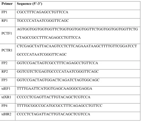
Table 2.2 List of primers used ...54
Table 2.3 List of peptides used for library screening ...55
Table 3.1 DNA Sequence of Sso6904 with mutated residues ...88
Table 3.2 List of primers used ...88
Table 3.3 Isopycnic density of subcellular organelles ...89
viii
LIST OF FIGURES
Figure 1.1 General overview of protein engineering by yeast surface display ...20
Figure 2.1 Characterization of the tSH2-WT biosensor ...47
Figure 2.2 Far western blotting analysis of cSH2 ...48
Figure 2.3 Analysis of intermediate cell populations during cSH2 library screening ...49
Figure 2.4 Characterization of specificity of the mSH2 biosensor ...50
Figure 2.5 Comparison of kinetics of EGFR phosphorylation detected using mSH2 and tSH2 –WT biosensors in c’1000 and c’1000F cell lines ...51
Figure 2.6 Comparison of kinetics of EGFR phosphorylation detected using mSH2 and tSH2 –WT biosensors in NR6 and NR6PAR cell lines ...52
Figure 2.7 Effect of changing binding affinity of biosensor-target interaction or biosensor concentration on readout ...52
Figure 2.8 Characterization of specificity of the SPY992 biosensor ...53
Figure 3.1 Mutated residues on Sso6904 used for library generation ...77
Figure 3.2 Schematic of Subcellular Fractionation using Sucrose Density Gradient Centrifugation ...78
ix
Figure 3.4 Characterization of VB15 binding to subcellular fractions ...80
Figure 3.5 Comparison of sequence of VB15 and the Sso6904 scaffold ...81
Figure 3.6 Far western blotting analysis of VB15...82
Figure 3.7 Immunoprecipitation of organelles from NIH3T3 cells using VB15 ...83
Figure 3.8 Immunoprecipitation of organelles from S2 cells using VB15 ...84
Figure 3.9 TEM image of a crosssection of NIH3T3 cells labeled using VB15 ...85
Figure 3.10 Immunoprecipitation of organelles from mouse tissues ...86
1
CHAPTER 1
2 1.1 Introduction
Protein-protein interactions are at the heart of most if not all biological processes in a
cell. The ability to control and study these interactions in a very specific fashion is important
for a mechanistic understanding of cellular processes. Some approaches currently used to
study these interactions include the use of genetic techniques (1–9) to increase or decrease a level of one or both the proteins. An alternate approach is the use of molecular recognition
i.e. specific non-covalent interactions, either through small molecules or antibodies, to either
affect protein function or in assays like microscopy, immunoprecipitation, western blotting
and flow cytometry to gain insight into these interactions.
While approaches like gene insertion, deletion or mutation offer specificity, the
ability to temporally affect function is lost. Deletion of genes coding for protein can have
undesirable effects and result in modification of pathways other than the one of interest.
Similarly, whereas the use of RNA interference using inducible expression (10–13) provides temporal control, it has issues with stability, delivery and off target effects (14–16). Small molecules represent an easy way of targeting intracellular protein-protein interactions in live
cell assays with the desired temporal control, potential for spatial control and ease of
delivery. However many small molecule drugs either do not have a primary target, or well-
3
majority of small molecules acting on conserved sites, such as the ATP binding site in
kinases which makes obtaining specificity a big challenge.
Antibodies are widely regarded as the gold standard for characterizing and studying
protein-protein interactions. However their use in live cell assays as a biosensor is limited by
challenges associated with delivery. Further many antibodies are polyclonal, have limited
shelf life, are often not sequenced and can have batch to batch variability. Additionally
tagging them with tags of interest or unnatural amino acids conferring interesting properties
is also a significant challenge. While progress is being made through development of
recombinant antibodies (22) to overcome these issues other alternatives are desirable.
Overall there is a need for new solutions to engineer not just specific inhibitors and
biosensors and but affinity reagents in general. While projects like the Human Protein Atlas
are developing affinity reagents for all human proteins (23) it is highly likely that different reagents maybe better suited for different applications and the ones generated may not be
ideally suited to study protein modifications. Scientists interested in targeting proteins for
e.g., only when the receptor is activated or a protein has a specific conformation or post
translational modification (24) require reagents developed using a customized approach and may not get the desired results from standard off the shelf reagents.
1.1.1 Protein Engineering
Protein engineering can help overcome a lot of challenges associated with the
4
engineered from mutagenesis of single chain variable fragment of antibodies (scFv) (25–28), small hyperthermophilic proteins (29, 30) or domains from other proteins like 10th type III domain of human fibronectin (Fn3), SH2 domain, PDZ domain, ankyrin repeats, armadillo
repeats, etc (31–37) represent another alternative to antibodies as reagents. The significant advantage of these proteins besides the cost and ease of production is their potential for use
in intracellular live cell assays as a biosensor.
Specificity to the target protein can be easily engineered using protein engineering by
targeting regions on a protein outside conserved regions, which are usually regions of low
homology across the protein family. The targeting of specific regions on a protein through
protein engineering facilitates the study of function of proteins in specific signaling
pathways. Identifying binding partners to these domains thus allows a more fine-grained
control of protein function. For example the protein Dishevelled interacts with over 50
different proteins (38) through three conserved domains (DIX domain, PDZ domain, DEP domain) and two conserved regions (basic region, proline rich region). Each of these
interactions is involved in regulating many different functions such as β-catenin stability,
secondary axis formation, axon differentiation, cell migration, etc and even in cell fate
decisions. Targeting specific regions either through inhibitors or specific biosensors
engineered using binding proteins could allow the study of the protein function without
perturbing other pathways potentially simplifying the analysis.
The use of hyperthermophilic proteins, specifically Sso7d (a DNA binding protein
5
been demonstrated in previous work (29). The basis for identification of desired proteins from combinatorial libraries is the linking of phenotype to genotype. This is achieved in cell
free systems like mRNA (39–41) or ribosome (42–44) display through linking of the protein to mRNA by covalent bond or to the ribosome. In cell based systems like phage(45–47), bacteria(48–50), yeast(51–53) and mammalian(54, 55) cells this link is established by fusing the protein of interest to a cell surface protein. While each display technique is advantageous
for a specific application, a combination of these techniques(56, 57) has also sometimes been used to take advantage of each. For example a combination of mRNA and yeast display used
to engineer binding proteins (30) enables a higher diversity library to be initially selected using mRNA display. The enriched binding pool from mRNA display is then converted to a
yeast display library to take advantage of affinity discrimination by FACS possible using
yeast display. A comparison of these techniques is also available to determine the most
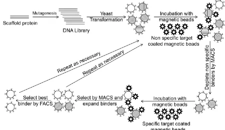
suitable method (58). An overview of yeast display which will be used in this study is presented in Figure 1.1
Unlike small molecules proteins are not normally cell-permeable. However
conjugation/fusion with cell penetrating/permeable peptides (CPP) or protein transduction
domains (PTD) like antennapedia, TAT, transportan or polyarginine allows delivery into the
cell (59–61) through active transport mechanisms. While the exact mechanism of transduction is not known it is thought to be through Clathrin-mediated endocytosis. Since
the efficiency of endosomal escape is also unknown it leads to challenges in identifying the
6
transfection of plasmid DNA coding for protein of interest through the use of cationic lipids
like Lipofectamine, electroporation, calcium phosphate precipitation or viral delivery (62– 64). Based on an analysis of literature it appears while direct protein delivery may be ideal it is not currently reliable or repeatable enough and plasmid delivery of binding protein (with
potential use of inducible expression) represents a realistic approach to achieve the desired
results.
1.1.2 Desired characteristics of engineered binding proteins
Binding proteins can be custom engineered for a variety of applications depending on
the affinity and specificity desired. For example an ideal biosensor should have an
intermediate affinity to its target. Whereas low affinity results in very little binding and low
signal, very high affinity may result in saturation of readout or perturbation of the target
protein depending on the expression levels (65). On the other hand for a reagent used for immunoprecipitation the affinity can be variable depending on the target. For example while
high affinity is desirable for immunoprecipitation of proteins it may not be essential for
immunoprecipitation of organelles where specificity is more important since there is likely
avidity between engineered binder on a bead and the organelle target which can amplify the
affinity of both desired specific and undesired non-specific interactions. While affinity and
specificity are key criterion depending on the application other characteristics may also
become essential. For example high contrast tumor imaging requires small size reagents for
rapid clearance in addition to high affinity whereas therapeutics require high serum half-life
7
been many engineered proteins developed as inhibitors (31, 33, 66–68), there is a dearth of specific intracellular biosensors for live cell imaging. Similarly western blotting and
immunoprecipitation reagents (32, 69–71) have been engineered but a systematic approach for engineering binding proteins for isolation of organelles is currently lacking.
1.1.3 Engineering reagents for isolation of secretory vesicles
Proteins secreted from the cell, like cytokines, growth factors and other signaling
molecules play a crucial role in many cellular processes. Of particular interest here is hESC
differentiation which is known to be significantly affected by the microenvironment.
Characterizing the secretome can help identify the signaling pathways responsive to the
microenvironment of hESCs leading to mechanistic insight into differentiation. Trophoblast
formation is a major process underlying implantation of the blastocyst in the endometrium
and development of the placenta. The cells of the trophectoderm (TE) are precursors of all
trophoblast cell types in the placenta. Identifying the secretome is especially important in
hESC differentiation to trophoblast where knowledge of molecular mechanisms is lacking.
We propose the engineering of binding protein that is capable of immunoprecipitating
secretory vesicles. This will allow us to probe the secretome of primary placental tissue, not
currently possible, allowing us to better understand trophoblast differentiation and improve
on the hESC-trophoblast model. Further the binder could also help in identifying cancer
biomarkers. Cancer cells acquire their malignant phenotype through the manipulation of
signaling processes involved in growth, proliferation, apoptosis, angiogenesis and one of the
8
microenvironment of cancer cells can provide insight into these mechanisms and help in
identification of potential biomarkers. Currently the cancer secretome is obtained through the
study of cancer cell lines, tumor proximal body fluids (73). While it is understood that the biology of these cell lines is often unrepresentative of the cells they are derived from (74) they currently represent the only option. Most importantly since these cell lines have already
acquired the malignant phenotype, they are unlikely to be useful in studying cancer
progression. The binder developed in this study can be used to isolate secretory vesicles from
tissue biopsies. This bypasses the need to culture the cells potentially altering their biology
and provides more physiologically relevant information and in identification of biomarkers.
1.1.4 Engineering phosphospecific biosensors
While the eventual goal is to apply protein engineering to study human embryonic stem
cell (hESC) differentiation, the tools for the development of these reagents needs to be
validated and characterized. One challenge towards this end is the ability to engineer binding
proteins to specific domains or epitopes on target proteins, especially when the target protein
is in a specific desired state. We propose the engineering and evaluation of a biosensor to
study the dynamics of phosphorylation and internalization of epidermal growth factor
receptor (EGFR). This biosensor allows us to validate our approach to engineering domain
and state specific binding proteins that eventually based on their affinity (65) could either be used as biosensors or inhibitors. EGFR represents a good target because its expression level
9
differentiation and failure to invade leading to preeclampsia (76). Thus the tools developed in this study can be developed further to study the pathway both in cultured trophoblast and
hESC derived trophoblast to improve the hESC – trophoblast model.
1.2 Thesis Overview
The engineering of binding proteins to secretory vesicles and phosphorylation sites on
EGFR is described. We explore the application of these binders derived using artificial
scaffolds to study biological processes in a cell in three different ways. First as a biosensor to
monitor EGFR internalization, second as an immunoprecipitation reagent to isolate secretory
vesicles to characterize the secretome of cells and tissues and third as a tool to study hESC
differentiation to trophoblast and potentially trophoblast biology.
Chapter 2 describes results pertaining to engineering and evaluation of a phospo and
site specific biosensor to EGFR. We constructed a combinatorial library through random
mutagenesis of the C-terminal SH2 domain (cSH2) of PLCγ1. The library was screened
using yeast surface display to isolate a mutant protein (mSH2) with enhanced specificity for
the Y992 phosphorylation site in EGFR (pY992). Accordingly, a biosensor based on mSH2
faithfully reports kinetics of EGFR phosphorylation in live-cell imaging experiments.
However mSH2 demonstrated non-specific binding to PDGFR (pY1021) since library was
based on a promiscuous SH2 scaffold. To overcome this a binding protein SPY992 was
10
considerations with a protein engineering strategy, can be generalized to design and evaluate
suitable biosensors for other intracellular targets.
Chapter 3 describes results pertaining to engineering and evaluation of a protein
capable of isolating secretory vesicles. We constructed a combinatorial library through the
introduction of degenerate NNK codons into a calcium binding protein from Sulfolobus
solfataricus, Sso6904. This library was screened using yeast surface display to isolate a
protein (VB15) that we then thoroughly characterized. We demonstrate the application of this
binder in isolation of secretory vesicles from NIH3T3, HEK293T and drosophila S2 cells.
Further we also demonstrate the application of this binder in isolation of secretory vesicles
from mouse pancreas, liver and placenta.
Chapter 4 gives an overview of previously described efforts to obtain trophoblasts
from hESCs. Generating trophoblast tissues from hESCs is of significant interest because
obtaining primary tissues from very early developmental stages in humans is challenging
which results in the molecular mechanisms being poorly understood. However whether
11 1.3 References
1. J. C. Miller, M. C. Holmes, J. Wang, D. Y. Guschin, Y.-L. Lee, I. Rupniewski, C. M. Beausejour, A. J. Waite, N. S. Wang, K. a Kim, P. D. Gregory, C. O. Pabo, E. J. Rebar, An improved zinc-finger nuclease architecture for highly specific genome editing., Nat.
Biotechnol.25, 778–785 (2007).
2. J. a Zuris, D. B. Thompson, Y. Shu, J. P. Guilinger, J. L. Bessen, J. H. Hu, M. L. Maeder, J. K. Joung, Z.-Y. Chen, D. R. Liu, Cationic lipid-mediated delivery of proteins enables efficient protein-based genome editing in vitro and in vivo., Nat. Biotechnol. , 1–10 (2014).
3. D. Seruggia, L. Montoliu, The new CRISPR-Cas system: RNA-guided genome
engineering to efficiently produce any desired genetic alteration in animals., Transgenic Res. (2014), doi:10.1007/s11248-014-9823-y.
4. H. Wang, H. Yang, C. S. Shivalila, M. M. Dawlaty, A. W. Cheng, F. Zhang, R. Jaenisch, One-Step Generation of Mice Carrying Mutations in Multiple Genes by CRISPR/Cas-Mediated Genome Engineering., Cell153, 910–8 (2013).
5. C. R. Hale, S. Majumdar, J. Elmore, N. Pfister, M. Compton, S. Olson, A. M. Resch, C. V. C. Glover, B. R. Graveley, R. M. Terns, M. P. Terns, Essential features and rational design of CRISPR RNAs that function with the Cas RAMP module complex to cleave RNAs., Mol. Cell45, 292–302 (2012).
6. D. H. Kim, J. J. Rossi, Strategies for silencing human disease using RNA interference., Nat. Rev. Genet.8, 173–84 (2007).
12
8. D. P. Bartel, MicroRNAs: Genomics, Biogenesis, Mechanism, and Function, Cell116, 281–297 (2004).
9. F. Stegmeier, G. Hu, R. J. Rickles, G. J. Hannon, S. J. Elledge, A lentiviral microRNA-based system for single-copy polymerase II-regulated RNA interference in mammalian cells., Proc. Natl. Acad. Sci. U. S. A.102, 13212–13217 (2005).
10. S. Freundlieb, C. Schirra-Müller, H. Bujard, A tetracycline controlled
activation/repression system with increased potential for gene transfer into mammalian cells., J. Gene Med.1, 4–12 (1999).
11. L. M. Fedorov, O. Y. Tyrsin, V. Krenn, E. V Chernigovskaya, U. R. Rapp, Tet-system for the regulation of gene expression during embryonic development, Transgenic Res10, 247–258 (2001).
12. A. R. Buskirk, D. R. Liu, Creating small-molecule-dependent switches to modulate biological functions, Chem. Biol. 12, 151–161 (2005).
13. M. K. Pastuszka, J. A. Mackay, Biomolecular engineering of intracellular switches in eukaryotes, J. Drug Deliv. Sci. Technol.20, 163–169 (2010).
14. M. Amarzguioui, J. J. Rossi, D. Kim, Approaches for chemically synthesized siRNA and vector-mediated RNAi., FEBS Lett.579, 5974–81 (2005).
15. D. Samarsky, C. Faherty, in RNA Interference: Principles and Applications, J. Rossi, Ed. (The Biomedical & Life Sciences Collection, 2007).
13
17. E. Gregori-Puigjané, V. Setola, J. Hert, B. a Crews, J. J. Irwin, E. Lounkine, L. Marnett, B. L. Roth, B. K. Shoichet, Identifying mechanism-of-action targets for drugs and probes., Proc. Natl. Acad. Sci. U. S. A.109, 11178–83 (2012).
18. T. Anastassiadis, S. W. Deacon, K. Devarajan, H. Ma, J. R. Peterson, Comprehensive assay of kinase catalytic activity reveals features of kinase inhibitor selectivity., Nat. Biotechnol.29, 1039–45 (2011).
19. M. I. Davis, J. P. Hunt, S. Herrgard, P. Ciceri, L. M. Wodicka, G. Pallares, M. Hocker, D. K. Treiber, P. P. Zarrinkar, Comprehensive analysis of kinase inhibitor selectivity., Nat. Biotechnol.29, 1046–51 (2011).
20. S. Davies, H. Reddy, M. Caivano, P. Cohen, Specificity and mechanism of action of some commonly used protein kinase inhibitors., Biochem. J.351, 95–105 (2000).
21. A. C. Dar, K. M. Shokat, The evolution of protein kinase inhibitors from antagonists to agonists of cellular signaling., Annu. Rev. Biochem.80, 769–95 (2011).
22. V. Marx, Finding the right antibody for the job, Nat. Methods10, 703–707 (2013). 23. V. Marx, Calling the next generation of affinity reagents, Nat. Methods10, 829–833 (2013).
24. L. Kummer, P. Parizek, P. Rube, B. Millgramm, A. Prinz, P. R. E. Mittl, M. Kaufholz, B. Zimmermann, F. W. Herberg, A. Plückthun, Structural and functional analysis of
phosphorylation-specific binders of the kinase ERK from designed ankyrin repeat protein libraries.Proc. Natl. Acad. Sci. U. S. A. (2012), doi:10.1073/pnas.1205399109.
14
26. G. Schaefer, L. Haber, L. M. Crocker, S. Shia, L. Shao, D. Dowbenko, K. Totpal, A. Wong, C. V Lee, S. Stawicki, R. Clark, C. Fields, G. D. Lewis Phillips, R. a Prell, D. M. Danilenko, Y. Franke, J.-P. Stephan, J. Hwang, Y. Wu, J. Bostrom, M. X. Sliwkowski, G. Fuh, C. Eigenbrot, A two-in-one antibody against HER3 and EGFR has superior inhibitory activity compared with monospecific antibodies., Cancer Cell20, 472–86 (2011).
27. G. Chao, W. L. Lau, B. J. Hackel, S. L. Sazinsky, S. M. Lippow, K. D. Wittrup, Isolating and engineering human antibodies using yeast surface display., Nat. Protoc.1, 755–68 (2006).
28. B. J. Hackel, J. R. Neil, F. M. White, K. D. Wittrup, Epidermal growth factor receptor downregulation by small heterodimeric binding proteins., Protein Eng. Des. Sel.25, 47–57 (2012).
29. N. Gera, M. Hussain, R. C. Wright, B. M. Rao, Highly stable binding proteins derived from the hyperthermophilic Sso7d scaffold., J. Mol. Biol.409, 601–16 (2011).
30. M. Hussain, N. Gera, A. B. Hill, B. M. Rao, Scaffold diversification enhances effectiveness of a superlibrary of hyperthermophilic proteins, ACS Synth. Biol.2, 6–13 (2013).
31. A. Stahl, M. T. Stumpp, A. Schlegel, S. Ekawardhani, C. Lehrling, G. Martin, M. Gulotti-Georgieva, D. Villemagne, P. Forrer, H. T. Agostini, H. K. Binz, Highly potent VEGF-A-antagonistic DARPins as anti-angiogenic agents for topical and intravitreal applications., Angiogenesis (2012), doi:10.1007/s10456-012-9302-0.
15
33. T. Kaneko, H. Huang, X. Cao, X. Li, C. Li, C. Voss, S. S. Sidhu, S. S. C. Li, Superbinder SH2 Domains Act as Antagonists of Cell Signaling, Sci. Signal. 5, ra68–ra68 (2012).
34. M. Ferrer, J. Maiolo, P. Kratz, J. L. Jackowski, D. J. Murphy, S. Delagrave, J. Inglese, Directed evolution of PDZ variants to generate high-affinity detection reagents., Protein Eng. Des. Sel.18, 165–73 (2005).
35. F. Parmeggiani, R. Pellarin, A. P. Larsen, G. Varadamsetty, M. T. Stumpp, O. Zerbe, A. Caflisch, A. Plückthun, Designed armadillo repeat proteins as general peptide-binding
scaffolds: consensus design and computational optimization of the hydrophobic core., J. Mol. Biol.376, 1282–304 (2008).
36. G. Varadamsetty, D. Tremmel, S. Hansen, F. Parmeggiani, A. Plückthun, Designed Armadillo Repeat Proteins: Library Generation, Characterization and Selection of Peptide Binders with High Specificity., J. Mol. Biol. , 1–20 (2012).
37. B. J. Hackel, A. Kapila, K. D. Wittrup, Picomolar affinity fibronectin domains
engineered utilizing loop length diversity, recursive mutagenesis, and loop shuffling., J. Mol. Biol.381, 1238–52 (2008).
38. C. Gao, Y.-G. Chen, Dishevelled: The hub of Wnt signaling., Cell. Signal.22, 717–27 (2010).
39. a D. Keefe, Protein selection using mRNA display., Curr. Protoc. Mol. Biol.Chapter 24, Unit 24.5 (2001).
40. T. T. Takahashi, R. J. Austin, R. W. Roberts, mRNA display: ligand discovery, interaction analysis and beyond., Trends Biochem. Sci.28, 159–65 (2003).
16
42. D. Lipovsek, A. Plückthun, In-vitro protein evolution by ribosome display and mRNA display., J. Immunol. Methods290, 51–67 (2004).
43. B. Ning, M. Liu, Y. Sun, Z. Sun, Y. Zhang, Construction of ribosome display library based on lipocalin scaffold and screening anticalins with specificity for estradiol, Analyst (2012) (available at http://pubs.rsc.org/en/content/articlehtml/2012/an/c2an16119b).
44. L. Lewis, C. Lloyd, Optimisation of antibody affinity by ribosome display using error-prone or site-directed mutagenesis., Methods Mol. Biol. (Clifton, NJ)805, 139–161 (2012). 45. H. R. Hoogenboom, Overview of antibody phage-display technology and its
applications., Methods Mol. Biol.178, 1–37 (2002).
46. F. Tian, M.-L. Tsao, P. G. Schultz, A phage display system with unnatural amino acids., J. Am. Chem. Soc.126, 15962–3 (2004).
47. W. J. J. Finlay, L. Bloom, O. Cunningham, in Methods in molecular biology, Methods in Molecular Biology. D. Walls, S. T. Loughran, Eds. (Humana Press, Totowa, NJ, 2011), vol. 681, pp. 87–101.
48. S. Ståhl, M. Uhlén, Bacterial surface display: trends and progress., Trends Biotechnol.15, 185–92 (1997).
49. P. S. Daugherty, G. Chen, M. J. Olsen, B. L. Iverson, G. Georgiou, Antibody affinity maturation using bacterial surface display., Protein Eng.11, 825–32 (1998).
50. J. Rockberg, J. Löfblom, B. Hjelm, M. Uhlén, S. Ståhl, Epitope mapping of antibodies using bacterial surface display, Nat. Methods5, 1039 – 1045 (2008).
17
52. E. T. Boder, K. D. Wittrup, Yeast surface display for directed evolution of protein expression, affinity, and stability., Methods Enzymol.328, 430–444 (2000).
53. L. Pepper, Y. Cho, A decade of yeast surface display technology: Where are we now?, Comb. Chem. High Throughput Screen.11, 127–134 (2008).
54. R. R. Beerli, M. Bauer, R. B. Buser, M. Gwerder, S. Muntwiler, P. Maurer, P. Saudan, M. F. Bachmann, Isolation of human monoclonal antibodies by mammalian cell display., Proc. Natl. Acad. Sci. U. S. A.105, 14336–41 (2008).
55. C. Zhou, F. W. Jacobsen, L. Cai, Q. Chen, W. D. Shen, Development of a novel mammalian cell surface antibody display platform., MAbs2, 508–18 (2010).
56. X. Hu, S. Kang, C. Lefort, M. Kim, M. M. Jin, Combinatorial libraries against libraries for selecting neoepitope activation-specific antibodies., Proc. Natl. Acad. Sci. U. S. A.107, 6252–7 (2010).
57. D. R. Bowley, T. M. Jones, D. R. Burton, R. a Lerner, Libraries against libraries for combinatorial selection of replicating antigen-antibody pairs., Proc. Natl. Acad. Sci. U. S. A. 106, 1380–5 (2009).
58. N. Gera, M. Hussain, B. M. Rao, Protein selection using yeast surface display, Methods 60, 15–26 (2013).
59. S. W. Jones, R. Christison, K. Bundell, C. J. Voyce, S. M. V Brockbank, P. Newham, M. a Lindsay, Characterisation of cell-penetrating peptide-mediated peptide delivery., Br. J. Pharmacol.145, 1093–102 (2005).
18
61. M. Konno, S. Masui, T. S. Hamazaki, H. Okochi, Intracellular reactivation of
transcription factors fused with protein transduction domain., J. Biotechnol. 154, 298–303 (2011).
62. J.-S. Kim, H. S. Chu, K. I. Park, J.-I. Won, J.-H. Jang, Elastin-like polypeptide matrices for enhancing adeno-associated virus-mediated gene delivery to human neural stem cells., Gene Ther.19, 329–37 (2012).
63. T. Helledie, V. Nurcombe, S. M. Cool, A simple and reliable electroporation method for human bone marrow mesenchymal stem cells., Stem Cells Dev.17, 837–48 (2008).
64. C. Magin-Lachmann, G. Kotzamanis, L. D’Aiuto, H. Cooke, C. Huxley, E. Wagner, In vitro and in vivo delivery of intact BAC DNA -- comparison of different methods., J. Gene Med.6, 195–209 (2004).
65. J. M. Haugh, Live-cell fluorescence microscopy with molecular biosensors: What are we really measuring?, Biophys. J.102, 2003–2011 (2012).
66. M. Kawe, P. Forrer, P. Amstutz, A. Plückthun, Isolation of intracellular proteinase inhibitors derived from designed ankyrin repeat proteins by genetic screening, J. Biol. Chem. 281, 40252–40263 (2006).
67. P. Amstutz, H. Koch, H. K. Binz, S. a. Deuber, A. Plückthun, Rapid selection of specific MAP kinase-binders from designed ankyrin repeat protein libraries, Protein Eng. Des. Sel. 19, 219–229 (2006).
19
69. L. Xu, P. Aha, K. Gu, R. G. Kuimelis, M. Kurz, T. Lam, A. C. Lim, H. Liu, P. a. Lohse, L. Sun, S. Weng, R. W. Wagner, D. Lipovsek, Directed evolution of high-affinity antibody mimics using mRNA display, Chem. Biol.9, 933–942 (2002).
70. E. Karatan, M. Merguerian, Z. Han, M. D. Scholle, S. Koide, B. K. Kay, Molecular recognition properties of FN3 monobodies that bind the Src SH3 domain, Chem. Biol.11, 835–844 (2004).
71. N. Gera, A. B. Hill, D. P. White, R. G. Carbonell, B. M. Rao, S. Karnik, Ed. Design of pH Sensitive Binding Proteins from the Hyperthermophilic Sso7d Scaffold, PLoS One7, e48928 (2012).
72. D. Hanahan, R. a Weinberg, Hallmarks of cancer: the next generation., Cell144, 646–74 (2011).
73. T. B. M. Schaaij-Visser, M. De Wit, S. W. Lam, C. R. Jiménez, The cancer secretome, current status and opportunities in the lung, breast and colorectal cancer context, Biochim. Biophys. Acta - Proteins Proteomics1834, 2242–2258 (2013).
74. J. R. Masters, Human cancer cell lines: fact and fantasy., Nat. Rev. Mol. Cell Biol.1, 233–236 (2000).
75. D. I. Sokolov, K. N. Furaeva, O. I. Stepanova, O. M. Ovchinnikova, L. P. Viazmina, G. R. Kozonov, T. U. Kuzminykh, S. a. Selkov, Changes in Functional Activity of JEG-3 Trophoblast Cell Line in the Presence of Factors Secreted by Placenta, Arch. Med. Res.46, 245–256 (2015).
76. D. R. Armant, R. Fritz, B. a. Kilburn, Y. M. Kim, J. K. Nien, N. J. Maihle, R. Romero, R. E. Leach, Reduced expression of the epidermal growth factor signaling system in
20
21
CHAPTER 2
DESIGN AND EVALUATION OF PROTEIN BIOSENSORS FOR
LIVE CELL IMAGING OF EPIDERMAL GROWTH FACTOR
RECEPTOR PHOSPHORYLATION
22 2.1 Introduction
Receptor tyrosine kinases (RTKs) are cell surface receptors that mediate several key
cellular processes including proliferation, differentiation and cell migration (1, 2). Aberrant activation of RTKs and their downstream signaling pathways is a hallmark of most human
cancers. Typically, ligand binding to the extracellular domain of the receptor results in
activation of tyrosine kinase function in the cytoplasmic domain, which in turn results in
phosphorylation of specific residues of the receptor and receptor-associated proteins. Thus,
ligand binding triggers RTK activation and initiation of downstream signaling pathways. The
ability to obtain spatially resolved measurements of receptor activation using live-cell
imaging can uniquely enable a mechanistic understanding of the relationship between
dynamic changes in signaling and resultant cell behavior. Yet, implementation of live cell
imaging using intracellular biosensors remains a significant challenge.
Intracellular biosensors contain a molecular recognition element that should
specifically recognize the target(s) of interest. However, there are only a small number of
highly specific protein biosensors currently available. A second and arguably greater
limitation is that biosensors may significantly interfere with or otherwise modulate the
signaling process they are meant to detect. In this latter context, we have previously outlined
a theoretical framework for understanding the relationship between fidelity of biosensor
readout and binding affinity of the biosensor for its target (3). Notably, an ideal biosensor should have an intermediate affinity to its target. Whereas low affinity results in very little
23
at high concentration, or perturb the target if the biosensor is present in excess. Conversely,
low affinities may result in decreased signal corresponding to the target. Thus, both
specificity and binding affinity of molecular recognition determines the fidelity of the
biosensor readout to dynamic changes in the concentration of the target species being
detected. Here we apply protein engineering tools in conjunction with the aforementioned
theoretical principles to design a protein biosensor targeting a specific phosphotyrosine (pY)
site in the EGFR.
Ligand binding to the extracellular domain of EGFR results in receptor dimerization
and subsequent phosphorylation of multiple tyrosine residues in the cytoplasmic domain of
EGFR, including Y992. We sought to design a protein biosensor to measure dynamic
changes in the concentration of EGFR with phosphorylated Y992 (pY992) in individual
cells. Fluorescent protein fusions of the pY-binding SH2 domains have been previously used
to study EGFR signaling (4, 5). Here, we evaluated the tandem SH2 domain from wild-type PLCγ1 (denoted tSH2-WT hereafter) that has been used to study the dynamics of EGFR
phosphorylation (4). Consistent with previous reports of promiscuous binding of SH2 domains (6–8), we show that tSH2-WT lacks specificity for EGFR pY992. Furthermore, the juxtaposition of two SH2 domains in tSH2-WT results in intramolecular avidity; the two SH2
domains can simultaneously bind pY-containing epitopes. This results in high effective
affinity of interaction between tSH2-WT and EGFR, which in turn distorts the readout
24
To overcome these limitations of tSH2-WT, we isolated a mutant SH2 domain by
screening a combinatorial library generated by random mutagenesis of the C-terminal SH2
domain in tSH2-WT. The isolated mutant (denoted mSH2) exhibits improved specificity for
the pY992 site in EGFR relative to tSH2-WT. Most importantly, the readout from mSH2 in
live-cell total internal reflection fluorescence (TIRF) microscopy experiments is consistent
with the expected dynamics of cell-surface EGFR phosphorylation and endocytosis.
However while mSH2 represented an improvement over tSH2-WT, it still
demonstrated non-specific binding to PDGFR. This is potentially because it was derived
from a promiscuous scaffold. While further negative selection may potentially result in an
improved SH2 mutant it is likely that it may still retain some promiscuous binding to a
receptor not tested. It is conceivable that using protein scaffolds with no native binding for
intracellular proteins is more likely to yield a biosensor that is more specific. To this end we
isolated a mutant SPY992 from a Sso7d library (9) that was shown in vitro to not have any binding to PDGFR. This approach, integrating theoretical considerations with a protein
engineering strategy, can be generalized to design and evaluate suitable biosensors for
specific intracellular targets.
2.2 Results
2.2.1 Modeling EGF Receptor dynamics
We adopt a highly simplified description of receptor dynamics similar to the classic
25
e.g., receptor dimerization. In the absence of the SH2 biosensor, the dynamic equations and
initial conditions are as follows.
dR
dt =Vsyn-ktR-kf[L]R+krCs; R(0)= Vsyn
kt dCs
dt =kf[L]R-krCs-keCs; Cs(0)=0 dCi
dt =keCs-kdegCi; Ci(0)=0
The model assumes that free, surface receptor (R) is produced via de novo synthesis at a constant rate, Vsyn. It is consumed via basal turnover of the membrane, with first-order
rate constant kt. These two parameters determine the initial density of R. At time zero, the
ligand EGF is added to the external medium with concentration [L], and this concentration is held constant in the model (i.e., its depletion is considered negligible). Formation of the
ligand-receptor complex on the cell surface (Cs) occurs by mass-action kinetics (forward rate
constant kf, reverse rate constant kr). Internalization of Cs is characterized by the first-order
endocytic rate constant, ke, which is greater than kt; thus, ligand binding induces overall
receptor downregulation. Finally, the internalized ligand-receptor complex (Ci) is degraded
with first-order rate constant kdeg. The values of the kinetic parameters described above were
assigned according to order-of-magnitude estimates: Vsyn = 1 nM/min (cytosolic volume
basis); kt = 0.01 min-1; kf = 0.1nM-1min-1; kr = ke = 0.1 min-1; kdeg = 0.05 min-1. The initial
receptor expression on the surface is Vsyn/kt = 100 nM, or roughly ~105 molecules/cell.
The SH2 biosensor binds to the phosphorylated EGF receptor. Receptor
26
ligated receptors are phosphorylated (or, rapidly come to an equilibrium of fast
phosphorylation and dephosphorylation rates). Conversely, we assume that dissociation of
ligand from the receptor, or of the biosensor from an unligated receptor, is followed by rapid
dephosphorylation. After internalization, the EGF-EGFR complex remains phosphorylated,
with its phosphorylation sites exposed to the cytoplasm (11). Phosphorylated receptors, whether on the surface or internalized, bind the SH2 biosensor with mass-action kinetics
(association rate constant kon, dissociation rate constant koff). With these assumptions in
place, the full set of equations and initial conditions are as follows. dR
dt =Vsyn-ktR-kf[L]R+krCs+koffR *
dCs
dt =kf[L]R-krCs-keCs-kon[B]Cs+koffCs *
dCi
dt =keCs-kdegCi-kon[B]Ci+koffCi *
dR*
dt = -kf[L]R *+
krCs* -koffR*
dCs*
dt =kon[B]Cs-koffCs *+k
f[L]R
*-k
rCs
*-k
eSH2Cs
*
dCi*
dt =kon[B]Ci-koffCi *+k
eSH2Cs
*-k
degCi
*
d[B]
dt = -kon[B]
(
Cs+Ci)
+koff R *+Cs
*+C
i
*
(
)
+kdegCi*In this model, the new species are the receptor species with asterisk superscripts,
signifying that the SH2 biosensor is bound, and [B], the concentration of biosensor in the cytosol. Besides the aforementioned kon and koff, the other new rate constant is keSH2, the
endocytic rate constant for the ligated receptor with SH2 bound. We fixed kon = 0.1 nM-1min
-1
27
the low-affinity variant was koff = 100 min-1 (KD = 1 μM). Note that, in both cases, koff >> kr;
therefore, the abundance of R* is negligible. As for the parameter keSH2, we investigated the
hypothesis that SH2 binding interferes with endocytosis and chose keSH2 = 0.1ke (i.e., keSH2 = kt). Finally, one must specify the initial value of [B]. This parameter was set in the range of
100 nM – 1 μM as is typical for expressed fluorescent protein fusions, as indicated in the
corresponding figure caption. All calculations were performed in the freely available Virtual
Cell software environment (12), using the Combined Stiff Solver (IDA/CVODE) solver option. The Biomodel ‘SH2 biosensor’ is publicly available via www.vcell.org (note that the
default units of VCell are μM and s, so we have externally converted those units to nM and
min).
2.2.2 The tSH2-WT biosensor lacks specificity and does not faithfully track kinetics of EGFR phosphorylation
The PLCγ1 SH2 domain pair tSH2-WT has been shown to bind phosphorylated
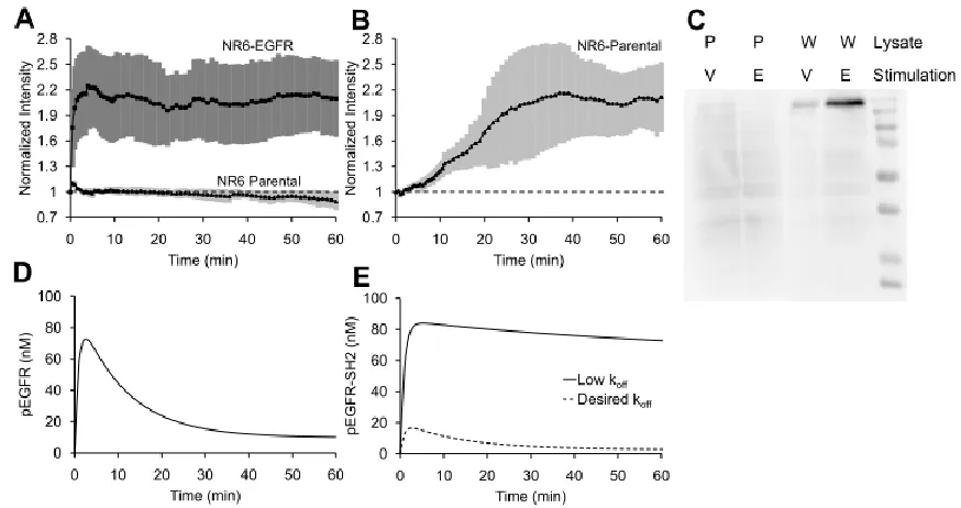
EGFR (pEGFR) and prevent tyrosine dephosphorylation in vitro, and pY992 has been identified as its major binding site(13). Further, tagged tSH2-WT has been used in live cell imaging studies(4, 14). Therefore, we chose to evaluate tSH2-WT as a biosensor to monitor dynamics of EGFR phosphorylation at pY992. Accordingly, an EGFP-tSH2-WT fusion was
expressed by transient transfection in parental NR6 cells (control) or NR6 cells expressing
EGFR. Subsequently, cells were treated with EGF, and membrane localization of tSH2-WT
was observed using TIRF microscopy. The fluorescence signal increases substantially in
28
2.1A), suggesting that tSH2-WT may be used for monitoring kinetics of EGFR phosphorylation; however, the critical attributes of specificity and fidelity have yet to be
characterized for this biosensor.
To assess the specificity of tSH2-WT, we monitored the fluorescence readout upon
treatment of NR6 parental cells with sodium orthovanadate, an inhibitor of protein tyrosine
phosphatases. Since NR6 parental cells lack EGFR, any recruitment of tSH2-WT is attributed
to recognition of other pY motifs. tSH2-WT produces a significant readout in
orthovanadate-treated parental cells, indicating poor specificity (Figure 2.1B). To further assess specificity of tSH2-WT, we conducted far-western blotting analysis. Lysates of NR6 parental cells
treated with sodium orthovanadate, and of parental and EGFR-expressing NR6 cells treated
with EGF, were probed with GST-tagged tSH2-WT (Figure 2.1C). Although a high-intensity band corresponding to EGFR was observed in the lysate of EGF-stimulated,
EGFR-expressing cells as expected; the presence of other bands confirms the promiscuity of
tSH2-WT binding.
To investigate the fidelity provided by tSH2-WT as a biosensor for live-cell imaging,
it is instructive to review the expected kinetics of EGFR phosphorylation, on which there is a
well-established literature(15–17). In response to a saturating dose of EGF, well above its apparent binding KD (~1 nM), receptor activation on the cell surface peaks rapidly and then
29
contrast, the experimental readout from the tSH2-WT sensor showed little adaptation (Fig. 2.1B). The cooperative binding of the two SH2 domains in tSH2-WT yields high binding avidity between the tSH2-WT biosensor and pEGFR(20, 21). Kinetic model calculations further suggest that the apparent lack of adaptation seen in the experimentsis consistent with
high overall binding affinity (low dissociation rate constant) of tSH2-WT for pEGFR (Figure 2.1E). Conversely, the model also predicts that a substantial increase of the dissociation rate constant (thus reducing the affinity) of biosensor binding to pEGFR results in a readout that
is consistent with the true time course of pEGFR (Figure 2.1E vs. Figure 2.1D).
Taken together, our experimental data and modeling analysis lead to two insights. First,
tSH2-WT is not a suitable biosensor, as it lacks specificity and does not faithfully track the
expected kinetics of pEGFR. Second, the lack of fidelity of the tSH2-WT biosensor can be
attributed to its high binding affinity for pEGFR; it is predicted that use of a biosensor with
lower affinity for pEGFR will alleviate the discrepancy.
2.2.3 The engineered mSH2 protein has increased specificity for pY992 of EGFR
Use of a single SH2 domain instead of the tandem SH2 domains in tSH2-WT should
eliminate the high affinity for pEGFR arising from the avidity effect. However, when a
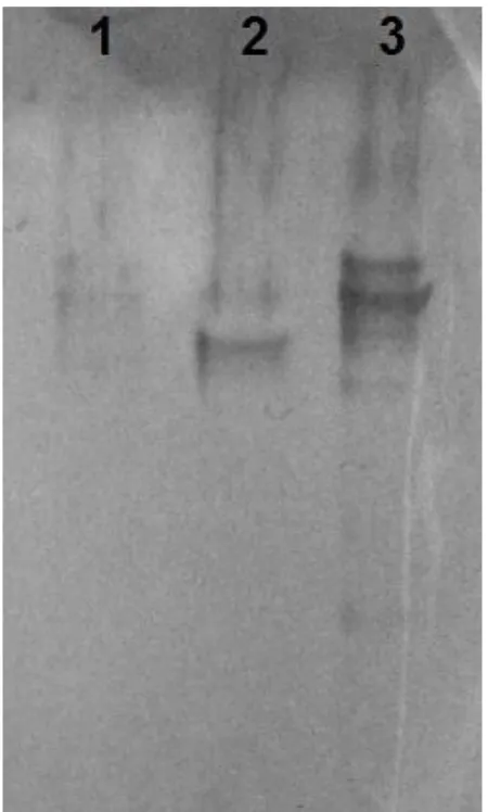
GST-tagged single SH2 domain – the C-terminal SH2 domain of the pair (cSH2) – was used to
probe cell lysates in far-western blotting assays, no specific EGFR band was detected (Fig 2.2). Further, SH2 domains are inherently promiscuous binders. To identify an improved biosensor, we generated a combinatorial library by random mutagenesis of cSH2 and
30
of ~ 109 mutants was generated using yeast surface display and screened using magnetic
selection and fluorescence-activated cell sorting (FACS) (22), with a synthetic peptide corresponding to the pY992 site as the target. Negative selection steps by magnetic selection
were used to eliminate binders to peptides that correspond to the unphosphorylated Y992
site, other phosphorylation sites in EGFR, and putative tyrosine phosphorylation motifs in
other proteins that have sequence homology to the pY motifs in EGFR. The population of
cells obtained after magnetic selection and two rounds of FACS was plated, and single clones
were analyzed. DNA sequencing identified a mutant (mSH2) with a single amino acid
substitution (C65G) relative to cSH2. Interestingly, this residue has been implicated in
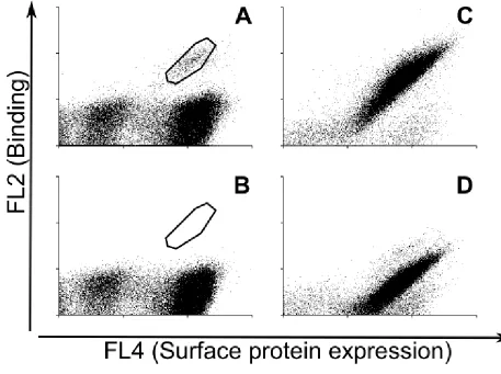
determination of specificity of binding of SH2 domains(23). The complete sequence of mSH2 is shown in Table 2.1. Analysis of intermediate cell populations during screening of the combinatorial library is shown in Fig 2.3.
We used yeast surface-displayed mSH2 and synthetic peptides to assess the
specificity of binding to the pY992 site in EGFR relative to other phosphorylation sites.
mSH2 exhibits > 10-fold greater signal for binding to the pY992 peptide relative to the
pY1148 peptide and no binding was detected for the pY1068 (Figure 2.4A). We further used far-western blotting analysis of NR6 parental and EGFR-expressing cells treated with
EGF, and NR6 parental cells treated with sodium orthovanadate. Cell lysates were probed
31
However, a single high molecular weight band is seen in orthovanadate-stimulated parental
NR6 cells. We hypothesized that this band could be the platelet derived growth factor β-receptor (PDGFR), since tSH2-WT is known to bind pY1021 of PDGFR(24–26). Consistent with this hypothesis, yeast cells displaying mSH2 (or tSH2-WT as a control) were labeled
with a synthetic peptide corresponding to the pY1021 site in PDGFR, and both mSH2 and
tSH2-WT were confirmed to bind pY1021 of PDGFR (Figure 2.4C). This non-specific binding of mSH2 to PDGFR is consistent with our experimental protocols for library
screening, wherein a negative selection step to eliminate binders to phosphorylation motifs
on PDGFR was not incorporated.
These results show that biosensors with improved specificity can be obtained through
random mutagenesis of native protein domains and combinatorial screening of variants with
prescribed properties. In particular, mSH2 exhibits improved specificity for EGFR relative to
tSH2-WT and binds selectively to the pY992 site relative to other EGFR phosphorylation
sites; however, like tSH2-WT, mSH2 retains affinity for the pY1021 site of PDGFR. It is
conceivable that using protein scaffolds with no native binding for intracellular proteins (27) may be more suited for engineering intracellular biosensors.
2.2.4 The mSH2 biosensor closely tracks the expected kinetics of EGFR phosphorylation and displays increased specificity for the pY992 site of EGFR in live-cell imaging
experiments
To evaluate mSH2 as a biosensor, an mSH2-tdTomato fusion was transiently
32
The cells were treated with EGF or sodium orthovanadate, and membrane localization of
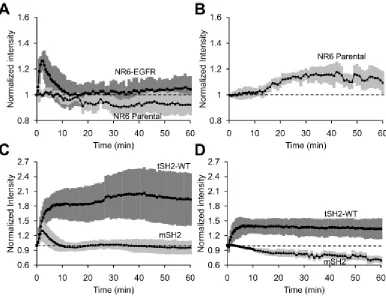
mSH2 was observed using TIRF microscopy. Compared with the EGF-stimulated
translocation of tSH2-WT in EGFR-expressing cells (Figure 2.1A), the corresponding time course of mSH2 recruitment shows a significantly lower fold change (Figure 2.5A; see Figure 2.6A for an overlay plot). A similar comparison holds true for sodium orthovanadate treatment of NR6 parental cells that lack EGFR (Figure 2.5B; overlay plot shown in Figure 2.6B). More importantly, unlike tSH2-WT, mSH2 exhibits the transient, EGF-stimulated response that is characteristic of pEGFR on the cell surface. Finally, the intracellular
concentration of the biosensor also affects readout. Nevertheless, it is important to note that
higher affinity of interaction between the biosensor and pEGFR results in significantly
greater distortion of the readout relative to the true pEGFR concentration, than changes in
biosensor concentration (Figure 2.7).
To further evaluate the specificity of mSH2 for EGFR pY992, we compared the
readout from mSH2 and tSH2-WT in NR6 cells expressing truncation mutants of EGFR, c’1000 or c’1000F. The cytoplasmic domain of the c’1000 mutant is truncated at residue
1000 of EGFR, leaving Y992 as the only major phosphorylation site. In the c’1000F variant,
Y992 is mutated to phenylalanine and therefore cannot be phosphorylated at that site;
however, it is established that c’1000F mediates intracellular signaling in response to EGF
stimulation(28), through heterodimerization with ErbB2. Similar to the responses in wild-type EGFR-expressing cells, there is a substantial difference between the EGF-stimulated
33
their magnitudes and shapes (Figure 2.5C). The most compelling contrast was observed in the c’1000F-expressing cells, however; whereas tSH2-WT was recruited in response to EGF,
albeit to a lesser extent, mSH2 showed no discernable response to the activated EGFR
lacking pY992 (Figure 2.5D).
Taken together, these results demonstrate the suitability of the engineered mutant,
mSH2, for live-cell imaging experiments. Unlike tSH2-WT, the mSH2 biosensor tracks the
transient kinetics of EGFR phosphorylation and exhibits greater specificity for its intended
target, pY992.
2.2.5 The engineered SPY992 protein has specificity for pY992 of EGFR with no affinity for pY1021 site ofPDGFR
To identify an improved biosensor with no affinity to PDGFR, we screened for
mutants from Sso7d library (9) was previously derived. Mutants that showed specific and detectable binding to pY992 of EGFR and no binding to pY1021 of PDGFR or Y992 of
EGFR were selected. A library of ~ 108 mutants generated using yeast surface display was
screened using magnetic selection and fluorescence-activated cell sorting (FACS) (22), with a synthetic peptide corresponding to the pY992 site as the target. Negative selection steps by
magnetic selection were used to eliminate binders to peptides that correspond to the
unphosphorylated Y992 site, other phosphorylation sites in EGFR, and putative tyrosine
phosphorylation motifs in other proteins that have sequence homology to the pY motifs in
EGFR and pY1021 of PDGFR. The population of cells obtained after magnetic selection and
34
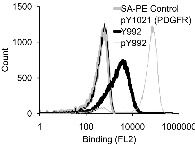
of SPY992 is shown in Table 2.1. Analysis of clone binding to phosphorylated and unphosphorylated peptide Y992 and phospho peptide pY1021 of PDGFR is shown in Fig 2.8. Thus biosensors with improved specificity can be obtained from of non native protein domains and combinatorial screening of variants with prescribed properties. In particular,
SPY992 exhibits improved specificity for EGFR and binds selectively to the pY992 site
relative to other EGFR phosphorylation sites. While SPY992 has no affinity for PDGFR it is
not as selective as SH2 domain for phosphotyrosine. Thus its suitability, for live-cell imaging
experiments as a phospho specific biosensor needs to be validated.
2.3 Discussion
Live-cell imaging studies of intracellular dynamics are presently limited by the
availability of suitable biosensors. Here we show that a previously described biosensor using
the tandem SH2 domains of PLCγ1 (tSH2-WT) suffers from two major limitations in the
context of studying EGFR phosphorylation dynamics. First, tSH2-WT lacks specificity,
consistent with the inherent promiscuity of SH2 domain recognition(6–8). Second, EGF-stimulated translocation of tSH2-WT, though robust in magnitude, exhibits kinetics that
qualitatively deviates from the expected transient kinetics of pEGFR at the cell surface.
Analysis of a basic kinetic model shows that the inability of tSH2-WT to faithfully
track cell-surface pEGFR can be explained by its high binding affinity and occupancy of
more than one site on the pEGFR. Pertinently, the avidity of the two linked SH2 domains in
tSH2-WT results in high overall binding affinity of pEGFR in cells. As discussed earlier,
35
readout or perturbation of the target’s dynamics(3). Indeed, mutant SH2 domains evolved for
high-affinity pY binding have been shown to antagonize pY-mediated signaling(29), and binding of tSH2-WT to pY992 from EGFR prevents tyrosine dephosphorylation in vitro(13). Our modeling results suggest that tSH2-WT perturbs EGFR dynamics by interfering with
endocytosis of the complex.
We applied protein engineering tools to overcome limitations associated with high
target binding affinity and lack of specificity of the tSH2-WT biosensor. Live-cell imaging
studies confirmed that an mSH2 biosensor indeed exhibits specificity for the Y992
phosphorylation site in EGFR. Most importantly, the readout from the mSH2 biosensor is
qualitatively consistent with the kinetics of EGFR phosphorylation in response to EGF
treatment. Phosphorylation of cell surface EGFR peaks and then shows adaptation, in large
part because of ligand-induced endocytosis. The plasma membrane translocation of mSH2
shows a nearly complete recovery during the course of chronic EGF stimulation, a much
greater extent of adaptation than previously inferred from population measurements for the
same cell line (30). In that study, a general anti-phosphotyrosine antibody was used to quantify pEGFR, and a mild acid strip was used to distinguish cell-surface and internalized
pEGFR. The quantitative differences between these measurements might reflect distinct,
site-specific kinetics of EGFR phosphorylation or differential endocytosis of the various
phospho-forms of EGFR. Development of other phospho-specific biosensors will allow such