Oncogene Co-operation in Kératinocytes
Elizabeth A. Roper
UNIVERSITY COLLEGE LONDON AND
IMPERIAL CANCER RESEARCH FUND, LONDON.
ICRF supervisor: Dr. Hartmut Land UCL supervisor: Dr. Alan Hall
A thesis submitted in partial fulfilment of the requirements for the degree
of Doctor of Philosophy, University of London.
All rights reserved
INFORMATION TO ALL USERS
The quality of this reproduction is dependent upon the quality of the copy submitted.
In the unlikely event that the author did not send a complete manuscript and there are missing pages, these will be noted. Also, if material had to be removed,
a note will indicate the deletion.
uest.
ProQuest 10010088
Published by ProQuest LLC(2016). Copyright of the Dissertation is held by the Author.
All rights reserved.
This work is protected against unauthorized copying under Title 17, United States Code. Microform Edition © ProQuest LLC.
ProQuest LLC
789 East Eisenhower Parkway P.O. Box 1346
Multi-stage tumourigenesis is associated with the accumulation of co-operating genetic lesions. One of the best studied models of carcinogenesis is experimentally induced tumours in the skin of mice. In tumorigenesis of the skin, activated Ras co-operates with mutations that inactivate the tumour suppressor p53. The absence of the cyclin dependent kinase inhibitor p2 1° ‘’\ a p53 target gene, has also been shown to co-operate with oncogenic ras in the induction of aggressive and relatively undifferentiated tumours in vivo. However, the molecular basis for these co-operations remains unresolved. Activation of oncogenes, including Ras, Myc and E l A, have been reported to stabilise and activate p53 via induction of the tumour suppressor pl9^"^. Therefore, it is thought that ARE might be the specific link between oncogene activation and induction of p53. Since most malignancies are epithelial in origin and ras and p53 mutations are most frequently associated with epithelial derived tumours, I investigated the molecular mechanisms involved in co-operation between Ras, p53 and p21^'^^ in the skin and the potential involvement of p i 9 " ^ .
T
a b l e
o f
C
o n t e n t s
Ab s t r a c t... 3
Ta b l e o f Co n t e n t s... 4
Ab b r e v i a t i o n s...1 3 Ac k n o w l e d g e m e n t s...1 6 Pu b l i c a t i o n s... 1 7 Ch a p t e r 1 - In t r o d u c t i o n 1 .1 Ov e r v i e w... 1 8 1 .2 C e ll cy cle c o n tr o l...18
1.2.1. Gj to S-phase tra n sitio n...19
1.2.2 The C K Is...21
1.2.2.1IN K 4 fa m ily ... 21
1.2.2.2 C ip /K ip fa m ily ... 22
1.2.3 pRb a n d E 2 F...23
1.2 .4 C yclin s D, E an d A...24
1.3 Ras... 26
1.3.1 Ras: a m o lecu la r sw itch...27
1.3.2 R a s/R a f/M A P K p a th w a y...29
1.3.2.1 R af as a Ras e ffe c to r... 29
1.3.2.2 Raf activation...30
1.3.2.3 The Raf/M EK /M A P kinase p ath w ay ... 34
1.3.2.4 ERK inactivation... 36
1.3 .3 O ther R a f e ffe c to r s...37
1.3 .4 A ltern a tiv e Ras e ffe c to rs...39
1.3.4.1 P I3 K ... 39
1.3.5 Ras fa m ily...42
1 .3 .6 R as su p e rfa m ily...42
1.3.7 Cellular Effects o f R a s...43
1.3.8 Ras and the cell c y c le...45
1.3.8.1 T he C D K Is... 45
1.3.8.2 Ras and the cell cycle: prohferative signals from Ras... 47
1.3.10 Ras and A poptosis...50
1.3.11 Ras and Differentiation...51
1 .4 RasA N D p53 ...5 1 1.5 P l 9 ^ ^ ... 5 2 1.6 Th e Ep id e r m is... 5 4 1 .6.1 The s k in...54
1.6 .2 The d e rm is...54
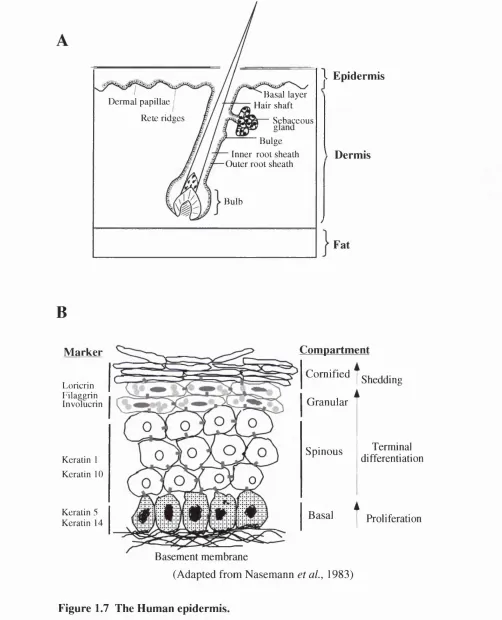
1.6.3 The epiderm is...54
1 .6 .3 .1 T h e B a s a i L a y e r ... 5 6 1 . 6 . 3 . 2 T h e S p i n o u s L a y e r ...5 7 1 .6 .3 .3 T h e G r a n u la r L a y e r ... 5 7 1 . 6 . 3 . 4 T h e c o r n i f i e d e n v e l o p e ...5 8 1.6.4 Terminal differentiation...58
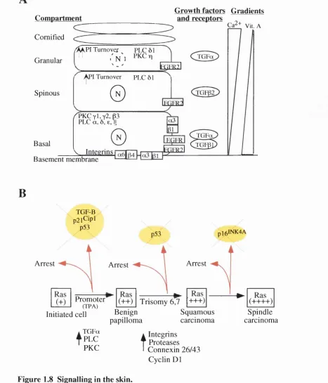
1 .6 .4 .1 C o m p a r tm e n ta lis a tio n a n d d iffe re n tia l e x p re s s io n o f s ig n a llin g m o le c u le s in s k i n ...5 8 1 . 6 . 4 . 2 C y c l e p r o t e i n s a n d D i f f e r e n t i a t i o n ... 61
1.7 C ANGERS OF THE EPIDERMIS... 6 2 1.7.1 Ras activation and loss o f p 5 3 in human skin cancers...63
1.8 Epid e r m isa sam o d e lf o rm u l t ist a g ec a r c in o g e n e s is...6 3 1.8.1 Transgenic mice, knockout mice andin v itr o keratinocyte culture...64
1 . 8 . 1 . 1 R a s ... 6 5 1 . 8 . 1 . 2 p 5 3 ... 6 6 1 . 8 . 1 . 3 C K I s ... 6 7 1 . 8 . 1 . 4 C y c l i n D ... 6 8 1.9 Mo u se Ke r a tin o c y tec u l t u r e... 6 8 1 .1 0 Ai m ...6 9 Ch a p t e r 2 ...7 0 Ma t e r i a l s a n d Me t h o d s 2 . 1 M a t e r i a l s... 7 0 2.1.1 Equipm ent...70
2.1.2. G eneral Cell C ulture S o lu tio n s...71
2.1.3 A ntibodies...73
2.1.4 Plasm ids and expression vectors used in this thesis...74
2 .1 .5 P a ren ta l c e lls-lin e s... 75
2.2.1 Mouse Keratinocyte culture medium (FAD+ FCS + HICE)...76
2.2.2 Isolation and culture o f prim ary mouse kératinocytes...77
2.2.2.1 Passaging of Kératinocytes... 78
2.2.2.2 Collagen coat for culture dishes/cham ber slides...78
2 .2 .2 .3 F reezin g o f c e lls ... 78
2.2.2.4 Retrieval of cell stocks from liquid nitrogen... 79
2 .2 .2 .5 Q uiescen ce o f c e lls ... 79
2.2.2.6 FACS sorting on the basis of EGFP fluorescence... 79
2.2.3 G eneral culture conditions fo r cell lines...79
2.2.3.1 M aintenance o f cell lin e s ... 79
2 .2 .3 .2 P assag in g o f c e ll-lin e s ... 80
2.2.3.3 Selection markers for cell lin e s...80
2.2.4 Transfection...80
2.2.4.1 Calcium phosphate transfections (after [Wigler, 1979 #3845])... 80
2 .2 .5 R e tro vira l in fe c tio n s...80
2.2.5.1 Preparation o f retroviral supernatants... 80
2.2.5.2 R etroviral infection o f c e lls... 81
2.2.5.3 Selection of stably transfected or infected cell lines...82
2.3. CELL CYCLE ANALYSIS...82
2.3.1 B rdU staining f o r im m unofluorescence...82
2.3.2 Cell cycle analysis by F A C s...83
2.3.3 Laser scanning cytometer: Proliferation assay...83
2.4 Proteinanalysis...87
2.4.1 . G eneral S o lu tio n s...87
2.4.2 Cell lysate preparation fo r Western blotting...88
2.4.3 Protein concentration determination...89
2.4.4 SDS-polyacrylamide gel electrophoresis (Laemmli, 1970)...89
2 .4 .5 W estern b lo ttin g...90
2.4.6 Membrane stripping...90
2 .4 .7 C yclin E K inase a ssa y...90
2.5 Molecular Biology...91
2 .5 .1. G eneral S o lu tio n s...91
2.5.2. G eneral DNA T ech n iq u es...92
2.5.2.1 R estriction enzym e d ig e stio n ... 92
2.5.3.2 A garose gel elec tro p h o resis... 92
2.5.2.3 E xtraction o f DNA from agarose g els... 93
2 .5 .2 .4 L ig atio n re a c tio n s ...93
2.5.2.5 D ephosphorylation o f vector D N A ...93
2.5.2.S Transformation of D H 5a E .c o li...94
2 .5.2.9 Q iagen Spin-prep M in i-p rep s... 95
2 .5 .2 .1 0 Q iagen M a x i-p re p ... 95
2.6 GEN O TY PIN G ... 95
2.6.1 M ouse tail genom ic DNA isolation:...95
2.6 .2 PC R f o r P53 G en o typ in g :...96
2.6.3 PCR fo r p l 9 ^ ^ G enotyping: (Kam ijo et al., 1997)...96
2.6.3.1 W ild type P C R ... 96
2 .6 .3 .2 K nock O ut P C R ... 97
2.6.4 PCRforp21^'^^ G enotyping: (W einberg et al., 1997)...97
2.6.4.1 W ild type P C R ... 97
2 .6 .4 .2 K nock O ut P C R ... 98
2.7 Northern BLOTTING... 98
2 .7.1 . G eneral S o lu tio n s...98
2 .7 .2 RN A E x tr a c tio n...99
2.7.3 Preparation o f RNA in Formaldehyde gel sample buffer...99
2 .7 .4 F orm a ldehyde RNA g e l...99
2 .7 .5 RNA gel e le c tro p h o re sis...99
2 .7 .6 N o rth ern b lo ttin g...100
2.7.7 Radiolabelling probes fo r Northern hybridisation... 100
2.7.8 Northern hybridisation and washing... 100
Ch a p t e r 3 Ra s o r Ra f a c t i v a t i o n i n p r i m a r y m o u s e k é r a t i n o c y t e s i n d u c e s a c e l l CYCLE ARREST AND DIFFERENTIATION 3 . 1 In t r o d u c t i o n... 1 0 1 3.2 Results...102
3.2.1. Introduction o f R a f into primary mouse kératinocytes... 102
3.2.2 RafFR is expressed and active in prim ary kératinocytes...103
3.2.3 R af activation in primary mouse kératinocytes induces a cell cycle arrest... 104
3.2.4 Analysis o f R a f induced growth arrest by the LSC and FACs...104
3.2.5 RafFR activation induces keratinocyte differentiation in v itro ... 106
3.2.6 Ras, like Raf, can induce a proliferation arrest and terminal differentiation o f primary mouse kératinocytes... 107
3.2.7 R a f activation induces p2ff'^^ and results in the loss o f cyclin D proteins.... 108
3.2.9 A more detailed kinetic analysis o f the induction o f cell cycle proteins and
d iffe ren tia tio n m a rkers... 109 3.2.10 R a f activation leads to a downregulation o f cyclin E-associated kinase
a c tiv ity... 110 3 .3 Su m m a r y... 1 1 0 3 .4 LSC: Ex pl o ita tio nint h efu t u r e. ... I l l
Ch a p t e r 4
Ra f-i n d u c e d c e l l c y c l e a r r e s t, b u t n o t d i f f e r e n t i a t i o n, i s p5 3 DEPENDENT
4 .1 iNTRODUCnON... 1 4 4
4 .2 Re s u l t s... 1 4 4 4.2.1 Genotyping and introduction o f R a f into mouse kératinocytes with different
p53 status...144 4.2.3 Raf-induced cell cycle arrest is p53 dependent...145 4.2.3 Raf-induction o fp21^‘^^ in p53-/~ cells is lo st...145 4.2.4 Confirming that the Raf-induced cell cycle arrest is p53 dependent using a
d iffe r e n t c o n str u c t...146 4.2.5 Raf-induction ofp21^‘’’^ in p53-/- cells is lost but Raf-induction o f cyclin D1 is
r e v e a le d... 146 4.2.5 R a f activation in p53-/- cells is not enough to push the cells into cycle 147
4.2.6 Raf-induced differentiation is independent o f p 5 3...148 4 .3 Effe c t so f Ra fa c t iv a t io no np5 3 t r a n s c r ip t io n... 148 4 .4 Su m m a r y... 14 9
Ch a p t e r 5
Ra f i n d u c e d c e l l c y c l e a r r e s t i s p1 9 i n d e p e n d e n t b u t p2 1 ^ * -DEPENDENT.
5 .1 In t r o d u c t io n... 16 2 5 .2 Re s u l t s... 163 5.2 .1 R a f activation has no effect on p l9 ^ ^ e x p re ssio n...163
5.2.2 Induction ofp53 and cell cycle arrest by R a fis pl9^^^-independent 163
5 .3 R AF ACTIVATION IN P 2 1 ^ A N D P 2 1 ‘^”’’- / + PRIMARY MOUSE KERATINOCYTES... 165
6 .1 T h e m e c h a n is m o f t h e R a f - i n d u c e d c e l l c y c l e a r r e s t in k é r a t i n o c y t e s is v i a a
P 5 3 - DEPENDENT UPREGULATION OF 183
6 .2 E v id e n c e t h a t R a f is a c t i n g t h r o u g h a n o v e l m e c h a n is m t o u p r e g u l a t e p53
INDEPENDENTLY OF p19^*^IN KERATINOCYTES... 18 4
6 .3 Pu t a t iv e MECHANISMS FOR P i 9 ^ ^ INDEPENDENT INDUCTION OF p5 3 ... 186 6 .4 Po ssib l ein v o l v e m e n to fP M L ...188 6 .5 Ra f LEADS TO DOWNREGULATION OF p27*^^... 189 6 .6 Loss OF p53 w a s s u f f i c i e n t t o r e n d e r c y c l i n D e x p r e s s io n r e s p o n s iv e t o R a f in KERATINOCYTES... 19 0
6 .7 Ra fc o u l dh a v eapo sitiv er o l eint h ed iffe r en t ia tio no fk e r a t in o c y t e s... 1 9 2 6 .8 G r o w t h ARREST AND DIFFERENTIATION c a n BE u n c o u p l e d ... 19 2
6 .9 p21^"'‘ AND ITS ROLE IN d iffe r e n t ia t io n... 193 6 .1 0 Ra s, l ik e Ra f, in d u c esg r o w t ha r r e s ta n dd iffe r e n t ia t io n... 193 6 .1 1 C O-OPERATION BETWEEN RAS AND P53 OR P 21^""^ IN THE SKIN... 1 9 4 6 .1 2 Co n c l u sio n s... 195 6 .1 3 Fu t u r ed ir e c t io n s... 195
T
a b l e
o f
F
ig u r e s
Figure 1.1 The Cell cycle...20
Figure 1.2 Activation of Ras...28
Figure 1.3 Activation of Raf...32
Figure 1.4 Signal transduction through the MAPK modules...38
Figure 1.5 Ras activation of multiple effector-mediated signalling pathways...41
Figure 1.6 Model of the effects of Ras/Raf activation in different cell types...46
Figure 1.7 The Human epidermis...55
Figure 1.8 Signalling in the skin...60
Figure 2.1 Operation of the LSC... 85
Figure 2.2 High resolution cell cycle analysis... 86
Figure 3.1 RafFR is expressed and active in primary keratinocytes... 113
Figure 3.2 Activation of Raf induces a growth arrest in primary keratinocytes... 115
Figure 3.3 An example of a typical LSC data print out... 117
Figure 3.4 Analysis of the Raf induced growth arrest by the LSC and FACs...119
Figure 3.5 Further analysis of Raf induced growth arrest by the LSC...121
Figure 3.6 RafFR activation induces a morphological change in keratinocytes... 123
Figure 3.7 RafFR activation induces keratinocyte differentiation...125
Figure 3.8 RafFR activation induces keratinocyte differentiation...127
Figure 3.9 RafFR activation induces Keratin 6... 129
Figure 3.10 Ras induces a morphological change and cell cycle arrest in keratinocytes.. 131
Figure 3.11 Ras induces the differentiation marker involucrin...133
Figure 3.12 Raf activation induces p21^'^' and results in the loss o f cyclin D proteins...135
Figure 3.13 Raf induces p53... 137
Figure 3.14 A more detailed kinetic analysis of the activation of Raf...139
... 141
Figure 3.16 Raf activation leads to a downregulation of cyclin E-associated kinase activity. ... 143
Figure 4.1 The introduction of Raf into mouse keratinocytes with different p53 status...151
Figure 4.2 Raf-induced p21^''’^ and cell cycle arrest is p53 dependent... 153
Figure 4.3 Raf activation in p53-/- cells is not enough to push the cells into cycle 155
Figure 4.4 Raf-induction o f p21^*'’^ in p53-/- cells is lost but Raf-induction o f cyclin D1 is revealed. Raf-induced differentiation is independent of p53...157
Figure 4.5 Effects of Raf activation on the morphology of mouse keratinocytes with different p53 status...159
Figure 4.6 Effects of Raf activation on p53 transcription... 161
Figure 5.1 Raf activation has no effect on p i 9'^’' expression...168
Figure 5.2 Genotyping of mouse keratinocytes with different p i 9 ^ status by PCR 170
Figure 5.3 Induction o f p53 and cell cycle arrest by Raf is p 19^-independent... 172
Figure 5.4 Induction of p53 by Raf is p 19*'^-independent... 174
Figure 5.5 Induction o f differentiation markers and alterations in cell morphology by Raf are p 19^-independent... 176
Figure 5.6 The Raf-induced cell cycle arrest is p21^'*’‘ dependent... 178
Figure 5.7 Raf-induced differentiation is independent of p21^''’^... 180
Figure 5.8 Effects o f Raf activation on the morphology of mouse keratinocytes with different p21^'‘’’status... 182
Figure 6.1 Model of the effects of Ras/Raf activation in primary mouse keratinocytes.... 185
T
a b l e s
Table 1.1 Members of the human Ras-like GTPase superfamily...43
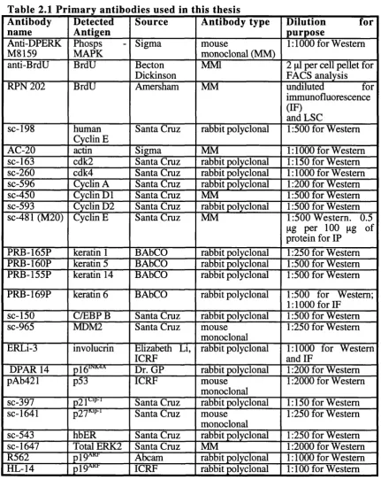
Table 2.1 Primary antibodies used in this thesis...73
Table 2.2 Secondary antibodies used in this thesis... 74
Table 2.3 Plasmids and expression vectors used in this thesis...74
Table 2.4 Parental cells and cell-lines used in this thesis...75
Table 2.5 Cell-lines used in this thesis...75
(d)NTP (2’ -deoxy)ribonuleoside 5 ’ triphosphate
aa amino acid residue
Amp Ampere
AR androgen receptor
ATP adenosine 5’triphosphate
BCC basal cell carcinoma
p-gal P-galactosidase
BM basement membrane
bp base pair
BrdU 5-bromo-2’ -deoxyuridine
BSA bovine serum albumin
Ca^+ calcium
CAK CDK-activating kinase
cDNA complementary deoxyribonucleic acid
CDK cychn dependent kinase
Ci Curie
GIF calf intestinal phosphatase
CKI cychn dependent kinase inhibitor
cm centimetre
CMV Cytomegalovirus
CR conserved region
CRD cysteine rich domain
dH^O distilled water
DMEM Dulbecco’s modified Eagles media
DMSO dimethylsulphoxide
DNA deoxyribonucleic acid
Dox doxycychne
DTAF dichlorotriazinyl amino fluorescein
DTT 1,4-dithiothreitol
E.coli. Escherichia coli
ECL enhanced chemi-luminescence
ECM extracellular matrix
EDTA ethylene-diamine-tetraacetic acid (disodium salt)
EOF epidermal growth factor
EMT epithehal-mesenchymal transition
ER oestrogen receptor
FACs Fluorescence activated cell sorting
FBS foetal bovine serum
Fig. figure
FITC fluoroescein isothiocyanate
G418 geneticin
g gramme
GAP GTPase activating protein
GDI guanine dissociation inhibitor
GDP guanosine diphosphate
GEF guanine exchange factor
GFP green fluorescent protein
Grb2 growth factor receptor-bound protein
GST glutathione S-transferase
GTP guanosine triphosphate
HBD hormone binding domain
HCl hydrochloric acid
HDAC histone deacetylation complex
HEPES N-2-hydroxy-ethyl-piperazine-N’ -2 e
HRP horse radish peroxidase
HSP heat shock protein
IF immunofluorescence
Ig immunoglobulin
IP immunoprécipitation
IVT in vitro translated
JAKS janus activated kinas
INK interacting protein
K keratin
KSR kinase supressor of ras
kbp kilo base pair
kDa kilo Dalton
1 litre
LB Luria-Bertani broth
LSC laser scanning cytometry
LTR long terminal repeat
micro
m milli
M molar
mA milli Ampere
MAPK MAP kinase
MAPKK MAP kinase kinase
MBP myehn basic protein
MEF mouse embryo fibroblast
MM mouse monoclonal
mm millimetre
MMTV mouse mammary tumour virus
Mo-MuLV Moloney murine leukaemia virus
mol mole
MPI MEK partner 1
Mr molecular weight
mRNA messanger ribonucleic acid
n nano
NCS new bom calf semm
NES nuclear-export sequence
nm nanometre
NP40 nonidet P-40
°C degrees centigrade
P pico
PAGE poly-acrylamide gel electrophoresis
PAK p21 activated protein kinase
PBSA phosphate buffered sahne A
PC12 phaeochromocytoma cell line
PCR polymerase chain reaction
PI propidium iodide
PI3K phosphoinositide 3-kinase
PKC protein kinase C
PLC phospholipase C
PM SF phenyl-methyl-sulphonyl fluoride
Raf-1 R af
RalG EF Ral-guanine nucleotide exchange factor
RED Ras binding domain
R EF rat embryo fibroblast
RNA ribonucleic acid
ROS reactive oxygen species
rpm revolutions per minute
RSV Rous sarcoma virus
RT room temperature
R TK receptor tyrosine kinase
SA P shrimp alkaline phosphatase
SDS sodium dodecyl sulphate
SH2 Src-homology 2
s e e squamous cell carcinoma
SV40 Simian virus 40
T A E tris-acetate-EDTA buffer
T B E tris-borate-EDTA buffer
TBS tris buffered saline
TB ST TBS with 0.1% Tween 20
T E tris EDTA buffer
TE M E D N,N,N',N',-tetramethylethylenediamine
TPA 12-0-tetradecanoyl-phorbol-13-acetate
Tris (Trizma base) Tris (hydroxymethyl)aminomel
Tween 20 polyoxyethylenesorbitan monolaurate
U unit (of enzyme activity)
u v ultra-violet light
V volts
v/v volume for volume
w/v weight for volume
W B western blot
wt wildtype
A
c k n o w l e d g e m e n t s
I would particularly like to thank Hucky and Fiona for giving me the opportunity to work in their laboratories and for their guidance, support and encouragement during my time at the ICRF. I would like to thank Alan Hall, Martine Roussel, Chuck Sherr, Esther van de Kamp, Weindy Weinberg, Martin McMahon and Gorden Peters for reagents and/or helpful discussions. The FACs laboratory (partically Derek Davis for all the interesting moments we had with the LSC) and the photography department at ICRF have also given invaluable assistance. I would especially like to thank Alison Lloyd for great conversations, inspiration, support and perspective.
I am also grateful to the members of both the Land and Watt Labs, as they have made my time during my Ph.D. most enjoyable and stimulating. Nacho, Bryony, Sharon, Susan, Soo, Sally, Betina, Laurent, Beatrice, Frank -thank you for everything! Nacho you were one of my guiding lights in the early years and helped me through some tough times. Simon, Liz, Richard and Douglas many thanks for your invaluable help. To the rest of the Watt Lab (to many to mention separately) I am grateful for everything! It was a very enjoyable adoption.
Outside of the lab, I especially would like to thank pigeon (Bjoem), Matt (Dobo), Ali, Avril and Gavin (foxy) for being there through it all and for being great friends.
Barry, what can I say, thank you for everything, for being there (putting up with me), for all the support and encouragment, and for being my kindred spirit.
Finally, I would particularly like to thank my family (including Rich) for their help, support and reassurance through out the past few years. This thesis is dedicated to my Dad and Mum without whom none of this would have been possible.
.. .they are ill discoverers that think there is no land when they can see nothing but sea.
Francis Bacon (1561-1626)
English Essayist, Philosopher, Statesman.
Some of the data described in this thesis has been presented in the following pubhcation:
Roper, E ., Weinberg W., Watt P.M. and Land H. (2001).
p l9 ^ -in d ep e n d e n t induction of p53 and cell cycle arrest by Raf in murine keratinocytes.
Embo Reports. In press.
C
h a p t e r
1 - I
n t r o d u c t io n
1.1 O verview
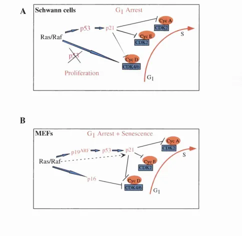
Tumorigenesis is a multi-step process that requires the co-operation of several distinct oncogenic mutations. This is reflected by many in vitro and in vivo studies which have demonstrated that more than one oncogene, or an oncogene and the loss of a tumour supressor gene, is required to fully transform most primary cell types (Hunter, 1991). For example, oncogenic Ras, or activated Raf, lead to a cell cycle arrest in Schwann cells or a cell cycle arrest and premature senescence in fibroblasts (Hirakawa and Ruley, 1988; Lloyd et al., 1997; Ridley et al., 1988; Sewing et al., 1994; Woods et al., 1997). However, this Ras/Raf-induced arrest is abrogated in the presence of certain oncoproteins, such as SV40 large T, adenovirus E l A and Myc, or in the absence of the tumour supressors p53 and p i 9"^^ (For review see Lowe, 1999 ), and, as a consequence, effective neoplastic cell transformation ensues. The work described in this thesis derives from investigations into the mechanisms of oncogene co-operation in epithelial cells. It is now clear that the cellular responses to the oncogene Ras differ depending on the cell-type, the presence of other oncogenes and the cellular environment. Ras can induce a variety of cellular responses, such as proliferation, cell cycle arrest, senescence, apoptosis and differentiation. To date most of our understanding of Ras function is derived from studies on rodent fibroblasts. However, most cancers are epithelial in origin and epithelial cells behave differently to fibroblasts (Shields et al., 2000). Thus heightening the importance of investigating the configuration of these interactions in epithelial cell types. This introduction will begin with a summary of the current understanding of the molecular mechanisms that regulate the cell cycle. Ras and its downstream targets will then be introduced, followed by an overview of the cellular effects o f Ras. Finally, the epidermis as a model for the investigation of the effects of Ras/Raf in epithehal cells will be discussed.
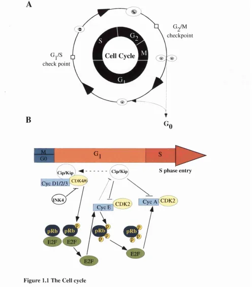
1.2 Cell cycle control
The eukaryotic cell cycle can be divided into four stages: G,, S, and M (Fig. 1.1 A). Gj is the gap phase where the cell integrates mitogenic and growth inhibitory signals and makes the decision to proceed, pause, or exit the cell cycle. It contains the restriction point at which a cell becomes committed to DNA replication. Gj is followed by S-phase during which the cellular DNA is duplicated. Following DNA duplication, cells enter a second growth phase called G^. During this time the cells duplicate their infrastructure.
including the amount of ribosomes, ribosomal RNA, cellular proteins, and other functional elements, in preparation for cell division. Following G2, the cells undergo mitosis (M), the phase in which replicated chromosomes are segregated into separate nuclei and cytokinesis occurs to form two daughter cells, completing the cell cycle. The cell can also exit from the cell cycle to differentiate or to enter a state referred to as quiescence (or the Gg phase). Cells can be induced to enter Gg through the removal of mitogens.
Progression of the cell cycle is controlled by multiple mechanisms and it has become clear that tumorigenesis involves the disruption of these controls. The work in this thesis is mainly concerned with the Gj to S-phase transition (which is summarised in Figure (Fig.)
1.1 B) and the current understanding of the mechanisms controlling this transition shall be discussed below.
1.2.1 . Gj to S-phase transition
Mitogen-dependent progression through the mammalian cell cycle is co-operatively regulated by a conserved family of serine/threonine kinases, the cyclin-dependent kinases (CDKs). CDKs activity is sequentially regulated by cyclin D, E and A proteins. In general, CDK activity requires cyclin binding, depends on both positive and negative regulatory phosphorylations, and can be constrained by at least two famihes of CDK inhibitors (CKIs) (Lees et al., 1992; Morgan, 1995; Nigg, 1995; Roussel, 1999; Sherr and Roberts, 1999). Each cyclin maintains a cell-cycle-specific pattern of accumulation and rapid proteolysis.
Cyclins function to bind and activate their specific CDK partner. D-type cyclins form active complexes with CDK4 and CDK6 (Matsushime et al., 1992; Meyerson and
Harlow, 1994). Cyclin E forms active complexes only with CDK2 (Dulic et al., 1992; Koff et al., 1992). Cyclin A, which is required for both entry into S-phase and onset of mitosis (Girard et al., 1991; Pagano et al., 1992), complexes with, and activates both CDK2 and CDKl (also known as Cdc2). Cyclin A/CDK2 complexes predominate in late Gj and S-phase whereas cyclin A/CDKl appears in G^.
G^/S
check point
B
checkpoint
G
0
_ M G i Ç
GO o
---- ^
S phase entry
Cip/Kip Cip/Kip
CDK4/
Cyc P l/2 /3
CVCÂKCDK2 CDK2
Figure 1.1 The Cell cycle
A) Stages of the cell cycle. B) The G j to S-phase transition
Mitogen signals promote the assem bly o f active cyclin D /C D K 4/6 com plexes and a Cip/Kip protein. Sequestration o f the Cip/K ip proteins facilitates the activation o f cyclin E/CD K 2 com plexes. Cyclin D and cyclin E dependent kinases contribute to sequential phosphorylation o f pRb, releaving its ability to repress E2F family members and activating the genes required for S-phase en t^ . This includes cyclin E and cyclin A. Active cyclin E-CDK2 com plexes
As well as binding to their specific cyclin partner, the CDK has to be phosphorylated on the threonine residue, T160 or T161, to fully open up the catalytic site. This phosphorylation is carried out by a CDK-activating kinase (CAK) (Harper and Elledge, 1998; Solomon et al., 1992). After the cyclin partner has been degraded, this phosphate is removed by the phosphatase KAP. In addition, phosphorylation of conserved tyrosine residues in the ATP binding site of the CDK can also inactivate the cyclin-CDK complex (Gu et al., 1992; Sebastian et al., 1993). In many CDKs it is Y15 that is phosphorylated by W eel/M ikl, and an adjacent threonine residue, T14, that is phosphorylated by M ytl. These phosphates are removed by members of the Cdc25 family of phosphatases resulting in activation of the complexes.
Recently, another level of regulation that has come to light is the possibility that a cyclin- CDK complex may be locally activated or inactivated due to compartmentalisation. For example, in some cases, two or more sets of regulators exist for the mitotic cyclin-CDK complexes, one nuclear and one cytoplasmic. Cdc25C is nuclear whereas Cdc25B is cytoplasmic. Similarly, W eel is nuclear but Mytl is an ER-associated protein. Furthermore, nuclear import and export play an important role in co-ordinating the components of the cell cycle machinery. The decision of whether to enter another round of DNA replication can be achieved by importing signalling mediators into the nucleus in response to a specific stimulus. For example, in resting cells, MAP kinase kinase (MAPKK) and MAP kinase (MAPK) are in a complex that is kept in the cytoplasm by active nuclear export, due to a nuclear-export sequence (NES) in MAPKK. However, growth factor stimulation of the MAPK pathway results in MAPKK phosphorylating and activating MAPK resulting in the two proteins dissociating. Activated MAPK is then imported into the nucleus where it is required for Gj progression (Brunet et al., 1999; Fukuda et al., 1997). Furthermore, a striking connection between nuclear transport and regulated proteolysis has become apparent for some key cell cycle regulators, see section
1.2.4 (for review see (Pines, 1999).
1 .2 .2 T h e C K Is
1 .2 .2 .1 INK4 fam ily
The CKIs fall into two distinct classes: the INK4A proteins and the Cip/Kip family. The specific inhibitors of the CDK4 (INK4) family consists of four members: plS^*^^"^® (Hannon and Beach, 1994); (Serrano et al., 1993); p i a n d p i91^x40
et al., 1995; Guan et al., 1994; Hirai et al., 1995). These proteins bind to CDK4 and CDK6 and prevent their interaction with cyclins. They do not bind other CDKs and thus
are specific for the inactivation of cyclin D-associated kinase activity (Hannon et al..
Ch a pt e r 1 - In t r o d u c t io n
1994). The signals that lead to the synthesis of INK4 proteins are poorly understood. p l6iNK4A accumulates progressively as cells age (senescence) (Palmero et al., 1997;
Serrano et al., 1997; Zindy et al., 1997). TGFp-induced Gj arrest is associated with its ability to induce p i ( H a n n o n and Beach, 1994; Reynisdottir and Massague, 1997). Importantly, overexpression of INK4 protein arrests the cell cycle in G^ phase, in a manner that depends on the integrity of pRb (Sherr, 1994; Weinberg, 1995a). However, a recent study has shown that plb^^^'^'^-induced arrest is not mediated exclusively by pRb, but also depends on the nonredundent functions of at least two pRb-family members, p i 07 and p i 30 (Bruce et al., 2000). Various studies suggest that increased INK4 synthesis results in CDK4 being redistributed from cyclin D-CDK4 complexes to INK4- CDK4 complexes, and unbound D-type cyclins are then rapidly degraded by the ubiquitin-dependent proteasome degradation pathway (Diehl et al., 1997b). As a result, release of Cip/Kip proteins that were previously bound to cyclin D/CDK4 occurs. These are now free to inhibit cyclin E/CDK2 and cyclin A/CDK2 complexes, thus, pRb becomes hypophosphorylated, binds to and represses E2F, which is required for progression through G^ (Sherr and Roberts, 1999).
Proper folding of CDK4 is catalysed by a 450 kDa complex composed of Hsp90 and p50 (Cdc37) but not the D-cyclins (Dai et al., 1996; Stepanova et al., 1996). Unassembled CDK4 is relatively unstable and Cdc37 has been shown to inhibit the interaction between CDK4 and INK4 proteins, in a concentration dependent manner (Lamphere et al., 1997; McConnell et al., 1999). It is not clear whether CDK4 has to be properly folded before it can interact with INK4 inhibitors, or whether it can interact with unfolded, unassembled CDK4. However, it is clear that CDK4 partitions between INK4 and Cip/Kip-bound states (McConnell et al., 1999; Parry et al., 1999).
1.2.2.2 Cip/Kip fam ily
The CDK proteins can also be constrained with the more broadly acting inhibitors of the Cip/Kip family. This family consists of p21^'^^ (Dulic et al., 1994; el-Deiry et al., 1993; Gu et al., 1993b; H arper et al., 1993; Noda et al., 1994; Xiong et al., 1993), p27^^^ (Polyak et al., 1994a; Polyak et al., 1994b; Toyoshima and Hunter, 1994) and p57^^^ (Lee et al., 1995; Matsuoka et al., 1995). These proteins affect the activities of cyclin D-, E-, and A-dependent kinases and can bind to both cyclin and CDK subunits (Fotedar et al., 1996; Gu et al., 1993b; Harper et al., 1993; Xiong et al., 1993).
Although first thought to be inhibitory of cyclins D, E and A, and have since been shown to have a positive role in the activity of cyclin D/CDK4 complexes (LaBaer et al., 1997) while remaining potent inhibitors of cyclin E- and A-dependent CDK2 activity. It has been shown that all of the cyclin D-CDK kinase activity in proliferating cells is found in complexes containing Cip/Kip (Blain et al., 1997; Cheng et al., 1999; Harper et al., 1995; LaBaer et al., 1997; Soos et al., 1996; Zhang et al., 1994), Furthermore, the assembly of the cyclin D-CDK complexes was impaired in primary p2iCipi_/_^ or p2 7^PL/_^ and p21‘^'‘’*-/-/p27^‘’^-/-, mouse embryo fibroblasts (MEFs), with
assembly of the complexes being restored by réintroduction of p21^'^^ or p27^^^ (Cheng et al., 1999). p27^^^ can still inhibit cyclin D-CDK complexes in vitro. However, it is more effective in antagonising cyclin E-CDK2. Recent reports have shown that a single molecule of p21^‘*’* is sufficient to inhibit the kinase activity of cyclin E/CDK2 and cyclin A/CDK2 complexes but not cyclin D/CDK2 (Hengst and Reed, 1998), Hence, it is now thought that cyclin D/CDK2 binds Cip/Kip proteins without being inhibited, whereas CDK2 complexes are inhibited by the same CKIs. It is likely that higher ratios of p21^'^^ or p27^'^^' are necessary to inhibit cyclin Dl/CDK. Furthermore, it has now been shown that C ip/K p proteins also promote activation of cyclin D-CDK4 complex by directing its nuclear import and by increasing its stability (Cheng et al., 1999; LaBaer et al., 1997).
The CKIs are also spatially regulated (Reynisdottir and Massague, 1997). p27*^*’^ is thought to be degraded upon entry into S-phase which depends on its nuclear export via Jab l (Tomoda et al., 1999; Yew and Kirschner, 1997). This degradation of p27^^^ is thought to be important for entry into the cell cycle and may be a key regulator of cyclin E/CDK2 activity (Sherr and Roberts, 1999). p21^'P^ expression is induced in early Gj- phase of the cell cycle and this depends on mitogens and ERK activity (Bottazzi et al., 1999). Its expression subsequently declines as cells reach mid-late G,-phase enhanced by cell anchorage and independent of ERK activity (Bottazzi et al., 1999). Thus, the extra cellular matrix (ECM) and growth factors act in parallel to regulate p21*^'^^ expression during Gj-phase. p2 1^'^^ can also inhibit cells in G^, this may be through binding to, and inhibiting, cyclin B-associated kinase activity (Medema et al., 1998).
1.2.3 pRb and E2F
Active CDKs exert their regulatory function through phosphorylation of key proteins involved in cell cycle progression. These include the retinoblastoma gene product, pRb, and the related proteins, p i 30 and p i 07, which constitute a family referred to as the pocket proteins. Active pRb binds to, and represses, E2F. E2F is bound to E2 elements in promoters of G/S-regulated genes, including cyclin E and cyclin A (DeGregori et al..
Ch a pt e r 1 - In t r o d u c t io n
1995; Harbour and Dean, 2000a; Nevins, 1992; Ohtani et al., 1995; Schulze et al., 1995; W eintraub et al., 1992). Inhibition of E2F directed transcription is thought to involve pRb-mediated histone deacetylation. This occurs, at least in part, through the recruitment of a histone deacetylation complex (HDAC). This is followed by chromatin condensation in the vicinity of the E2F site, which also results in inhibition of other transcription factors on the promoter (Harbour and Dean, 2000b). Recent reports have supported the idea that pRb forms a repressor complex containing HDAC and hSWI/SNF (a nucleosome remodeling complex), which inhibits the transcription of cyclin E and A. Zhang et al. found that phosphorylation of pRb by cyclin D1 disrupted association with HDAC, relieving the repression of the cyclin E gene. However, pRb-SWI/SNF complex persisted and was sufficient to maintain repression of cyclin A and CDC2 genes, inhibiting exit from S-phase, thus maintaining the order of cyclin E and cyclin A expression during the cell cycle (Zhang et al., 2000). The importance of regulated E2F activity is indicated by overexpression studies in mammalian cells, or during Drosophila embryogenesis, where deregulated E2F disrupts the normal control of the cell cycle and drives cells into S-phase (DeGregori et al., 1995; Duronio et al., 1995; Lukas et al., 1996). E2F proteins are also thought to have an important role in tumour suppression since mice which are null for E2F1 are tumour prone (Field et al., 1996; Yamasaki et al., 1996). This may be related to the ability of E2F to promote apoptosis, which can be separated from its ability to induce DNA synthesis (Phillips et al., 1997), or its role as a transcriptional repressor (Helin, 1998). Recently, it was reported that E2F activity may also be directly regulated by phosphorylation via cyclin E/CDK2 complexes, leading to increased E2F-dependent transcription (Morris et al., 1999).
1 .2 ,4 C y c lin s D, E a n d A .
D-type cyclins connect extracellular signalling pathways to the cell cycle machinery and, as such, they are growth factor sensors, with cyclin D transcription, assembly, spatial location and turnover being mitogen-dependent steps. When cells are stimulated to enter the cell cycle from quiescence, D-type cyclins (D l, D2 and D3) are induced. They then assemble with CDK4 and CDK6 as the cells progress through G j. Assembled complexes enter the nucleus where they are phosphorylated by CAK in order to become active. Studies from knockout mice demonstrate that cyclin D l, D2 and D3 are, for the most part, functionally redundant but that each has unique tissue-specific functions (Lam et al., 2000; Sicinski et al., 1996; Sicinski et al., 1995). Cyclin D promoters respond to a variety of mitogenic signals, such as those transduced through the Ras/Raf/MAPK pathway and APC-|3-catenin-TCF/LEF pathways (Morin, 1999; Tetsu and McCormick,
1999). Furthermore, the Ras/Raf-1/M APK pathway promotes assembly of the newly
synthesised cyclin/CDK complexes (Peeper et al., 1997), and cyclin D requires an active MAPK pathway for nuclear import in the -phase of the cell cycle (Diehl et al., 1998; Elledge and Harper, 1998). During S-phase, D-type cyclin/CDK complexes are exported into the cytoplasm and this correlates with their ubiquitin-mediated proteolysis. Phosphorylation of cyclin D1 by GSK-3P on T286 enhances the shuttling of cyclin D1 into the cytoplasm and accelerates its degradation. This can shorten cyclin D1 half-life to 10 min. Cyclin D1 proteolysis is accelerated by mitogens (Diehl et al., 1998; Diehl et al., 1997b). The Ras pathway inhibits the phosphorylation of cyclin D1 by GSK-Sp (Diehl et al., 1998).
In fibroblasts and epithelial cells, mitogens and the extracellular matrix (ECM) are jointly required to induce the expression of cyclin D1 mRNA (Bohmer et al., 1996; Radeva et al., 1997; Resnitzky, 1997; Zhu et al., 1996a). This is linked to a role of integrin signalling in sustaining ERK activity throughout G ^-phase (Roovers and Assoian, 2000; W eber et al., 1997). Cyclin D1 translation also depends on cell adhesion (Huang et al.,
1998; Zhu et al., 1996a)
The major role for D-type cyclins in cell cycle progression appears to be the phosphorylation of pRb. It is generally accepted that cyclin D-dependent kinases initiate pRb phosphorylation in mid Gj-phase. D-type cyclins, initially, partially phosphorylate pRb, inactivating it sufficiently to free enough E2F to transcribe cyclin E. Cyclin E/CDK complexes then become active and complete this process by phosphorylating pRb on additional sites (Hinds et al., 1992; Zarkowska and Mittnacht, 1997). This leads to complete inactivation of pRb and thus a higher level of E2F activity, which is sufficient for cyclin A transcription. Cyclin A/CDK2 complexes then maintain this level of pRb phosphorylation beyond G,.
Unlike cyclin E, ectopic expression of cyclin D1 is unable to overcome cell cycle inhibition caused by a constitutively active mutant of pRb (Lukas et al., 1997). Furthermore, pRb-/- fibroblasts, or cells with inactivated pRb, no longer require D-type cyclins for S-phase entry (Lukas et al., 1995; Lukas et al., 1994a; Lukas et al., 1994b). Thus, it appears that once pRb is inactivated and E2F transcription can ensue, D-type cyclins are no longer required. In support of this view, ectopic expression of E2F is able to promote S-phase entry even when D-type cychn activity is suppressed, either by expression of inhibitor proteins specific for CDK4 and CDK6, or by microinjection of
cyclin D1 neutralising antibodies (Lukas et al., 1996; Mann and Jones, 1996). A second non-catalytic function of cyclin D-CDK4 complexes is the binding and sequestration of
Ch a pt e r 1 - In t r o d u c t io n
CKIs, such as p 27^^ and (Perez-Roger et al., 1999; Sherr and Roberts, 1995; Sherr and Roberts, 1999), thus relieving cychn E from Cip/Kip constraint.
Cyclin B and cyclin A have important roles in ceU cycle progression, and appear to regulate aspects of cell cycle control different from D-type cyclins. Upregulation of cychn B protein and associated activity occurs later than D-type cyclins, and peaks just prior to S-phase entry. This activity is required for S-phase entry and seems to have two major roles in this process (Duronio et al., 1996). Firstly, cychn B/CDK2 participates together with cychn D/CDK4 in the control of transcriptional processes that are critical for cell cycle progression i.e. control of B2F transcription factors via the phosphorylation of pRB family members.
Cychn B/CDK2 can also function in an B2F-independent manner to activate DNA replication. Ectopic cychn B/CDK2 expression can bypass the requirement for pRb inactivation and B2F activation for S-phase entry (Leng et al., 1997; Lukas et al., 1997; Ohtsubo et al., 1995). Cychn A was originally thought to have its main role in Gj since that is where its protein level and activity peaks. However, accumulation of cychn A occurs prior to S-phase entry (Duhc et al., 1994; Duhc et al., 1992) and cychn A- associated kinase activity is required for entry into S-phase, completion of S-phase and entry into M phase (Girard et al., 1991; Pagano et al., 1992; Resnitzky et al., 1995).
1.3 Ras
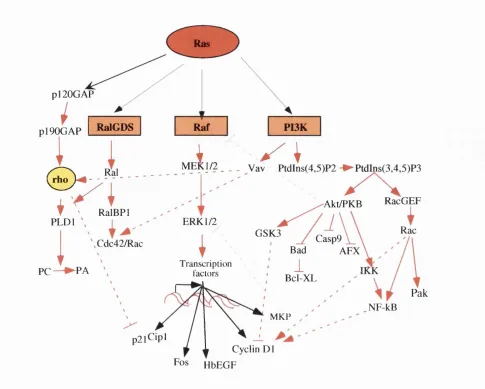
Mutations in a ras allele occur in 30% of all human tumours (Bos, 1989b; McCormick and Wittinghofer, 1996). This makes ras the most widely mutated human proto oncogene, highlighting the importance of this gene. This is mirrored in the multiple effects that Ras expression can have on all facets of cell behaviour, including both positive and negative effects on cell proliferation, apoptosis, differentiation and senescence. With such a diverse spectrum of cellular responses to Ras activation, it is not surprising that Ras also uses a multitude of downstream effectors. Overwhelming evidence identified the Raf/MAPK pathway as a key effector in Ras signalhng (For reviews see (Campbell et al., 1998; Marshall, 1996; McCormick, 1999; Shields et al., 2000). Ras binds to at least two other types of effector protein: phosphoinositide 3- kinases (PI3K) and members of the Ral-guanine nucleotide exchange factor (RalGBF) family. In this thesis, I will concentrate on the Ras/Raf/MAPK pathway but it is important to remember that other Ras targets are indispensable in ehciting a full Ras biological response and these will be discussed briefly.
1.3.1 Ras: a molecular switch.
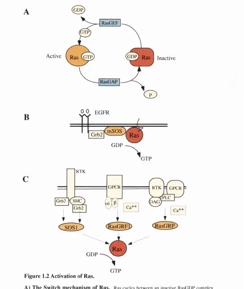
Ras proteins act as molecular switches, cycling between inactive GDP and active GTP- bound states, and function as important relays in the transduction of signals from membrane receptors. Regulation of this cychng is aided by opposing effects of GTPase activating proteins (GAPs) (Trahey and McCormick, 1987), which increase the intrinsic GTPase activity of Ras, and by Guanosine nucleotide exchange factors (GEFs) (Prive et al., 1992; Schaeffer et al., 1998), which promote exchange of GDP for GTP (Fig. 1.2 A). Two GAPs which show catalytic activity towards Ras are p i 20°^^ (Adari et al.,
1988; Trahey and McCormick, 1987; Vogel et al., 1988) and N Fl (Ballester et al., 1990; M artin et al., 1990; Xu et al., 1990a). GEFs recruitment to the plasma membrane activates Ras signalling (Quilliam et al., 1995). The three major manunalian GEFs in Ras signal transduction are SOS 1 and 2 (Bowtell et al., 1992; Chardin et al., 1993), RasGRFl and RasGRF2, and RasGRP (Fam et al., 1997; Farnsworth et al., 1995). The role o f other putative Ras GEFs, such as Vav and Smg-GDS, is more controversial (Bustelo et al., 1994; Gulbins et al., 1993; Gulbins et al., 1994; K hosravi-Far et al., 1994; M izuno et al., 1991; Takai et al., 1993). GAPs and GEFs enzymatic activity responds to extracellular stimuli such as growth factors (Lowy and Willumsen, 1993; McCormick, 1993). Most receptor tyrosine kinases (RTK) do not bind to GEFs directly but do so through adaptor proteins, such as the mammalian protein growth factor receptor-bound protein 2 (Grb2).
Ras genes become constitutively activated when point mutated in specific amino acid residues. The most common mutations in human tumours are 12,13 and 61 (Lowy and W illumsen, 1993). Ras proteins activated by these mutation become immune to the effects of GAPs since they have lost their intrinsic GTPase activity and so become GTP bound (Adari et al., 1988; Scheffzek et al., 1997; Trahey and McCormick, 1987; Vogel et al., 1988). Mutants that act in a dominant negative fashion towards Ras family members such as Ras^*^ have a very low affinity for guanosine nucleotides and may act by sequestering RasGEFs and preventing normal Ras molecules from utilising GEF exchange activity (van den Berghe et al., 1997).
Activation of Ras occurs in response to a wide variety of stimuli, such as growth factors, cytokines, hormones, and neurotransmitters. These stimuli signal to transmembrane receptors such as RTKs, non-receptor tyrosine kinase-associated receptors and G-protein coupled receptors (GPCR). One of the best-characterised Ras-mediated signal transduction pathways is the activation of the epidermal growth factor
Active
RasGEF
G T P
nGDP) Ras
I
InactiveRasGAP
C D
B
mSOS
Grb2 Ras
GDP
GTP
RTK
GPCR RTK H GPCR PLC
Grb2 SHC DAG
Grb2
(^asGR^
<IasGRe>
SOSl
Ras GDP
GTP Figure 1.2 Activation of Ras.
A) The Switch m echanism of Ras. Ras cycles between an inactive RasGDP com plex and an active RasGTP complex in a regulated manner depending on the opposing actions o f GEFs and GAPs.
B) Activation of Ras by the EG FR.
C) Activation of Ras by different receptors and RasG EFs. Three distinct Ras GEFs have been identified; SOS 1/2, R asG R Fl/2 and RasGRP. RTK activation leads to recruit ment o f Grb2, com plexed to SOS, to the plasma membrane. This facilitates SOS activation of Ras. GPCRs that signal through heterotimeric G proteins consisting o f a i and Py subunits active RasGRF. This involves calcium associated RasGRF-associated calmodulin. Receptor activation
(EOF) receptor. This is depicted in Fig. 1.2 B. The EGF receptors are single transmembrane domain proteins that dimerize and transphosphorylate upon binding to their native ligand. These phosphorylated residues then provide binding sites for the Src- homology 2 (SH2) domains of Grb2 (Lowenstein et al., 1992; Matsuoka et al., 1995). Grb2 is in complex with the carboxy terminus of SOS via its SH3 domains. Therefore, SOS is recruited to the EGF receptor complex at the plasma membrane, where it can activate plasma membrane associated Ras via catalysing Ras-GTP exchange (McCormick, 1993; Moodie and Wolfman, 1994). Subsequently, Ras associates with the kinase Raf-1, initiating the Raf/MEK/ERK/MAPK cascade, eventually leading to gene expression in the nucleus (See section 1.3.2). Feedback phosphorylation of SOS by the activated ERK pathway induces the disassembly of the SOS complex and termination of Ras activation. Other growth factor receptors use similar mechanisms (Fig. 2.2 C). For example, the SOS-Grb2 complex binds to the insulin receptor via another adaptor protein. She and the insulin receptor substrate (IRS-1) (Skolnik et al., 1993). A similar though more complex mechanism is used by FGF receptors (for review see (Olson and Marais, 2000). GPCR that signal through G proteins consisting of a i and Py subunits activate RasGRF. This involves calcium (Ca^^) association of RasGRF-associated calmodulin. Receptor activation (GPCR and RTK) of PLC, leading to the production of DAG and the release of intracellular calcium, may stimulate RasGRP activation of Ras (reviewed in (Luttrell et al., 1999; Reuther and Der, 2000). An increase in GEF activity is just one mechanism by which Ras is activated. Phorbol ester treatment of T cells leads to Ras activation involving no change in guanine nucleotide exchange activity, and instead coincides with downregulation of RasGAP activity (Downward et al., 1990). Furthermore, in adipocytes, Ras activity is regulated by PI3K-mediated inhibition of GAP activity (DePaolo et al., 1996).
1.3.2 Ras/Raf/MAPK pathway
1.3.2.1 R a f as a Ras effector
The Raf/MAPK pathway remains one of the key signalling pathways important for Ras biology, ra/genes encode serine/threonine-specific kinases that integrate upstream input signals and as such have complex regulation. Raf kinases were first discovered as gain of function mutants with the ability to induce growth and morphological transformation of established cell lines. Mammals possess three R af proteins: Raf-1 (also referred to as c-Raf) A-Raf and B-Raf (Bonner et al., 1984; Huleihel et al., 1986; Ikawa et al., 1988). Raf-1 protein is ubiquitously expressed. On the other hand, B-Raf and A -raf isoforms exhibit more restricted expression profiles, being predominately expressed in neuronal and urogenital cells (Storm et al., 1990; Wadewitz et al., 1993). The different phenotypes of
Ch a pt er 1 - In t r o d u c t io n
the Raf-1, A -Raf and B-R af knockout mice strongly suggest that these proteins are non- redundant and serve distinct functions ( Pritchard et al, 1996; Wojnowski et al, 1997;
Wojnowski et al, 1998). A-Raf-/- mice produce intestinal and/or
neurological defects depending on the genetic background. B-Raf-/- mice have neuroepithelial differentiation defects as well as defects in maturation and maintenance of endothelial cells. They die in utero due to vascular haemorrhage. In contrast, Raf-1-/-mice die during midgestation, in an inbred background, and, die shortly after birth, in an outbred strain. The latter show general growth retardation and developmental defects, the most apparent in the placenta, lung and skin. This indicates a general role of Raf-1 in morphogenesis in comparison to the specialised functions of A-Raf and B-Raf. Raf was originally placed downstream of Ras because genetic evidence from Drosophila and C.elegans showed that Raf is essential for Ras signalling in eye and vulval development, respectively (Dickson et al., 1992; Han et al., 1993). Raf was subsequently shown to be necessary for Ras signalling in mammalian cells since dominant negative Raf molecules blocked Ras-induced gene transcription (Bruder et al., 1992) and proliferation (Kolch et al., 1991). D irect binding of Raf-1 to Ras was dem onstrated using yeast two hybrid analyses and direct in vi/ro binding assays. Specifically, the N-terminal portion of Raf-1 binds directly to the effector domain of Ras-GTP (Moodie et al., 1993; Van Aelst et al., 1993; Vojtek et al., 1993; W ame et al., 1993; Zhang et al., 1993). Moreover, Raf-1 can reproduce many of the cellular responses of Ras in mammalian cells. For example, both Ras and Raf-1 can activate ERK, and dominant negative versions of both can block growth factor induced ERK activation (de Vries-Smits et al., 1992; Schaap et al., 1993). Furthermore, the Ras^’^°^^ effector domain mutant, which can no longer bind to Raf-1, is defective in ERK activation and this impairment can be complemented by mutations in Raf-1 which restore an interaction with Ras^^^^^^ (White et al., 1995). In Schwann cells, P C -12 cells, and fibroblasts, Raf-1 has been shown to mimic many of the effects of activated Ras (discussed in Section 1.3.7) (Lloyd et al., 1997; Noda et al., 1985; Serrano et al., 1997; Sewing et al., 1997; Woods et al., 1997; Zhu et al., 1998). These observations provide strong evidence that Raf-1 is a genuine effector of Ras signalling responsible for the activation of the ERK/MAPK pathway, with Raf-1 being able to elicit many of the Ras phenotypes (reviewed in (Katz and McCormick, 1997; Marshall, 1999; Marshall, 1996; Shields et al., 2000).
13.2.2 R a f activation
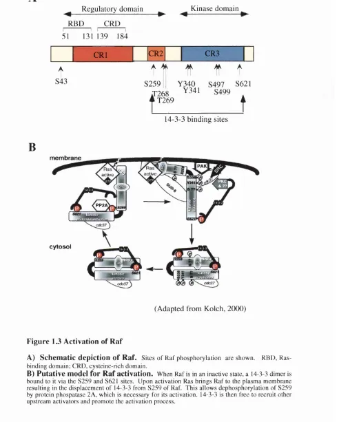
Raf-1 activation appears to be a complex, multi-step process involving many components and the mechanism remains to be fully deciphered (for review see (Kolch, 2000; Morrison and Cutler, 1997). A current model of Raf-1 activation is depicted in Fig. 1.3 B. Raf-1 is composed of two functional domains, the amino terminal regulatory domain (conserved regions, C Rl (aa 62-194) and CR2 (aa 254-269)) and the carboxy terminal
kinase domain (CR3 (aa 330-627)) which interacts with downstream targets of Raf-1 (Fig. 1.3 A). The amino terminal domain of Raf-1 supresses its catalytic activity and deletion of part of this amino-terminal domain results in a constitutively activated Raf-1 mutant (Morrison and Cutler, 1997).
In its inactive state, Raf-1 is normally located in the cytosol in a multi-protein complex of 300-500 kDa (Wartmann and Davis, 1994). Ras is known to play a key role in Raf-1 activation by directly interacting with and translocating Raf-1 to the plasma membrane from the cytoplasm (Traverse et al., 1993). Signalling that activates Ras results in the formation of Ras-Raf-1 complexes. Mutations in either Ras or Raf-1 that block this interaction, or inhibit Ras function, prevent Raf-1 activation (Luo et al., 1997; Marais et al., 1998; Marais et al., 1995; Rodriguez-Viciana et al., 1997). However, the discovery that purified recombinant Ras is not sufficient to activate Raf-1 in vitro eluded to other components being involved (Traverse et al., 1993; Zhang et al., 1993). Furthermore, cytosohc Ras (membrane localisation blocked by post-translational modification inhibitors) is still able to complex with Raf-1, but these cytosolic complexes are inactive (Kikuchi and Williams, 1994; Lemer et al., 1995; Okada et al., 1996). It is now known that Ras-GTP cannot activate Raf-1 unless Ras-GTP is membrane bound and an unidentified cytosolic factor is present (Dent and Sturgill, 1994; Stokoe and McCormick,
1997; Tamada et al., 1997).
Ras can interact with two domains in the Raf-1 N-Terminus: the Ras binding domain (RED), aa 50-40, and the Cysteine rich domain (CRD), aa 139-186. These have a low affinity for RasGDP and a high affinity for RasGTP. The RED alone is sufficient for translocation of Raf-1 from the cytosol to the membrane. The CRD is necessary for efficient activation (Hu et al., 1997; Luo et al., 1997; Roy et al., 1997). This is consistent with the finding that Raf-CAAX (which tethers Raf to the membrane) results in only partial activation and that it can be further stimulated by growth factors or RasGTP (Leevers et al., 1994; Marais et al., 1998; Marais et al., 1995; Stokoe et al., 1994). This appears to be mediated by Ras directly binding to Raf-CAAX, as well as by other Ras- initiated signalling processes (Mineo et al., 1997; Sun et al., 2000). Thus Ras supplies both direct and indirect activation signais
Regulatory domain Kinase domain
-► ^
RBD CRD
51 131 139 184
E
CRl CR2 CR3t
S43t
S259 268 T269
It
If
t
Y 340 S497 S621 Y 341 S499
t
14-3-3 binding sitesB
membrane
active y
PP2A
cytosol
(Adapted from Kolch, 2(XX))
Figure 1.3 Activation of R af
A) Schem atic depiction of Raf. Sites o f Raf phosphorylation are shown. RBD, Ras-binding domain; CRD, cysteine-rich domain.
The observation that Raf-1 becomes hypeiphosphorylated in response to upstream signalling and that Raf-1 activity can be abolished by protein phosphatases suggested that phosphorylation plays a role in regulating Raf-1 activity (Morrison and Cutler, 1997). Both serine/threonine and tyrosine phosphorylation appear to be important. The binding of 14-3-3 proteins to Raf-1 is also essential for regulating its activity and seems to be linked to the phosphorlyation of Raf-1 (Fantl et al., 1994; Fu et al., 1994; Tzivion et al., 1998). 14-3-3 are reported to assemble as dimers (Liu et al., 1995c; Luo et al., 1995). The cores of two 14-3-3 binding sites in Raf-1 are formed by phosphorylation of S259 and S621. Dephosphorylation of Raf-1 disrupts 14-3-3 binding (Tzivion et al., 1998). The binding of 14-3-3 to the N-terminus of Raf-1 appears to inhibit Raf-1 activity whereas C-terminal binding is essential for activity (Clark et al., 1997; Rommel et al., 1997; Tzivion et al., 1998). Furthermore, removal of 14-3-3 by competition with synthetic phosphopeptides disabled both basal and induced Raf-1 activity. Re-addition of recombinant 14-3-3 could revive Raf-1, but only if it had been activated previously. This leads to a proposed role of 14-3-3 to stabilise both the inactive and the activated conformations of Raf-1. Importantly, Ras interferes with the interaction between amino terminal of Raf-1 and 14-3-3 (Rommel et al., 1996). Furthermore, déphosphorylation of S259 is one of the first changes in Raf-1 phosphorylation during mitogen induced activation and is required for activation (Abraham et al., 2000). This déphosphorylation o f S259 appears to be carried out by protein phosphatase 2A (Abraham et al., 2000). Inhibition of protein phosphatase 2A prevents both dephosphorylation and activation of Raf-1. It is thought that on activation, Raf-1 is brought to the membrane via RasGTP resulting in the displacement of 14-3-3 from S259 and allowing the necessary dephosphorylation of the S259 site by protein phosphatase 2A. 14-3-3 is then free to possibly recruit other upstream activators and promote the activation process (Drugan et al., 1996). The exact role of S621 is as yet unclear. The presence of S621 is required for full CR3 activation by stimulatory factors and the continuous presence of 14-3-3 at this site is necessary for retaining activity once the kinase is activated (Yip-Schneider et al.,
2000).
Activated R af is also phosphorylated at residues Y340 and Y341 residues
(Fabian et al, 1993; Chow et al, 1995; Marais et al ,1995; Mason et al, 1999). This is dependent upon Ras activity in mammalian cells and is performed by tyrosine kinases such as the Src family. Furthermore, abl (Skorski et al., 1995; Weissinger et al., 1997) and the Janus ædvated kinases (JAKs) also induce Raf-1 activation and tyrosine phosphorylation (Stancato et al., 1997; Xia et al., 1996). It has been suggested that tyrosine phosphorylation of Raf-1 is also regulated by Raf-1 itself and by the phosphatase Cdc25A (Xia et al., 1999). In addition, Ras also indirectly regulates Raf-1 activation through the activation of PI3K. Active PI3K leads to p21 activated protein kinase.
Ch a pt er 1 - In t r o d u c t io n
PAK3, directly phosphorylating Raf-1 on S338 (Chaudhary et al., 2000; Sun et al.,
2000).
There appears to be a role for the Wnase supressor of Ras (KSR) in Raf-1 regulation (Therrien et al., 1995; Therrien et al., 1996). Several groups suggest KSR interacts (possibly via 14-3-3 proteins) with and activates Raf-1 in a membrane bound multi protein signalling complex (Michaud et al., 1997; Xing et al., 1997; Zhang et al., 1997). Other groups show that KSR does not interact with Raf-1, but does interact with MEK and ERK in yeast two hybrid screens and by inununoprecipitation of endogenous proteins from P C I2 cells (Denouel-Galy et al., 1998; Yu et al., 1998). The physiological role for KSR remains enigmatic as both activating and inhibitory effects of KSR have been reported (Denouel-Galy et al., 1998; Michaud et al., 1997; Therrien et al., 1996; Xing et al., 1997; Yu et al., 1998). Different MAPK modules in yeast share components, and the specificity of signalling in response to stimuli is thought to be achieved by the use of scaffold or adaptor proteins (For review see(Kolch, 2000). KSR could be a candidate scaffold protein for the Raf/MEK/ERK module that binds to MEK and ERK constitutively, but only to Raf-1 at the cell membrane. There are also indications that KSR is a substrate for ERK, which may be an event connected to a regulatory feedback loop (Cacace et al., 1999). Furthermore, a novel gene, connector enhancer of KSR {cnk) is also required for efficient signal transmission within the Ras/ERK cascade. CNK is a possible target of tyrosine phosphorylation and contains several protein-protein interaction domains, suggesting it functions also as a multi valent adapter protein (Therrien et al.,
1998). Other Raf-1 binding proteins, such as the molecular chaperone heat shock proteins (HSP), HSP 90 and HSP 50, may have roles in maintaining Raf-1 protein stability and its proper localisation. Their binding to Raf-1 is irrespective of its activation state (Schulte et al., 1995; Schulte et al., 1996). Finally, forced dimérisation of Raf-1 proteins has been demonstrated by two groups to activate Raf-1 signalling (Farrar et al., 1996; Luo et al., 1996) and SUR- 8 can also form a ternary complex with RasGTP and
Raf-1, enhancing Raf activation (Li et al., 2000b). Future experiments are necessary to elucidate an exact mechanism of Raf-1 regulation.
1.3.2.3 The Raf/MEK/MAP kinase pathway.
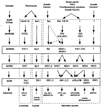
Raf-1 controls a cascade of dual specificity kinases in which Raf-1, a MAP kinase kinase kinase, phosphorylates and activates MEK, a MAP kinase kinase. This in turn phosphorylates, on both threonine and tyrosine residues, and activates p42 and p44 ERK serine/threonine kinases (MAPK) resulting in their translocation into the nucleus and the activation of transcription factors (see Fig. 1.4). This sequence of events is generally
termed the MAP kinase (MAPK) cascade (Reviewed in (Campbell et al., 1998; Marshall, 1996).
ERKs, once in the nucleus, phosphorylate more than 50 substrates. These include cytoskeletal proteins, kinases, phosphatases, enzymes and transcription factors (reviewed in (Treisman, 1996)). For example, those in the Ets family (such as Elkl; Ets transrepressors e.g. ERF; ATFs; c-fos; c-myc and the estrogen receptor) and bZIP and MADS box containing transcriptional regulators (Lewis et al., 1998). TBP can be upregulated by Ras via either the Raf-1 and RalGDS pathways (Johnson et al., 2000). In addition, ERKs can activate protein kinases referred to as MAPK activated protein kinases (MAPKAPK) 1, 2 and 3 and Mnks 1 and 2. Some MAPKAPKl substrates are involved in transcription and include cAMP response-element binding protein (CREE), CREE binding protein (CEP), c-fos, Nurr 77 and serum response factor (SRF) Chen, 1996a #2567; (Nakajima et al., 1996; Tan et al., 1996; Xing et al., 1996). MAPKAPK2 and MAPKAPK3 phosphorylate HSP27 which is believed to be an actin capping protein (Clifton et al., 1996; Engel et al., 1995; McLaughlin et al., 1996; Stokoe et al., 1992). M nkl and 2 phosphorylate Elongation Initiation Factor 4E (EIF4E), implying that they have a role in translational control (Waskiewicz et al., 1997). ERKs also activate phospholipase A2 which suggests a role for ERKs in agonist stimulated arachidonic acid release (Lin et al., 1993; Nemenoff et al., 1993). Furthermore, the duration and intensity of MAPK activation may be important in the downstream effectors activated. It has been shown that active R af-1 can elicit either a mitogenic response or cell cycle arrest in NIH3T3 cells, depending on the level of pathway activation (Sewing et al., 1997). Indeed, it has been demonstrated that the duration of MAPK signalling determines the repertoire of Fos and Jun proteins upregulated. The choice of substrates for ERK 1/2 are potentially controlled by a number of mechanisms including cell-type and situation specific expression. Positive feedback loops also occur within the cascade. For instance ERKs can phosphorylate M EK l (Gotoh et al., 1994; Zheng and Guan, 1993), Raf-1 (Anderson et al., 1991; Kyriakis et al., 1993; Lee and Fain, 1991) and KSR, and M EKl can phosphorylate Raf-1 through an ERK dependent pathway (Zimmermann et al.,
1997). Docking domains such as KIM and DEE, which act respectively as ERK specific and MAPK generic binding sites and guide the appropriate MAPK to its phosphorylation target, could also help achieve specificity (reviewed in (Kolch, 2000)).
Similar to the scaffold protein KSR, adapter proteins have been reported to have a role in the proper physical interaction between the components of the MAPK cascades. Recently, two non-enzymatic adapter proteins have been identified in mammalian cells;