Modelling T Cell Activation
Cliburn Chan Chi Wei
Thesis submitted for the degree of
Doctor of Philosophy
Centre for Nonhnear Dynamics and its Applications
University College London
ProQuest Number: U642209
All rights reserved
INFORMATION TO ALL USERS
The quality of this reproduction is dependent upon the quality of the copy submitted.
In the unlikely event that the author did not send a complete manuscript
and there are missing pages, these will be noted. Also, if material had to be removed,
a note will indicate the deletion.
uest.
ProQuest U642209
Published by ProQuest LLC(2015). Copyright of the Dissertation is held by the Author.
All rights reserved.
This work is protected against unauthorized copying under Title 17, United States Code.
Microform Edition © ProQuest LLC.
ProQuest LLC
789 East Eisenhower Parkway
P.O. Box 1346
A cknow ledgem ents
I am extremely grateful to my two supervisors, Jaroslav Stark and Andrew George, who have provided wonderful support and encouragement for my work. One could not ask for better supervisors.
Thanks to all the folk in the Centre for Nonlinear Dynamics and its Appli cations who have made my stay there so enjoyable, especially Rob, Ben, Sakse, Alex, Ricardo, and Winston. Thanks also to David and Anita, who ‘suffered’ the same MSc program in Chaos and Nonlinear dynamics together with me, for the many, many coffee breaks we had. Steve Baigent and Steve Bishop a t the Centre taught me much about mathematical biology and modelling r^pectively.
Andrew Yates at the Institute of Child Health has provided many stimulating discussions, use of his awesome Silicon Graphics workstation, and introduced me to the best banana shake in central London. He also read through and provided useful feedback on a draft of this thesis, for which I am truly grateful. Thanks, Andy!
The immunologists at the Hammersmith Hospital have been surprisingly tol erant of my modelling work, and extremely friendly as well. Nothing motivates me to clarify my thoughts more than having to present my work to this partic ular audience. Thanks especially to Robert Lechler and Mary Ritter, for their encouragement of this work. Robert Lechler, Mary Ritter and Ron Germain (at the National Institute of Health) also read through and commented on an early version of Chapter 6.
I would like to thank my parents for their years of care and love, my brothers for their support and visits, and my nieces and nephews for the happy noises during those visits.
Finally, I gratefully acknowledge the financial support provided by an ORS and a UCL graduate school scholarship.
A b s tr a c t
The recognition of foreign antigen by T cells is the foundation of the adaptive inunune response, and this has been demonstrated experimentally to be an extremely sensitive, specific and reliable process. In this thesis, models for T cell signalling based on recent experimental data are constructed to understand how these properties arise.
Contents
1 Introdu ction 12
1.1 O v e rv ie w ... 12
1.2 Chapter o u t l i n e s ... 16
2 B iology o f A ntigen R ecognition and T C ell A ctivation 18 2.1 Introduction... 18
2.2 An overview of the immune s y s te m ... 19
2.2.1 The Anatomy of A c tiv a tio n ... 20
2.2.2 Antigen Processing and P re sen ta tio n ... 21
2.2.3 T Cell D ev e lo p m e n t... 26
2.2.4 Peripheral Circulation of T C e l l s ... 29
2.2.5 T Cell A ctivation... 30
2.2.6 Ligand Quantity and TCR Signalling ... 36
2.2.7 Ligand Quality and TCR S ig n a llin g ... 37
2.3 S u m m a r y ... 38
3 M odelling T cell activation and antigen recognition 39 3.1 Models for TCR triggering... 40
3.1.1 Conformational change ... 40
3.1.2 M ultimerisation... 42
3.1.3 TCR clu ste rin g ... 44
3.1.4 Lipid r a f ts ... 45
3.1.5 Kinetic Segregation ... 47
3.2 Models for T cell se n sitiv ity ... 48
3.2.1 Serial lig a tio n ... 48
3.2.2 Tunable activation th r e s h o l d ... 49
3.3 Models for T ceU sp ecificity... 51
3.3.1 Kinetic proofreading... 51
3.4 Models for T cell re lia b ility ... 55
3.4.1 Extrinsic n o i s e ... 55
3.4.2 Intrinsic n o ise ... 56
3.4.3 R o b u s tn e s s ... 57
3.5 S u m m a r y ... 57
4 A n exten d ed analysis o f kinetic proofreading in T cells 59 4.1 Introduction... 59
4.2 Biological plausibility of kinetic p ro o fre a d in g ... 59
4.3 Kinetic proofreading - a view from the T C R ... 61
4.3.1 Waiting time and TCR activation ... 61
4.3.2 The effect of stochastic ligand dissociation... 65
4.3.3 Sequential versus parallel m o d ific a tio n s... 67
4.3.4 Lack of robustness to parameter variations ... 68
4.4 C onclusions... 69
5 Feedback in T C R signalling 72 5.1 Introduction... 72
5.2 Feedback in TCR sig n a llin g ... 73
5.2.1 Basic signalling p a th w a y s ... 74
5.3 Lck recruitment m o d e l... 76
5.3.1 Model assum ptions... 76
5.3.2 Model d e s c r ip tio n ... 77
5.3.3 Model r e s u lt s ... 80
5.4 Model v a r ia tio n s ... 92
5.4.1 No negative feedback... 94
5.4.2 No reciprocal activation of Lck by ZA P-70... 94
5.4.3 No reciprocal activation of Lck by ZAP-70 and no negative feedback... 97
5.5 Stochastic m o d e llin g ... 101
5.5.1 R a tio n a le ...101
5.5.2 Description of alg o rith m ...102
5.5.3 Stochastic model re s u lts ... 103
5.6 C onclusions...109
6 C ooperative enhancem ent o f T C R specificity 111 6.1 Introduction...I l l 6.2 M o d e l... 112
6.3.1 Poor specificity in absence of signal spread ... 115
6.3.2 Spreading inhibition improves recognition specificity . . . 116
6.3.3 Spreading protection improves s e n s itiv ity ...118
6.3.4 Varying neighbourhood size reveals trade-off between sen sitivity and sp e c ific ity ... 120
6.3.5 Reliability of signalling in presence of cooperativity . . . . 120
6.3.6 Rebinding enhances sensitivity to weak lig a n d s ... 123
6.4 D iscussion...126
6.4.1 Altered lig a n d s ... 126
6.4.2 Thymocyte developm ent...126
6.4.3 Model lim itations... 127
6.5 C onclusions...130
7 C onclusions 131 7.1 R eview ...131
7.2 Future w o rk ...132
A A nalysis and num erics for nonlinear O D Es 136
List o f Figures
2.1 Antigen presentation by dendritic c e lls ... 22
2.2 Peptide-MHC:TCR in te r a c tio n ... 23
2.3 MHC class I and II antigen processing p a t h w a y s ... 24
2.4 Selection in the t h y m u s ... 28
2.5 MIRRs and their IT A M s... 31
2.6 Immune s y n a p s e ... 32
2.7 Proximal T cell s ig n a llin g ... 34
3.1 Models for TCR triggering... 41
3.2 Clustering induced C u n f o ld in g ... 46
3.3 Tunable activation threshold sim ulations... 50
3.4 Kinetic proofreading m o d e l ... 51
3.5 Kinetic discrimination m o d e l ... 54
3.6 Kinetic differentiation model ... 54
4.1 Kinetic proofreading m o d e l ... 60
4.2 Improving the resolution of kinetic proofreading... 63
4.3 Rescaling in kinetic p ro o frea d in g ... 64
4.4 Activation probabihty of l i g a n d s ... 67
4.5 Kinetic proofreading without obligatory sequence of modifications 68 4.6 Increasing activation threshold decreases sensitivity and hgand discrim ination... 70
5.1 TCR activation Model 1 ... 78
5.2 Hysteresis in TCR a c tiv a tio n ... 81
5.3 Phosphatase inhibition can activate T C R ... 82
5.4 Ligand discrimination is robust to threshold tu n in g ... 83
5.5 Ligand discrimination in feedback m o d e l... 84
5.6 Rebinding and hysteresis can sustain sig n a llin g ... 85
5.8 Threshold hierarchy from multiple kinases (II) ... 91
5.9 Effect of rebinding on multiple kinase m o d e l... 93
5.10 TCR activation Model 2 ... 94
5.11 Ligand discrimination from Model 2 ... 95
5.12 TCR activation Model 3 ... 96
5.13 Phase plane analysis for Model 3 ... 98
5.14 TCR activation Model 4 ... 99
5.15 Phase plane analysis for Model 4 ( I ) ...100
5.16 Phase plane analysis for Model 4 ( I I ) ...101
5.17 Stochastic simulation reproduces threshold e f f e c t ... 105
5.18 Hysteresis reduces c h a t t e r ... 106
5.19 Stochastic simulation suggests role for ITAM multiphcity . . . . 108
6.1 Possible state transitions for the T C R ...113
6.2 Model for spreading inhibition and p ro te c tio n ... 114
6.3 TCR neighbourhood and signal s p re a d in g ...115
6.4 Effect of spreading inhibition and protection ... 117
6.5 Effect of Hill coefficient on ligand d isc rim in a tio n ... 119
6.6 Effect of neighbourhood s iz e ... 121
6.7 Spreading signaUing and re lia b ih ty ...122
6.8 Possible state transitions (rebinding m o d e l) ... 124
6.9 Effect of r e b in d in g ...125
6.10 Simulation of maturation with single h g a n d ... 128
List of Abbreviations
A P I Activating protein 1 A F C Antigen presenting cell A P L Altered peptide ligands B C R B cell receptor C b p Csk binding protein C D Cluster of differentiation.
C D F Cumulative distribution function
C L IP Class Il-associated invariant chain-derived peptide C sk C-terminal Src kinase
C T L Cytotoxic T lymphocyte D C Dendritic cell
D P Double positive refers to thymocytes expressing both CD4 and CD8 D N A Deoxyribonucleic acid
E R Endoplasmic reticulum
E R K Extracellular signal-regulated kinase F R E T Fluorescence resonance energy transfer H L A Human leukocyte antigen
H S P Heat-shock protein
IS Immune synapse
IT A M Immimoreceptor tyrosine-based activation motif Ig Immunoglobulin
II Invariant chain
J N K Jun N-terminal kinase LA T Linker of activation for T cells LFA Lymphocyte function-related antigen LH S Left-hand side
L P S Lipopolysaccharide M A P Mitogen activated protein M A P K MAP kinase
M C P Monocyte chemoattractant protein M H C Major histocompatibility complex M I R R Multichain immune recognition receptor N F Nuclear factor
N FA T Nuclear factor of activated T cells N F Nuclear factor
P A G Phosphoprotein associated with glycosphingolipid-enriched microdomains P A M P Pathogen associated molecular patterns
P D F Probability density function P I Phosphatidylinositol
P L C Phospholipase C P K C Protein kinase C
P T P Protein tyrosine phosphatase R H S Right-hand side
S H P Protein tyrosine phosphatases with SH2 domains S H Src homology
SLT Secondary lymphoid tissues
S L P -76 SH2 domain containing leukocyte phosphoprotein of 76 kDa S M A C Supramolecular activation clusters
S N R Signal to noise ratio
S P Single positive - refers to thymocytes expressing only CD4 or CDS T A P Transporter associated with antigen processing
To Cytotoxic T cell T C R T cell receptor T L R Toll-like receptor T h Helper T cell
Chapter 1
Introduction
“The immunocosmologies of the 1970s, based on creative interpreta tion in the absence of reliable biochemistry or genetics, have largely fallen into disfavor, taking with them the arcane circuitries of sup pressor cells, helper factors, and antiidiotypic networks.”
- Christophe Benoist and Diane Mathis, Chapter 11 Page 398 (T- Lymphocyte Differentiation and Biology) of Fundamental Immunol ogy, Fourth Edition (Lippincott-Raven, 1999)
1.1
O verview
The immune system is generally considered to have irmate and specific immune responses (Paul, 1999). Innate immunity is more ancient in an evolutionary sense, and provides the early lines of defence against pathogens. It comprises physical and chemical barriers to pathogen entry, the complement system (and other blood proteins involved in inflammation), phagocytic cells (neutrophils and macrophages) and other leucocytes (e.g., natural killer cells). The responses of innate immunity are stereotyped, and largely based on the recognition of invariant pathogen associated molecular patterns (PAMPs) (Janeway, Jr., 2001). In contrast, adaptive immunity is characterised by memory and specificity. The adaptive immune response ‘remembers’ previously encountered pathogens, such th a t subsequent encounters result in increasingly effective defence mechanisms (memory). In addition, the nature of the adaptive immune response varies according to the type of pathogen (e.g., intra- or extra-cellular), and is tailored to eliminate it effectively (specificity).
presenting cells, the dendritic cells (DCs), sit and wait at the interfaces be tween the body and the environment. DCs constantly sample the environment, and then migrate to the secondary lymphoid organs (lymph nodes and spleen) where they present their captured antigens to the T cells. If the T cell receptor (TCR) recognises the ligand presented by the DC, and the DC provides the right costimulatory signals, the T cell then prohferates and mounts an appropriate immune response.
One of the major discoveries in immimology was th at the TCR only recog nises ligand in the form of short peptides presented in the groove of a molecule known as the major histocompatibility complex (MHC). There are two forms of this molecule; MHC class II is only found on antigen presenting cells and is recognised by a subclass of T cells known as CD4 T cells, while MHC class I is found on aU nucleated cells and is recognised by CD8 T cells. The essential difference between the MHC molecules is th a t class I presents peptides derived from the cell interior, while class II presents peptides captured from the envi ronment. Somewhat surprisingly, cells do not appear to distinguish between self and pathogen protein when cleaving them up into peptide fragments suitable for MHC presentation, and most of the peptides presented on the ceU surface by MHC molecules are actually derived from self proteins.
T cells axe extremely sensitive to the presence of their cognate peptide pre sented on MHC molecules. It is known, for example, th a t CD4 T cells can proliferate in response to as few as 10-100 MHC bearing foreign peptide, while CD8 T cells appear to be even more sensitive (Harding and Unanue, 1990; De- motz et al., 1990; Sykulev et al., 1996). Since T cells can respond appropriately even though self peptides outnumber the foreign peptides on the cell surface by a factor of 1000 to 10000, there is little margin for error and the TCR must be an exquisitely specific biosensor.
The aim of this thesis is to model some of the events occurring at the T cell-.APC interface th a t may be responsible for the amazing fidelity of the T cell in the presence of a high signal to noise ratio (SNR). Obviously, these properties of the T cell sensing system are critical for the proper functioning of the immune system and indeed, the organism itself. Errors in one direction would lead to failure to control pathogen, while errors in the other direction result in auto-immunity, a condition where the immune system attacks self tissue with consequent morbidity and sometimes mortality.
the computer in these modem times) can complement 96 multi-well plates (and surface plasmon resonance biosensors in these modem times). The first bene fit of modelling and computer simulation is th a t it enforces clarity of thought. Assumptions used in any model must be explicitly formulated, and often this allows one to see implications or make predictions which may not otherwise be obvious. This is particularly tm e of nonlinear systems (just about everything in immunology) which often have unexpected or even counter-intuitive behaviour. A second benefit of modelling is th a t it is often not possible to isolate par ticular biological subsystems of interest to analyse; this is usually trivial in a mathematical model or computer simulation. Finally, it is sometimes possible to model events beyond the resolution of current laboratory instruments, for example, stochastic fluctuations of signalling components about a single T cell receptor. In this case, modelling is the only recourse we have at present for understanding the implications of the phenomenon.
In terms of basic science, a good model of the mechanisms underlying T cell specificity and sensitivity would be of benefit simply because it provides insight into the foundation of the adaptive immune response. Additionally, it could help us understand T cell developmental biology and how the T cell can respond to the same or a similar set of antigens with different responses (positive and negative selection) during thymic development.
In terms of applications, understanding the characteristics of ligands th at excite T cells would clearly have the potential to lead to better therapeutics. It would benefit rational vaccine design, since epitopes could be synthesised to optimise T cell response to various infectious agents or tumours. On the other hand, understanding the characteristics of ligands that result in them being ignored by T cells would also be helpful. It could result in a better theoretical understanding for how altered peptide figands (APL) work, which has important clinical implications (Sloan-Lancaster and Allen, 1996). For example, APL have been suggested as therapy to down-regulate unwanted specific immune responses in the case of autoimmune disease. Another example of the clinical importance of understanding how APL work is in the field of infectious disease, where HIV often manages to subvert the immune response by expressing antigens similar to antagonist APL. Finally, there is the possibility of applying the model for sensitivity and specificity to the design of better biosensors, which would be of commercial importance.
mathematical analysis; much of the analytical effort is in trying to relate the model to the immunology, seeing if it can shed any hght on experimental puz zles, or make testable predictions, or suggest therapeutic applications. There is a deUberate effort to make the models comprehensible and relevant to immu nologists, since they are obviously the only people qualified to test such models. The thesis itself integrates a small number of themes - how stochastic events, signal amplification, feedback control and cooperativity can result in the ‘emer gent’ T ceU properties of sensitivity, specificity and robustness. While stochas- ticity, amplification and feedback are characteristic of networks at the level of the gene, protein and cell, this thesis focuses mainly on protein interactions, although the modelling issues are very similar for genetic and cellular networks as well.
The first step in this analysis is to study the TCR as an autonomous unit, subject to stochastic fiuctuations in both the external and internal environ ment. The external environment refers to both the quality (mainly determined by hgand t i ) and quantity (rate of encounter) of peptide-MHC complexes. The internal environment refers to the signalling and adaptor molecules which inter act with a TCR once it has engaged figand.
The widely accepted kinetic proofreading model (McKeithan, 1995) which is typically presented in terms of equilibrium distributions of peptide-MHC:TCR complexes is re-interpreted for a single TCR and the implications of this con sidered. An alternative model for TCR figand discrimination based on feed back control of signalling pathways is then proposed, and an analysis of this model shows th at it can have the same strengths in enhancing specificity as ki netic proofreading. However, the analysis also shows clearly th a t in the face of stochastic figand dissociation, TCR functioning as independent signalling units cannot possibly account for the specificity seen experimentally.
The next step was then to consider a population of TCR on the cell-surface, locally coupled to their neighbours via ‘horizontal’ positive and negative sig nalling pathways. This was done by a Monte Carlo simulation, which showed th a t the recently discovered ‘spreading signals’ (Dittel et al., 1999) could signif icantly enhance specificity at a modest cost to sensitivity.
modelling and computer simulation. The advantages of such a combined ap proach in generating new hypotheses and making experimental predictions will be discussed in the concluding chapter.
1.2
Chapter outlines
C h a p te r 2 This is an introduction to T cell immunology, paying particular attention to what happens at the T cell-APC interface and what happens within the T cell after hgand engagement. T ceU development, activation and differentiation will be covered here. P art of this introduction was previously pubhshed (Yates et al., 2001).
C h a p te r 3 This is a review of previous modelling and computer simulation of T cell recognition and activation. We start by looking a t models for how the TCR is actually ‘triggered’ by hgand engagement to give a signal, then go on to discuss models for the critical T ceh properties of sensitivity, specificity and rehabihty. Much of the analysis focuses on the two fundar mental models for sensitivity and specificity th a t are widely beheved to be true, i.e., kinetic models and serial hgation. More recent theoretical contributions will also be covered.
C h a p te r 4 This chapter considers the kinetic proofreading theory for T cells in detail, and discusses several theoretical and practical problems with the original model. A stochastic version of the kinetic proofreading model for an individual TCR is constructed, and the imphcations examined. C h a p te r 5 A new model for specificity of hgand discrimination a t the level of
the individual TCR is described. The model is based on current under standing of how signalling is initiated, as well as the proximal intracehular signalling and feedback events. The model suggests a novel role for self peptide-MHC in sustained signalling, and possible experimental tests of the model are discussed. A stochastic version of the model suggests a possible role for immunoreceptor tyrosine-based activation motifs (ITAM) multiphcity in suppressing stochastic fluctuations.
dissociation. Recent experimental evidence for both positive and negative cross-talk between TCR, when built into the model, result in improved specificity at a modest cost to sensitivity. The implications of the model for understanding APL and thymocyte development are also discussed. Much of this chapter has been previously published (Chan et al., 2001). C h a p te r 7 This final chapter reviews the results of the previous work, consid
Chapter 2
B iology o f A ntigen
R ecognition and T Cell
A ctivation
2.1
Introduction
If there is a fundamental feature of adaptive immunology, it must be the recog nition of ligand by the multichain immune recognition receptors (MIRR), since this is the basis for specificity. The MIRR family includes the TCR, BCR, and receptors for IgG (FcyRIII) and IgE (FccRI), but this thesis will just focus on the T cell. However, it is likely th at the members of the MIRR share most of their functionality, and some of the models may be generalisable to other MIRRs.
While many factors contribute to overall T cell signalling, the primacy of TCR hgand recognition is acknowledged in the term signal 1 given to the in tracellular signals resulting from the peptide-MHCiTCR interaction. For an adaptive immune response in vivo, a signal 2 or costimulatory signal result ing from the hgation of co-receptors (e.g., CD28) is also necessary. Signal 2 presumably, provides a context for the recognition of hgand by the TCR.
secretion without proliferation) and full agonist. A rough categorisation of hg ands by their duration of binding can be made as follows:
Dissociation time Ligand < 2 s Null 2 — 5 s Antagonist 5 —10 s Partial agonist > 10 s Agonist
Models for how and why this is so wiU be presented in the next chapter, but this chapter will provide a summary of the biology of antigen recognition between T ceh and APC. An understanding of the specific type of response resulting from TCR engagement will also require a description of the possible micro-environmental contexts in which the antigen recognition event occurs. This chapter does not aim to be a comprehensive survey of immunology, only those parts necessary for understanding the biological basis of the models in the thesis are included.
2.2
A n overview o f th e im m une system
The immune system consists of a wide range of distinct cell types, but the lymphocytes have a central role because they confer specificity on the immune system, and are therefore responsible for adaptive immunity. Two broad classes of lymphocytes are the B lymphocytes which produce antibody, and the T lym phocytes which have important regulatory and effector roles. One subset of T lymphocytes is characterised by a cell surface marker known as CD4, and are sometimes also known as helper T cells (Th). Helper T cells provide help to B cells to prohferate and produce specific antibody and stimulate infiammar tion. Another subset of T cells are characterised by expression of CD8, and are also known as cytotoxic T cells (Tc). These serve to monitor somatic cells for infection and tumours, and can kill their target cells by inducing apoptosis.
Another important class of immune cells are the antigen presenting cells (APC), which present antigen to the T lymphocytes and mediate the immune response. Examples of APC include monocytes/macrophages, dendritic cells and the closely related Langerhans’ cells. Antigen is presented in the form of short peptides held in the groove of a cell surface molecule known as the major histocompatibility complex (MHC). The peptide-MHC complex is the ligand for the TCR.
naive T cells, and naive T cells are responsible for the primary response to an infection. After receiving stimulation from an APC expressing the appropriate specific peptide-MHC, the naive T cells are activated, and proliferate to become effector and memory T cells. The memory T cells proliferate more rapidly in response to the same antigen, and are responsible for the secondary response to an infection. Induction of immune memory is the basis for long-term im munity to pathogens we have already encountered, either through infection or vaccination.
Both the primary and secondary immune responses result in the production of specific antibodies made by B lymphocytes and by the expansion and dif ferentiation of effector and regulatory T lymphocytes. This response aimed at clearing the infectious agent is coordinated by a network of highly specialised cells th a t communicate through cell surface molecular interactions and through a complex set of intercellular communication molecules known as cytokines and chemokines.
The adaptive immune response is highly specific, and antibodies and T cells generated in response to one pathogen generally fail to respond to antigens from unrelated pathogens. In addition, the immune system is able to discriminate between self and foreign antigens. The processes involved in this tolerance to self antigens include deletion, anergy and active regulation. Failure of these safety mechanisms can result in autoimmunity, in which the immune response is directed towards the host tissue.
2.2.1
The A natom y o f A ctivation
A specific immune response begins with the activation of a naive T cell by a professional APC in the environment of a lymph node or the spleen (Manickas- ingham and e Sousa, 2001). Naive T cells are T cells which have not previously been activated; they are made in the primary lymphoid tissues (i.e., bone mar row), migrate to the thymus where they are ‘educated’ and subsequently are mostly found in the secondary lymphoid tissues (i.e., lymph nodes and spleen), where they proliferate and differentiate to memory and effector T cells following activation. Between periods of antigen sampling in the lymph nodes, T lym phocytes re-circulate in the bloodstream. Finally, tertiary lymphoid tissues are the immune effector sites, and essentially consist of all the other tissues of the body, though the most important sites are obviously tissues with direct contact with the external environment (skin and mucosal surface of the gastrointestinal, pulmonary and genitourinary tracts).
cytes. The lymph node has defined areas for naive T cells (the T zones in the cortex), and the blood vessels there are specialised to recruit naive T lympho cytes. In the T zone, the naive T lymphocytes encounter interdigitating cells which present antigen, and if a particular naive T cell encounters its cognate ligand in such a context, it then undergoes changes in its activation apparatus, effector machinery and homing capabihties to become a memory or effector T cell. Unlike B cells, T cells do not appear to change their antigen recognition capability after activation.
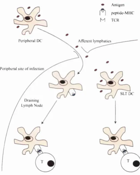
The interdigitating cells th a t present antigen to the naive T lymphocytes in the lymph nodes are derived from dendritic cells in the tertiary lymphoid tissues (Banchereau et al., 2000). Immature DCs collect and process antigen from the skin, lungs, gut etc. In response to signals from pathogens (e.g., LPS ,hpoproteins, peptidoglycans and nonmethylated DNA via toll-like recep tors (TLR)), endogenous infiammatory stimuli (e.g., heat-shock proteins (HSP) released by necrotic cells via CD91) or T cell feedback (e.g., TN F-a, type I in terferons, and interleukin-1 (IL-1)), DCs then migrate to the lymph nodes where they serve as professional APCs for sensitising naive T lymphocytes (Pulendran et al., 2001; Lanzavecchia and Sallusto, 2001). En route, DCs increase their expression of MHC, adhesion and costimulatory molecules and become more potent at stimulating T-ceUs, a process known as maturation. The secondary lymphoid tissues also contain resident DCs th a t are able to capture antigens draining there passively (Manickasingham and e Sousa, 2001). Other APCs like macrophages and B cells can stimulate effector and memory T cells, but do not appear to be capable of stimulating naive T cells because they either lack or are deficient in particular costimulatory molecules. So the dendritic cell serves a critical role in the capture, transport and presentation of antigen. See Figure 2.1 for an illustration of how antigen finds its way to the T cell.
2.2.2
A ntigen Processing and P resen tation
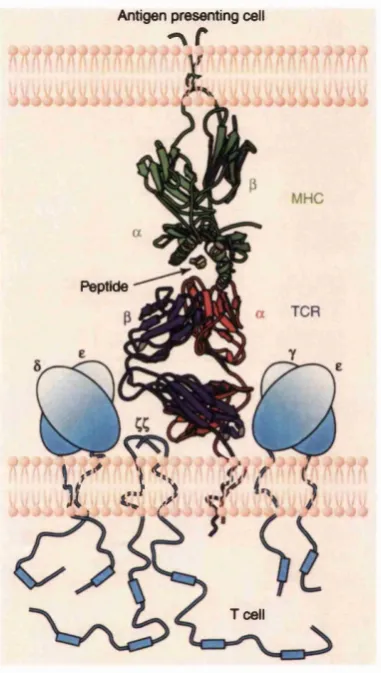
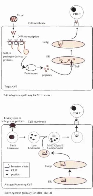
Since T cells do not recognise proteins or peptide firagments in isolation but only peptides bound to an MHC molecule (Figure 2.2), it is crucial to understand how and why antigens are processed in a form suitable for co-presentation with MHC molecules (Germain, 1999). To complicate matters, there are two classes of MHC molecules, class I and II, which are recognised by CDS and CD4 T cells respectively. Each also has a different antigen processing pathway, as illustrated in Figure 2.3.
A n tig en p cp tid c -M M C T C R
P e rip h e ra l D C A ftcrciU ly m p h a tic s
P e rip h e ra l s ite o f 'in f e c tio n
S L T D C D ra in in g
L y m p h N o d e
Antigen presenting cell
- \ r
Peptide
V' 3 >
Tcell
v ir u s
w
♦ D NA IranscnfHion
Target C ell
( A ) e n d o g e n o u s p a thw ay Tor M H C c lass I
H ndocyiosis o f
p athogen o r p ro tein s C e ll m em brane
r " #
\ Invariant chain
A ntige n Presenting C ell ( B | l-ixogenous p a th w ay li>r M H C c lass II
protein, followed by protease cleavage into short peptides, which then associate with MHC molecules and are transported to the cell surface. This allows the T cell to recognise peptides irrespective of their original location in the native folded protein, making it diflBcult for a pathogen to avoid immune detection by mutating non-essential surface side-chains. The functional difference between the MHC I and II systems is th a t the former is specialised to present peptides present in the cytosol, while the latter is specialised to present peptides captured from outside the cell.
The class I pathway begins with the breakdown of cytosoUc proteins (typ ically marked by ubiquitination) by a highly complex molecule known as the proteasome. Peptide fragments resulting from this proteolysis are then trans ported into the endoplasmic reticulum (ER) via a transport molecule known as transporter associated with antigen processing (TAP). TAP preferentially allows the entry of peptides of a length suitable for binding to MHC class I molecules (i.e., less than or equal to 12 amino acids). These peptides are then transferred with the help of a molecule known as tapasin to the newly synthesised MHC I molecules in the ER. The peptide-MHC I complex then passes to the cell surface via the usual cell secretory pathway. Binding to peptide stabilises MHC class I, such th a t MHC I which fail to bind to peptide are very short-Uved and do not make it to the cell surface.
In practice, dendritic cells also present phagocytosed antigens via MHC class I, in a process known as cross-presentation. This appears to be necessary for a cytotoxic immune response to allotransplants, tumour ceUs or viruses th a t avoid infecting APCs, since non-dendritic cells are unable to stimulate resting naive T lymphocytes (Heath and Carbone, 2001).
It has recently been discovered th a t immature DCs (but not mature ones) present a large proportion of empty MHC II molecules on their surface, which can be loaded in vitro with antigenic peptides to stimulate T cells (Santambrogio et al., 1999b). The physiological relevance of this is probably to allow the presentation of labile peptides th a t would normally be degraded via the usual endocytic pathway (Santambrogio et al., 1999a), but it has also been exploited for its therapeutic potential. Monocyte derived immature DCs can be loaded in vitro with antigen from synthetic peptides, dead tumour cells or infected cells, induced to mature and then re-injected to patients to stimulate the desired immune response (Théry and Amigorena, 2001).
Experimentally, it is now possible to produce homogeneously occupied class I and II molecules, both in soluble forms and on Uving cells. For class I MHC, refolding recombinant forms of the class I components in the presence of a spe cific peptide will give high uncontaminated yields of a particular peptide-MHC I complex. TAP deficient cells incubated with specific peptide and exogenous /02-microglobulin wiU achieve the same effect in living cells. For class II MHC, it is possible to produce peptide covalently tethered to the P chain, which will re-assemble in the presence of wild type a chain to produce the desired class II dimer. In hving cells, CLIP is often inserted instead of the tethered peptide in the ER, leading to cleavage of the tethered peptide in the endosome and loading of contaminating peptides upon CLIP removal. However, knocking out li pro duction results in the expression of a single peptide-MHC II on the cell surface, although a t a lower density than wild-type cells. Similarly, knocking out DM allows the generation of APCs where nearly all the surface MHC II molecules present only CLIP, with MHC class II densities similar to wild-type cells. Such simplified systems will be crucial for testing the predictions of any model of T cell antigen recognition and activation.
2.2.3
T Cell D evelopm ent
natural question - how then do T cells tell the difference between self peptide- MHC and foreign peptide-MHC complexes? The key to understanding this lies with the intricacies of T cell development (Benoist and Mathis, 1999).
T cells derive from stem cells in the foetal liver and adult bone marrow. Subsequently, the immature thymocytes migrate to the thymus, where the gene rearrangements th at result in the synthesis of a huge number of both a and ^
chains through combinatorial recombination of the V, D, and J genes occurs. More diversity is added by essentially random junctional nucleotide addition and deletion during imprecise joining of the V, D and J genes. The control of this process of gene rearrangement is extremely complex and poorly understood; for our purposes, it is sufficient to know th a t a huge repertoire of TCRs is potentially available. An added complication is th a t although 95% of TCRs are a/3, 5% of them are ')8. Since the role of 7(5 T cells is poorly understood, we will only consider a/3 cells in this thesis.
The process of selecting a peripheral T cell repertoire from the immature thymocytes also occvus in the thymus as illustrated in Figure 2.4. Here, T cells leam to adapt to the variabihty presented by both MHC polymorphism and TCR somatic diversity, and somehow a repertoire capable of distinguishing be tween self and foreign antigens emerges. This education can be very crudely divided into two stages - positive selection occurs first in the cortex, followed by negative selection in the medulla (Sezbda et al., 1999). SimpUstically, thymo cytes appear to follow a ‘Goldilocks’ principle - if the TCR signal is too weak, die from failure of positive selection; if the TCR signal is too strong, die from negative selection; if the TCR signal is just right, mature and be exported to the periphery.
When they enter into the stage of positive selection, thymocytes express both CD4 and CDS molecules and are known as double positive (DP) cells. If they survive positive selection, they become committed to either the CD4 or CDS Uneages, and are known as single positive (SP) cells. Essentially, it seems th a t some minimal signal resulting from the TCR binding with peptide-MHC is necessary for DP thymocytes to progress through positive selection - those th a t fail to muster this minimal signal die by apoptosis (Sezbda et al., 1999). The nature of the ligands th a t give such a minimal signal is not clear, beyond the general agreement th a t peptide is required. Observations demonstrating a specific influence of peptide sequence on selection efficiency suggests th a t direct recognition of both peptide and MHC are essential.
Apoptosis
(Failure o f positive selection)
CD4 CD4
CDS CDS
DP DP
Cortex
Positive Selection
CD4 or CDS
f
gpMedulla
(Negative selection)
CD4 or CDS
Surviving cells leave the thymus
SP - single positive thymocytes DP - double positive thymocytes
Cortical epithelial cell
Medullary DC or macrophage
Thymocyte
Very low/No affinity peptide
Intermediate affinity peptide
High affinity peptide
have matching MHC specificities receive a survival signal (stochastic selection model) (Chan et al., 1993; Davis and Littman, 1994) or th a t CD4/CD8 binding transduce lineage specific signals (instruction model) (Seong et al., 1992; Robey and Fowlkes, 1994). However, the data suggesting th a t the only difference in signals in the presence of CD4 or CDS coreceptors is in signal strength (Lck preferentially associates with CD4 in DP thymocytes) led to the model th at strong TCR signals induce differentiation into the CD4 lineage and vice-versa (strength of signal model) (Itano et al., 1996). More recently, it was discov ered th a t DP thymocytes appear to pass through an obligatory CD4“*"CD8“ intermediate stage, from which they differentiate into the CD4 lineage if TCR signals persist, and CDS if not (kinetic signalling model) (Brugnera et al., 2000; Bosselut et al., 2001).
The same DP thymocytes are also subject to negative selection. It is es tim ated th a t approximately 2/3 of the thymocytes th a t are positively selected will subsequently be deleted by negative selection. While there appears to be a temporal and spatial separation of positive and negative selection events, it is still a paradox how essentially similar sets of antigens presented by the thymic epithehal cells and therefore presumably generating similar signals do not cancel each other out. The end result of this extremely wasteful process is th a t only 3-5% of thymocytes successfully make the transition from DP to SP. Those th a t survive appear to undergo at least 6 cell divisions before export to the periphery,
therefore there are likely to be multiple copies of each specific naive T cell. W hat is the purpose of thymic education? Negative selection seems straight forward enough; it deletes cells th a t might otherwise cause autoimmunity. The traditional explanation for positive selection is the deletion of ‘useless’ T cells (von Boehmer et al., 1989); if this is so, then affinity for MHC alone should be a suf ficient criteria for survival which would result in a larger T cell repertoire, and it is not clear why positive selection requires affinity to the self peptide-MHC complex. It is therefore at least plausible th at selection of T cells with some affinity for self ligands is important in its own right.
2.2.4
Peripheral C irculation o f T Cells
1 week before re-emerging as effector/memory T cells. Unlike naive T cells, these activated T cells can migrate across endothelia to reach extrarlymphoid sites; in addition, this homing ability tends to correlate with the site of primary activation.
2.2.5
T C ell A ctivation
W hat happens when a T cell meets an APC with a matching peptide-MHC and becomes activated? The initial formation of the T cell-APC interface is probably independent of the nature of the ligand, and comes about from non-specific cell adhesion interactions. This creates an interface which probably excludes large inflexible molecules like CD43, allowing the relatively short TCR and MHC molecules to meet. If the TCR encounters its cognate ligand, the adhesion be tween T cell and APC is strengthened, possibly by an ‘inside-out’ mechanism, where signalling by the TCR results in ceU surface adhesion molecules having increased affinity for their counterpart on the APC. If the TCR fails to meet its cognate ligand, the T cell will separate from the APC and move on to sample another one. The mechanisms underlying T cell activation are highly complex, and involve a whole host of dynamic molecular events both on the plasma mem brane as well as intracefiularly (Weiss, 1999).
Signal Transduction I - E vents at th e P lasm a M em brane
In addition to the a and f3 chains which form the Ti dimer, the TCR com plex includes the invariant CD3 molecule as well as the disulphide linked CC homo-dimer or hetero-dimer (Davis and Chien, 1999). CD3 typically con sists of 7e and eS hetero-dimers. It seems th a t the functionality of the TCR is
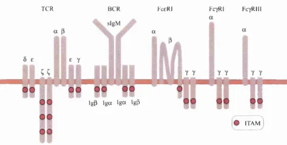
modular: the Ti dimer recognises antigen, while the CD3 and C chains serve as independent signal transducers. Signal transduction appears to be mediated by a common sequence motif known as the immunoreceptor tyrosine-based ac tivation motif (ITAM), which can interact with further downstream signalling molecules. ITAMs are not unique to the TCR, but are also associated with, amongst others, the B ceU receptor (BCR), FceRl and Fc^RIII. Since the 5, e, and 7 chains each have a single ITAM while each ( chain has three, the TCR has a total of ten ITAM copies (See Figure 2.5). The reason for this ITAM multiplicity is not clear, since the FceRl makes do with two, and the BCR with four.
TCR FceRl FctRI FcyRIM
m
IgP I g a I g o Igp
itamJ
Figure 2.5: Schematic of the multi-chain immune recognition receptor (MIRR) family and number of immunoreceptor tyrosine-based activation motifs (ITAMs). Among the MIRR family, the TCR complex has the most ITAMs. After (Turner et al., 2000).
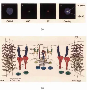
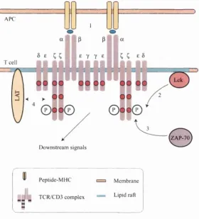
The initial TCR triggering leads to a complex re-distribution of cell sur face molecules mediated by the actin cytoskeleton, resulting in the formation of supramolecular activation clusters (SMACs) and the immune synapse (IS) (Monks et al., 1998; Grakoui et al., 1999; Dustin and Cooper, 2000; Bromley et al., 2001). The formation of the mature IS takes several minutes, and involves the sequen tial stages of junction formation, peptide-MHC transport and stabilisation. The mature IS has a central region (cSMAC) enriched in TCR, peptide-MHC, CD28, CD80, and signalling molecules including Lck, Fyn and PKC-0. There is a ring of molecules surrounding this called the peripheral SMAC (pSMAC), consisting of LFA-1, ICAM-1 and talin. CD2 and its ligand CD48 appear to cluster at the interface between the cSMAC and pSMAC. (Pictures of the IS shown in Figure 2.6(a) with a schematic cross-sectional view in Figure 2.6(b)).
lCAM-1
C-SMAC
pSMAC
O verlay
(a)
CD80
MUCH
CD48
iC AM -1
^ Glycoproteins
GD45, CD43 CD4*Tcell
(b)
S ignal T ra n s d u c tio n I I - E v e n ts in th e C ell
The most proximal biochemical changes after the TCR engages its hgand are the activation of the src kinases Lck and Fyn. Lck then tyrosine phosphorylates the ITAMs in the C chains and CD3. Biphosphorylated ITAMs recruit the Syk family kinase ZAP-70, and in turn, activated ZAP-70 phosphorylates the raft associated molecule Linker of Activation for T cells (LAT), which is associated with a diverse set of downstream signaUing and adaptor proteins, and may serve as a scaffold protein integrating signals from several pathways. See Figure 2.7 for a schematic of the proximal signalling pathways.
More distal signalling events get extremely complex and are not well under stood, but eventually get funnelled into the phosphatidylinositol (PI) pathway, the Ras pathway, and regulation of the actin cytoskeleton. Activation of the PI pathway results in release of Ca^+ from intracellular stores and this drives the translocation of cytoplasmic nuclear factor of activated T cells (NFAT) into the nucleus, where it regulates the transcription of many cytokine genes including IL-2. The P I pathway also results in the activation of protein kinase C (PKC) isoenzymes, which play an important role in T cell activation. The isoform PKC-0 is particularly fascinating, since it appears to integrate TCR and CD28 co-stimulatory signals (Altman et al., 2000). It is the only isoform to localise to the cSMAC, plays a key role in the activation of the transcription factors activatmg-protein 1 (AP-1), and nuclear factor-K/) (NF-k/?), and therefore is the leading candidate for the second TCR signal required for IL-2 induction. Activation of the Ras pathway leads to the mitogen activated protein (MAP) kinase cascade; this results in the induction of the Fos and Jun-related proteins which together make up AP-1. Although both PKC-Ô and Ras play a role in activating AP-1, the exact relationship between them is not known. Finally, TCR triggering results in an association of the ^ chain and CD3e with the actin cytoskeleton, which leads to T cell-APC conjugates and the formation of the IS.
A PC
1
T cell
Z A P-70
D ow nstream signals
P eptide-M H C
T C R /C D 3 com plex
M em brane
L ipid raft
As an example of positive feedback, it is known th at in addition to providing SH2 docking sites for ZAP-70 by phosphorylating ITAMs, Lck also directly activates ZAP-70 by phosphorylation of critical tyrosine residues and also by binding via the SH2 domain (Chan et al., 1995; Yamasaki et al., 1996; Bartolo et al., 1999; Visco et al., 2000; Acuto and Cantrell, 2000). Conversely, ZAP-70 also has a positive feedback eflFect on the recruitment (Thome et al., 1995) and possibly catalytic activity of Lck (Couture et al., 1994).
There is also good evidence for the negative regulation of Lck and ZAP- 70. One possible candidate is the protein tyrosine phosphatase SHP-1, which is activated by Lck (and possibly ZAP-70) and in turn dephosphorylates and inactivates Lck and ZAP-70 (Lorenz et al., 1996; Plas et al., 1996; Raab and Rudd, 1996; Mary et al., 1999). Interestingly, it appears th a t the action of SHP-1 is not confined to the engaged TCR, but affects neighbouring receptors as well (Germain and Stefanova, 1999; Dittel et al., 1999; Chan et al., 2001). Another documented negative feedback loop involved in TCR signalling is the kinase-phosphatase complex of Csk-PEP, which is recruited by Lck and inacti vates both Lck and ZAP-70 through a cooperative mechanism. Csk decreases the catalytic activity of Lck by phosphorylating its negative regulatory tyro sine (Bergman et al., 1992; Chow et al., 1993; Bougeret et al., 2000; Amrein et al., 1998), while P EP dephosphorylates positive regulatory tyrosines on both Lck and ZAP-70 (Cloutier and Veilette, 1999; Gjorloff-Wingren et al., 1999). Cbl (Thien et al., 1999), CD148 (Tangye et al., 1998) and other protein tyro sine phosphatases (Jin et al., 1998) may also be involved in negative feedback on Lck/ZAP-70, but these are currently not well characterised.
Why does the T cell require sequential activation of kinases (i.e., Lck —» ZAP- 70)? Syk is expressed in T cells, able to phosphorylate ITAMs in the absence of src kinases, as well as initiate the same downstream signals as ZAP-70, and is more eflScient than ZAP-70, yet T cells have an absolute requirement for ZAP-70 for both positive and negative selection. In fact, overexpression of Syk in ZAP- 70“ /~ mice restores peripheral T cell numbers to normal, showing th a t Syk can in some sense replace ZAP-70 (Turner et al., 2000). One possibility is th a t the Lck/ZAP-70 mechanism allows additional feedback circuitry, which results in a more flexible TCR signalling machinery, both in terms of the response threshold and signal amplitude. This will be further discussed in Chapter 5.
ity. Association with signalling molecules may be constitutive or inducible. These proteins direct the appropriate localisation of enzymatic complexes, am- pUfy signalling pathways and integrate the functions of distinct signalling com plexes (Kelly and Chan, 2000). Scaffold proteins are adaptors th a t serve to assemble several components of a signalling complex. By coupling disparate el ements together, scaffolds are beheved to increase effective substrate specificity of the enzymes and minimise unwanted cross-talk (Pawson and Scott, 1997). Adaptor and scaffold proteins are sometimes also known as linker proteins for obvious reasons.
Linker proteins may enhance T cell signalling by bringing elements of a signal cascade into close proximity and hence increasing the reaction rate (Levchenko et al., 2000). The effect of binding to a linker protein may also induce a confor mational change which results in catalytic activation of a signalling molecule. However, linker proteins can also have negative regulatory effects. Examples of how negative regulatory linker proteins can work include the recruitment or activation of negative regulatory effector molecules (Kawabuchi et al., 2000; Takeuchi et al., 2000; Brdicka et al., 2000), displacement of positive regulatory effector molecules, elimination of essential components of the signalling machin ery (e.g., by inducing degradation) (Joazeiro et al., 1999) and sequestration of rate limiting signalling components (Boussiotis et al., 1997).
2.2.6
Ligand Q uantity and T C R Signalling
While the total number of peptide-MHC on the surface of an APC is about 10^-10®, the minimum number of specific peptide-MHC on the APC needed to activate the T cell is about 100-200 for CD4 cells (Harding and Unanue, 1990; Demotz et al., 1990), and possibly as low as 1 for CDS cells (Sykulev et al., 1996). This suggests th at each individual TCR must have a false positive rate of lower than 1 / 1 0 0 0 or 1 / 1 0 0 0 0 if it is to avoid being swamped by signals from self peptide-MHC molecules.
There are two conflicting sets of data regarding the minimum number of TCRs in vivo th a t have to be triggered for a T cell response. While some studies have suggested th a t 100-200 TCR are sufficient in vivo (Schodin et al., 1996; Wei et al., 1999; Labrecque et al., 2001), this contrasts with the in vitro
2.2.7 Ligand Q uality and T C R Signalling
The discovery of altered peptide ligands (APL) added a new dimension of complexity to the understanding of TCR signalling (Evavold and Allen, 1991; Evavold et al., 1993; Sloan-Lancaster et al., 1993). M utating a single amino acid residue of a peptide known to activate a particular T cell could sometimes convert it into a nuU hgand, an antagonist or a partial agonist. Nuh ligands, as expected, basically do not appear to have any effect when bound to TCR. Par tial agonists result in a subset of the changes seen on full activation, for example, cytokine expression without proliferation. Antagonists inhibit T cell activation when simultaneously presented with the wild-type peptide (agonist). A s im ilar range of signaUing responses can be seen if the wild-type peptide was presented by m utant MHC. BiochemicaUy, it was shown th a t partial agonists caused a pattern of C phosphorylation and defective CD3e and ZAP-70 phosphorylation not seen with low concentrations of agonist, suggesting a quaUtative difference in signalling at the individual TCR level (Itoh and Germain, 1997; Itoh et al., 1999).
Further experiments with APL suggested th a t there was a hierarchy of TCR responses (Itoh and Germain, 1997). Cytotoxicity, cytokine expression, prolifer ation and differentiation is the order of responses evoked by hgands of progres sively increasing ‘quaUty’. Several experiments with soluble receptors at 25°C suggest th a t the most important determinant of hgand ‘quaUty’ is simply the duration of time th at it remains engaged with the T C R (Matsui et al., 1994; Corr et al., 1994; Sykulev et al., 1994; Alam et al., 1996). In general, the longer the duration of binding, the more efficacious the hgand, although it has recently been reported th at there is an optimal binding time, and hgands which bind for too long are not as effective (Hudrisier et al., 1998; Kalergis et al., 2001). Also, it is possible th a t hgand rebinding has an effect, since the fcon can also affect the quality of TCR signaUing (Garcia et al., 2001).
Obvious hmitations of these studies are th at TCR-MHC interactions occur in vivo at 37°C, on apposed plasma membranes, and in the context of co-receptors and other co-stimulatory molecules. Other factors, including conformational change may also determine the nature of the T ceU response, b ut it is generaUy agreed th a t the dissociation rate or half-life of binding is probably the most important.
Kersh et al., 2001). This is quite remarkable, since ligand dissociation times are stochastic events, and even for agonists, last only for seconds. Given th a t the typical number of specific peptide-MHC on an APC is vastly outnumbered by self peptide-MHC which are positively selected to have a minimum afiinity for the TCR, how can the T cell be so effective at Ugand discrimination?
2.3
Sum m ary
Chapter 3
M odelling T cell activation
and antigen recognition
This chapter reviews published models for the sensitivity and specificity of T cell activation, and also discusses some of the concepts fundamental to this enterprise. Since the purpose of a model is to provide a conceptual framework for interpreting experimental results and making new predictions, not all the models described are necessarily explicitly mathematical.
The first issue in modeUing T cell activation is to understand how ligand engagement leads to TCR activation. Next, we need models to explain how the T cell can amplify the signals from a few specific peptide-MHC molecules into a robust cellular response (sensitivity). We also need to understand what the TCR actually 'measures' during hgand engagement, and how it can use this binding property to rehably discriminate between different hgands (specificity).
FinaUy, there is the issue of how the T ceh copes with both extrinsic and intrinsic noise (reliability), where extrinsic noise refers to the variations in the antigenic profile presented to the T ceh, and intrinsic noise refers to fluctuations in TCR signalling due to low reactant numbers.
I have therefore decided to classify the models into four groups - TCR trig gering, sensitivity, specificity and rehabihty. Such a classification is intuitively appealing since these are the issues th a t intrigue immunologists, although there is some overlap between the models for these categories.
which are referred to as sub-sensitive, hyperboUc, or ultra-sensitive depending on whether the slope is shallower, the same, or steeper than for a Michaelis- Menten type reaction (Koshland, Jr. et al., 1982).
Specificity is another confusing term. It does not imply th a t a single TCR can only bind to a particular ligand, but rather th a t any given peptide-MHC is likely to trigger only a small number of T cell clonotypes. While TCRs are highly cross-reactive (Mason, 1998), because the universe of peptides which can be presented by MHC is so large, there is a very small probability th a t a T cell will cross-react with any one chosen at random. In terms of the binding property measured during hgand engagement, each TCR is specific because it has stringent requirements for th a t property before it wiU signal, but any hgand th a t meets those criteria can trigger the TCR, hence the cross-reactivity.
Finally, rehabihty refers to the abUity of T cells to respond in a consistent way to the same hgand under noisy conditions. For example, T cehs respond to the presence of foreign antigen on an APC by activation and proliferation and not do so in their absence, and they tend to be very good at this even though the density of foreign antigen on an APC and the molecules involved in the T CR signalling machinery are subject to stochastic fluctuations.
3.1
M odels for T C R triggering
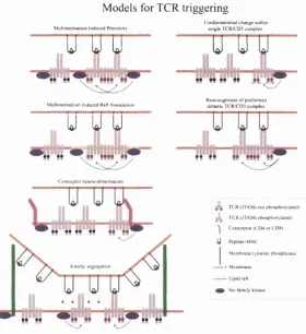
There are several types of models for how hgand engagement results in TCR signahing including conformational change, multimerisation, clustering and seg regation models (van der Merwe et al., 2000; van der Merwe, 2001). These are illustrated in Figure 3.1.
3.1.1
Conform ational change
M odels for TCR triggering
M u h im en sa tio n Induced P roxim ity
C onlo rm a lio n a l change w ithin single T C R C D 3 com plex
u y y
M ullim criM tion Induced Kat^ A xsocw non
u y g
C ore ce p to r hclero -d im cn satio n
K inetic segregation
c n j
jrw-ipïç*-R e arrangem ent o f p rctb n n e d d im e n c com plex
TC R tlT A M s not p h osphoryla ted) j L T C R (IT A M s phtwphory lated)
^ C o rc c e p lo r(C 'l> 4 o rC D « ) U Peptide-M H C
I M em brane tyrosine pU ^sphatase — ■ - - M em brane
— Lipid raft Src tam ily kinase
The dimer conformational change model suggests th at simultaneous bind ing of both q/3 chains on pre-formed (0 ^ ) 2 TCR dimers changes the relative
orientation of the two TCRS (Ding et al., 1999; Reth, 2001), and is consis tent with the possibility th a t the cell surface TCR forms a complex of two a/9 units (Femandez-Miguel et ai., 1999). However, a recent report showed th at TCR activation was largely independent of the orientation of biochemically coupled MHC dimers, which argues against the dimer conformational change model (Cochran et al., 2001b).
3.1.2
M ultim erisation
The first crystallographic studies of the MHC II molecule suggested th at it formed a dimer of dimers (Stern et al., 1994), and generated interest in the idea th a t TCR dimérisation or multimerisation was necessary for activation, driven by the tendency for MHC to self dimerise (Schafer et al., 1995). There is evidence for the formation of MHC II molecules in detergent lysates of im m u n e cells, demonstrating an intrinsic ability of MHC II to assume the double dimer conformation (Schafer et al., 1998). There is also evidence for spontaneous clustering of a fraction of the MHC I and II molecules on the surface of APC, determined using Fluorescence Resonance Energy Transfer (FRET) and long- range electron transfer (Jenei et al., 1997; Smith et al., 1999; Rezso Gâspâr et al., 2001). Finally, recent evidence showing th a t mutations in the putative dimer of dimers interface inhibited T cell proliferation and IL-2 secretion suggests th at MHC II driven TCR cross-hnking contributes to T cell activation (Lindstedt et al., 2 0 0 1).
However, the role of MHC self-dimerisation is complicated by the fact th at other MHC II structures crystallised do not form the same kind of dimer of dimers in their crystals (Fremont et al., 1996), and neither do the MHC I crystals studied so far. In addition, crystal structures of a half dozen peptide-MHC:TCR structures also fail to show dimérisation (Garcia et al., 1999; Baker and Wiley, 2001). Even if MHC molecules do form dimer of dimers spontaneously in vivo, the fact th a t specific peptides are effective when present at a frequency of 1
in 1 0^ or less means th at the probability of there being two specific peptides within one such MHC dimer of dimers is in the range of 1 in 10®. Since there are only about 10® MHC molecules on the surface of an APC, the relevance of such dimer of dimers for T cell activation is questionable.
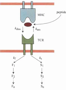
Thus most multimerisation models propose th a t Ugand engagement results in an affinity increase of one peptide-MHC:TCR for another, i.e.,
pMHC + TCR # pMHC-TCR
pMHC-TCR + pMHC-TCR (pMHC-TCR)2 ^ 2TCR*
*3"
where fcon and kofi depict on- and oflF-rates, respectively, of the peptide- MHC:TCR interaction, and kr is the rate constant at which the engaged TCR is marked for downregulation. A:^” and k ^ depict the rate constants of the monomer/dimer equiUbrium (Bachmann and Ohashi, 1999). Induced proximity of signalling modules then triggers signalling. These models are supported by experiments showing th a t soluble oUgomeric peptide-MHC oUgomers are more effective at activating T cells than monomers (Boniface et al., 1998; Cochran et al., 2 0 0 0).
Using the principle of mass action, we can convert the above reactions into first order ordinary differential equations (Bachmann and Ohashi, 1999):
^pM H C -T C R = AbnpMHC • TCR - AbffpMHC-TCR
^(pM H C -T C R)2 = jfe2"(pMHC-TCR)2 - A:f (pMHC-TCR)2 - Av(pMHC-TCR)2
At steady state.
^pM H C -T C R = ^(pM H C -T C R)2 = 0
and the rate of downregulation of TCR is given by
2
= 2 k r - ^ (pMHC-TCR)2 = TCR^
dt ' ^ ^ \ k o s J
So if the TCR dimérisation is necessary before downregulation, the rate of downregulation should be proportional to the square of the TCR density. Analysing one set of experimental data measuring the rate of TCR internalisa tion under high Ugand densities, it was found th at the data was most consistent with a simple model in which peptide-MHC :TCR complexes form dimers or trimers which were internalised after productive Ugand dissociation (Bachmann et al., 1998).
it was suggested th a t at least four mechanisms were necessary to produce a good fit (Sousa and Cameiro, 2000). These were the presence of a significant background rate of TCR turnover independent of hgand, th a t the rate of TCR internalisation was much slower than th a t of triggering, a high kinetic order > 5 (a possible interpretation of which is the necessity of n-mers for triggering where n > 5), and the existence of a pool of TCRs in dynamic equilibrium such th a t one pool had access to hgand but not the other. However, the vahdity of inferring such mechanisms is questionable, given the paucity of experimental data on which the model was fitted. For example, the data sets used in the second study consisted of only six data points eeudi.
Such multimerisation models would require th a t a highly variable peptide- MHCrTCR interface lead to consistent binding-induced TCR association. A second problem (which also apphes to the dimer conformational change model) is th a t the requirement for multimerisation seems incompatible with the fact th a t TCR triggering is most efiicient at very low peptide-MHC densities (Lan zavecchia et al., 1999), in the sense th a t the number of TCRs down-regulated per agonist peptide is highest. A recent report using kinetic, saturation bind ing, and fight scattering techniques also found no evidence for dimérisation or oligomerisation of human soluble peptide-MHC:TCR complexes (Baker and Wi ley, 2001). Also, no dimers have been observed in any of the X-ray structures of peptide-MHCia/JTCR complexes characterised so far (Garcia et al., 1999; Baker and Wiley, 2001).
C o-receptor hetero-dim erisation
The co-receptors CD4 and CDS are associated with the src kinase Lck, and it has been proposed th at association of CD4 or CDS can trigger TCR activation by bringing Lck into close proximity with the TCR ITAMs (Delon et al., 199S). While this model overcomes the problems of multimerisation discussed above, it is known th a t TCR triggering can occur in the absence of CD4/CDS (Abraham et al., 1991), ruling it out as a general mechanism.