Vol. 14, 2984 –2998, July 2003
Functional cdc25C Dual-Specificity Phosphatase Is
Required for S-Phase Entry in Human Cells
Patric Turowski,*
†‡Celine Franckhauser,*
†May C. Morris,
§Philippe Vaglio,*
¶Anne Fernandez,* and Ned J. C. Lamb*
储*Cell Biology Unit, Institut de Genetique Humaine, Centre National de la Recherche Scientifique
Unite´ Mixte de Recherche 1142, F-34396 Montpellier cedex 5, France; and
§Centre de Recherche
Biochimie Macromole´culaire, Centre National de la Recherche Scientifique Formation de Recherche en
Evolution 2593, F34293 Montpellier, cedex 5 France.
Submitted August 19, 2002; Revised March 18, 2003; Accepted March 18, 2003 Monitoring Editor: Tony Hunter
In view of the common regulatory mechanism that induces transcription of the mitotic
phospha-tase cdc25C and cyclin A at the beginning of S-phase, we investigated whether cdc25C was
required for S-phase transit. Here, we show that in both nontransformed human fibroblasts and
HeLa cells, cdc25C protein levels significantly increased concomitant with S-phase onset and
cyclin A synthesis. Activity measurements on immunoprecipitates from synchronized HeLa cells
revealed a sharp rise in cdc25C-associated phosphatase activity that coincided with S-phase.
Microinjection of various antisense-cdc25C molecules led to inhibition of DNA synthesis in both
HeLa cells and human fibroblasts. Furthermore, transfection of small interfering RNA directed
against cdc25C specifically depleted cdc25C in HeLa cells without affecting cdc25A or cdc25B
levels. Cdc25C RNA interference was also accompanied by S-phase inhibition. In cells depleted of
cdc25C by antisense or siRNA, normal cell cycle progression could be re-established through
microinjection of wild-type cdc25C protein but not inactive C377S mutant protein. Taken together,
these results show that cdc25C not only plays a role at the G2/M transition but also in the
modulation of DNA replication where its function is distinct from that of cdc25A.
INTRODUCTION
Cyclin-dependent kinases (cdks) play a central role in the regulation of eukaryotic cell cycle progression (Morgan, 1995). Regulatory mechanisms responsible for their con-trolled activation include combinatorial assembly of cdks with one of a number of different cyclins, activating phos-phorylation of a conserved threonine residue (T161 in hu-man cdk1/cdc2), inhibition by association with specific in-hibitors, and inhibitory phosphorylation of a conserved tyrosine (and adjacent threonine) in the amino-terminal ATP-binding region. Ultimately cdk/cyclin complexes are rapidly activated by dephosphorylation of this tyrosine (and
threonine), which is brought about by cdc25 dual specificity phosphatases (for review, see Nilsson and Hoffmann, 2000). Cdc25 phosphatases belong to the tyrosine phosphatase family. Their active site loop, containing the HCX5R motif, is similar to that of tyrosine phosphatases but their overall topology is different (Faumanet al.,1998).
In human cells, cdc25 proteins are encoded by a multigene family, consisting of cdc25A, cdc25B, and cdc25C (Sadhuet al.,1990; Galaktionov and Beach, 1991; Nagataet al.,1991). All three display more than 60% identity to each other within the carboxyl terminal, catalytic domain. The less-conserved amino terminal domains are multiply phosphor-ylated and harbor distinct regulatory and substrate specific-ity determinants (Hoffmann et al., 1994; Strausfeld et al.,
1994; Gabrielliet al.,1997). Amino terminal domains are also subject to alternative splicing giving rise to two, three, and five variants of cdc25A, B, and C, respectively (Baldinet al.,
1997; Bureik et al., 2000; Wegener et al., 2000). Cdc25A is required for G1/S transition (Jinnoet al.,1994) and seems to be regulated in part by mitogenic signaling (Galaktionovet al.,1995).
More recent data suggest that in humans a stable form of cdc25A may also play a role in mitosis (Mailandet al.,2002) and checkpoint signaling (Zhao et al., 2002). Cdc25B is
Article published online ahead of print. Mol. Biol. Cell 10.1091/ mbc.E02– 08 – 0515. Article and publication date are available at www.molbiolcell.org/cgi/doi/10.1091/mbc.E02– 08 – 0515.
储Corresponding author. E-mail address: [email protected]. †Both authors contributed equally to this work.
Present addresses:‡Institute of Ophthalmology, University Col-lege London, 11– 43 Bath Street, London EC1V 9EL, UK;¶Dana Farber Cancer Institute, 44 Binney Street, Boston, MA 02115.
present in S- and G2-phase cells. Overexpression of cdc25B in mammalian cells rapidly induces mitotic entry, overrid-ing most cell cycle checkpoints (Karlssonet al.,1999). Indeed cdc25B is required for the G2/M progression and a number of reports suggest that cdc25B is the initial activator of cdc2/cyclin B at mitotic entry (Gabrielliet al., 1996; Nishi-jimaet al.,1997; Lammeret al.,1998).
Likewise cdc25C has been associated with mitotic activa-tion. There is strong evidence that cdc25C is involved in mitotic entry by dephosphorylating T14 and Y15 of cdc2/ cyclin B. Its inhibition through microinjection of antibodies blocks entry into mitosis (Millaret al.,1991; Sekiet al.,1992). Cdc25C becomes phosphorylated and activated at mitosis by cdc2/cyclin B itself and a positive feedback loop has been proposed (Hoffmann et al., 1993; Izumi and Maller, 1993; Strausfeldet al.,1994). Indeed, cdc25C phosphorylated by cdc2/cyclin B is capable of inducing partial mitotic activa-tion (Hoffmannet al.,1993; Strausfeldet al.,1994). However, cdc25C is also subject to other site-specific phosphorylation events at mitosis, particularly by polo-like kinase 1 (Plk1; Toyoshima-Morimotoet al., 2002) and undergoes pin1-de-pendent proline isomerization (Zhouet al.,2000). Moreover, cdc25C is a converging point of several signal transduction cascades and links the mitotic activation of cdc2/cyclin B to the DNA replication and repair checkpoint (for review, see Walworth, 2001). Muller and colleagues have reported that cdc25C gene transcription is restricted to the S- and G2-phase, and the underlying transcriptional regulation is the same as found for cyclin A (Zwickeret al.,1995). Cyclin A protein appears exclusively during S and G2 and is required for the initiation of DNA replication (Girardet al.,1991). In light of these parallels to cyclin A expression we have ad-dressed the question whether cdc25C had a role during S-phase and provide experimental evidence that in human fibroblast and HeLa cells, active cdc25C protein is required for proper G1-S progression even in the presence of cdc25A.
MATERIAL AND METHODS
Cell Culture, Synchronization, and Cell Cycle
Analysis
Nontransformed human fibroblasts Hs68 (CRL-1365) and HeLa cells were cultured in DMEM supplemented with 10% fetal calf serum. Hs68 fibroblasts were synchronized in G1 by serum depri-vation and resynchronized for S and G2 using hydroxyurea (HU) as described elsewhere (Girardet al.,1992). HeLa cells were synchro-nized by blocking the cells with thymidine at G1/S as described elsewhere (Steinet al.,1998). Briefly, asynchronous HeLa cells were supplemented with 2 mM thymidine for 24 h. Cells were released from the thymidine block by incubation in PBS for 30 s and 5 min at 37°C followed by incubation in serum containing DME supple-mented with 24M deoxycytidine for 15 min. Finally, cells were refed with fresh media supplemented with 24M deoxycytidine and grown for 12–14 h. At this point cells were refed with fresh media containing 2 mM thymidine for 24 h and subsequently re-leased as described above. In some experiments mitotic cells were mechanically collected by mitotic shakes 10, 11, and 12 h after double thymidine block. Cell cycle distribution was determined by fluorescence-activated cell sorting (FACS). Cells were trypsinized, and nuclei were stained for DNA in PBS, 0.1% Triton X-100, 5% glycerol, and 50g/ml propidium iodide. Subsequently, fluores-cence from propidium iodide–DNA complexes was determined using a FACScan flow cytometer and CellQuest analysis software (Becton Dickinson, Franklin Lakes, NJ).
Semiquantitative RT-PCR
Total RNA from synchronized or randomly growing Hs68 fibro-blasts or HeLa cells was prepared using the RNeasy kit (Qiagen, Courtaboeuf, France). Total RNA, 0.25g, was reverse-transcribed using a RNA PCR kit (Perkin Elmer-Cetus, Boston, MA). The re-sulting cDNA was then subjected to PCR amplification in 100-L reactions containing 10 mM Tris/Cl, pH 8.3, 1.5 mM MgCl, 50 mM KCl, 0.2 mM of each dATP, [␣-32P]dCTP (50 mCi/mmol), dGTP, and dTTP, 0.5M forward oligonucleotide, GATGCCAGAGAACT-TGAAC (annealing to nucleotides 858 – 866 of cdc25C), 0.5 M reverse oligonucleotide, TGAAACCTAATCCATTCCC (annealing to nucleotides 1779 –1797 of cdc25C), and 2.5 U ofTaqpolymerase. These oligonucleotides were designed to amplify a fragment in the 3⬘region of cdc25C, which is common to all described cdc25C splice variants (Bureiket al.,2000; Wegeneret al.,2000). Twenty-five am-plification cycles with the following temperature profile were per-formed: denaturation at 95°C for 15 s, primer annealing at 55°C for 30 s, and primer elongation at 72°C for 2 min. These conditions allowed detection of cdc25C transcripts during all cell cycle stages and were in the linear range of amplification (our unpublished results). Reactions were analyzed on ethidium-bromide– containing 1.4% agarose gels or on 5% polyacrylamide gels followed by auto-radiography.
Antibodies and Immunoblot Analyses
Antiserum was raised against the peptide MSTELFSSTREEGSSGS-GPS corresponding to the N-terminus of cdc25C. Immunizations and subsequent affinity purification of the antibody N20 were per-formed as previously described (Turowskiet al.,1999). This anti-body was highly specific for in vitro–translated or recombinant cdc25C as well as the endogenous protein when used in immuno-precipitations or immunoblots (see also Figure 2F).
Total extracts of HeLa or Hs68 cells were electrophoretically separated on 10% SDS-polyacrylamide gels and immunoblotted as described (Turowskiet al.,1999). Primary antibodies against cdc25A (sc7389, Santa Cruz Biotechnology, Santa Cruz, CA), cdc25B (sc326, Santa Cruz Biotechnology, and clone 23, Transduction Laboratories, Becton Dickinson, San Diego, CA), cdc25C (C20: sc327, Santa Cruz Laboratories, and N20), anti-cdk2 (M2, sc163, Santa Cruz Laborato-ries), and cyclin A (Girardet al.,1991) were used at 1:500. Anti-tubulin ascites (clone DM1A; Bloseet al.,1984) was used at 1:10,000.
Immunoprecipitation and Phosphatase Activity
Measurement
Active cdc25C was immunoprecipitated from double thymidine synchronized HeLa cells using anti-cdc25C-C20 or N20 antibodies. Briefly, 5g anti-cdc25C were incubated with 20L protein G-sepharose (AP-Biotech, Amersham, UK) for 2 h at 4°C. Synchro-nized HeLa cells from a 100-mm culture dish were washed twice in ice-cold PBS, scraped into PBS, and collected by centrifugation at 1000⫻gfor 5 min. Cells were lysed in RIPA buffer (50 mM Tris/Cl, pH 8.0, 50 mM NaCl, 1.0 mM EDTA, 0.5% sodium deoxycholate, 1.0% Triton X-100, 50 nM calyculin A, 2g/ml aprotinin, 4 ⌴
leupeptin, 1 mM PMSF, 3M pepstatin), and the soluble lysate was precleared with protein G-Sepharose for 30 min at 4°C. Subse-quently 20L of antibody-protein G-Sepharose complex was added to lysate containing 500g of protein. After incubation for 2 h, antibody-protein G complexes were collected by centrifugation and washed twice with RIPA buffer, twice with RIPA containing 400 mM NaCl, twice in TBS (50 mM Tris/Cl, pH 7.5, 150 mM NaCl), and twice in TBS containing 1.0 mM EDTA before activity measure-ments were made.
resuspended in 300L of 3-OMFP assay buffer with and without 2 mM sodium orthovanadate. After incubation at 30°C for 30 min fluorescence was measured on a Wallac Victor2 fluorescence plate reader (Perkin Elmer-Cetus Life Sciences, Boston, MA) using 489-nm excitation and recording at 531 nm. Experiments were done in duplicates and repeated at least three times. Fluorescent emis-sions values were corrected for background activity, which was immunoprecipitated from HeLa cells double blocked in thymidine and which generally gave the same values as immunoprecipitates without antibody.
Microinjection and Immunofluorescence
Cdc25C antisense oligonucleotides AS1 (TGAGAAGAGTTCCGTA-GACAT), AS2 (CCTTGAATTTTTCCACCTGCT), the corresponding sense oligonucleotides S1 and S2, and cdc25B antisense oligonucle-otide (CCGGCCCCGCCGCGATGGAGGTGC) were synthesized using phosphorothioate modification of end bases to increase in vivo half-life and purified by reversed-phase chromatography (Eu-rogentec, Seraing, Belgium). For the sense and antisense cdc25C expression vectors p25Cs and p25Cas, the open reading frame (nu-cleotides 211-1632) from human cdc25C cDNA (Sadhuet al.,1990) was subcloned into pJ3 (Morgenstern and Land, 1990).
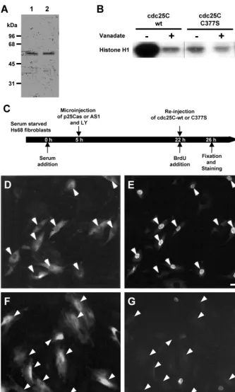
Synchronized HS68 or HeLa cells were microinjected as described previously (Lamb and Fernandez, 1997). Before injection, plasmids, oligonucleotides, and proteins were diluted into injection buffer containing 0.5 mg/ml inert marker antibodies (to act as a marker for injected cells). Plasmids were injected at⬍100 copies per cell (ca. 10 nM in the needle) and oligonucleotides at concentrations of⬍5 nM. Higher concentrations tended to affect S-phase nonspecifically. Cdc25C-wt and C377S proteins were at a concentration of ca. 0.2 mg/ml after dilution into marker-containing injection buffer. Cells were microinjected at indicated times throughout G1 or early S-phase and further subcultured in presence of bromodeoxyuridine (BrdU). At various time points after microinjection, cells were fixed and stained for incorporated BrdU, DNA, and microinjection marker (Girardet al.,1991). Cells were mounted and photographed as described elsewhere (Turowskiet al.,1999).
Recombinant cdc25C Protein
Human Cdc25C subcloned into the T7-inducible bacterial expres-sion vector pRK171 was a kind gift of Paul Russell (Scripps Research Institute, La Jolla, CA). Recombinant cdc25C was expressed using BL21 (DE3) bacteria transformed with the pRK171-hcdc25C con-struct and cdc25C-containing inclusion bodies isolated as previ-ously described (Strausfeldet al.,1991). Instead of the renaturation procedure described before, inclusion bodies were solubilized in guanidinium-containing buffer, and proteins were renatured by quick dilution. After overnight dialysis cdc25C was further sepa-rated from contaminants and improperly folded protein by anion exchange chromatography on a Mono Q column. Details of this procedure will be published elsewhere and are available on request. The plasmid pRK171-hcdc25C was mutated using the Quickchange Mutagenesis kit (Stratagene, La Jolla, CA) and mutant oligonucleo-tides (top strand: ATAATCATCGTGTTCCACAGTGAGT-TCTCCTCAGAGAGGG and the corresponding lower strand oligo-nucleotide). The mutated construct was verified by sequencing and encoded human cdc25C with a single cysteine to serine change at amino acid position 377 (cdc25C-C377S). Subsequently, the C377S protein was expressed and purified in the same way as wild-type protein. Specific phosphatase activities were measured using para-nitrophenylphosphate (pNPP) as a substrate (Gottlinet al., 1996). Wild-type cdc25C had a specific activity of 46 nmol/min/mg, whereas the C377S mutant protein displayed no activity toward pNPP at all.
RNA Interference
The small interference RNA (siRNA) duplexes were 21 base pairs. They were selected from within the open reading frames of cdc25A,
B, and C⬃70 base pairs downstream of the ATG at the next double adenine. The sequences of the coding strands of the human cdc25A, B, and C siRNA duplexes were AAGGCGCUAUUUGGCGCUUCA, AAGAGCGAGGCGGGCAGUGGA, and AAAAAUGUUGCCUC-GAUCUUUC, respectively. The control siRNA used was AC-CCGCGCCGAGGUGAAGUU, targeting enhanced green fluores-cent protein (Cortez et al., 2001). siRNA were synthesized and annealed by Dharmacon Research Inc, (Lafeyette, CO). For RNA interference siRNA duplexes were resuspended in RNAse-free wa-ter at 20 M and transfected as described elsewhere (Liu and Erikson, 2002). Alternatively, cells were transfected using calcium phosphate (Alessiet al.,1996). Cells were incubated in the presence of the antisense RNA for 24 h. Cells were washed and further cultured in the absence or presence of BrdU for 12–24 h before they were processed for immunofluorescence, microinjection, or Western blotting. To determine transfection efficiency, cells were transfected with siRNA directed against GFP coupled to fluorescein. After 24-h transfection, cells were washed once in PBS, and the proportion of cells positive for fluorescein was determined.
RESULTS
Cell Cycle Analysis of cdc25C Expression and
Activity
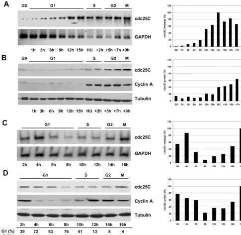
cdc25C, and notable de novo synthesis of RNA and cdc25C protein occurred at the G1/S-phase boundary. Likewise in cycling HeLa cells, we observed a clear decrease of cdc25C during G1 and induced expression at the G1/S boundary.
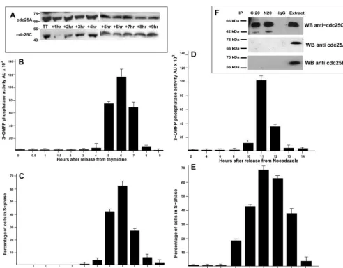
As shown in Figure 2A, thymidine-blocked HeLa cells (TT) contained steady state levels of cdc25C similar to those observed in G1 cells. However, on release from the thymi-dine block, cdc25C levels rapidly decreased for 60 min be-fore de novo synthesis occurred, followed by a consistent
increase in cdc25C levels over the next 8 h. In contrast, levels of cdc25A remained constant during the same period. We next asked if these changes in cdc25C expression were also reflected by changes in cdc25C activity. Considering the low abundance of cdc25C, we chose to immunoprecipitate the protein from synchronized human fibroblasts or HeLa cells and to measure activity using the highly sensitive fluores-cent substrate 3-OMFP (Gottlin et al. 1996). As shown in Figure 2B, as HeLa cells were released from a thymidine
block, no cdc25C activity was observed during the first 4 h. However, cdc25C activity increased sharply 5– 6 h after thy-midine release before falling again. This peak in cdc25C activity coincided with maximal S-phase activity as deter-mined by BrdU incorporation (Figure 2C). A similar result was obtained in HeLa cells released from a nocodazole block (Figure 2D). Again the peak of cdc25C activity coincided with the peak of S-phase activity (Figure 2E). Cdc25C phos-phatase activity at S-phase constituted only a fraction of that measurable at mitosis. Typically, cdc25C immunoprecipi-tated from M-phase extracts (blocked and released from nocodazole) and measured under the same conditions as for S-phase showed 3–5-fold higher activity than peak S-phase samples (our unpublished results). However, this increase in activity at S-phase reverted to background levels as cells entered G2. As shown in Figure 2F, the phosphatase activity measured was specific to cdc25C because our cdc25C anti-bodies quantitatively immunoprecipitated cdc25C but not even traces of cdc25A or B. A similar increase in cdc25C activity was also detected in synchronized HS68 cells, with the peak of cdc25C activity detectable 18.5 h after refeeding (corresponding to mid–S-phase, our unpublished results). These data show that cdc25C is specifically synthesized at S-phase entry in refed cells and in cycling cells. In addition, this de novo synthesis of cdc25C is accompanied by a peak in cdc25C activity, which also coincides with maximal DNA synthesis. Interestingly, our observation that this activity was restricted to S-phase decreasing as cells transit into G2, strongly suggests regulation by posttranslational modifica-tion because the levels of cdc25C protein continue to in-crease, whereas the S-phase activity declines (see also DIS-CUSSION).
Antisense Depletion of cdc25C in Human Cells
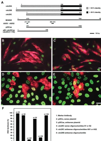
With low levels in G1, induction during S and maximal levels during G2/M, the observed cdc25C expression pat-tern is reminiscent of that seen with cyclin A (Pines and Hunter, 1990). Cyclin A has been shown to be required for S-phase initiation (Girard et al., 1991), whereas no such role has so far been described for cdc25C. To examine if the induction of cdc25C protein expression and activity was associated with a potential role of cdc25C at S-phase, we chose to specifically interfere with its protein resyn-thesis using different antisense approaches (Figure 3A). On the one hand we used the expression constructs p25Cs and p25Cas, containing the full-length open reading frame of human cdc25C under the control of an SV40 promoter in sense or antisense orientation, respectively. Alternatively, because mammalian cdc25s share strong sequence identity within their catalytic domain, antisense oligonucleotides targeting the most specific and divergent regions of cdc25C were designed. The sequences of single-stranded DNA oligonucleotides AS1 and AS2 were unique to cdc25C and absent from cdc25A or B. To act as controls, oligonucleotides encoding the sense strands were used.
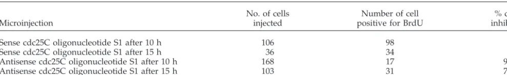
To analyze the effect of cdc25C antisense tools on S-phase transition, HS68 fibroblasts were synchronized by serum deprivation and microinjected at different times after entry into G1. Cells were fixed and analyzed 20 –22 h later when most cells have completed DNA replication (Girard et al.,
1992). Transition through S-phase was assessed by
monitor-ing BrdU incorporation. Microinjection of oligonucleotides AS1 or AS2 effectively prevented BrdU incorporation, whereas microinjection of either control oligonucleotides S1 or S2 did not affect S-phase transition (Figure 3, B, C, and F). Notably, microinjection of oligonucleotide AS1 still blocked S-phase entry when injected as late as 15 h after serum stimulation (Table 1), which corresponded to the time in late G1 just before the marked increase in cdc25C protein (Figure 1B). Similar S-phase inhibition was observed in cells micro-injected with the antisense-cdc25C expressing construct p25Cas (Figure 3F). Microinjection of either the sense con-struct p25Cs or marker antibodies had no effect on DNA synthesis. When microinjected into synchronized HeLa cells p25Cas, AS1 or AS2 efficiently blocked S-phase as well (Fig-ure 3, D and E). This inhibition of S-phase was specific to antisense cdc25C and was not observed using antisense cdc25B. Neither microinjection of an antisense-cdc25B RNA expressing construct nor cdc25B antisense oligonucleotides prevented S-phase transition in Hs68 or HeLa cells (Figure 3F and our unpublished results).
SiRNA to cdc25C Specifically Suppresses cdc25C
Expression
the cases where a cdc25 isoform was targeted did we see changes in the levels of the other cdc25 isoforms when compared with GFP siRNA-transfected or nontransfected cells (Figure 4, A–D).
Cyclin A and cdk2 were also examined after cdc25C siRNA transfection: Cyclin A was strongly reduced in cells transfected with siRNA to cyclin A as expected, but un-changed in cells transfected with cdc25C (Figure 4E). We also examined the phosphorylation state of cdk2 as a poten-tial substrate for cdc25C at S-phase. HeLa cells were de-pleted of cdc25C by siRNA, and cdk2 was examined by Western blot using long gels run at pH 8.6. This has previ-ously been shown to allow immunodetection of two bands
for cdk2: the top band being the dephosphorylated active form and the lower band corresponding to the phosphory-lated, inactive form (Dulicet al.,1993). As shown in Figure 4F, mainly the 33-kDa top band was detectable in control cells transfected with siGFP. When cells are depleted of cdc25C, the majority of cdk2 still resolves as the slow-mi-grating, active form, but a small proportion now clearly resolves in the fast migrating form. Membranes were re-probed for cdc25C to ensure that effective knockdown of cdc25C had occurred (our unpublished results). These re-sults suggest that under these conditions, cdk2 was not a direct target for cdc25C. In support of this we did not find significant changes in cdk activity in cyclin A or cdk2
im-Figure 3. Microinjection of antisense-cdc25C prevents S-phase entry. (A) cdc25A, B, and C protein sequences are schematically aligned to each other. Numbers correspond to the amino acids of the published sequences (Gen-Bank accession numbers M81933, M81934, and M34065, respectively). The catalytic do-mains (black) display more than 58% identity, the amino terminal domains (white)⬍38% identity. The cDNA of cdc25C (Sadhuet al., 1990; M34065) is depicted underneath to illus-trate the position of the antisense and sense (control) oligonucleotides AS/S1 and AS/S2 and the fragments used for the antisense vec-tors p25Cas. The positions in base pairs are given relative to M34065. (B and C) Hs68 fi-broblasts were synchronized by serum starva-tion and refed with medium containing se-rum and BrdU. Ten hours after sese-rum readdition, cells were microinjected with cdc25C antisense AS1 (B) or sense S1 (C) oli-gonucleotide, together with inert rabbit IgG. Cells were fixed 22 h after refeeding (when
⬎95% of cells have initiated or completed DNA synthesis) and double-stained for the rabbit marker antibodies (red) and BrdU in-corporation (green). (D and E). Mitotic HeLa cells were obtained using thymidine followed by nocodazole treatment. Three hours after relieving the mitotic block, cells were micro-injected with oligonucleotides AS1 (D) or S1 (E) and marker antibody. BrdU was added and the cells were cultured for another 12 h before fixation. Shown are merged micro-graphs of cells double-stained for marker an-tibody (red) and BrdU (green). Scale bars, 10 m. The histogram in F summarizes the data obtained from four to six independent sets of microinjection experiments performed with synchronized Hs68 fibroblasts. The experi-mental outline was as described in B and C, except that p25Cs (2) and p25Cas (3) were injected in the first 5 h after the refeed to allow efficient expression. In all cases cells were fixed 22–24 h after the refeed. Values in col-umns 4 (and 5) represent data from experi-ments performed with either oligonucleotides S1 or S2 (or AS1 or AS2). Percent of DNA synthesis refers to the percentage of microin-jected cells that have incorporated BrdU. n⫽
munoprecipitates isolated from cdc25C depleted cells (our unpublished results).
SiRNA to cdc25C Inhibits DNA Synthesis
We next examined the consequences of cdc25C siRNA on DNA synthesis. As shown in Figure 5, HeLa cells transfected with cdc25C siRNA failed to incorporate BrdU during the following 24-h period. Little or no BrdU staining was seen in cells transfected with siRNA to cdc25C (Figure 5, C and D). The only cells positive for BrdU were in mitosis. In compar-ison, control cells transfected with EGFP siRNA showed BrdU staining in⬃40% of cells (Figure 5, A and B). In all experiments the levels of transfection were very high (ca. 95%), as assessed using a fluorescently tagged siRNA tar-geting GFP. Transfections for shorter periods of time before BrdU addition resulted in a higher background levels of BrdU incorporation, most likely reflecting the half-life of the cdc25C protein present at the time of transfection. A similar failure to undergo DNA synthesis was also observed in cells transfected with cdc25A (Figure 5, E and F) but not cdc25B siRNA (our unpublished results), consistent with their pre-viously reported functions at G1/S and mitosis, respectively (Jinnoet al.,1994; Gabrielliet al.,1996).
Cdc25C Protein Injection into cdc25C-depleted Cells
Restores S-Phase
Although the described cdc25C-depletion experiment seemed to specifically target cdc25C with respect to the other cdc25 isoforms, there remained the possibility that they interfered with another as yet unidentified cellular process. To further specify the role of cdc25C at S-phase entry, functional rescue experiments were performed. Hu-man wild-type (wt) cdc25C was expressed inEscherichia coli
and purified to apparent homogeneity (Figure 6A, lane 1). For catalysis all dual-specificity protein phosphatases de-pend on an essential cysteine residue in their active site (see Faumanet al.,1998 and references therein). We mutated the catalytic cysteine in human cdc25C (C377) to serine to pro-duce cdc25C-C377S. The C377S protein was expressed and purified in a way similar to that of the wt protein (Figure 6A, lane 2). Phosphatase activity of both proteins was assessed using inactive cdc2/cyclin B complexes immunoprecipitated from S/G2 HeLa cells.
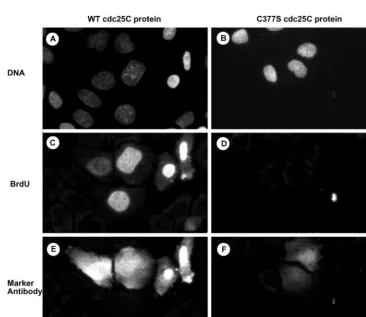
As shown in Figure 6B, wild-type cdc25C but not C377S mutant protein strongly activated the histone H1 kinase activity of cdc2/cyclin B in a vanadate-sensitive manner, showing the functional integrity of these proteins. Hs68 fibro-blasts were microinjected with p25Cas together with Lucifer Yellow to act as an injection marker. Twenty-two hours after injection when cells were blocked before S-phase, fluorescent cells were reinjected with wt or C377S cdc25C, and subsequent DNA synthesis was monitored (Figure 6, D–G). Active, wild-type cdc25C specifically restored the ability to enter S-phase (Figure 6, D and E; Table 2). In contrast, inactive cdc25C-C377S could not relieve the S-phase block in antisense cdc25C-depleted cells (Figure 6, F and G; Table 2), suggesting that cells need active cdc25C phosphatase to enter S-phase. Similar re-sults were also obtained in cell microinjected with antisense DNA oligonucleotides (Table 2).
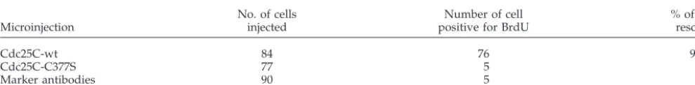
Functional rescue experiments were also performed in cells depleted of cdc25C through RNA interference. As shown in Figure 7 and in confirmation of the results shown in Figure 6, microinjection of active cdc25C into Hela cells effectively overcame the cell-cycle block induced by cdc25C siRNA because injected cells underwent DNA synthesis within 4 h (Figure 7, A, C, and E; Table 3). Again the microinjection of inactive cdc25C-C377S mutant protein was unable to restore DNA synthesis (Figure 7, B, D, and F; Table 3).
Taken together, these data show that the failure to progress through S-phase in human nontransformed cells after ablation cdc25C results from the specific lack of cdc25C activity. This activity is required despite the presence of unaltered levels of cdc25A, suggesting a function distinct from that of cdc25A.
DISCUSSION
Cdc25C mRNA was originally described to be expressed primarily in the G2-phase of the cell cycle (Sadhuet al.,1990; Jinno et al., 1994). Studies based on more sensitive PCR technology showed that cdc25C transcripts appear from late G1 onward and that high levels are already reached in S-phase (Lucibelloet al., 1995). We observed a similar ex-pression pattern in synchronized human fibroblasts Hs68 and HeLa cells. This stringently controlled, cell cycle-depen-dent transcription, which is also found for the cdc2 and cyclin A genes, is the result of repression by a cell-cycle–
Table 1. Inhibition of DNA synthesis by antisense-cdc25C oligonucleotides in mid- and late-GI
Microinjection
No. of cells injected
Number of cell positive for BrdU
% cells inhibited
Sense cdc25C oligonucleotide S1 after 10 h 106 98 8
Sense cdc25C oligonucleotide S1 after 15 h 36 34 6
Antisense cdc25C oligonucleotide S1 after 10 h 168 17 90
Antisense cdc25C oligonucleotide S1 after 15 h 103 31 70
dependent element during G1 (Zwickeret al.,1995). Cdc25C protein levels were reported to fluctuate very little during cell cycle progression, but a certain increase has always been observed at S-phase (Millaret al.,1991; Patelet al.,1999).
We report here that in highly synchronized Hs68 fibro-blasts cdc25C protein levels were markedly induced at the G1/S transition, subsequent to the rise in mRNA levels and parallel to cyclin A accumulation. Moreover, we found a similar induction at the G1/S transition of postmitotically synchronized HeLa cells or HeLa cells released from a thy-midine block. Changes in cdc25C protein levels were not as marked as those found for cyclin A, especially in cycling HeLa cells (Figure 1D). Also, de novo cdc25C protein syn-thesis in refed Hs68 occurred significantly later than the induction of the RNA (Figure 1, A and B). This raises the possibility that cdc25C unlike cyclin A is additionally regu-lated on the translational level. However, in contrast to the
rapid protein degradation typically seen for cyclins (Mor-gan, 1995), the decrease of cdc25C after mitosis was slow.
We present experimental evidence that cdc25C displays significant phosphatase activity coinciding with DNA syn-thesis. Activity started to be detected at cell cycle times when protein levels also markedly increased. However, ac-tivity induction was much stronger than the increase in protein. Moreover cdc25C phosphatase activity was re-stricted to S-phase despite protein levels continually rising throughout G2. Therefore and in analogy to mitotic activa-tion of cdc25C, posttranslaactiva-tional modificaactiva-tion appears to regulate its S-phase–specific activity as well. It will be inter-esting to determine whether cdc25C activity at S-phase is modulated by direct posttranslational modifications such as phosphorylation as is the case in mitosis (Strausfeldet al.,
1994), changes in conformation such as isomerization (Zhou
et al.,2000), or binding to 14.3.3 (Penget al.,1997). We used
the broad specificity phosphatase substrate 3-OMFP princi-pally because of its acute sensitivity. However, like pNPP, 3-OMFP presents the drawback of giving no indication about the nature of the in vivo substrate. Current experi-ments are underway to determine which proteins associate with cdc25C during S-phase with the objectives of determin-ing the intracellular target activated at S-phase.
The marked increase in cdc25C protein levels and activity at the S-phase entry led us to examine whether there was functional requirement for this mitotic phosphatase as early as S-phase. For this we chose to interfere with cdc25C ex-pression using antisense approaches. A well-documented problem with the use of antisense molecules is that nonspe-cific effects can cause artifacts (for review, see Branch, 1998). The antisense oligonucleotides were designed for stability and specificity with a length of⬃20 base pairs, phosphoro-thioate modifications at their ends, and by avoiding any of the sequence motifs previously described to induce nonspe-cific antiproliferative effects. Furthermore, we used a num-ber of different antisense oligonucleotides and vectors cov-ering distinct regions of cdc25C, and all of them similarly affected entry into S-phase, whereas neither sense cdc25C nor antisense cdc25B molecules induced such effects. Be-cause mammalian cdc25 gene products display a high
de-gree of homology and cdc25A appears as an essential regu-lator of G1 progression and/or S-phase entry (Jinnoet al.,
1994; Blomberg and Hoffmann, 1999; Sexlet al.,1999), it was important to discount the possibility that cdc25C antisense molecules affected cdc25A nonspecifically. For this we used siRNA to specifically inhibit cdc25C expression (Elbashiret al., 2001). SiRNA-induced suppression of cdc25C was ac-companied by a block at G1/S in the presence of unchanged levels of cdc25A or B proteins, proving that only cdc25C was targeted but not cdc25A or B.
We also show that the effects of cdc25C antisense or siRNA molecules on G1-S transition could be specifically attributed to a lack of cdc25C phosphatase activity because microinjection of active cdc25C phosphatase was required to restore S-phase transit. This role of cdc25C is distinct from that of cdc25A since cdc25A levels were unaffected in cdc25C-depleted cells. In addition we have observed that, unlike siRNA to cdc25C that left levels of cyclin A un-changed, siRNA to cdc25A resulted in a strong reduction in cyclin A levels (Figure 4E), further supporting that cdc25A and C have distinct roles at S-phase and that in human cells, cdc25A cannot substitute for cdc25C at S-phase. Finally, we showed that cdc25A ablation like cdc25C also arrested cells before S-phase. This arrest was not due to nonspecific effects
of siRNA because siRNA targeting cdc25B had no effect on S-phase entry while ablating cdc25B expression. A similar lack of effect was observed when cells were transfected with siRNA targeting human MEF2 (our unpublished results).
Previous studies have concluded that the principal cdc25 phosphatase active at S-phase entry is cdc25A (Jinnoet al.,
1998). Importantly, antisense cdc25B or cdc25C oligonucle-otides affected mitotic transit when microinjected into G2 cells (Lamb, N.J.C., unpublished result) demonstrating that they also inhibited previously described functions of these two phosphatases. A role of cdc25C during S-phase appears unprecedented, but a number of technical and/or functional reasons could explain why such a role may have been sys-tematically overlooked in previous studies. First, cdc25C being required at two points in the cell cycle is more difficult to demonstrate than with a single cell cycle function, espe-cially because most previous studies concentrated
essen-tially on mitosis using synchronized cells in S and G2 (Millar
et al., 1991; Hoffmann et al., 1993; Strausfeld et al., 1994; Gabrielliet al.,1996). Second, our initial results of S-phase arrest after cdc25C depletion were obtained in Hs68 fibro-blasts, microinjected early in G1 when they contained little or no cdc25C protein. In contrast, in HeLa cells low cdc25C levels were only observable during a short time in late G1 (Figures 1 and 2). We also show that cdc25C is degraded during the first 60 min after thymidine release, and this brief window may well have been overlooked in previous stud-ies. Lastly, some of the multiple phosphorylation sites
im-Figure 7. Active cdc25C over-comes the block of DNA synthesis in siRNA-cdc25C–transfected cells. Asynchronously growing HeLa cells were transfected with siRNA duplexes targeting cdc25C. Thirty-six hours after transfection, cells were microinjected with either ac-tive cdc25C-wt protein (A, C, and E) or the inactive C377S mutant protein (B, D, and F). Cells were further cultured in the presence of BrdU for 4 h before fixation. Subse-quently cells were costained for DNA (A and B), BrdU (C and D), and rabbit marker antibodies (E and F; included in the microinjec-tion solumicroinjec-tion). Scale bar, 10m.
Table 2. DNA synthesis in antisense-cdc25C injected cells following cdc25C protein microinjection.
Microinjection
No. of cells reinjected
Number of cell positive for BrdU
% cells positive for BrdU
Cdc25C-wt into antisense plasmid p25as cells 22 18 82
Cdc25C-wt into antisense oligo AS1 cells 49 30 62
Cdc25C-C377S into antisense oligo AS1 cells 42 3 7
plicated in the mitotic activation of cdc25C (Hoffmannet al.,
1993; Strausfeldet al.,1994; Lammeret al.,1998) may not be restricted to mitosis, because we have recently observed phosphorylation of some “mitotic” sites as early as S-phase using phosphorylation site-specific antibodies (our unpub-lished observations).
Our data imply that a significant difference must exist between mice and humans with respect to cdc25C. In mice, data from gene knockouts suggests that cdc25C has a non-essential and redundant role (Chenet al., 2001). We have shown here that in both normal and transformed human cells, cdc25C, in addition to cdc25A, is required for entry into S-phase. These functions are clearly nonredundant be-cause human cells blocked in the absence of cdc25C have wild-type levels of cdc25A and vice versa. Analysis of the protein product from the mouse cdc25C gene suggests that it differs significantly from its human counterpart. This is particularly notable with respect to the cytoplasmic anchor-ing region required for bindanchor-ing cdc25C to 14.3.3 and the putative NLS (amino acid region 200 –250 in human cdc25C, absent in mouse cdc25C). In addition, key sites for cell cycle checkpoint kinase signaling cascades are also absent in mouse cdc25C. Murine cdc25C protein shows many similar-ities with the alternatively spliced forms of human cdc25C identified in human cancer cells (Bureiket al.,2000; Wegener
et al.,2000). These forms are thought to promote deregulated proliferation due to the absence of key N-terminal regula-tory sequences. It is therefore possible that in mice, the key functions of cdc25 phosphatases have been taken over by cdc25A with cdc25C playing a dispensable role similar to the divergent forms seen in human cancers. In addition, there is also a possibility that, unlike the analysis in living organisms where the time scale after knock-out allows establishing compensatory mechanism(s), antisense and siRNA targeting studies leave no time for such compensation to develop.
The finding that cdc25C is specifically synthesized and required at S-phase, raises the question of what role it plays. Our data show that cdc25C activity is induced during S-phase with a peak coinciding closely with maximal DNA synthesis, suggesting that cdc25C functions in the course of DNA replication. However, very much as for cyclin A (Gi-rardet al.,1991), cells lacking cdc25C do not initiate S-phase because we did not even observe a punctate BrdU staining. There is accumulating evidence of cellular mechanisms to measure the presence of essential cell cycle components well before they are required. For instance centrosomes need to
be present in G1 for the cell to commit to S-phase (Hinch-cliffe et al., 2001). Therefore the lack of cdc25C could be detected by a sensor mechanism, such as the S-phase check-point. Our results also show that catalytically active cdc25C is required to enter S-phase, whereas inactive cdc25C polypeptide is not sufficient to relieve the S-phase block. Thus this checkpoint would rather measure phosphatase activity than the mere presence of protein. We are currently examining the status of the human checkpoint kinases in cdc25C-depleted cells with the objective of determining whether they are involved in detecting the absence of cdc25C activity. Most importantly, the requirement for ac-tive cdc25C to enter S-phase suggests a role in the activation of a cdk-cyclin complex, and future work will concentrate on the identification of such a complex. Cdk2 could be a can-didate molecule: its activating role in S-phase induction is well documented (Ekholm and Reed, 2000). However, we did not find significant changes in either cdk2 activity or its phosphorylation status when cells were depleted of cdc25C by siRNA. Cdc25A has been implicated in the activation of cdk2 (Blomberg and Hoffmann, 1999), but a mechanism involving direct dephosphorylation of cdk2 by cdc25A has been challenged (Sexlet al.,1999). Cdc25A and/or C may still dephosphorylate a specific subpopulation of cdk2 for a precise function and such subtle changes may not be readily observable with the currently available techniques. Yet an-other possible function of cdc25C upstream of cyclin A synthesis seems unlikely because cyclin A is solely regulated by transcription (Pines and Hunter, 1990), and cdc25C tran-scription underlies the same mechanism. Alternatively, cdc25C could modulate the half-life of cyclins or other cell cycle proteins at S-phase. Indeed the finding that cdc25C is specifically lost in cycling late G1 HeLa cells suggests that a degradation mechanism plays a key function in modulating G1 to S-phase progression. A recent study has shown that hEmi1, originally identified as an inhibitor of APC20 ubiq-uitination in mitosis, is also transcriptionally induced at S-phase entry where it inhibits APCcdh1, and consequently promotes cyclin A accumulation (Hsuet al.,2002). It will be interesting to investigate whether cdc25C is implicated in this mechanism either as a target for the APC or in modu-lating the activation of hEmi1.
In conclusion, we demonstrate that like cdc25A, but un-like cdc25B, cdc25C is necessary for G1-S transition in trans-formed and nontranstrans-formed human cells. This previously unidentified function sheds new light on the biology of
Table 3. DNA synthesis in cdc25C siRNA-transfected cells following cdc25C protein microinjection.
Microinjection
No. of cells injected
Number of cell positive for BrdU
% of cells rescued
Cdc25C-wt 84 76 90
Cdc25C-C377S 77 5 6
Marker antibodies 90 5 5
cdc25 phosphatases, like a number of recent reports that implicate cdc25B in mitotic induction (Nishijimaet al.,1997; Lammeret al.,1998; Karlssonet al.,1999) or cdc25A during G2/M transition (Mailandet al.,2002). Taking into account the increasing number of cdc25 splice variants found in mammals (Baldinet al.,1997; Bureiket al.,2000; Wegeneret al.,2000), the original roles assigning respectively cdc25A, B, and C to G1/S, S/G2, and G2/M progression and the pro-posed substrates for these different dual specificity phospha-tases need to be reexamined. It appears feasible that more than one cdc25 phosphatase is implicated in the control of a given cell cycle transition and that they are imbedded in several parallel and hierarchical networks.
ACKNOWLEDGMENTS
We thank Dr. Paul Russell for the cdc25C-containing plasmids BSK1 and pRK171, Cosette Rebouissou for help with FACS analyses, and Djamila Pavlovic for cell culture; Drs. Pete Adamson and Rob Blaber for critical reading of the manuscript; and Dr. Jean-Marc Brondello for scientific discussions concerning cdk2 activation. This work was supported by grants from Hoechst Marion Roussel (now Aventis), the EEC (Grant BIO4-CT96 – 0517 and Biomed 2, no. BMHa-CT98 – 3328) and the Association pour la Recherche contre le Cancer (con-tract 4459).
REFERENCES
Alessi, D.R., Andjelkovic, M., Caudwell, B., Cron, P., Morrice, N., Cohen, P., and Hemmings, B.A. (1996). Mechanism of activation of protein kinase B by insulin and IGF-1. EMBO J.15,6541– 6551. Baldin, V., Cans, C., Superti-Furga, G., and Ducommun, B. (1997). Alternative splicing of the human CDC25B tyrosine phosphatase. Possible implications for growth control? Oncogene14,2485–2495. Blomberg, I., and Hoffmann, I. (1999). Ectopic expression of Cdc25A accelerates the G(1)/S transition and leads to premature activation of cyclin E- and cyclin A-dependent kinases. Mol. Cell Biol. 19, 6183– 6194.
Blose, S.H., Meltzer, D.I., and Feramisco, J.R. (1984). 10-nm filaments are induced to collapse in living cells microinjected with monoclo-nal and polyclomonoclo-nal antibodies against tubulin. J. Cell Biol.98,847– 858.
Branch, A.D. (1998). A good antisense molecule is hard to find. Trends Biochem. Sci.23,45–50.
Bureik, M., Rief, N., Drescher, R., Jungbluth, A., Montenarh, M., and Wagner, P. (2000). An additional transcript of the cdc25C gene from A431 cells encodes a functional protein. Int. J. Oncol.17,1251–1258. Chen, M.S., Hurov, J., White, L.S., Woodford-Thomas, T., and Piwnica-Worms, H. (2001). Absence of apparent phenotype in mice lacking Cdc25C protein phosphatase. Mol. Cell Biol.21,3853–3861. Cortez, D., Guntuku, S., Qin, J., and Elledge, S.J. (2001). ATR and ATRIP: partners in checkpoint signaling. Science294,1713–1716. Dulic, V., Drullinger, L.F., Lees, E., Reed, S.I., and Stein, G.H. (1993). Altered regulation of G1 cyclins in senescent human diploid fibro-blasts: accumulation of inactive cyclin E-Cdk2 and cyclin D1-Cdk2 complexes. Proc. Natl. Acad. Sci. USA90, 11034 –11038.
Ekholm, S.V., and Reed, S.I. (2000). Regulation of G(1) cyclin-de-pendent kinases in the mammalian cell cycle. Curr. Opin. Cell Biol. 12,676 – 684.
Elbashir, S.M., Harborth, J., Lendeckel, W., Yalcin, A., Weber, K., and Tuschl, T. (2001). Duplexes of 21-nucleotide RNAs mediate
RNA interference in cultured mammalian cells. Nature411,494 – 498.
Fauman, E.B., Cogswell, J.P., Lovejoy, B., Rocque, W.J., Holmes, W., Montana, V.G., Piwnica-Worms, H., Rink, M.J., and Saper, M.A. (1998). Crystal structure of the catalytic domain of the human cell cycle control phosphatase, Cdc25A. Cell93,617– 625.
Gabrielli, B.G., Clark, J.M., McCormack, A.K., and Ellem, K.A. (1997). Hyperphosphorylation of the N-terminal domain of Cdc25 regulates activity toward cyclin B1/Cdc2 but not cyclin A/Cdk2. J. Biol. Chem.272,28607–28614.
Gabrielli, B.G., De Souza, C.P., Tonks, I.D., Clark, J.M., Hayward, N.K., and Ellem, K.A. (1996). Cytoplasmic accumulation of cdc25B phosphatase in mitosis triggers centrosomal microtubule nucleation in HeLa cells. J. Cell Sci.109,1081–1093.
Galaktionov, K., and Beach, D. (1991). Specific activation of cdc25 tyrosine phosphatases by B-type cyclins: evidence for multiple roles of mitotic cyclins. Cell67,1181–1194.
Galaktionov, K., Jessus, C., and Beach, D. (1995). Raf1 interaction with Cdc25 phosphatase ties mitogenic signal transduction to cell cycle activation. Genes Dev.9,1046 –1058.
Girard, F., Strausfeld, U., Cavadore, J.-C., Russel, P., Fernandez, A., and Lamb, N.J.C. (1992). cdc25 is a nuclear protein expressed con-stitutively throughout the cell cycle in nontransformed mammalian cells. J. Cell Biol.118,785–794.
Girard, F., Strausfeld, U., Fernandez, A., and Lamb, N.J. (1991). Cyclin A is required for the onset of DNA replication in mammalian fibroblasts. Cell67,1169 –1179.
Gottlin, E.B., Xu, X., Epstein, D.M., Burke, S.P., Eckstein, J.W., Bal-lou, D.P., and Dixon, J.E. (1996). Kinetic analysis of the catalytic domain of human cdc25B. J. Biol. Chem.271,27445–27449. Hinchcliffe, E.H., Miller, F.J., Cham, M., Khodjakov, A., and Sluder, G. (2001). Requirement of a centrosomal activity for cell cycle pro-gression through G1 into S phase. Science291,1547–1550. Hoffmann, I., Clarke, P.R., Marcote, M.J., Karsenti, E., and Draetta, G. (1993). Phosphorylation and activation of human cdc25-C by cdc2-cyclin B and its involvement in the self-amplification of MPF at mitosis. EMBO J.12,53– 63.
Hoffmann, I., Draetta, G., and Karsenti, E. (1994). Activation of the phosphatase activity of human cdc25A by a cdk2-cyclin E depen-dent phosphorylation at the G1/S transition. EMBO J.13, 4302– 4310.
Hsu, J.Y., Reimann, J.D., Sorensen, C.S., Lukas, J., and Jackson, P.K. (2002). E2F-dependent accumulation of hEmi1 regulates S phase entry by inhibiting APC(Cdh1). Nat. Cell Biol.4,358 –366. Izumi, T., and Maller, J.L. (1993). Elimination of cdc2 phosphoryla-tion sites in the cdc25 phosphatase blocks initiaphosphoryla-tion of M-phase. Mol. Biol. Cell4,1337–1350.
Jinno, S., Suto, K., Nagata, A., Igarashi, M., Kanaoka, Y., Nojima, H., and Okayama, H. (1994). Cdc25A is a novel phosphatase function-ing early in the cell cycle. EMBO J.13,1549 –1556.
Karlsson, C., Katich, S., Hagting, A., Hoffmann, I., and Pines, J. (1999). Cdc25B and Cdc25C differ markedly in their properties as initiators of mitosis. J. Cell Biol.146,573–584.
Lamb, N.J.C., and Fernandez, A. (1997). Microinjection of antibodies into mammalian cells. Methods Enzymol.283,72– 83.
Cdc25b phosphatase is required for resumption of meiosis during oocyte maturation. Nat. Genet.30,446 – 449.
Liu, X., and Erikson, R.L. (2002). Activation of Cdc2/cyclin B and inhibition of centrosome amplification in cells depleted of Plk1 by siRNA. Proc. Natl. Acad. Sci. USA99,8672– 8676.
Lucibello, F.C., Truss, M., Zwicker, J., Ehlert, F., Beato, M., and Muller, R. (1995). Periodic cdc25C transcription is mediated by a novel cell cycle-regulated repressor element (CDE). EMBO J. 14, 132–142.
Mailand, N., Podtelejnikov, A.V., Groth, A., Mann, M., Bartek, J., and Lukas, J. (2002). Regulation of G(2)/M events by Cdc25A through phosphorylation-dependent modulation of its stability. EMBO J.21,5911–20.
McGowan, C.H., Russell, P., and Reed, S.I. (1990). Periodic biosyn-thesis of the human M-phase promoting factor catalytic subunit p34 during the cell cycle. Mol. Cell Biol.10,3847–3851.
Millar, J.B.A., Blevitt, J., Gerace, L., Sadhu, K., Featherstone, C., and Russel, P. (1991). p55cdc25 is a nuclear protein required for the initiation of mitosis in human cells. Proc. Natl. Acad. Sci. USA88, 10500 –10504.
Morgan, D.O. (1995). Principles of cdk regulation. Nature374,131– 134.
Morgenstern, J.P., and Land, H. (1990). A series of mammalian expression vectors and characterisation of their expression of a reporter gene in stably and transiently transfected cells. Nucleic Acids Res.18,1068
Morice, W.G., Brunn, G.J., Wiederrecht, G., Siekierka, J.J., and Abra-ham, R.T. (1993). Rapamycin induced inhibition of p34cdc2 kinase activation is associated with G1/S-phase growth arrest in T lym-phocytes. J. Biol. Chem.268,3734 –3738.
Morris, M.C., and Divita, G. (1999). Characterization of the interac-tions between human cdc25C, cdks, cyclins and cdk-cyclin com-plexes. J. Mol. Biol.286,475– 487.
Nagata, A., Igarashi, M., Jinno, S., Suto, K., and Okayama, H. (1991). An additional homolog of the fission yeast cdc25⫹gene occurs in humans and is highly expressed in some cancer cells. New Biol.3, 959 –968.
Nilsson, I., and Hoffmann, I. (2000). Cell cycle regulation by the Cdc25 phosphatase family. Prog. Cell Cycle Res.4,107–114. Nishijima, H., Nishitani, H., Seki, T., and Nishimoto, T. (1997). A dual-specificity phosphatase Cdc25B is an unstable protein and triggers p34(cdc2)/cyclin B activation in hamster BHK21 cells ar-rested with hydroxyurea. J. Cell Biol.138,1105–1116.
Patel, R., Holt, M., Philipova, R., Moss, S., Schulman, H., Hidaka, H., and Whitaker, M. (1999). Calcium/calmodulin-dependent phos-phorylation and activation of human Cdc25-C at the G2/M phase transition in HeLa cells. J. Biol. Chem.274,7958 –7968.
Peng, C.Y., Graves, P.R., Thoma, R.S., Wu, Z., Shaw, A.S., and Piwnica-Worms, H. (1997). Mitotic and G2 checkpoint control: reg-ulation of 14 –3-3 protein binding by phosphorylation of Cdc25C on serine-216. Science277,1501–1505.
Pines, J., and Hunter, T. (1990). Human cyclin A is adenovirus E1A-associated protein p60 and behaves differently from cyclin B. Nature346,760 –763.
Sadhu, K., Reed, S.I., Richardson, H., and Russel, P. (1990). Human homolog of fission yeast cdc25 mitotic inducer is predominantly expressed in G2. Proc. Natl. Acad. Sci. USA87,5139 –5143. Seki, T., Yamashita, K., Nishitani, H., Takagi, T., Russell, P., and Nishimoto, T. (1992). Chromosome condensation caused by loss of RCC1 function requires the cdc25C protein that is located in the cytoplasm. Mol. Biol. Cell3,1373–1388.
Sexl, V., Diehl, J.A., Sherr, C.J., Ashmun, R., Beach, D., and Roussel, M.F. (1999). A rate limiting function of cdc25A for S phase entry inversely correlates with tyrosine dephosphorylation of Cdk2. On-cogene18,573–582.
Stein, G.S., Stein, J.L., Lian, J.B., Last, T.J., Owen, T.A., and McCabe, L. (1998). Synchronization of normal diploid and transformed mam-malian cells. In: Cell Biology: A Laboratory Manual, ed. J.E. Celis. San Diego: Academic Press, 253–260.
Strausfeld, U., Fernandez, A., Capony, J.-P., Girard, F., Lautredou, N., Derancourt, J., Labbe´, J.-C., and Lamb, N.J.C. (1994). Activation of p34cdc2 protein kinase by microinjection of human cdc25C in mammalian cells. J. Biol. Chem.269,5989 – 6000.
Strausfeld, U., Labbe´, J.-C., Fesquet, D., Cavadore, J.-C., Picard, A., Sadhu, K., Russel, P., and Dore´e, M. (1991). Dephosphorylation and activation of a p34cdc2/cyclin B complex in vitro by human CDC25 protein. Nature351,242–245.
Takizawa, C.G., and Morgan, D.O. (2000). Control of mitosis by changes in the subcellular location of cyclin-B1-Cdk1 and Cdc25C. Curr. Opin. Cell Biol.12,658 – 665.
Toyoshima-Morimoto, F., Taniguchi, E., and Nishida, E. (2002). Plk1 promotes nuclear translocation of human Cdc25C during prophase. EMBO Rep.3,341– 8.
Turowski, P., Myles, T., Hemmings, B.A., Fernandez, A., and Lamb, N.J.C. (1999). Vimentin dephosphorylation by protein phosphatase 2A is modulated by the targeting subunit B55. Mol. Biol. Cell10, 1997–2015.
Walworth, N.C. (2001). DNA damage: Chk1 and Cdc25, more than meets the eye. Curr. Opin. Genet. Dev.11,78 – 82.
Wegener, S., Hampe, W., Herrmann, D., and Schaller, H.C. (2000). Alternative splicing in the regulatory region of the human phospha-tases CDC25A and CDC25C. Eur. J. Cell Biol.79,810 – 815. Zhao, H., Watkins, J.L., and Piwnica-Worms, H. (2002). Disruption of the checkpoint kinase 1/cell division cycle 25A pathway abro-gates ionizing radiation-induced S and G2 checkpoints. Proc. Natl. Acad. Sci. USA99,14795– 800.
Zhou, X.Z., Kops, O., Werner, A., Lu, P.J., Shen, M., Stoller, G., Kullertz, G., Stark, M., Fischer, G., and Lu, K.P. (2000). Pin1-depen-dent prolyl isomerization regulates dephosphorylation of Cdc25C and tau proteins. Mol. Cell6,873– 83.