1
1
Neisseria meningitidis Serogroup B Lipopolysaccharides Induce a Lower Pro-Inflammatory Effect within the Proteoliposome Used in Cuban Anti-Meningococcal Vaccines
Oliver Péreza*, Sarah Grahamb, Miriam Lastrea, Charles D. Ellisb,c, Raúl Ramos Pupoa; Tellez-Martínez Dd; Alexander Batista-Duharted
aDepartment of Immunology, University of Medical Science, Havana, Cuba.
bDepartment of Microbiology and Immunology, University of Newcastle upon Tyne, The Medical School, Framlington Place, Newcastle upon Tyne, NE2 4HH, UK; where the work was conducted. cUniversity of Sheffield, D Floor, School of Medicine and Biomedical Sciences, Beech Hill Road, Sheffield, S10 2RX. UK
d São Paulo State University (UNESP), School of Pharmaceutical Sciences, Department of Clinical Analysis, Araraquara, SP, Brazil
2
ABSTRACT
Neisseria meningitidis outer membrane vesicles or proteoliposomes (PLs) has been used as vaccines
and adjuvant. Despite the presence of potentially toxic amounts of lipopolysaccharide (LPS), they have been shown to be safe, well tolerated, and immunogenic. This suggests that LPS-PL may have reduced LPS toxicity. We show here that the ability of PL to induce pro-inflammatory cytokine production in human U937 histiocytic cell line is significantly lesser than that of an equivalent concentration of purified LPS, thus confirming that certain components or physical properties of PL reduce the pro-inflammatory activity of their endogenous LPS. To investigate the mechanisms responsible for this protective effect, PLs were fractionated and assayed the ability of the resulting fractions to induce inflammatory cytokine expression. Several individual PLs fractions were more potent inducers of pro-inflammatory cytokine production than the unfractionated PLs. The majority of the pro-inflammatory activities appeared to be mediated by the presence of LPS in the fractions, as shown by the ability of an anti-CD14 antibody to block it. However, in two PL fractions, the production of IL-8 and to a lesser extent IL-6 was not inhibited by anti-CD14 treatment, indicating that pro-inflammatory components other than LPS could also be present in PL. Eight proteins present in the fractions were identified by n-terminal sequencing. Our results suggest that two of them PorB and particularly the RmpM protein may also contribute to the pro-inflammatory activity of N. meningitidis PL. Our results could support the development of PLs as vaccine adjuvant.
Key words: proteoliposome, Neisseria meningitidis, LPS, proinflammatory cytokines, adjuvant.
3
1. INTRODUCTION
Adjuvants play a determinant role in vaccine formulations23,18. As part of their immunostimulant effect, adjuvanted vaccines can over-stimulate the inflammatory mechanisms, with high production of TNF and other mediators, such as tumor necrosis factor-alpha (TNF-α), interleukin 1 beta (IL-1 β), IL-2 and IL-6. They can generate a variety of symptoms, according to the quantity and the time they last in the plasma, ranging from a transitory hyperthermia to a state of shock, similar to that observed in sepsis4. Thus, adjuvant are determinant factors in the efficacy-toxicity ratio of the contemporary vaccines2.
Outer membrane vesicles (OMV) also named Proteoliposomes-based vaccines of several types, based on different strains and specific antigens, have offered effective protection against B serogroup through the induction of mucosal and systemic bactericidal antibody responses mainly to outer membrane porins, PorA and PorB. N. meningitides (PLs) are regarded a valuable platform for a new generation of antimeningococcal vaccines5. PLs are extracted by detergents or obtained as blebs in the supernatant of Neisseria meningitidis serogroup B cultures. PLs has several proteins, lipoproteins, and lipopolysaccharide (LPS) or lipooligosaccharide included in the lipids constituents that form nanoparticles8,9. In mice and humans, PLs preferentially induces a cellular (Th1) immune response characterized by delayed-type hypersensitivity response, bactericidal and opsonic antibody subclasses, and interferon-IFN- and IL-2 production. Specific IgE, anaphylaxis responses, IL-5, and IL-4 production, characteristic of Th2 response, are not induced by this PLs20,21.
4 recognition receptors (PRR), which specifically binds to MAMPs. These receptors include Toll-like receptors (TLR), collectins, CD14, and mannose binding protein 16,32. Among the members of the TLR family described, TLR4 is known as the receptor for some LPS and TLR2 for peptidoglycan and bacterial lipoproteins, particularly PorB of N. meningitides30,15.
5
2. MATERIAL AND METHODS
2.1 Preparation of Proteoliposome. PL was prepared from the outer membrane of serogroup B N. meningitidis strain Cu 385-83 (B:4:P1.19.15;L3,7,9) by detergent extraction8,9. Briefly, PLs were obtained from live bacteria by gentle extraction with 10% deoxycholate (DOC) (Merck, Darmstadt, Germany). Bacterial debris was removed by sequential centrifugation steps at 20 000 x g for 30 min. Following ultracentrifugation at 125 000 x g for 2 h, the pelleted PLs was homogenised in phosphate-buffered saline (PBS), pH 7.2, and nucleic acids were removed by enzymatic treatment with deoxyribonuclease and ribonuclease (5 g/mL) (Merck, Darmstadt, Germany). PL was purified by gel filtration chromatography on Sephacryl S-300 (Pharmacia Fine Chemicals, Uppsala, Sweden). Then, PLs were conserved in a buffer containing DOC. PL is a membrane vesicle that contains major outer membrane proteins (P1 [class 1, PorA] and P3 [class 3, PorB]), a complex of proteins from 65 to 95 kDa, LPS, and phospholipids. LPS inserted in PLs were measured by determination of 2-keto-3-deoxyoctonate by the thiobarbituric acid method17 and represented the 4±2% of PL proteins content.
6
2.3 SDS-PAGE analysis and sequence analysis of PLs. SDS-PAGE analysis of PLs was performed on 12% polyacrylamide gels14. PLs and purified LPS were used at 10 and 2 g per track, respectively. A control of WCL was included at 20 L per track. The gels were stained with Coomassie to visualise proteins or silver stained to visualise LPS bands31. Low and high molecular weight standards were included in each gel (Bio-Rad). Molecular weights of PLs components were calculated using the molecular analyst program (Bio-Rad). For n-terminal sequencing, PLs and fractions 11-13 and 18 were separated on 12% polyacrylamide gels and then proteins were transferred onto nitro-cellulose, stained with Coomassiee and cut to sequence. The sequences obtained by n-terminal analysis were then compared with protein sequences in a data bank (www.ncbi.nlm.nih.gov) to elucidate the identity of the protein.
2.4 Electro elution of proteins and lipopolysaccharide from Proteoliposome. PLs were first separated using 12% polyacrylamide SDS-PAGE gels11, and electroeluted (LKB 2014 Extraphor, Bio-Rad) at 100-200 V for 30 min. The fractions collected (5 mL each one) were then concentrated 5 times by using Amicon and the protein concentration of each fraction was determined by micro BCA protein assay (Pierce). Aliquots of each fraction were stored at -20ºC with or without 10% foetal calf serum (FCS) until use. The PLs fractions were analyzed by electrophoresis under reducing conditions, on 12% SDS polyacrylamide gels followed by Comassie blue or silver staining.
7 containing PMA was removed and replaced with fresh medium without PMA. Cells were stimulated with PL (500, 250, or 125 ng of proteins/mL) diluted in DOC 0.5% or with each fraction (100 L/well). The fractions were used in the same proportion as they were extracted from the
proteoliposome’s elution (170 ng in F4, F5, and F7; 500 ng in F8, F17, F18, and F19; 750 ng in F9 and F14; 900 ng in F10; 1300 ng in F13; 2500 ng in F11 and F12; and in the rest of the fractions they were less than 50 ng) in triplicate wells. LPS (10 ng/mL) or WCL (1:20 000) from N. meningitidis serogroup B were used as positive controls. DOC was used as negative control. Cells in six wells were not stimulated as additional controls. In parallel, plates were treated with an anti-CD14 mouse monoclonal antibody (mAb, Monosan, Uden, Netherlands) for 30 min before the addition of stimulants. Samples of culture supernatants were obtained before stimulation and 24 h post stimulation. At these time points, the cell culture and media were removed from the corresponding wells and centrifuged at 13 000 rpm for 5 min and the supernatants were harvested and stored at -20°C until tested.
8 (clone MQ2-39C3) or mouse anti-human IL-8-mAb (clone G265-8); 100 µL/well at 1 µg/mL in PBS-10% FBS was then added for 1 h at 37° C, after which 100 µL of peroxidase-labelled streptavidin per well at 2.5 µg/mL (Sigma) in PBS-10% FBS was added for 45 min at room temperature. Ortho-Phenylenediamine (Sigma) (1 mg/mL in 0.2 M Na2HPO4 - 0.1 M citrate buffer) was used to develop the plates in the presence of H2O2. The reaction was stopped with 15 µL of 3 M H2SO4 per well and the optical density was read at 490 nm.
2.7 Statistic analysis. Significant differences between the means of two groups and different groups were determined by a Student T test and Tukey multiple comparison test using the Graph Pad Prism 4 software (Calif.), respectively. A p-value of <0.05 was considered statistically significant.
3. RESULTS
3.1 PLs induce lower pro-inflammatory cytokines production than equivalent amounts
of pure lipopolysaccharide. The human U937 histiocytic cell line was differentiated and stimulated with PL or LPS at indicated concentrations and pro-inflammatory cytokine production (TNF, IL-1, IL-6, and IL-8) was measured by ELISA. LPS (10 ng/mL) induced production of all the pro-inflammatory cytokines tested. This effect was significant inhibited (p<0.05), as expected, by pre-treatment of the cells with anti-CD14 blocking mAb. Detergent extracted PLs obtained from N. meningitidis serogroup B was used as stimulant at 500, 250, or 125 ng/mL, containing LPS at the
9 of IL-1, IL-6, and IL-8 were reduced by 94.3, 88.6 and 74.8%, respectively. Overall, PLs induces lower pro-inflammatory cytokines production than equivalent amounts of pure LPS from the same bacteria.
3.2 Fractionation of PL by slot elution and identification of major proteins by end
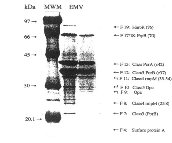
terminal sequencing. Twenty fractions (F1-F20) from low to high molecular weight components were obtained by SDS-PAGE followed by slot electroelution separated according with their gel migration characteristics. SDS-PAGE and silver staining of the fractions showed that LPS was present in F1 and F2, while the principal meningococcal outer membrane proteins in the various fractions could be only tentatively identified at this stage. A protein with the migration characteristics of Class 1 (PorA) was detected in fractions F10 to F14, peaking at F13. Class 3 (PorB) appeared to be in F9 to F13 but predominated in F11 and F12, while Class 4 (rmpM) was mostly in F11, Class 5 (Opc) was in F9 and F10, and FrpB (70 kDa) was mostly in F18 (Fig. 2).
Eight major meningococcal outer membrane proteins present in the PL and its main fractions were identified using end terminal sequencing. Two of these appeared to be in more than one fraction obtained by slot elution (Fig. 3). We found that PLs contained surface protein A (NspA), identified as a band of approximately 18-22 kDa in fractions F4 and F5; Class 3 (PorB, 37-42 kDa) in F7 and F12; Class 4 (RmpM, 33-34 kDa) in F8 and F11; Class 5 (Opa, 22-33 kDa) in F9 and F10; Class 1 (PorA, 44-47 kDa) in F13; FrpB (70 kDa) in F17 and F18; and HmbR 76 kDa in F19. We found that the protein representation and concentration between independent N. meningitidis PLs vaccine batches are very similar (data not shown). Overall, PLs was separated in 20 fractions and there main proteins were identified still some of them were present in several fractions.
10 particular, F1-F4, F8, F9, F11, and F18 were high inducers giving four distinct peaks of TNF, IL-1, and IL-6 production, while other fractions showed lower levels of induction. Additional fractions appeared to induce IL-8, although the pattern of IL-8 production broadly followed that of the other cytokines tested (Fig. 4). Taking in consideration that each fraction contains different protein concentration and that we used the same volume of each fractions to stimulate the differentiated cell line, a parallel experiment was conducted to evaluate these possible influence in the cytokine production. The independence of the different concentrations used was demonstrated where similar cytokine production behavior were found (data not shown).
11 4. DISCUSSION
4.1 The LPS of PLs has significantly reduced inflammatory effects
The present evaluated the individual participation of each PL component in the pro-inflammatory effect of PL and to understand why native LPS, is well tolerated and immunogenic in small animals and human when it is inserted in PLs. Vaccine toxicity is associated with local inflammation and the production of pro-inflammatory cytokines such as TNFα, IL-1β, IL-6, and IL-83. We first evaluated the production of these cytokines by differentiated U937 histiocytic human cell line stimulated with either purified LPS or PLs, which contains inserted native LPS. The production of pro-inflammatory cytokines by PLs was dose-dependent. A dramatic reduction in the production of TNF and IL-6 and a reduction in the production of IL-1 and IL-8 were observed in samples stimulated with PLs, in comparison with samples stimulated by matching amounts of LPS. These results suggest that LPS inserted in PL have a different behaviour than when it is free. A recent study using The Hen’s Egg Test on Chorioallantoic Membrane (HET-CAM) for a comparative evaluation of the irritating effect of different adjuvants, showed that PL and cochleates derived from N meningitidis B induced a low irritant effects with minimal local toxicity1.
4.2 Some PLs fractions were more pro-inflammatory than LPS or whole PLs
12 This indicates that the ability of F1 and F2 and probably F3 to strongly induce cytokine production is due to its LPS content. Indeed, in agreement with this conclusion, we found that the ability of the fractions to induce cytokine expression was almost completely blocked by anti-CD14.
In addition to LPS, the PLs contains lipoproteins and peptidoglycan which are activators of TLR2 and could therefore also be involved in PL-induced cytokine production12. We used CD14 depletion to test whether or not activation of cytokine production by the fractions occurred thought CD14 and TLR4 receptors. Although, LPS could be only detected by sugar silver staining of the gels in the first 3 fractions and not in the others, in most cases their ability to induce cytokine expression was also blocked by anti-CD14. This suggests that the ability of these fractions to induce cytokine expression can be owing to the presence of contaminating LPS. The remarkable cytokine induction by F18 in relationship with its low concentration and its completely blockage with anti-CD14 suggest that the protein present in this fraction is sticking more LPS than other proteins. The presence of FrpB, 70 kDa in this fraction in the PL might have implication in it immunopotentiator ability.
13 non-responder mice26. Quarkyi et al. demonstrated that LPS inserted in this kind of PL structures is less reactive by chromogenic LAL assay, less inductor of shock in animals and less pyrogenic in rabbits than purified LPS25.
4.3 Identification of Class 4 protein as a major component in F11
15
Conflicts of interest
The authors declare no commercial or financial conflict of interest.
Acknowledgements
16
Figure 1. LPS inserted in Proteoliposome (PLs) induced levels of cytokine response
dramatically lowers than purified lipopolysaccharide (LPS). Differentiated human U937 histiocytic cell line was incubated with PL (250 ng/mL) or LPS (10 ng/mL). LPS concentration in the PL was 10 ng/mL. Fig. 1A shows TNFand IL1 production (pg/mL) and Fig. 1B shows IL6 and IL8 production (ng/mL) 24 h after stimulation as determined by ELISA. Data are presented as mean cytokine concentration in supernatants ± standard deviation of al least three different experiments. Significant differences between the means of two different groups were determined by a Student t test using statistical analyses with Graph Pad Prism 4 software (Calif.) and a p-value of <0.05 was considered statistically significant.
Figure 2. Twenty fractions were obtained by slot elution following SDS-PAGE separation of
17 weight standard was included in each gel. White lines were included to separate the fractions (1-20). Molecular weights were calculated with the molecular analyst program (Bio-Rad). Lipopolysaccharide (LPS) was clearly present in fractions 1 and 2, while the principal meningococcal outer membrane proteins where present in various fractions, but mainly in one of them and could be only tentatively identified at this stage. PL was included as control in line 21. Main proteins were also marked in the middle of the figure.
Figure 3. Eight meningococcal outer membrane proteins were identified in Proteoliposome
18 2 were also used to verify that each band correspond to the PL proteins. The sequences were identified comparing with those of the data bank (www.ncbi.nlm.nih.gov).
Figure 4. Several individual fractions induced dramatically higher levels of cytokine production
19
Figure 5. Pre-incubation of the cells with anti-CD14 mAb considerably reduced induction of
20
REFERENCES
1. Batista-Duharte A, Jorge Murillo G, Pérez UM, Tur EN, Portuondo DF, Martínez BT, Téllez-Martínez D, Betancourt JE, Pérez O. The Hen's Egg Test on Chorioallantoic Membrane: An Alternative Assay for the Assessment of the Irritating Effect of Vaccine Adjuvants. Int J Toxicol. 2016 Nov;35(6):627-633.
2. Batista-Duharte A, Lastre M, Pérez O. Immunological adjuvants. Determinant factors in the efficacy-toxicity ratio of the contemporary vaccines. Enferm Infecc Microbiol Clin. 2014;32(2):106-14.
3. Batista-Duharte A, Portuondo D, Carlos IZ, Pérez O. An approach to local immunotoxicity induced by adjuvanted vaccines. Int Immunopharmacol. 2013;17(3):526-36.
4. Batista-Duharte A, Portuondo D, Pérez O, Carlos IZ. Systemic immunotoxicity reactions induced by adjuvanted vaccines. Int Immunopharmacol. 2014;20(1):170-80.
5. Collins B.S. Gram-negative outer membrane vesicles in vaccine development. Discovery medicine 2011, 12(62), 7-15.
6. Geurtsen J, Banus HA, Gremmer ER, Ferguson H, de la Fonteyne-Blankestijn LJ, Vermeulen JP, Dormans JA, Tommassen J, van der Ley P, Mooi FR, Vandebriel RJ. Lipopolysaccharide analogs improve efficacy of acellular pertussis vaccine and reduce type I hypersensitivity in mice. Clin Vaccine Immunol. 2007;14(7):821-9.
21 8. Huergo CC, Sierra G, Gutierrez MM, Bisset G, García LG, Puentes G, et al. Method of
producing Neisseria meningitidis B vaccine, and vaccine produced by method. 1998. European patent 885900077.8.
9. Huergo CC, Sierra G, Gutierrez MM, Bisset G, García LG, Puentes G, et al. Method of producing Neisseria meningitidis B vaccine, and vaccine produced by method. 1997. United States patent 5,597,572.
10. Kattner, C., Toussi, D.N., Zaucha, J., Wetzler, L.M., Rüppel, N., Zachariae, U., Massari, P., and Tanabe, M.. Crystallographic analysis of Neisseria meningitidis PorB extracellular loops potentially implicated in TLR2 recognition. J Struct Biol. 2014;185(3):440-7.
11. Laemmli UK. Cleavage of structural proteins during the assembly of the head of bacteriophage T4. Nature. 1970;227:680-5.
12. Liang MD, Bagchi A, Warren HS, Tehan MM, Trigilio JA, Beasley-Topliffe LK, Tesini BL, Lazzaroni JC, Fenton MJ, Hellman J. Bacterial peptidoglycan-associated lipoprotein: a naturally occurring toll-like receptor 2 agonist that is shed into serum and has synergy with lipopolysaccharide. J Infect Dis. 2005;191(6):939-48.
13. Liu G, Zhang L, Zhao Y Modulation of immune responses through direct activation of Toll-like receptors to T cells. Clin Exp Immunol. 2010;160(2): 168–175.
14. Lugtenberg B, Mejers J, Peters R, van der Hoek P, van Alphen L. Electrophoretic resolution of the major outer membrane protein of E. coli K12 into four bands. FEBS Lett. 1975;58:254-8.
22 16. Muñoz-Wolf N, Lavelle EC. Innate Immune Receptors. Methods Mol Biol.
2016;1417:1-43
17. Osborn MJ, Rosen SM, Rothfield L, Horechker BL. Studies on the Gram-negative cell wall. Evidence for the role of 2-keto-3-deoxyoctonate in the Lipopolysaccharide of Salmonella thyphimurium. Proc Natl Acad Sci USA. 1963;50:499-506.
18. Pasquale AD, Preiss S, Silva FT, Garçon N. Vaccine adjuvants: from 1920 to 2015 and beyond. Vaccines (Basel). 2015;3(2):320-343.
19. Pennock ND1, White JT, Cross EW, Cheney EE, Tamburini BA, Kedl RM. T cell responses: naive to memory and everything in between. Adv Physiol Educ. 2013 Dec;37(4):273-83.
20. Pérez O., Romeu, B., del Campo J., Zayas C., Lastre M. Proteoliposome and polysaccharide-based meningococcal vaccine are immunogenic in infants and toddlers and primes for memory against serogroup C polysaccharide. World Journal of Vaccines, 2013, 3, 77-87. 21. Pérez O, Lastre M, Lapinet J, Pérez A, Díaz M, Zayas C, Batista A.; Quintero Y; Aguiar F;
Sánchez R; Sierra G. Long-lasting cellular immune response in babies, children, and pre-teenagers vaccinated with a proteoliposome based anti-meningococcal BC vaccine. Inmunología 2001,20(4):177-183.
22. Pérez O, Romeu B, Cabrera O, González E, Batista-Duharte A, Labrada A, Pérez R, Reyes LM, Ramírez W, Sifontes S, Fernández N, Lastre M. Adjuvants are Key Factors for the Development of Future Vaccines: Lessons from the Finlay Adjuvant Platform. Front Immunol. 2013;4:407.
23 24. Platt, A., MacLeod, H., Massari, P., Liu, X., and Wetzler, L. In vivo and in vitro
characterization of the immune stimulating activity of the Neisserial porin PorB. PLoS One 2013. 8(12):e82171.
25. Quarkyi EK, Hochstein HD, Tsai CM. Modulation of the biological activities of meningococcal endotoxins by association with outer membrane proteins is not inevitably linked to toxicity. Infect Immun 1997;65:1972-79.
26. Rodríguez T, Pérez O, Ménager N, Ugrinovic S, Bracho G, Matroeni P. Interactions of proteoliposome from serogroup B Neisseria meningitidis with bone marrow-derived dendritic cells and macrophages: adjuvant effects and antigen delivery. Vaccine 2015; 23:1312–21. 27. Sierra GV, Campa HC, Varcacel NM, Garcia IL, Izquierdo PL, Sotolongo PF, et al. Vaccine
against group B Neisseria meningitidis: protection trial and mass vaccination results in Cuba. NIPH Ann. 1991;14:195-210.
28. Sumiya YU, Inoue T, Ishikawa M, Inui T, Kuchiike D, Kubo K, Uto Y, Nishikata T. Macrophages exhibit a large repertoire of activation states via multiple mechanisms of macrophage-activating factors. Anticancer Res. 2016;36(7):3619-23.
29. Sun R, Zhu Z, Su Q, Li T, Song Q. Toll-like receptor 4 is involved in bacterial endotoxin-induced endothelial cell injury and SOC-mediated calcium regulation. Cell Biol Int. 2012 May 1;36(5):475-81.
30. Stefanelli P, Neri A, Tanabe M, Fazio C, Massari P. Typing and surface charges of the variable loop regions of PorB from Neisseria meningitidis. IUBMB Life. 2016;68(6):488-95
24 32. Vajjhala PR, Ve T, Bentham A, Stacey KJ, Kobe B. The molecular mechanisms of
signaling by cooperative assembly formation in innate immunity pathways. Mol Immunol. 2017;86:23-37 .
33. Venier C, Guthmann MD, Fernández LE, Fainboim L. Innate-immunity cytokines induced by very small size proteoliposomes, a Neisseria-derived immunological adjuvant. Clin Exp Immunol. 2007;147(2):379-88.