DOI: 10.1534/genetics.108.097345
Horka
D, a Chromosome Instability-Causing Mutation in Drosophila,
Is a Dominant-Negative Allele of
lodestar
Tamas Szalontai,* Imre Gaspar,* Istvan Belecz,* Iren Kerekes,*
Miklos Erdelyi,
†Imre Boros
‡and Janos Szabad*
,1*Department of Biology, Faculty of Medicine, University of Szeged, H-6720 Szeged, Hungary,†Institute of Genetics, Biological Research Center of the Hungarian Academy of Sciences, H-6726 Szeged, Hungary and‡Department of
Biochemistry and Molecular Biology, University of Szeged, H-6726 Szeged, Hungary Manuscript received October 10, 2008
Accepted for publication November 21, 2008
ABSTRACT
Correct segregation of chromosomes is particularly challenging during the rapid nuclear divisions of early embryogenesis. This process is disrupted byHorkaD, a dominant-negative mutation inDrosophila melanogaster
that causes female sterility due to chromosome tangling and nondisjunction during oogenesis and early embryogenesis.HorkaDalso renders chromosomes unstable during spermatogenesis, which leads to the
for-mation of diplo//haplo mosaics, including the gynandromorphs. Complete loss of gene function brings about maternal-effect lethality: embryos of the females without theHorkaD-identified gene perish due to
disrupted centrosome function, defective spindle assembly, formation of chromatin bridges, and abnormal chromosome segregation during the cleavage divisions. These defects are indicators of mitotic catastrophe and suggest that the gene product acts during the meiotic and the cleavage divisions, an idea that is supported by the observation that germ-line chimeras exhibit excessive germ-line and cleavage function. The gene affected by theHorkaDmutation islodestar, a member of the helicase-related genes. TheHorkaDmutation
results in replacement of Ala777 with Thr, which we suggest causes chromosome instability by increasing the affinity of Lodestar for chromatin.
M
OST of the proteins required in early embryo-genesis are maternally provided; it is generally agreed that little if any zygotic gene expression occurs during the onset of embryogenesis (Derenzo andSeydoux2004; Tadrosand Lipshitz2005). To dissect
the commencement of embryogenesis in Drosophila melanogaster, we isolated dominant female-sterile (Fs) mutants (Erdelyiand Szabad1989; Szabadet al.1989)
and focused our attention on those that terminate embryogenesis at or shortly after fertilization.
HorkaD
is one suchFsmutation (Erdelyiand Szabad
1989). It is a gain-of-function mutation (Erdelyi and
Szabad1989) that results in chromosome
nondisjunc-tion and renders chromosomes unstable during sper-matogenesis, causing them to be lost in the resulting zygotes (Szabadet al.1995). Loss of the chromosomes
leads to the formation of diplo//haplo mosaics, in-cludingXX//X0, female//male mosaics, and gynandro-morphs (Szabadet al.1995). (Xrepresents chromosomes
derived from theHorkaDmales.) In fact,HorkaDhas been used as a ‘‘tool’’ to generate genetic mosaics (Szabadand
Nothiger1992; Zallenand Wieschaus2004; Villanyi
et al.2008).
To determine the function of the gene carrying the HorkaD
mutation, we first mappedHorkaD
by screening
for duplications that can ameliorate the HorkaDmutant
phenotype in embryos of theHorkaD/1/1females. This revealed the dominant-negative (antimorphic) nature of the mutation. We generated horkarvP P -element-induced alleles (hereafter called pseudorevertants) that no longer exhibit the dominant mutant phenotype and used them to map and then isolate the gene. We discovered thatHorkaDis an allele oflodestar(lds), which encodes a member of the Snf2 family of the helicase-related genes (Girdham and Glover1991; Liu et al.
1998; Flaus et al. 2006). Our results suggest that the
lodestar (LDS) protein is involved in progression from metaphase to anaphase of the cell cycle. We propose that the lodestar protein altered by HorkaD disturbs chromatin organization and segregation and renders chromosomes unstable. It appears thus that the LDS protein is one of the many components engaged in maintaining genome integrity (Takada et al. 2003;
Allardet al.2004; Musacchioand Salmon2007).
MATERIALS AND METHODS
HorkaD,horkarv, andHorkaRRalleles:HorkaDwas induced by
EMS on an isogenic third chromosome labeled with themwh and theerecessive marker mutations (Erdelyiand Szabad 1989). For an explanation of the genetic symbols, see FlyBase at http://flybase.bio.indiana.edu. Thehorkarvrevertant alleles
were generated through second mutagenesis ofHorkaD: the
horkarvE1allele by EMS (Erdelyiand Szabad1989) and the 1Corresponding author: Department of Biology, Faculty of Medicine,
University of Szeged, Somogyi Str. 4, H-6720 Szeged, Hungary. E-mail: [email protected]
horkarvP alleles through mutagenesis with the normal P
elements. For induction of the horkarvP alleles, dysgenic
HorkaD/TM3, Sb Sermales were mated withTM3, Ser/TM1,
Mevirgin females. The Pelements were hopping in these males and might have become inserted into theHorkaDallele.
(The dysgenicHorkaD/TM3, Sb Sermales were generated by
crossingMcytotypeTM3, Sb Ser/TM6B, Tbfemales with P cytotypeHorkaD/TM3, Sb Sermales. The latter males resulted
from a cross betweenPcytotypeCxD/TM3, Sb Serfemales and HorkaD/TM3, Sb Sermales.) Since theTM3, Sb Ser/TM3, Ser
and theTM3, Sb Ser/TM1, Mecombinations are lethal, only the HorkaD/TM3, Ser and the HorkaD/TM1, Me offspring
survive. The resulting females, who mated with the sibling males, were screened for offspring production. Only the horkarvP/TM3, Serand thehorkarvP/TM1, Mefemales give rise
to progeny, allowing a direct selection of thehorkarvP
pheno-typically revertant alleles. (To avoid the isolation of clusters of thehorkarvPalleles, groups of 10 dysgenic males were mated
withTM3, Ser/TM1, Mefemales and the descendants from the parallel crosses were screened separately.)
The P-element insertion sites in the horkarvP alleles were
determined by standardin situhybridization on salivary gland chromosomes, using DIG-labeledP-element DNA probe.
To remobilize thePelements in thehorkarvPrevertants and
isolateHorkaRRalleles (revertant alleles of the horkarvP
rever-tants), we constructedhorkarvP/TM3,D2-3females and males.
TheHorkaRRoriginated most likely through precise excision of
thePelement from thehorkarvP alleles. TheHorkaRR alleles,
which behaved asHorkaD, were used inin situhybridization
studies on salivary gland chromosomes.
The chromosome destabilizing effect of HorkaD and the
horkarvPalleles was analyzed in outcrosses withy v f malfemales
and measured through the frequency of XX//X0, female// male mosaics among the descendingXXzygotes (cf.Szabad et al.1995).
Drosophila cultures used in the study were kept at 25°.
The HorkaD/Dp(3;3) combinations:HorkaD was mapped to
the right arm of the third chromosome (Erdelyiand Szabad 1989). To determine the approximate location and the nature of HorkaD (whether it is antimorphic or neomorphic), we
constructed HorkaD/Dp(3;3) females and males by crossing
Dp(3;3)/TM3females withHorkaD/TM3, Sb Sermales.Dp(3;3)
stands for 18 tandem duplications, which cover—bit by bit—the right arm of the third chromosome. The resulting HorkaD/Dp(3;3)females were mated with wild-type males and
the fate of their resulting embryos was monitored. Males were mated withy v f malfemales and the subsequent generation was screened forXX//X0 mosaics.
Localizing the horkarv alleles and complementation
analy-sis:To locate thehorkarvalleles and to determine the
loss-of-function mutant phenotype, we combined thehorkarvalleles
(as well asHorkaD) withDf(3R)deficiencies and analyzed the
horkarv/(and theHorkaD/) flies. (Thesymbol stands for
either of the deficiencies that remove theHorkaD-identified
locus.) The studiedHorkaD/and thehorkarv/hemizygotes
were produced by crossingDf(3R)dsx15/TM6B, Tbfemales with
horkarv/TM6B, Tbor withHorkaD/TM6B, Tbmales.
To determine whether thehorkarvalleles identify a gene with
already existing mutant alleles, we carried out complementa-tion analyses betweenhorkarand mutant alleles of the nearby
genes,lds,dsx, andCG10445(see Figure 3). (Mutant alleles of theCG10445gene were generated in our laboratory; I. Belecz and J. Szabad, unpublished observations.)
Characterization of mutant phenotypes: To describe the HorkaD- and the horkarv-associated mutant phenotypes,
ova-ries, testes, and eggs/embryos of HorkaD/1; HorkaD/– and
horkarvP2/– females and males were dissected and fixed
ac-cording to Gonza´ lez and Glover (1993). The stage 14
oocytes were immunostained according to Tavosaniset al. (1997). The eggs and the embryos were prepared as follows: the chorion was removed by Clorox, the dechorionated embryos were fixed in a 1:1 mixture of 4% paraformaldehy-de:heptane or in a 1:1 mixture of methanol:heptane, and the vitelline membrane was removed subsequently by agitation in a mixture of heptane and methanol. To block nonspecific staining, the embryos were incubated in 1% BSA (Sigma, St. Louis) in PBST for 90 min at room temperature.
For immunological detection of the microtubules, we used the DM1A monoclonal anti-a-tubulin antibody (1:1000, over-night at 4°; T6199, Sigma). The centrosomes were detected using an anti-centrosomin antibody (Heueret al. 1995). The LDS protein was detected by polyclonal anti-LDS rabbit antibody raised against the almost complete LDS protein, a generous gift from David Glover’s laboratory (Girdhamand Glover 1991). The anti-LDS antibody was present in the serum from which the nonspecific components were depleted through preincubation of the serum in dechorionated and heptane-permeabilized eggs ofhorkarvP2/– females, which do
not contain LDS protein. The anti-LDS antibody was applied at a 1:200 dilution in 1% BSA in PBST. The embryos were incubated in secondary antibodies for 3 hr at room temper-ature or overnight at 4°. The secondary antibodies were either anti-mouse or anti-rabbit IgG (Sigma) and were labeled with FITC, Texas-Red, or Alexa Fluor-633. To detect DNA, the embryos were stained with DAPI following incubation with the secondary antibody. After several rinses in PBST, the embryos and the testes were mounted in Aqua PolyMount (Polyscien-ces, Warrington, PA). The immunostained preparations were analyzed either in an Olympus IX71 fluorescent microscope with a cooled CCD camera or through optical sections col-lected in an Olympus FV1000 confocal microscope.
We also prepared and analyzed cuticles of the dead embryos as described in Wieschausand Nusslein-Volhard(1989).
Cytoplasm injections:To analyze the effect ofHorkaDon the
cleavage divisions, we injected300 pl of cytoplasm (3% of the total egg volume) from eggs of wild-type (as the control) andHorkaD/1females into the posterior region of embryos in
which the microtubules were highlighted by Jupiter-GFP and the nuclei by histone-RFP (Karpovaet al.2006; Schuhet al. 2007). The donor embryos were a maximum of 30 min old and the injected embryos were in the 9th–11th cleavage cycle of embryogenesis. Effect of the injected cytoplasm was followed in time through a series of optical sections generated in an Olympus FV1000 confocal microscope. The injections were carried out at 25°.
Germ-line chimeras:To determine whether theHorkaD-and
thehorkarvP2/-related defects originate from altered function
of the germ line and/or the soma, we constructed different types of germ-line chimeras through the transplantation of pole cells, embryonic precursor cells of the future germ line. Tables 2 and 3 list the crosses from which the donor and the host embryos originated. Pole cells were collected from single blastoderm-stage donor embryos and transplanted into two to three host blastoderm embryos. While pole cells do not develop in the embryos of thetropomyosin-IIgs(tmIIgs)
homo-zygous females, the somatic cells function normally (Erdelyi et al.1995).Fs(1)K1237(also known asovoD1) is an X-linked
dominant female-sterile mutation (Komitopoulouet al.1983; Perrimon1984). Although theFs(1)K1237/1host females do not produce eggs of their own, their soma provides a normal environment for development of the received female pole cells. Pole cells of y v f malembryos were transplanted into HorkaD/1andhorkarvP2/host embryos, and the developing
transplantation were mated with appropriate partners, as de-scribed in Tables 2 and 3, and tested for germ-line chimerism.
Inverse PCR:To clone theHorkaD-identified gene, we used
the inverse PCR technique and amplified DNA sequences flanking thePelements in three of thehorkarvPalleles. Briefly,
we isolated DNA fromhorkarvP-carrying males and digested the
DNA withHinPI or withMspI. The digested genomic DNA was ligated overnight at 4°, ethanol precipitated, and resuspended in distilled water. Two PCR reactions were conducted next. In the first reaction, the outward primers were designed on the basis of the terminal sequences of theP-element adjacent to the cut site. (The primers are described in supplemental Table 2.) Because the first PCR did not yield sufficient amounts of DNA for sequencing, a second, so-called nested PCR reaction was conducted using primers complementary to slightly more interior sequences in thePelement. (See supplemental Table 2.) Products from the second PCR reactions were isolated, purified, and sequenced in an IBI automated sequenator on both strands. The resulting sequence information allowed us to precisely position the P-element insertion sites on the Drosophila genome sequence (Adamset al.2000).
Molecular cloning and sequencing of HorkaD: DNA of HorkaD/– andmwh e(as the control) males served as a template
in a set of PCR reactions to produce DNA fragments for sequencing. The PCR primers were designed on the basis of theldsgene sequence (EMBL nucleotide sequence database, accession no. X62629). Sequencing of the PCR products was carried out in an IBI sequenator on both strands.
The Horka1
(TG1
) and theHorkaD (TGHD) transgenes: To characterize theHorkaD-identified gene, we generated aHorka1 transgene (TG1
) in which a 5.1-kb genomic segment included both the regulatory and the structural parts of theldsgene (see Figure 3). The 5107-bp genomic sequence was cloned into the CaSpeRvector with themini-whitemarker gene and a germ-line transformant transgenic line was generated on aw1118
back-ground by standard procedures. TheTG1
transgene became inserted into the second chromosome. TheTG1
transgene was combined, in appropriate genetic crosses, with theHorkaD, the
horkarv, and theldsmutant alleles to determine whether the
TG1
transgene can overcome the mutant phenotypes associ-ated with theHorkaDand thehorkarvalleles.
To generate transgenes that carryHorkaD(theTGHD
trans-genes), we PCR amplified a 5107- and a 5499-bp genomic segment that included the promoter and the structural parts of theHorkaDallele (see Figure 3). The DNA was isolated from
HorkaD/Df(3R)dsx15males. The two transgene types correspond
to the twoldsmRNAs that differ by500 nucleotides in their 39-UTR (Girdham and Glover 1991; see Figure 3). Stable germ-line transformant lines were generated through stan-dard procedures.
RESULTS
HorkaD
disrupts the meiotic and the early cleavage divisions: Although the HorkaD
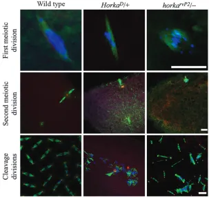
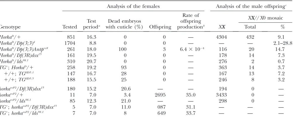
/1 females deposit normal numbers of normal-looking eggs (fertilized as in wild type), cleavage divisions do not commence in .90% of the eggs. Moreover, when cleavage divisions are seen, only 12 scattered chromosomes appear, along with unusual microtubule bundles (Figure 1). As expected, cuticle fragments, indicators of develop-ment beyond the blastoderm stage, never form inside the eggs of theHorkaD/1females (Table 1). Abnormal segregation of the chromosomes is already apparent during both the first and the second meiotic divisions in egg primordia of theHorkaD
/1females (Figure 1). The mutant phenotypes suggest involvement of theHorkaD
-identified normal gene product in chromosome orga-nization, stability, and/or segregation.
HorkaD
has been reported to be a gain-of-function mutation (Erdelyi and Szabad 1989). We have now Figure 1.—Meiotic and cleavage divisions in the egg primordia and in eggs of wild-type, HorkaD/1, andhorkarvP2/females. In the optical
sections the microtubules appear in green, the centrosomes and the spindle pole bodies in red, and the DNA in blue. Detachment of one of the spindles (dashed circle) is a typical feature of the second meiotic division in theHorkaD/1
females. Although the meiotic divisions proceed as in wild type in70% of the cases, abnormal meiotic spindles develop in a number of egg pri-mordia in thehorkarvP2/females. Note that most
confirmed this observation by cytoplasm injection studies. When cytoplasm taken from newly deposited eggs of theHorkaD/1 females was injected intohorka1 embryos in which the chromosomes were highlighted by RFP-tagged histones and the microtubules by GFP-tagged tubulins, it induced chromosome tangling during ana-and telophase, the formation of chromatin bridges, abnormally shaped and positioned nuclei (which usually drop inside the egg cytoplasm during the upcoming cleavage mitosis), and free centrosomes. (See Videos 1 and 2 in the supplemental material.) Toxicity of the HorkaD
-derived egg cytoplasm is best illustrated by the fact that not a single embryo survived the cytoplasm injections. (Injection of wild-type egg cytoplasm did not alter progression of the cleavage cycles, and larvae hatched from almost all of the injected embryos.)
HorkaDresides between 84D5–8 and 85F5–8:
HorkaD has been mapped to the right arm of the third chro-mosome (Erdelyiand Szabad 1989). To more
accu-rately locate HorkaD, we generated a series ofHorkaD/ Dp(3;3)flies and analyzed the embryos of the females
and searched for XX//X0 mosaics among the XX off-spring of the males. If HorkaD is a dominant-negative mutation, (i) a less severe mutant phenotype was ex-pected to develop inside eggs of the HorkaD/Dp(3;3)1 females (compared to theHorkaD/1control) and (ii) reduced frequency of theXX//X0 mosaics was expected to appear in the offspring of theHorkaD/Dp(3;3)1
males. We used 18 Dp(3;3) tandem duplications that, in aggregate, fully covered the entire right arm of the third chromosome. Of these duplications tested, only Dp(3;3)Antprv8
ameliorated theHorkaD
imposed defects, such that embryogenesis inside eggs of the HorkaD
/ Dp(3;3)Antprv8
females proceeded well beyond the initial steps. Not only did cuticle fragments form in almost 100% of the eggs, but also three offspring were produced by the HorkaD/
Dp(3;3)Antprv8 females (Table 1). Thus the results of mapping locatedHorkaD within the 84D5–8 and 85F5–8 cytological interval (Fig-ure 2). Moreover, the ability ofDp(3;3)Antprv8to amelio-rate the dominant-negative nature ofHorkaDargues that the mutant and normal gene products participate in the
TABLE 1
Features of theHorkaD- and thehorkarvP2-carrying females and males
Analysis of the females Analysis of the male offspringc
Test perioda
Dead embryos with cuticle (%)
Rate of offspring productionb
XX//X0 mosaic
Genotype Tested Offspring XX Total %
HorkaD/1 851 16.3 0 0 — 4304 432 9.1
HorkaD/Dp(3;3)d 1704 8.8 0 0 — — — 2.1–28.8
HorkaD/Dp(3;3)Antprv8 261 18.0 100 3 6.43104 116 20 14.7
HorkaD/Df(3R)dsx15 161 19.5 0 0 — 178 14 7.3
HorkaD/lds98.1 310 20.7 0 0 — 276 2 0.7
TG1;HorkaD/1 258 19.2 93 0 — 363 14 3.7
1/1;TGHD5.1 147 16.7 28 0 — 167 13 7.2
1/1;TGHD5.5 188 15.5 25 0 — 246 8 3.2
horkarvP2/Df(3R)dsx15 180 15.2 20.6 — — 194 0 —
horkarvP2/1 11 7.0 3.4 2695 35.0 3433 0 —
horkarvP2/lds98.1 85 12.3 21.0 — — 298 0 —
TG1;horkarvP2/Df(3R)dsx15 5 7.0 11.0 087 31.1 — — —
TG1
;horkarvP2/lds98.1 7 7.0 8 649 33.7 — — —
horkarvP2is a functionally null allele andlds98.1is alodestarnull allele (Girdhamand Glover1991).
aAverage test period per female (days). bOffspring/(female3day).
cThe males were mated withy v f malfemales (XX) and theXXoffspring flies were screened forXX//X0 mosaics. d
Pooled data from 17Dp(3;3)tandem duplications with the exception ofDp(3;3)Antprv8.
Figure 2.—Duplication and deficiency map-ping of the HorkaD and the horkarvP2mutations.
The thick bar represents the Dp(3;3)Antprv8
same process and the mutant gene product is truly antimorphic (i.e., it impedes function of the normal counterpart).
We cannot discern whether theHorkaD-related dom-inant paternal effect is also of domdom-inant-negative nature because the frequency of the XX//X0 mosaics varied between 2.1 and 28.8% in the XX offspring of the HorkaD/Dp(3;3)males. The variation in theXX//X0 mosaic frequencies is most likely related to the different genetic backgrounds of theHorkaD
/Dp(3;3)males (Szabadet al.
1995).
Thehorkapseudorevertant
(horkarv
) alleles:To learn the func-tion of the gene carrying the HorkaD
mutation we inducedhorkarvP
pseudorevertant alleles through muta-genesis ofHorkaD
with a normalPelement. Of the 15,600 P-element-mutagenized females tested, 9 independent ones were fertile and gave rise to onehorkarvP pseudor-evertant allele each. All thehorkarvalleles are lethal both in homozygotes and intrans-heterozygotes due to one or more second-site lethal mutations induced during EMS induction of HorkaD (cf. Erdelyi and Szabad 1989).
To characterize thehorkarvPalleles, we crossedhorkarvP/ TM3, Sb Sermales from each of the ninehorkarvPlines withy v f mal females and searched the offspring for XX//X0 mosaics (see Table 1). Mosaics appeared (though with very low frequencies) among the offspring of three of the ninehorkarvP
pseudorevertants (horkarvP5 , horkarvP6
, and horkarvP8
), suggesting that these alleles retain some feature ofHorkaD. Their incomplete loss of the HorkaD phenotype is also shown by the strongly
reduced fertility of the heterozygous females. Mosaics did not appear in the offspring of the males that were heterozygous for the other sixhorkarvPalleles and larvae hatched from the vast majority of the eggs deposited by the heterozygous females, indicating the loss-of-function nature of six of thehorkarvPmutations. One of the alleles, horkarvP2, is a complete loss-of-function mutation (Table 1 and see below). The concurrent loss of dominant female sterility and dominant paternal effect in six of the ninehorkarvP
alleles shows that the HorkaD
-related dominant mutant phenotypes stem from the same mutation.
The horkar
mutations reside within the 84E1–84E8 cytological interval:Thehorkarvalleles (and also
HorkaD)
were combined with deficiencies that remove well-defined regions around the 84E cytological region (Fig-ure 2). Thehorkarv/
–hemizygous combinations are via-ble and the flies develop with the expected frequencies. (The – symbol stands for either of the deficiencies that remove thehorkarvidentified locus.) Thehorkarv/– hemi-zygous females either are completely sterile (horkarvP2/–; Table 1) or possess reduced fertility: progeny develop from 4–21% of the zygotes in all the other horkarv
/– combinations. The deficiencies located the horkarv
-identified locus within the 84E1–E8 cytological region (Figure 2).
The fertility ofhorkarvP2
/– males is also very strongly reduced (see supplemental Table 2). The cross in which several hundred horkarvP2
/– males were mated with several hundredy v f malfemales yielded only few off-spring, none of which wasXX//X0 mosaic (Table 1).
TheHorkaD/
–flies are also viable and emerge with the expected frequency. The females deposit normal num-bers of normal-looking eggs in which, although normally fertilized, embryogenesis never commences. Fertility of theHorkaD/–males is also strongly reduced (supplemen-tal Table 2). However, a few offspring derived from a cross between several hundredHorkaD/– males andy v f mal females and 6.7% (14/192) of theXXoffspring flies were XX//X0 mosaics (Table 1).
It appears that reduced fertility of the HorkaD also exhibits a dominant-negative effect on male fertility, as evidenced by the observation that, when sired by HorkaD/–,
HorkaD/1, or
HorkaD/
Dp(3;3)Antprv8 males, 92, 71, and 59% of the embryos perished during embryogenesis (supplemental Table 2). Because there was no sperm in 98.5% (446/453) of the eggs in which embryogenesis did not commence, the reduced fertility of the HorkaD-carrying males is most likely the conse-quence of abnormal spermatogenesis. Remarkably, the egg production rate of the partnery v f malfemales was not significantly different from the control (supplemen-tal Table 2) and thusHorkaDandhorkarvP2do not seem to affect other fertility-related features than sperm pro-duction, suggesting thatHorkaD
has little if any effect on the somatic cells (cf.Liuand Kubli2003).
In situhybridizations confirm that theHorkaD -identi-fied gene resides in 84E:To locate the gene carring the HorkaDmutation we probed salivary gland chromosomes
Figure3.—Organization of the region around thelodestargene in the 84E5 cytological region. The lds gene encodes the formation of two mRNAs that differ in the last500 nucleotides. Stippled boxes correspond to sequences that en-code the 59and the 39untranslated regions of the ldsmRNAs, and open and solid boxes represent introns and exons, respectively. The P-element insertion sites inhorkarvP3, horkarvP9, and horkarvP2
are labeled and also the position of theHorkaD
in the nine horkarvP revertants with labeled
P-element DNA. There were three to sixP-element insertions in the right arm of the third chromosome in the different horkarvPalleles. The only common P-element insertion site appeared in 84E, suggesting that the gene resides in 84E.
ThePelements of thehorkarvP7and thehorkarvP9alleles were successfully remobilized. As a result, twoHorkaRR alleles (revertant alleles of the horkarvP
mutations) emerged from the 135 chromosomes tested. The HorkaRR
alleles behaved as HorkaD
: HorkaRR
/1 females are sterile and embryos perish inside their eggs, essentially as described for the HorkaD/1 females. Among the progeny of theHorkaRR/1 males and
y v f malfemales, 13.9% (14/85) and 13.0% (25/164) of the XX zygotes developed as XX//X0 mosaics in the two HorkaRR alleles. More important, the P-element inser-tions in 84E were absent in theHorkaRRalleles, suggest-ing that the gene carrysuggest-ing theHorkaDmutation indeed resides at 84E. The HorkaRR alleles underline the common origin of theHorkaD-related dominant defects.
HorkaD
and its revertant alleles identify the lodestar
gene:We exploited thePelements inhorkarvP2
,horkarvP3 , and horkarvP9
(each with as few as three P elements inserted into 3R) to identify the gene carryingHorkaD
. We amplified sequences adjacent to thePelements in an inverse PCR. The DNA sequence of the PCR products was determined, and we analyzed only those originating from 84E. ThePelement resides in the leader sequence of theldsgene inhorkarvP3and in
horkarvP9(Figure 3 and supplemental Figure 3). InhorkarvP2, the
P element is inserted between nucleotides 3,746,261 (G) and 3,746,262 (A) in the first exon of the open reading frame of theldsgene. On the basis of this mutant lesion and the observation that the LDS protein is also missing from ovaries of the horkarvP2/ females (data not shown), we conclude thathorkarvP2
is a null allele of the ldsgene.
The positions of thePelements in thehorkarvP alleles identify the lds gene (Girdham and Glover 1991).
Indeed, thehorkarv
and the lds alleles do not comple-ment, so we conclude thatHorkaD
is a dominant-negative ldsallele (Table 1). ThehorkarvP2
/lds98.1
and thehorkarvP2 / lds298.8combinations are female sterile and are as strong as thehorkarvP2/condition. The
lds98.1and the lds298.8 alleles are complete loss-of-function alleles as there is no LDS protein in ovaries of thelds98.1/and thelds298.8/ hemizygous females (Girdham and Glover 1991).
Although embryogenesis proceeds beyond the blasto-derm stage in60% of the eggs of thehorkarvP2/, the horkarvP2/lds98.1, and the horkarvP2/lds298.8 females and fragments of cuticles appear in 21% of their eggs, larvae never hatch. The females are semisterile in all the furtherhorkarv
/ldscombinations.
We crossed several hundred horkarvP2 /lds98.1
males, which are almost completely sterile, with several hun-dredy v f malfemales. None of the recovered 298XX
offspring wereXX//X0 mosaic (Table 1). However,XX// X0 mosaics appeared among offspring of theHorkaD
/ lds98.1
males (Table 1).
An lds1
-bearing transgene (denoted TG1
) rescues loss-of-function horkamutants:To confirm thatHorkaD and horkarv
alleles indeed identify the lds gene we
Figure4.—Impaired centrosome function develops in late cleavage embryos of thehorkarvP2/females. Time-lapse
opti-cal sections were collected from embryos that derived from
1/ (control) and fromhorkarvP2/ females. The
generated a stable transgenic line (TG1
, inserted into a second chromosome) that covers a 5.1-kb genomic sequence and includes the normal lds gene (except the last 500 bp; Figure 3). Although theTG1
;HorkaD /1 females are sterile, the effects ofHorkaDare ameliorated and cuticle fragments develop inside 93% of their eggs (Table 1). TheTG1transgene overcomes the sterility of the horkarvP2/,horkarvP2/lds, and lds98.1/lds298.8 females (Table 1). In the presence of TG1, fertility of the
horkarvP2/ and thehorkarvP2/lds98.1males is essentially as in wild type (see supplemental Table 2). Evidently, HorkaDandhorkarvare alleles of theldsgene.
HorkaD
originated through a transition: The HorkaD mutation is a single-nucleotide change G2424 / A,
resulting in the replacement of Ala777 by Thr in the
lodestar protein (Figures 3 and 5).
Lodestartransgenes that carry the G2424/A mutation
(Figure 3) render females sterile. Although cuticle fragments appear inside 25–28% of their eggs, larvae never hatch (Table 1). Crosses betweeny v f malfemales
and1/1;TGHD
males yieldedXX//X0 mosaics among theXXoffspring (Table 1). Thus,HorkaD
is a dominant ldsmutant allele and theHorkaD
phenotypes originated from the same mutation.
Phenotypic analysis of the horkarvP2
null allele:
Cytological analysis revealed abnormal chromosome segregation in 30% (14/47) of the horkarvP2/
– egg primordia during the first meiotic division (Figure 1). Similarly,38% (5/13) of the second meiotic divisions are unusual as shown by the abnormalities in both chromosome segregation and the formation of unusual spindles (Figure 1).
All eggs of thehorkarvP2
/– females appear normal and are fertilized as in wild type, and although cleavage divisions commence inside 60% of the eggs, larvae never hatch. Once started, the cleavage divisions pro-ceed more or less normally and cells may form over relatively large areas in the egg cortex and differentiate as indicated by the cuticle fragments that form inside 20.6% of the eggs (Table 1). Although the cuticle
Figure5.—Domain organization of the Rad54, part of the LDS, and the A777T-LDS proteins. (A) The nucleotide triphosphate-binding so-called helicase motifs (I–VI) appear in shaded boxes and the E–N conserved domains are in open boxes in zebrafish Rad54A, a typical member of the Snf2 family of the helicase-related proteins (Flauset al.2006). (NLS, putative nuclear locali-zation signal.) (B) The region including the J and the C boxes forms protrusion 2 that is composed of thea17 anda18 helices and the connecting short stretch of amino acids. Protrusion 2 was proposed to interact with the DNA (Thomaet al. 2005; Flauset al. 2006). (See the inset and see www.sanger.ac.uk/cgi-bin/Pfam/swisspfamget.pl?name¼P34739.) The presence of the B, the J, and the C boxes and thea17 anda18 helices is apparent in the LDS protein. The KK amino acids near the C box (labeled ** and also present in LDS) have been implemented in protein–DNA interaction (Thomaet al. 2005). In the LDS protein more amino acids compose the sequence that connects thea17 and thea18 helices as in Rad54. Presence of ana-helix is predicted inside this interconnecting region in the LDS protein. This a-helix became longer by two amino acids in the HorkaD encoded
fragments are usually poorly differentiated, every larval cuticle landmark develops, albeit in different embryos. Although daughter centrosomes separate appropri-ately, several of them lose the ability to nucleate micro-tubules. The centrosome defects lead to the formation of abnormal spindles, which then bring about a dis-torted arrangement of the chromosomes, a defect known as mitotic catastrophe (Figures 1 and 4; Sibon
et al.2000; Takadaet al.2003; Wichmannet al.2006).
While the nuclei close to the abnormal centrosomes drop from the egg cortex inside the egg cytoplasm, the centrosomes remain in place. Most of the free centro-somes nucleate microtubules and bring about further abnormalities by disturbing the nearby cleavage spin-dles. The impaired centrosome function may be related to one or more problems: DNA damage, incomplete replication of the DNA, abnormal chromatin conden-sation, and/or chromosome segregation. Thus, the loss-of-function mutant phenotype suggests involvement of lodestarin the maintenance of genomic integrity.
Characteristic types of defects appear during sper-matogenesis in theHorkaD/
–males. As a consequence of nondisjunction, larger- and smaller-than-normal onion stage spermatid nuclei appear side by side (see supple-mental Figure 2;cf.Szabadet al.1995). Several of the
sperm nuclei are displaced from their sperm tip posi-tion, and a good number of sperm tails bear no nucleus (see supplemental Figure 2).
Although the onion stage spermatid nuclei appear in thehorkarvP2
/–males as in wild type, the sperm bundles are abnormal: individualization of the sperm is incom-plete, a few of the sperm heads are dislocated, and the sperm head is missing from several sperm tails (see supplemental Figure 2). Yet some of the sperm must be functional as thehorkarvP2/– males are not completely sterile (supplemental Table 2).
The analysis of germ-line chimeras inHorkaD/1and
horkarvP2/– flies:The viability and sterility of theHorkaD/1 and the horkarvP2/females and reduced fertility of the males suggest that the function oflodestaris required
TABLE 2
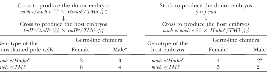
Features of theHorkaD/1germ-line chimeras
Cross to produce the donor embryos Stock to produce the donor embryos mwh e/mwh e$$3HorkaD/TM3## y v f mal
Y Y
Cross to produce the host embryos Cross to produce the host embryos tmIIgs/tmIIgs$$3tmIIgs/TM6## mwh e/mwh e$$3HorkaD/TM3##
Genotype of the transplanted pole cells
Germ-line chimera
Genotype of the host embryos
Germ-line chimera
Femalea Maleb Femalec Malec
mwh e/HorkaD 3 3 mwh e/HorkaD 4 2d
mwh e/TM3 8 4 mwh e/TM3 5 2
a
The females were mated withmwh e/mwh emales.
b
The males were mated withy v f malfemales.
c
Mated withy v f malpartner.
d
Many more offspring originated from they v f malthan from their ownHorkaD/1germ-line cells.
TABLE 3
Features of thehorkarvP2/germ-line chimeras
Cross to produce the donor embryos Stock to produce the donor embryos horkarvP2/TM6B$$3Df(3R)dsx15/TM3## y v f mal
Y Y
Cross to produce the host embryos Cross to produce the host embryos w/w$$3Fs(1)K1237/Y## horkarvP2/TM6B$$3Df(3R)dsx15/TM3##
Genotype of the transplanted pole cells
Germ-line chimera
Genotype of the host embryos
Germ-line chimera
Femalea Malea
horkarvP2/TM3 8 horkarvP2/TM3 2 2
Df(3R)dsx15/TM6B 5 Df(3R)dsx15/TM6B 3 4
TM3/TM6B 1 TM3/TM6B 1 2
horkarvP2/Df(3R)dsx15 3 horkarvP2/Df(3R)dsx15 1 1
Arrows symbolize the direction of pole cell transplantations.HorkaDwas induced by EMS on anmwh-and
e-labeled isogenic chromosome (Erdelyiand Szabad1989).
aThe chimeras produced
only in the gonads. To determine whether function of the gene is required in the germ line or in the somatic components of the gonads, we constructed germ-line chimeras through the transplantation of pole cells. First, pole cells ofHorkaD/1embryos were transplanted into host embryos that did not have pole cells yet provided a normal environment for development and function of the donor pole cells (Table 2). Three of the female germ-line chimeras produced eggs, and the fate of embryos inside these eggs was essentially identical to that described for embryos of the HorkaD
/1 females. Three sibling male germ-line chimeras were generated and then mated with y v f mal females. On average, 3.1% of theirXXzygotes developed asXX//X0 mosaics (Table 2). Features of the chimeras clearly show that the HorkaD-induced defects originate from altered function of the germ-line cells.
We also usedHorkaD/1females and males as host for normal germ-line cells. Apparently fully functional germ cells developed from the transplanted pole cells in the HorkaD/1 environment and offspring derived from the chimeras that carried normal germ-line cells (besides their own) and HorkaD
/1 soma (Table 2). Features of the latter types of germ-line chimeras not only revealed the germ-line autonomous nature effect ofHorkaD
but also showed that theHorkaD
/1 gonadal soma functions normally.
In the second set of experiments, pole cells of horkarvP2/ embryos were transplanted into Fs(1)K1237/1host embryos. Of the developing chime-ras three carriedhorkarvP2/germ-line cells (Table 3). They deposited normal-looking eggs from which larvae never hatched. Cuticle fragments were present in 21% of the eggs, as inside eggs of the horkarvP2/ females (Table 1). We also transplanted normal pole cells into horkarvP2/host embryos and analyzed the developing female and male germ-line chimeras. The horkarvP2
/
flies produced offspring from the implanted germ-line cells (exclusively), showing that the horkarvP2
/ soma
provides full support for the normal germ-line cells (Table 3). It appears that function oflodestaris primarily required in the germ line.
DISCUSSION
Nature of the HorkaD-encoded A777T-LDS protein:
HorkaDis an allele oflodestar, which encodes a member of the Snf2 family of the helicase-related proteins that are involved in transcription regulation, DNA repair, re-combination, and chromatin unwinding (Flaus et al.
2006). The helicase motifs and the other conserved domains contribute to distinctive features in the Snf2 protein family (Figure 5). In Rad54, the only member of the family of known structure, two of thea-helices (a17 anda18) and a short interconnecting region compose protrusion 2, the part of the protein that interacts with
DNA (Thomaet al. 2005; Flauset al. 2006; Figure 5).
The a17 and the a18 helices are present in the LDS protein but the interconnecting region is longer than in Rad54 and contains an a-helix (Figure 5).HorkaDis a G2424/A transition that results in replacement of Ala777
by Thr in the interconnecting region. It appears that this amino acid replacement expands thea-helix by two amino acids (see supplemental Figure 1).
Possible function of the LDS protein: The LDS protein is cytoplasmic during interphases of the cleav-age mitoses, enters the nucleus during prometaphase, and becomes associated with the chromosomes through-out mitosis, suggesting an involvement of the LDS protein in chromatin/chromosome surveillance during mitosis (Girdham and Glover 1991; supplemental
Figure 1). This idea is supported by the loss-of-function mutant phenotype in embryos of the horkarvP2/ fe-males: abnormal assembly of the chromosomes during meiosis and mitosis, formation of anastral centrosomes and abnormal spindle apparatus, failures in the cleav-age mitoses, fallout of the abnormal cleavcleav-age nuclei, and eventual death of the embryos. Similar, if not identical, defects have been reported in embryos of those females defective in (i) spindle assembly check-point functions or (ii) the mitotic catastrophe avoidance mechanism (Castedo et al. 2004; Musacchio and
Salmon2007; Vakifahmetogluet al.2008; Yuenand
Desai 2008). The latter mechanism operates through
the activation of checkpoint kinase 2 (Chk2), whereby the damaged or the incompletely replicated DNA leads to Chk2 activation and resulting inactivation of the centrosomes and the spindles. These events in turn result in blocked chromosome segregation during anaphase and the eventual elimination of those nuclei from the embryonic precursor pool. The Chk2-based mechanism is especially important for maintaining genomic stability during genotoxic stress (Masrouha
et al. 2003; Takada et al. 2003; Brodsky et al. 2004;
Wichmannet al.2006; Larocqueet al.2007). Defects in
the Chk2-based mechanism cause mitotic catastrophe (Vakifahmetogluet al.2008).
LDS does not likely function in the spindle assembly checkpoint because—in contrast to the LDS and the Chk2 proteins—the spindle checkpoint proteins have been shown to bind to the kinetochores (Gillettet al.
2004; Musacchioand Salmon2007). The
abnormali-ties that emerge in embryos of thehorkarvP2/females exhibit all the distinctive features of mitotic catastrophe. Largely identical defects have been described for checkpoint kinase 1 (grapes,grp), checkpoint kinase 2 (lokormaternal nuclear kinase,mnk), and Ataxia telangi-ectasia-related mei-41 mutant alleles (Masrouhaet al.
2003; Brodskyet al.2004; Royouet al. 2005; Takada
et al.2003, 2007; Wichmannet al.2006; Larocqueet al.
elimination of inappropriate nuclei from the pool that serves as a source of the blastoderm cells following the cleavage cycles (Takadaet al.2003; Larocqueet al.2007).
It is possible that the LDS protein might be involved in the same pathway as Chk2, because a few of the embryos of the mnk/mnk; horkarvP2/– females (lacking both the Chk2 and the LDS proteins) develop to adulthood (our unpublished results). Such an event never happens to embryos of thehorkarvP2
/– females. The role of the LDS protein in chromatin surveillance and cell-cycle pro-gression regulation, however, remains to be clarified.
The requirement oflodestar in the germ line and in the soma: Remarkably, lds gene function is indispens-able in the germ line but not in the soma: although flies develop normally without LDS protein, the meiotic divisions are abnormal in horkarvP2/– females, as are the cleavage mitoses in their embryos (Girdham and
Glover1991 and this article). These defects are
germ-line autonomous. In fact, complete or almost complete maternal-effect lethality is a characteristic feature of females that are homozygous for mutant alleles of the genes engaged in the G2/M transition control (Henderson 1999). For example, only 20% of
em-bryos hatch from eggs of females that are both homozy-gous for the strong mnk mutant alleles and lack Chk2 function (Xuet al.2001; Masrouhaet al.2003; Takada
et al.2003; Xuand Du2003; Brodskyet al.2004). Similarly,
thegrphomozygous females, which lack checkpoint kinase 1, are sterile; their embryos suffer from abnormal cortical nuclear divisions and do not cellularize (Yuet al.2000;
Jaklevicet al.2006; Takadaet al.2007). Females
homo-zygous for the Ataxia telangiectasia-relatedmei-41strong mutant alleles are, in effect, sterile (Laurenconet al.2003;
Larocqueet al.2007).
We thank David Glover for the lds alleles and the anti-lodestar antibody, Thomas C. Kaufman for the anti-CNN antibody, Alain Debec for Jupiter-GFP, Stefan Heidmann for Histone-RFP, the Bloomington Drosophila Stock Center for stocks, and Kissne´ Ani and Kiaspatine´ Margo´ for technical assistance. Support for this research was provided by The Cell Cycle Network/66090, the Maternal-Effect and Embryo-genesis Research Group of the Hungarian Academy of Sciences (T9544 and 1348), and three grants of the Hungarian Scientific Research Fund (T16737, T43158, and NI69180) as well as from the Graduate Student Program of the University of Szeged.
LITERATURE CITED
Adams, M. D., S. E. Celniker, R. A. Holt, C. A. Evans, J. D. Gocayne
et al., 2000 The genome sequence of Drosophila melanogaster. Science287:2185–2195.
Allard, S., J. Y. Massonand J. Cote, 2004 Chromatin remodeling
and the maintenance of genome integrity. Biochim. Biophys. Acta1677:158–164.
Brodsky, M. H., B. T. Weinert, G. Tsang, Y. S. Rong, N. M. McGinnis
et al., 2004 Drosophila melanogaster MNK/Chk2 and p53 regu-late multiple DNA repair and apoptotic pathways following DNA damage. Mol. Cell. Biol.24:1219–1231.
Castedo, M., J. L. Perfettini, T. Roumier, K. Yakushijin, D.
Horneet al., 2004 The cell cycle checkpoint kinase Chk2
is a negative regulator of mitotic catastrophe. Oncogene23:
4353–4361.
DeRenzo, C., and G. Seydoux, 2004 A clean start: degradation of
maternal proteins at the oocyte-to-embryo transition. Trends Cell Biol.14:420–426.
Erdelyi, M., and J. Szabad, 1989 Isolation and characterization of
dominant female sterile mutations ofDrosophila melanogaster.I. Mutations on the third chromosome. Genetics122:111–127. Erdelyi, M., A. M. Michon, A. Guichet, J. B. Glotzerand A. Ephrussi,
1995 Requirement for Drosophila cytoplasmic tropomyosin in os-kar mRNA localization. Nature377:524–527.
Flaus, A., D. M. Martin, G. J. Barton and T. Owen-Hughes,
2006 Identification of multiple distinct Snf2 subfamilies with conserved structural motifs. Nucleic Acids Res.34:2887–2905. Gillett, E. S., C. W. Espelinand P. K. Sorger, 2004 Spindle
check-point proteins and chromosome-microtubule attachment in bud-ding yeast. J. Cell Biol.164:535–546.
Girdham, C. H., and D. M. Glover, 1991 Chromosome tangling
and breakage at anaphase result from mutations in lodestar, a Drosophila gene encoding a putative nucleoside triphosphate-binding protein. Genes Dev.5:1786–1799.
Gonza´ lez, C. G., and D. M. Glover, 1993 Techniques for studying
mitosis in Drosophila, pp. 143–175 inA Practical Approach: The Cell Cycle, edited by P. B. R. Fantesand R. Brooks. Oxford University
Press, London/New York/Oxford.
Henderson, D. S., 1999 DNA repair defects and other (mus)takes
in Drosophila melanogaster. Methods18:377–400.
Heuer, J. G., K. Liand T. C. Kaufman, 1995 TheDrosophila
home-otic target genecentrosomin(cnn) encodes a novel centrosomal protein with leucine zippers and maps to a genomic region re-quired for midgut morphogenesis. Development 121: 3861– 3876.
Jaklevic, B., L. Uyetake, W. Lemstra, J. Chang, W. Learyet al.,
2006 Contribution of growth and cell cycle checkpoints to radi-ation survival in Drosophila. Genetics174:1963–1972. Karpova, N., Y. Bobinnec, S. Fouix, P. Huitoreland A. Debec,
2006 Jupiter, a new Drosophila protein-associated with microtu-bules. Cell Motil. Cytoskeleton63:301–312.
Komitopoulou, K., M. Gans, L. H. Margaritis, F. C. Kafatosand
M. Masson, 1983 Isolation and characterization of sex-linked
female-sterile mutants inDrosophila melanogasterwith special at-tention to eggshell mutants. Genetics105:897–920.
LaRocque, J. R., B. Jaklevic, T. T. Su and J. Sekelsky,
2007 Drosophila ATR in double-strand break repair. Genetics
175:1023–1033.
Laurencon, A., A. Purdy, J. Sekelsky, R. S. Hawleyand T. T. Su,
2003 Phenotypic analysis of separation-of-function alleles of MEI-41, Drosophila ATM/ATR. Genetics164:589–601. Liu, H., and E. Kubli, 2003 Sex-peptide is the molecular basis of the
sperm effect inDrosophila melanogaster.Proc. Natl. Acad. Sci. USA
100:9929–9933.
Liu, M., Z. Xieand D. H. Price, 1998 A human RNA polymerase II
transcription termination factor is a SWI2/SNF2 family member. J. Biol. Chem.273:25541–25544.
Masrouha, N., L. Yang, S. Hijal, S. Larochelle and B. Suter,
2003 The Drosophila chk2 gene loki is essential for embryonic DNA double-strand-break checkpoints induced in S phase or G2. Genetics163:973–982.
Musacchio, A., and E. D. Salmon, 2007 The spindle-assembly
checkpoint in space and time. Nat. Rev. Mol. Cell Biol.8:379– 393.
Perrimon, N., 1984 Clonal analysis of dominant female-sterile,
germ-line-dependent mutations inDrosophila melanogaster. Genet-ics108:927–939.
Royou, A., H. Maciasand W. Sullivan, 2005 The Drosophila Grp/
Chk1 DNA damage checkpoint controls entry into anaphase. Curr. Biol.15:334–339.
Schuh, M., C. F. Lehnerand S. Heidmann, 2007 Incorporation of
Drosophila CID/CENP-A and CENP-C into centromeres during early embryonic anaphase. Curr. Biol.17:237–243.
Sibon, O. C., A. Kelkar, W. Lemstraand W. E. Theurkauf, 2000
DNA-replication/DNA-damage-dependent centrosome inactivation in Drosophila embryos. Nat. Cell Biol.2:90–95.
Szabad, J., and R. Nothiger, 1992 Gynandromorphs of Drosophila
Szabad, J., M. Erdelyi, G. Hoffmann, J. Szidonyaand T. R. Wright,
1989 Isolation and characterization of dominant female sterile mutations of Drosophila melanogaster. II. Mutations on the sec-ond chromosome. Genetics122:823–835.
Szabad, J., E. Matheand J. Puro, 1995 Horka, a dominant
muta-tion of Drosophila, induces nondisjuncmuta-tion and, through pater-nal effect, chromosome loss and genetic mosaics. Genetics139:
1585–1599.
Tadros, W., and H. D. Lipshitz, 2005 Setting the stage for
develop-ment: mRNA translation and stability during oocyte maturation and egg activation in Drosophila. Dev. Dyn.232:593–608. Takada, S., A. Kelkar and W. E. Theurkauf, 2003 Drosophila
checkpoint kinase 2 couples centrosome function and spindle as-sembly to genomic integrity. Cell113:87–99.
Takada, S., S. Kwak, B. S. Koppetsch and W. E. Theurkauf,
2007 grp (chk1) replication-checkpoint mutations and DNA damage trigger a Chk2-dependent block at the Drosophila mid-blastula transition. Development134:1737–1744.
Tavosanis, G., S. Llamazares, G. Goulielmosand C. Gonzalez,
1997 Essential role for gamma-tubulin in the acentriolar female meiotic spindle of Drosophila. EMBO J.16:1809–1819. Thoma, N. H., B. K. Czyzewski, A. A. Alexeev, A. V. Mazin,
S. C. Kowalczykowski et al., 2005 Structure of the SWI2/
SNF2 chromatin-remodeling domain of eukaryotic Rad54. Nat. Struct. Mol. Biol.12:350–356.
Vakifahmetoglu, H., M. Olssonand B. Zhivotovsky, 2008 Death
through a tragedy: mitotic catastrophe. Cell Death Differ. 15:
1153–1162.
Wieschaus, E., and C. Nusslein-Volhard, 1989 Looking at
em-bryos, pp. 179–214 inDrosophila: A Practical Approach, Ed. 2, edi-ted by D. B. Roberts. Oxford University Press, Oxford.
Villanyi, Z., A. Debec, G. Timinszky, L. Tirianand J. Szabad,
2008 Long persistence of importin-beta explains extended sur-vival of cells and zygotes that lack the encoding gene. Mech. Dev.
125:196–206.
Wichmann, A., B. Jaklevicand T. T. Su, 2006 Ionizing radiation
in-duces caspase-dependent but Chk2- and p53-independent cell death inDrosophila melanogaster.Proc. Natl. Acad. Sci. USA103:
9952–9957.
Xu, J., and W. Du, 2003 Drosophila chk2 plays an important role in a
mitotic checkpoint in syncytial embryos. FEBS Lett.545:209– 212.
Xu, J., S. Xinand W. Du, 2001 Drosophila Chk2 is required for DNA
damage-mediated cell cycle arrest and apoptosis. FEBS Lett.508:
394–398.
Yu, K. R., R. B. Saintand W. Sullivan, 2000 The Grapes checkpoint
coordinates nuclear envelope breakdown and chromosome con-densation. Nat. Cell Biol.2:609–615.
Yuen, K. W., and A. Desai, 2008 The wages of CIN. J. Cell Biol.180:
661–663.
Zallen, J. A., and E. Wieschaus, 2004 Patterned gene expression
directs bipolar planar polarity in Drosophila. Dev. Cell6:343– 355.