The AutosomaLChorion
Locus
of
the
Medfly
Ceratitis
ar.tutu.
I.
Conserved Synteny, Amplification
and
Tissue
Specificity
but
Sequence Divergence and Altered Temporal Regulation
Dina
Vlachou,*
Mary
Konsolalti,+ Peter P. T ~ l i a s , ~
Fotis C.Kafatoss and
Katia Komitopoulou*
*Department of Biology, Division of Genetics and Biotechnology, University of Athens, Panepistimiopolis, Kouponia, Athens 15701, Greece, tDepartment of Molecular Biology, Princeton University, Princeton, New Jersq 08544, fPublic Health Research Institute,
New York, New York 10016 and &European Molecular Biology Laboratory, 691 17 Heidelberg, Gennany
Manuscript received October 28, 1996 Accepted for publication September 17, 1997
ABSTRACT
We report the isolation, full sequence characterization, amplification and expression properties of medfly chorion genes corresponding to the autosomal chorion locus of Drosophila. These genes are found adjacent to the paramyosin gene and are organized in the same order and tandem orientation as their Drosophila homologues, although they are spaced further apart. They show substantial sequence divergence from their Drosophila homologues, including novel peptide repeats and a new spacing of the tyrosines, which are known to be cross-linked in Dipteran chorion. The genes are amplified and expressed during oogenesis, as in Drosophila. Three of them are expressed in the same relative temporal order as in Drosophila but the fourth gene, the homologue of s15, shows a clear shift to an earlier expression period. This is the first known instance of changed temporal regulation in dipteran chor- ion genes.
T
HE autosomal Drosophila gene cluster encodingthe s18, s15, s19 and s16 chorion proteins is a
favorite system for comparative chromosomal analysis and molecular evolution. This locus has been fully se- quenced in four distant species of the genus Drosophila widely diverged during a period of 60-80 million years
(LEVINE and SPRADLING 1985; WONG et aL 1985; MARTI- NEZ-CRUZADO et aL 1988; FENERJIAN et al. 1989; SWIMMER
et al. 1990). Despite considerable sequence diversifica- tion, this locus has remained highly conserved in orga- nization throughout the genus: the same four chorion genes are found in all four species in the same order, in a compact tandem organization spanning approxi- mately the same length of DNA. Moreover, all four spe- cies possess an unrelated highly conserved gene just downstream of the chorion cluster; this gene, originally described as NGORF (MARTINEZ-CRUZADO et al. 1988), has been identified recently as the paramyosin gene (VINOS et al. 1991, 1992; BECKER et aL 1992; MAROTO et
aL 1995,1996). The highly conserved gene organization of the locus is paralleled by conserved developmental regulatory mechanisms. Throughout the genus, these chorion genes amplify differentially as a single unit in the follicular epithelial cells during late oogenesis
(SPRADLING and MAHOWALD 1980; SPRADLING 1981; GRIFFIN~HEA et al. 1982; DELIDAKIS and KAFATOS 1987; SWIMMER et aL 1990). Moreover, these genes are ex-
hspondnigazcth: Katia Komitopoulou, Department of Biology, Division of Genetics and Biotechnology, University of Athens, Panep istimiopolis, Kouponia, Athens 15701, Greece.
Email: [email protected]
Genetics 147: 1829-1842 (December, 1997)
pressed at characteristic, brief periods during late oo-
genesis, which are gene-specific and completely con- served within Drosophila (FENERJIAN et aL 1989). Possi- bly associated with these conserved regulatory features is the presence of short conserved sequence motifs in the proximal 5’ flanking DNA, embedded within other- wise highly diverged DNA sequences (MARTINEZ-CRU- ZADO et aL 1988; FENERJIAN et aL 1989; SWIMMER et al.
1990). The autosomal chorion cluster also has been studied in more closely related taxa of Hawaiian Dro- sophila, confirming the above features and illuminating the processes of its sequence evolution, including an unusually rapid evolutionary rate of divergence of the encoded proteins (MARTINEZ-CRUZADO 1990).
Recently, the Mediterranean fruitfly Ceratitis capitata (Wiedermann), frequently called medfly, has become a favorite experimental system for molecular studies in Diptera, both fundamental and applied in orientation. The applied interest results from its nature as one of the most destructive and costly agricultural pests, causing immense devastation to coffee and >ZOO fruit crops
1830 D. Vlachou et al.
lection and release of only male sterile flies (LOUIS et
ul. 1987), have been enhanced by the recent develop- ment of a successful transformation system for
C.
cupz- tutu (LOUKEFUS et ul. 1995). This breakthrough also makes the medfly especially attractive for fundamental comparative studies of insect gene regulation and mo- lecular evolution. Such studies are facilitated by the ease of rearing the medfly (wherever this is not impru- d e n t or illegal for reasons of agricultural quarantine), the ease of collecting and injecting its embryos, a n d its phylogenetic position, which is close enough to Dro- sophila to facilitate gene isolation by cross-hybridization o r PCR but far enough to be informative. The medfly belongs to Tephritidae, a family of Diptera related to but distinct from the drosophilids, a n d is thought to have shared a last common ancestor with Drosophila ca. 120 mya (BEVERLEY a n d WILSON 1984) or approxi- mately double the time since the separation of the an- cestors of Drosophila melanogaster a n d D. virilis.In previous studies, we have isolated four medfly chorion cDNAs. Two of them, C2 and C5, were found to cross-hybridize with and correspond to the sex-linked s36 a n d s38 chorion genes of Drosophila (KONSOLAKI et al. 1990; TOLIAS et ul. 1990); they were renamed Ccs36 a n d Ccs38. These genes are clustered at position 70B
of the X chromosome of C. cupitutu, which has been homologized with the Xchromosome of D. melunoguster
(ZACHAROPOULOU 1990; ZACHAROPOULOU et ul. 1992).
Ccs36 and Ccs38 also show sequence conservation a n d the same regulatory properties as their homologues in Drosophila: they are expressed with strict develop- mental specificity in early stages of choriogenesis and are differentially amplified in the ovarian follicle cells
(KONSOLAKI et ul. 1990; TOLIAS et ul. 1990; M ~ U Z A I U a n d MARGARITIS 1991).
In contrast, we had been unable to isolate the autoso- mal chorion genes or their cDNAs by cross-hybridiza- tion. The CI a n d C4 chorion cDNAs are encoded by genes that are clustered at 88B of the
6L
chromosome (ZACHAROPOULOU et ul. 1992) and are amplified and expressed in late choriogenesis, as are the products of the Drosophila autosomal chorion cluster. However, their size and expression patterns correlate with the synthesis of polypeptides of substantially greater appar- e n t size, a n d preliminary partial sequencing of C1 a n dC4 did not reveal obvious similarities to the Drosophila autosomal chorion cluster. Therefore, we resorted to an indirect cloning strategy, which is explained below a n d which proved successful. This permitted complete characterization of the structural and regulatory p r o p erties of the autosomal chorion genes of the medfly, which show both conserved a n d novel features relative to the Drosophila homologues.
MATERIALS AND METHODS
Screening of the genomic library: The medfly genomic li-
brary ( R I N A and SAVAKIS 1991) was a kind gift of the authors.
Approximately 300,000 plaques were screened in two batches by hybridization at 55" (CHURCH and GILBERT 1984) to an 1.7-kb EcoRI-BamHI genomic fragment, containing the two
last exons of D. melanogaster paramyosin (described as NC- ORF) (FENERJIAN et al. 1989). Preparation of phage and plas- mid DNA, agarose gel electrophoresis of DNA and labeling of DNA probes by nick-translation were carried out using standard procedures (SAMBROOK et al. 1989). Southern blot analysis was performed on GeneScreen nylon membrane (Du- Pont), using the hybridization conditions described by CHURCH and GILBERT (1984).
Construction of cDNA clones: The Ccsl8 and Ccsl9 cDNA clones were previously identified as C4 and CI (TOLIAS et al. 1990) and had been selected from a Ceratitis ovarian cDNA library. cDNA clones of the Ccsl6 and Ccsl5 chorion genes were prepared by the rapid amplification of cDNA ends (RACE) method as described (FROHMAN 1990). Briefly, 1 pg total RNA extracted from Ceratitis ovaries was reverse tran- scribed using a primer consisting of oligo(dT) (17 residues) linked to a unique 17 nucleotide adapter. Amplification of
CcsI6 and Ccsl5 cDNAs was subsequently performed using
the universal dT,radapter primer, and a gene-specific primer corresponding to the ATG region of each gene. The se- quences of the specific primers, defined from genomic se- quencing, were 5' ATGCTCCGCTTCACGGTT 3' for Ccsl6, and 5' ATGCAACGATTCATTTGC 3' for Ccsl5. The PCR products were directly subcloned into Pcr I1 vector, using the TA cloning kit (Invitrogen) and amplified after transforma- tion as described by the manufacturer.
Sequence analysis and alignments: Genomic and cDNA clones were mapped with various restriction enzymes and sub- cloned into M13mp18 and M13mp19 or pBluescript I1 KS (Stratagene). Further subcloning was performed after dele- tions of clones and subclones using Exonuclease 111 (HENI- KOFF 1987). Clones and subclones were amplified after trans- formation with CaCl,/RbCI, and DNA was extracted from miniplasmid preparations by alkaline lysis (SAMBROOK et al. 1989). Overlapping restriction fragments were sequenced in both directions using the chain termination procedure (SANGER et al. 1977) with [35S-dATP] (NEN) (BICGEN et al. 1983) and Sequenase V2.0 (U.S. Biochemicals) (TABOR and RICHARDSON 1987). Samples were resolved in 4 or 5% poly- acrylamide, 7.5 M urea gels, and autoradiography was per- formed using Kodak X-Omat film. Each strand was sequenced at least three times. Sequences were identified by comparison with data from the SWISSPROT database using the BLAST program (ALTSCHUL et al. 1990). Other computer programs used were CLUSTAL W (THOMPSON et al. 1994), DNA analysis programs developed by PUSTELL and KAFATOS (1982, 1984) and the GCG secondary structure predictions programs (ver- sion 8, Genetics Computer Group, Inc).
Amplification analysis: Genomic DNA was prepared from male flies or handdissected ovaries from 3-May-old female flies as described (HOLMES and BONNER 1973). The DNA was restricted with EcoRI or HindIII, electrophoresed in Tris- borate agarose gels and transferred to nylon membranes (Genescreen). The filters were hybridized (CHURCH and GIL BERT 1984) with '*P-labeled EcoRI restriction fragments con- taining chorion genes by nick translation (RIGBY et al. 1977) or random hexanucleotide priming (FEINBERG and VOCELSTEIN 1983). Hybridizations were carried out at 72".
RNA blot and developmental Northern analysis: RNAs were extracted from male flies, whole ovaries or stage-specific follicles of the medfly, according to theJAC:OBS-LORENA proto- col (1980) as modified by H. BOUHIN et al. (1992). For RNA dot blots, performed according to KAFATOS et al. (1979), 20 male flies or 10 hand-dissected ovaries were homogenized in
CC
n u
cc Dm
cc
Ihn
CC
Ihn
NGELEEVRS~LDSAVRAKRTVELQYEEAQTRINELSTVNVNLVSLKSKLE~ELS~~DYEEVTKEL
NGELEEVRSHLDSANRAKRTVELQYEEAASRINELTTANVSLVSIKSKLEQELSVVASDYEEVSKEL
612
V V
RISDERYQKVQVELKHTIEVVHEEQERIVKLETIKKSLEVEVKNLSIRLEEVELNAVAGSKRIISKL RISDERYQKVQVELKHVVEQVHEEQERIVKLETIKKSLEVEV~NLSIRLEEVELNAVAGSKRIISKL
67 9
EARVRALELELEEEKRRHAETIKILRKKERTVKEVMVQCEEDQKNIILLQEALDKSVAKVNLFKRQL EARIRDLELELEEEKRRHAETIKILRKKERTVKEVLVQCEEDQKNLILLQDALDKSTAKINIYRRQL 7 4 6
V
AEQEGMSQQSVTRVRRFQRELEAAEDRAVSAETNLNMIRAKHRTFVTTSTIPGSQVYIQETTRTITE SEQEGVSQQTTTRVRRFQRELEAAEDRADTAESSLNIIRAKHRTFVTTSTVPGSQVYIQETTRTITE
813
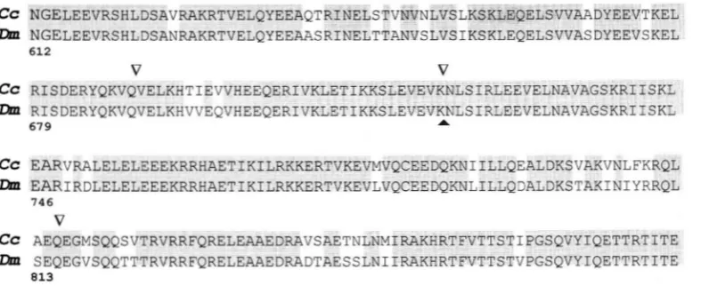
FIGURE 1.-Sequence alignment of the Ctermi- nal regions of the de-
duced D. melanogaster and C. capitata paramyosin prc- teins. Residues identical in the two species are shaded. Amino acids are numbered relative to the Drosophila sequence. In- tron positions of the para- myosin gene in C. capitata
(V)
and D. melanogaster(A) are indicated.
EDTA, 5% ethanol containing 2 mg of heparin and 1.5 mg of proteinase K) and extracted twice with 1 vol phenol (satu- rated with 1' M Tris, pH 9.5)/0.5 vol chloroform, once with 0.5 vol phenol/l vol chloroform and once with 1 vol chloro- form. After ethanol precipitation, total nucleic acids were dis- solved in double distilled water, incubated for 1 hr at 37" with RNAase-free pancreatic DNAase I to a final concentration of 2 pg/ml and reprecipitated with ethanol. For developmental Northern analysis, RNAs were extracted from pools of 50 de- veloping follicles staged according to their size, proportion of oocyte vs. nurse cells, and formation of the chorion layers
(MOUZAKI and MARGARITIS 1991). Samples were resolved by electrophoresis on 1.5% agarose gels containing formalde- hyde according to SAMBROOK et al. (1989). RNA blot analysis was performed using rapid alkaline transfer to a neutral nylon membrane (Genescreen) as described by Low and RAUSCH (1994). The membranes were baked at 80" for 2 hr and hy- bridized as previously using as probes cDNA chorion clones labeled by random hexanucleotide priming (FEINBERG and VOCELSTEIN 1983). Used blots were stripped of probes by boiling for 10-30 min in a solution of 0.010 M Tris-HC1, pH 7.5-8.0, 0.001 M EDTA, 1% SDS, before reusing.
RESULTS
Isolation of the autosomal chorion gene cluster in C.
cupitata Initial attempts to clone the medfly homo-
logues of the Drosophila autosomal chorion locus by low stringency hybridization, using Drosophila chorion genes as probes, were unsuccessful. This was not unex- pected considering the rapid rates of divergence within the genus Drosophila (MARTINEZ-CRUZADO 1990). On the assumption that the paramyosin gene is well con- served in the medfly as in other organisms (BECKER et al. 1992; VINOS et al. 1992) and that this gene is closely linked to a chorion gene cluster as in all Drosophila species examined (FENERJIAN et al. 1989), we screened for the paramyosin gene to gain an indirect entry into the chorion locus.
The medfly genomic library was screened under per- missive criteria with a genomic fragment containing the last two exons of the D. melanogaster paramyosin gene. Two positive lambda clones were isolated and subjected to Southern hybridization with the same probe, and the cross-hybridizing restriction fragments were mapped, subcloned and partially sequenced. Indeed they encom-
passed the 3' terminal region of the paramyosin gene, as shown by alignment of the encoded protein with the corresponding D. melanogaster sequence (89% identity and 97% similarity over 264 amino acid residues, aligned without gaps; Figure 1). In the sequenced re- gion, one intron was found at the same location as in
D. melanogaster, while two others were unique to the medfly (Figure 1 ) . Once the position and the orienta- tion of the Ceratitis paramyosin homologue had been determined by sequencing, all EcoRI fragments down- stream of its last intron were tested for putative chorion genes (which should only be expressed in the female ovary) (TOLIAS et al. 1990; MOUZAKI and MARGARITIS
1991) by being used as probes in dot hybridization of RNAs from Ceratitis males and ovaries (see below). Four successive EcoRI fragments hybridized exclusively with ovarian RNA. The fragment most distant from the paramyosin gene was used to isolate a third chromo- somal clone bearing one additional fragment that showed ovarian expression. Plasmid subclones were constructed, restriction mapped and partially se- quenced, resulting in the detection of four putative chorion genes and their positioning on the 26-kb map of this chromosomal walk (Figure 2).
In situ hybridization to polytene chromosomes, with a biotinylated genomic fragment containing the 3' ex- ons of the cloned C. capitata paramyosin gene, revealed a single band at position 88B of the 6L chromosome
(A. ZACHAROPOULOU, personal communication), where the genes corresponding to the previously isolated CI
1832 D. Vlachou et al.
Ccs18 Ccsl5 Ccsl9 Ccs16
__* -b
-
"*KC C R CR K R K C R C
I 1 I 1 U I I 1 I 1 1
cch37
Prm
CR K R K C R C R C
>'
-b
-
4II I I I
Cchs
Yl Y l V I
4 .",
+v,"
K C R C R C C R C R
I I I I I I I 1 I I
Cch7
r l l l l l l l l l l l l l l l l I l l I I I I I I I
0 1 2 3 4 5 6 7 8 9 10 1 1 12 13 14 15 16 17 18 19 20 21 22 23 24 25 26kb
RGURE 2.-A chromosomal walk consisting of three lambda clones and spanning five genes. Partial restriction maps of cloned segments were generated by standard prosedures (E, EcoRI; C , C M ; K, W I ) . The location and direction of transcription of the paramyosin gene
(m)
were defined by cross-hybridization to the Drosophila homologue and sequence analysis of the appropriate restriction fragments. Note that the sequenced segment of this gene contains four exons (Named xl, x2 and yl, y2 relative to the previously described x, y exons of Drosophila; FENERJIAN et al. 1989), which are shown as an arrow interrupted by three introns. Chorion genes were identified downstream ofe,
by dot blot hybridization to ovarian RNA, and then fully characterized by subcloning and sequencing. Their transcriptional orientations are indicated by solid arrows.+1 ATTCAATAATATTATTAAACTAGAATTACGCAAA ATG AAT AGA TTC CTT TGT ACC TTT GCC GCT ATT GTC GCC
J .1
Y N R F L C T F A A I V A
+74 GTT GCC AAT GGT T C GCA GTT GGT GGA GGT GGT GGT T T GGT GGT CGC GGT GGT AGC GGT ACC GTA
V A P C ~ A V G G G G G ~ G G R G G S Q T V
+140 ATC GGT GGA CAA GCC TAC CAA ATT TTG CCT GCT TTG CAA GTG CAA ACT ATT GCT GCC GCT GGT GGC
) I Q Q Q A Y Q I L P A L Q V Q T I ~ A A A G G
+206
+272
+338
+I04
+470
+53s
+602
+668
+734
+eo0
+E66
+932
+998
+lo73
TCC TCA GCT GGT TAT GGT GGC TCC TCA GCT GGT TAT GGT GCA TCG TCG GGT AGC T T GGT GCA TCG
8 8 A G ~ G G S 8 A G ~ G A S 8 G 8 ~ G A S
TCG GGT GGT TAC GGT GGC TCA TCG AAT GGA TAT GGT GCG TCC AGT GCA CCA TCG AT? GAT ATT GGA
8 G G ~ G G B S N G ~ G A S S A P S I D I G
CAA TTA TTG GCC GCC GTT GGT GGT GAC CTT ACC GCA CAA GAA GCT GCC CAA TTA GTA AAT TCT TTG Q L L A A V G G D L T A Q E A A Q L V N S L
CCT TCA GCT GGC GGT CCA ATC ATT GAC ACC AGT GGT TCA TCG GCT GGT AGC AGC CAT CAA GGT TCA P 8 A O O P I I D T S G S B A G 8 S H Q G 8
TAC CCT TCG GGC GGT AAT CTA GCT T T GTT ATT CAA TCT GGT GGT AGC TCA TAC TCT GCA CCA GCA ~ P 8 G G N L A g V I Q S G G 8 ( 8 O 8 A P A
CCT GCT GCT TCA TAC AGT GCC CCA GCA CCA GCG CCA GCT GCT TCG T C AGT GCT CCA GCT CCT TCT P I A - _ P - I S
8
8 A P A P I"""""""_
A P A A 18&
8 A P AT C AGT GCT CCA GCA CCA GCG CCA GCT CCT TCT T C AGT GCT CCA GCT CCT TCT T C AGT GCT CCA
68
A P A P I A P A P I S 6 8 A P A P I 1 8 6 8 A PGCT CCT TCT T C AGT GCT CCA GCA CCA GCG CCC GCA CCA GCT GCA T T AGT GCT CCT GCA CCT GCT A P I I S
6
8 A P A P I A P A P A I A6
8 A P A P I AGTA T C AGT GCT CCT GCA CCA GCT GCA T T AGT GCT CCT GCA CCT GCT GTA T C AGT GCT CCA GCA I V Q S A P A P
"-
I A k Q 8 A P A P I"-
A I0 V Q S A P ACCA GCG CCC GCA CCA GCT GCA TAT AGT GCT CCA GCA CCT GCT GCA T C AGT GCT CCG GCT CCT GCT P A P A P , ] A @ . A P A P l A k & S A P A P I A
GCA T C AGT GCT CCT GCA TCA TCT GGC T T GGT GCC TCA GCT CCT GCC GCC GCT GCT CCA GCT GCA
I A & S A P A 8 1 8 G Q G A 8 A P A A A A P A A
"""""""-
"""""""""_
0"-
A
""""""""_"
0"-
"_
GCA CAT CAG CCA TCA GCA GCC GCT GCT CGC AGC T T ATC TCA GGC AGT TAC GGT GCT
"""""""""""""""""~
A l i Q P S A A A A R S ~ I S G 8 Y G A
"-
CCC GCA CCC GCC CCT GCT GCT GGT GGT GCT TAT TAA ATCTGTGATATTAAATATTTTCACTAAATTCAGTGTTCG
P A P A P A A G G A Y @
ATCAAAAGATAGMTAAAACGCGATATTTCGAACAAAG
"""""""""""""
FIGURE 3.-Complete cDNA sequence of medfly Ccsl8 (C4 cDNA clone) and the encoded protein. Numbers refer to the nucleic acid sequence. The putative polyadenyla- tion signal AATAAA is bold and underlined. The best conserved peptide sequence detected after alignment with all Dro- sophila s18 proteins is boxed, and 14 imperfect tandem repeats of the h e p tapeptide SysAPAp are bracketed; deviations from
this consensus are dotted. The repeats are spaced by pure alanine and proline segments, which are also found elsewhere in the
1 1 1
TGC ATC ATC TTT TCT ATG CTC GCT CTA GTG CAT GCT TCT CCT TAT GGA AGC
C I I F 8 M L A L V E A S P @ G S
GGT GGA TCA GCT CCA GCT CCA GCA CCA GCA CCA GCG CCT TAC AGC AGT GGT
I G G 8 A P A P I A P A P A P o 8 8 G
T T GAA CCT CCA GTT TTA TAT AAT ATT CCG GCA CCA ACG GCT TGG AAT TTA
g l P P V L @ N I P A P T A W N L
GGT GCA GCG TTG ACA CCA GTT AAT GCT CGC ACC ATC TCA GGA GAT GGT ACA
G A A L T P V N A R T I S G D G T
GGC ATT TCA TCA CAG CCT AGC GCT GCT GCT GCT GCT GCT TCC GCT AAT GTA
G I S S Q P S A A A A A A S A N V
"""""""""""
1 ATG CAA CGA TTC ATT
M Q R F I
67 AGC AGC GGT ACG T C
8 8 G T &
133 CAG GTT GTG ATT TCA
Q V V I S
199 TCA CCA CGT CAT GCC
S P R B A
265 CCT GGC TCT TTG GTC
P G S L V
331 T T T T T C AT CCC AAT GTG GTG GGA
P L N A G S ( Y ) K L W V V P 8(Y)E V P N V V G
P T S G @ A G Q L S ~ G G G Q 5 @ G A Q A P 397 CCT ACT AGC GGC TAT GCT GGT CAA TTA TCG T T GGT GGT GGT CAA TCA TAC GGC GCT CAA GCA CCT
FIGURE 4.-cDNA and deduced animo acid se- quence of medfly Ccsl5. This cDNA was produced by the RACE-PCR method (MATERIALS AND METHODS),
using as gene-specific pri- mer the oligonucleotide ATGCAACGATTCATTT GC, defined by preliminary genomic sequencing of the beginning of the OW. An-
notations as in Figure 3.
463 GCC GCT CCA TCT T T GGC GCC CAG TCA TCC AGT TAC TAA TTGTTGTCGTTGGCTGCTTGAAATTTGAMTMGG
A A P S & G A Q S S S @ . n d
537 GAAACTTAAATTGAAATATAAGCTCTAAGAGCAAAATCCTT
+1
+85
+151
+217
+283
+349
+415
+481
+s47
+613
+679
+745
+811
+077
+943
+loo9
+lo79
+1166
+1253
ATTAGTACGAATTGTGGAAGCCAAAGAACTGCACACAACTC~CTCGCCGAACCGCTGAACCGCTGAACCGCA ATG CAT
M E
1 ATC AAT ATG AAA CAG TTC ATG TGT CTT TCT TTG GCA CTT TTC GCT TCC GTC GCT GTG GCA CAG CCC
I N M K Q F M C L S L A L F A S V A V A Q P
TAT GGA CAA TCT CCG GCA TCC TAT GGT GCA CCA GCA CCA GCA CCA GCT CCT GCT GCC AGT CTT GCC
@ G Q 8 P A I S @ G A P A P I A P A P A A 8 L A AGT TTG GTG AAT TTG GAG GCG CTC GAA CGG TAC CTA AAT GCT CCA GCT CCA GCT CCA GCA GCG TAT
S L V N L E A L E R Y L N A P A P A P A A O
GGT GGC TCT AGC ACA T T TCT CAG GCA AGA TAT GTT TCA GCT GGT GGT TCG CCC AGC GCA GGT CCA
G G 8 8 T ~ S Q A R Y V 8 A G G 8 P 8 A G P
GTA ACT TCA ACT AGC TAC AGT GCT GCA CCA GCA TCT TCG GCT GGC AGT TCT GCG CCG TCC GGT GGT
V T S T 8 Y S A A P A 8 5 A G 5 8 A P 8 G G
"""_
"""""""""_
""""""""""""""-"
TAT GGC GGT TCA GAT GAG CCA ATT CTT TTT GTC AAT GAA ATA CCT GCC GCT GGT TAC TCA TCA TCT
Y G G 8 D L P I L F V N E I P A A G @ 8 8 8
GGC GGT TAC TCA GCA CCC GCA CCT GCA CCT GCT GCT AGC TAT TCC GCT CCA GCT CCT AGC TAC ACT
G I G @ 8 A P A P I A P A A 1 8 @ 8 A P A P I I S
@ I
GCA CCA GCT CCT AGC T T TCC GCA CCA GCT CCA AGC T C TCT GCT CCA GCT GCT ACC T C TCT GCT A P A P I I S
6
8 A P A P I I S6
8 A P A A l l :6
5 A CCA ACT GCT ACC TAC TCG GTT CCA GCT CCC AGC TAC TCT GCT CCA GCT CCT AGC TAC TCT GCA CCAP
: all:
@ 8 V P A P I 1 8 @ 8 A P A P I I S @ 8 A PGCT CCT AGC T T TCC GCA CCA GCT CCA AGC T T GCT GCA CCT GCT A P I I S
6
8 A P A P I186.
A P A CCA GCC AGC TAT TCA GCT CCC GCT CCC GCT CCT GCT GCT GGT TAT GGT GCT GCC CCA TCA TCT GGTP I A L 5 @ 8 A P A P I A P A A G O G A A P 5 8 G
0 0 0
"""""""_
0
"""""""_
T T GGT GGT GCT TCT GCT GCT CCC AAA TAC ACC GTA CTC CCA GCA ACT ATT ACA CAG CTC AAC CCC
Q G G A 8 A A ( P X ~ T V L P A T I T Q L N P
GGC AAG ACT AGC TAC AAA ACC TAC CAG TCT CCC ATC CAA TAC AGC ACT GTC ACC CTT CCA GTT ACT
G K T S ( Y ) K T m Q 8 P I Q m S T V T L P V T
G T T T TA T GCC GGT GGT AGT T T GGT GCA TCT GCT
E
G:G?Cz
G:GG?yTFgGV
"FY?l
A G Q 5 G A 5 AGGT T T GGT AGC AGC TCA GGT AGC AGC TCC T T GGT GGT TCC AGC TAT TAG AGAAAAAAACAAACAAGAA
Q Q G 8 8 5 G 5 8 S 6 6 G 5 5 r e n d
C T T A A G T C T T A A A G A C G T T A G T T T T A G T T C A T A A T A G A A C T G T T C A G A G T G C T T G T G A C C A G T T A T G ~ T T C ~ T T C ~ T T
T T A C T T T G A G C G A G T T T T G T G T G T A A A T A T T A T T T G T T T G G C ~ T C C T T T T T A T A T T C T T G T T T T T G G T ~ ~ T G A A A G A
TGTTTTTAAGATT
1834 D. \lachou c/ nl.
J J J J
1 ATG CTC CGC TTC ACG GTT TGC TTG GTG GCC ACG GCC GTG TGT ATA TGC ACA TCC GCG TTG GCT TCT
M L R F T V C L V A T A V C I C T S A L A S
67 CCA CAA TAT GGC AGC GCT CCA ACT CCA GCA GCA TCC TCG TAT GGC GGG AGT GGT GGT TAT GCC GGT
P Q Y G S A P T P A A S S @ G G S G G @ A G
133 GCC GGT GCG AGT GGC GCA CCA GCA GCT CAG GTA ATT GAA TTG CCC GCC AAT GTT GAA GCT CAA GCG
A G A S G A P A A Q V I E L P A N V E A Q A
199 GCT GCT TTG ACA RAT GCT GCC GGC GCT AAG GCG ACA GCT GCC AAA ATC AAT GCA CTC AAA TGG GAT
A A L T N A A G A K A T A A K I N A L K W D
265 TTG CTC AAT TTG TAC GGT TAT GAA ATC GGT TAT CCA TTG TTG GTG AAG AAA TAC GGA CCA TTG ACC
L L N L O G Y E I G O P L L V K K O G P L T
331 TCA TTG TTC TCC GCC TCG TTG CCA CCA CGC AGT TTT GTT GGA AAG ATC GAT CCT GCT TTC TTG AAA
S L F S A S L P P R S F V G K I D P A F L K
397 GAC TCT TAC GGC AAG ATC ?@A TAT GTC GGC GAA AGT TCG ATC GGT GTT GCG GCT ATT TAA AATCGTG
D S Y G K I K Y V G E S S I G V A A I e n d
464 C G C C T A A A A C G C G A C T T G T A A A T T A T T G T T A A A G T A G C T A A ~ T ~ C T A T A C T T T T T T ~ G T G G
F K X ' R F 6.-cDNA se-
quence o f medfly ( h l h . This
cDNX w a s produced by the
RACE-PCR method. using as
gene-specific primer, the oli- gonucleotide ATGCTCCG
<;TT<;I\CGGTT, dcfined by
preliminary genomic se-
quencing of the kginning
of the ORF. Annotations as
in Figurc 3.
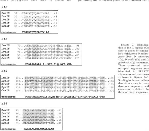
according to their order in the locus relative to the SM'ISSPROT database through the RZASTX program
paramyosin gene. (&.TS(:I-IL*L ut 01. 1990). The closest matches were de-
Characterization of the medfly proteins: The pre- tected with the Drosophila s18, ,715, s19, sl6 proteins, dicted polyeptides were used to search the permitting the C. mo/)itrr/n genes to be renamed Ccsl8
sl8
-18 3 2
...
VGGYAYQVQPALTVKAI...
48Dss18 3 2
...
IGAYAYQVQPALTVQAI...
48Dvs18 3 0
...
VGGYAYQVQPALTVKAI...
46Dgsl8 30
...
VGGLAYQVQPALTVSSI...
46Ccs18 36
...
IGGQAYQILPALQVQTI...
5 2consensus VGGYAYQVQPALTV-AI
s15
-15 70
...
GSAAAAAAASAAAVNPGTYKQYAIPSYEL...
98Dssl5 7 1
...
SRASAAAASAAAAIAPGSYSQYAIPRYEI...
99Dvsl5 71
...
PSASAAAAAASAGIRPGRYEQAAVIGYDL...
99Dpsl5 5 1
...
PSAAAAAAAASSGVNSGLYNQRGVIGYEL...
85CCSl5 99
...
PSAAAAAASANVPLNAGSYKLWVVPSYEV..127consensus PSAAAAAAAA-A--NPG-Y-Q-AVP-YEL
sl9
-19 106
...
PRWTVQPAGATLLYPGQNNYKAYVSPPEYSKVILPIRPAAPVAKLFVPEN...
155 Dssl9 107...
PRWTVQPAGATLLYPGQNNYRAYVSPPEYTKVVLPVRPAEPVAKLYIPEN...
156 m S 1 9 134...
PRWSVQPAGTTLLYPGQNSYRRYASPPEYTKVVLPVRAAPPVAKLYLPEN...
183 DgSl9 139...
PRFTVQPAGATLLYPGQNSYRRISSPVEYSKVILPVRAAAPVAKLYIPQN...
188 cCSl9 252...
PKYTVLPATITQLNPGKTSYKTYQSPIQYSTVTLPVTAAGPVANLYVPEN...
301consensus PRWTVQPAGATLLYPGQNSPR-Y-SPPEYSKV-LPVRl#i-PVA?CLY-PEN
sl6
-16 45..
Dssl6 46..
Dvsl6 45..
Dgsl6 4 8 . .
Ccsl6 63.
.
_ _ _ _
L _ _ I _ _..EAQASALTNAAGAAASAAK. ..EAQAAALTNAAGAAASAAK. ..EAQAAALTNAAGAAASSAK- ..EAQAAALTNAAGAAAASAK.
-
.
.63.
.64.
.63.
.66.
.81FIGURE 'i.-IIdentifica-
tion o f the C. cn/,itn/n ( C c )
chorion genes, b y compar-
ison with known I). mdnno-
g n s / ~ r ( D m ) , D. subohsrurn
(Lh), D. rririlis ( D o ) and D.
gn'n~shnroi (Dg) sequences. These consened, unin- terrupted segments were
idcntified in RIASTX
alignments and are shown
as boxes in Figures 3-6.
Shading indicates residues
that do not vary in more than one sequence, and
consensus is defined by
three or more sequences.
Autosomal Chorion Locus of Medfly
TABLE 1
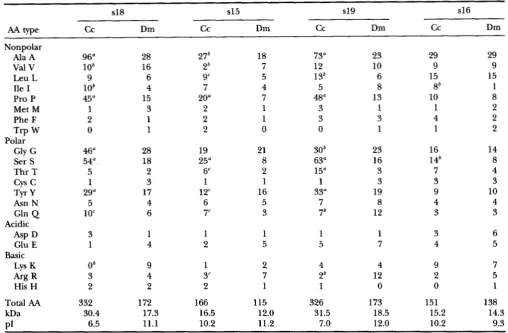
Comparison of amino acid residues in chorion proteins of C. r n p i h t ~ and D. m k t ~ e
s18 s15 s19 s16
AA c c w e Dm c c Dm c c Dm c c Dm
Nonpolar
Ala A 96" 28 27' 18 73" 23 29 29
Val V lob 16 2b 7 12 10 9 9
Ile I
lo*
4 7 4 5 8 8b 1Pro P 45" 15 20" 7 48" 13 10 8
Met M 1 3 2 1 3 1 1 2
Phe F 2 1 2 1 3 3 4 2
1 2
Gly G 46" 28 19 21 30' 23 16 14
Ser S 54" 18 25" 8 63" 16 14b 8
Thr T 5 2 6" 2 15" 3 7 4
Leu L 9 6 gc 5 13b 6 15 15
Trp
w
0 1 2 0 0 1Polar
cys
c
1 3 1 1 1 3 3 3TYr y 29" 17 12" 16 33" 19 9 10
Asn N 5 4 6 5 7 8 4 4
Gln Q 10" 6 7" 3 76 12 3 3
Acidic
Asp D 3 1 1 1 1 1 3 6
Lys K O b 9 1 2 4 4 9 7
Arg R 3 4 3" 7 2b 12 2 5
Glu E 1 4 2 5 5 7 4 5
Basic
His H 2 2 2 1 1 0 0 1
Total AA 332 172 166 115 326 173 151 138
kDa 30.4 17.3 16.5 12.0 31.5 18.5 15.2 14.3
PI 6.5 11.1 10.2 11.2 7.0 12.0 10.2 9.3
a, b, and c indicate differences in numbers of specific residues between C. capitata and D. melanogaster chorion proteins in either direction. a, >10 residues; b, >4 residues; and c, >3 residues.
( C 4 ) , CcslS, Ccsl9 ( C l ) , and Ccsl6, respectively, in in- creasing proximity to the paramyosin gene. Figure 7 presents the most conserved peptide sequences that show diagnostic similarities to the aligned sequences of the corresponding proteins in four diverse Drosophila species (LEVINE and SPRADLING 1985; WONG et al. 1985; WTINEZ-CRUZADO et al. 1988; FENERJIAN et al. 1989; SWIMMER et al. 1990), unequivocally establishing the identity of the medfly genes. These diagnostic se- quences are boxed in Figures 3-6. Additional similari- ties exist, but the fruitfly and medfly genes are highly diverged for much of their length, consistent with our inability to identify them by cross-hybridization. For convenience, we will use the term "autosomal chorion locus" for this medfly gene cluster, as in Drosophila.
The most striking difference is that the medfly chorion proteins are invariably longer than their Dro- sophila homologues: by 9% in the case of Ccsl6, 44% for Ccsl5, 88% for Ccsl9and 93% for Ccsl8 (Table 1). This disparity would be even greater if the putative sig- nal peptides, which are usually of similar length, were omitted from the comparison (Figures 3-6). It should be noted that all C. capitata chorion components mi-
grate on SDS gels more slowly than their expected mo- lecular weights, but C c s l 9 ( C l ) and Ccsl8 (C4) show an unusually large retardation: they behave as proteins of 68 and 59 kD, respectively (according to TOLIAS et al.
1990) or 66 and 61 kD (according to MOUZAKI and
MARGARITIS 1991), although they are 31.5 and 30.4 k D ,
including the signal peptide, as determined by sequenc- ing (Table 1). Slower migration on SDS gels has been noted in some gel systems for
D.
melanogash- chorion proteins (WARING and MAHOWALD 1979) and may largely reflect unusual features of sequence and struc- ture. However, early attempts at detecting chorion gly- cosylation showed the presence of 6% sugars (PETRI etal. 1976), and
KOCH
and SPITZER (1982) reported that chorion was heavily labeled after in vivo incubation with radioactive sugars.1836 D. Vlachou et al.
Dmsld DmslSDmsI9 Dmsld
+ + 3
+ *
f?
i i i i i i i i i i - 1 i i i
0 1 2 3 4 5 6 7 8 9 10 11 12 13 14 15 16 17 18 19 20 21
amino acid each: threonine in the case of Ccsl9, and glycine in the case of Ccsl8. In both Ccsl9 and Ccsl8,
a substantial part of the increased length is due to a novel segment consisting of imperfect tandem repeats of the same heptapeptide consensus sequence, SYSA- PAP (bracketed in Figures 3 and 5 ) . There are 13 such repeats in the case of Ccsl9 and 14 in Ccsl8, either directly abutting each other or separated by one to five residues, which are invariably alanine or proline (dashed underlining in Figures 3 and 5). Only one imperfect repeat of this consensus is encountered in
CcslS and is followed by three repeats of
AP
(Figure4). The msApAp consensus is not encountered in Ccsl6
(Figure 6 ) nor in any of these four chorion proteins in any Drosophila species that has been studied to date.
In addition to the interrelated differences in length, amino acid content and occurrence of SYSAPAP-like repeats, the medfly chorion proteins differ from their
D. melanogaster homologues in isoelectric point. Ccsl8
and Ccsl9 are substantially less basic than their D. mela- nogaster homologues, and CcslS is also somewhat less basic. Again, Ccsl6 is deviant in that it is actually more basic than D m l 6 .
The locus as a whole differs substantially in length, because of substantially longer intergenic DNA in C. capitata as compared with D. melanogaster or other Dro- sophila species (MARTINEZ-CRUZADO et al. 1988). Thus the distance from the 5' end of s18 to the 3' end of the paramyosin gene is 6.9 kb in D . melanogaster and 17.2 kb in C. capitata (Figure 8). Nevertheless, the order and orientation of the genes, with respect to each other as well as to the neighboring paramyosin gene, are per- fectly conserved (Figure 8). Each medfly gene has a single intron, at the same position as the Drosophila homologue (data not shown).
The autosomal chorion genes are amplified in C. capi tats: In all Drosophila species examined, the autosomal chorion genes amplify differentially in the ovarian folli- cle cells during oogenesis (SPRADLING and MAHoWALD 1980; SPRADLINC 1981; ORR et al. 1984; ~ T I N E Z - C R U -
ZADO et al. 1988). This is also true in C. capitata (Figure
FIGURE 8.-Compari- son of the autosomal chorion locus in D. mela- noguster and C. cupitata en- compassing the paramyo- sin gene
( p )
and four similarly organized chor- ion genes. In Ceratitis, the locus shows size expan- sion in both coding (espe- cially s18 and s 1 9 ) and in- tergenic regions. Note that the sequenced por- tion of the paramyosin gene shows three introns as compared with one in the Drosophila homo- kb logue (see Figure 1 ) .9). Genomic DNA was prepared from either males or total choriogenic ovaries. Equal amounts of each DNA were digested with EcoRI and blot hybridized with three cloned fragments encompassing chorion genes. In each case, autoradiography revealed amplification in ovarian relative to male DNA (Figure 9A). This was confirmed (Figure 9B) by comparison of blots of HindIII-di- gested DNA hybridized with chorion probes and a sin- gle copy control probe (the Adh2 cDNA isolated by BOURTZIS and S A V ~ S , unpublished results). Surpris- ingly, several amplified bands were seen with each of the chorion probes (Figure 9A), suggesting the exis- tence of restriction fragment length polymorphism in the autosomal chorion locus of C. capituta; the different intensities of the bands presumably correspond to the frequencies of the alleles in the medfly population. The polymorphism was confirmed by analysis of additional C. capitata genomic clones and by Southern analysis from ovaries of individual females (D. VLACHOU and K KOMITOPOULOU, unpublished results) and is interesting in view of the commonly held view that medfly popula- tions have a low degree of polymorphism.
The chorion genes are expressed specifically in the
ovary but show some changes in temporal expres- sion: To test whether expression of autosomal chorion genes is ovarian-specific in the medfly, as it is in Dro- sophila (GRIFFIN-SHEA et al. 1982; FENERJIAN et al. 1989), equal amounts of
RNAs
from choriogenic ovaries and from adult males were subjected to dot blot analysis. We used as probes the same three cloned fragments encompassing chorion genes as in the previous experi- ment. Indeed, expression was abundant in the ovaries but not detectable in males (Figure 10).Detailed comparisons of the temporal specificities of the four chorion genes in D. melanogastmand C. capitata
showed clear evidence of regulatory evolution (Figure 11). Staging the choriogenic follicles (egg chambers) in a comparable manner was not straightforward because their morphology differs in the two species (MARGA-
RITIS 1985). We used the staging criteria established by
Autosomal Chorion Locus of Medfly
A Ccsl6, Ccsl9 Ccsl5 Ccsld
d ov
d
ovd
ov6.0 +
4.8 + 3.5,
3
6.5-5.0,
3.5,
2.7
-.
1.2-
e
B
Adh2
Ccs36
Ccsl8
w
-
4.0
FIGURE 9.-Amplification of the medfly chorion genes in choriogenic ovaries. (A) High molecular weight genomic DNAs were isolated from males and choriogenic ovaries (ov) from C. capitataand digested with EcoRI; equal aliquots were blot hybridized as described in the text. "P-labeled EcoRI fragments, encompassing Ccsl6 plus Ccsl9, Ccsl5 and Ccsl8, respectively, as defined by ovarian RNA hybridization (Figure 10) were used as probes. Note that each probe hybridizes to more than one EcoRI band in both males and female ovaries and that these bands are all specifically amplified in ovaries. The weakest bands seen in the ovarian lanes were also seen in the male lanes upon long exposure (data not shown). Additional studies showed that this band multiplicity is due to restriction fragment length polymorphism in the autosomal locus (data not shown). The varying intensities of hydridizing bands indicate different representation of the alleles in the C. cupitatu population. (B) An additional Southern blot hybridization experiment using Hind111 digested genomic DNA. Note that both sex-linked (Ccs36) and autosomal (Ccsl8) chorion genes are amplified in the ovary, unlike the Adh2 single copy control.
proportion of the oocyte us. nurse cells, and formation of the chorion layers. The clearly stronger stage 12 ex- pression of s19 in medfly as compared with Drosophila should be viewed with caution because of this uncer- tainty in absolute equivalence of the stages. However, we were able to compare with complete confidence the
relative temporal specificities of the four genes relative to each other by the simple expedient of using the same RNA blot for all of them. Following the procedure described by FENERJIAN et al. (1989), we isolated RNAs from pools of large numbers (50) of staged follicles of
C. capitata, fractionated the RNAs by electrophoresis, transferred them to nylon filters (MATERIALS AND METH-
ODS), and probed the filters sequentially with the four chorion genes. Thus the temporal RNA pattern of each gene served as an internal reference for the others, permitting consistent analysis of the relative timing of gene expression. In D. melanoguster (Figure 11, right),
the genes are expressed in the order s19, s16, s18 and
s15, from earliest to latest expressed gene. In particular, the mRNA abundance ratios at stages 13 and 14 demon- strate that s15 is expressed in an overlapping but clearly later manner relative to s18. The same temporal order is preserved in all Drosophila species ( F E N E R ~ N et al.
1989). In contrast, repeated experiments in C. capitata
(Figure 11, left and data not shown) revealed a clear alteration of the gene expression order. In particular,
Ccsl5 has the same RNA expression pattern as CcslG
and is clearly earlier than Ccs18.
DISCUSSION
Extensive conservation of chromosomal linkage groups has been observed in Diptera, including both Drosophila and Ceratitis (FOSTER et al. 1981; LOUW
1838 D. Vlachou et nl.
d
ov
* I ,
18s
0
Ccsld
,
Ccsl9
0
CCSIS
0
c c m
FIGURE 10.-Dot blot analysis of RNAs isolated from males and female ovaries (MATERIAIS AND METHODS). 4 1 EcoRI re- striction fragments mapping downstream of the paramyosin gene (see Figure 2) were used as probes and showed hybrid- ization with ovarian but not male RNA, demonstrating the ovarian specificity expected of chorion genes. Ribosomal 18s
hybridization served as a control demonstrating comparable RNA loading.
al. 1989; ZACHAROPOULOU 1990). This synteny occurs despite the occurrence of a number of inversions and other chromosomat rearrangements; for example, the haploid female genome in the medfly includes three metacentric, two submetacentric and one acrocentric chromosome, while in Drosophila it includes two meta- centric, one acrocentric and one dot chromosome. The possibility of retention of close linkage with the paramy- osin gene was the basis of our successful strategy for cloning the chorion locus. First we cloned the paramyo- sin gene by homology and showed that it is extensively conserved relative to the singlecopy paramyosin gene of Drosophila (BECKER et aL 1992; VINOS et al. 1992), which maps close to the autosomal chorion genes (FEN-
ERJIAN et nl. 1989). As predicted, a short chromosomal walk and characterization of the transcription units downstream of the paramyosin gene revealed the exis- tence of homologues of the four Drosophila autosomal genes, two of which were identified subsequently with the C1 and C4 sequences.
The study of this gene cluster complements earlier studies of the evolution of the autosomal chorion locus in diverse species of the genus Drosophila (MARTINEZ-
CRUZADO et al. 1988; FENER~IAN et al. 1989; MARTINEZ-
CRU7UDO 1990; SWIMMER et al. 1990). By comparison of the organization and sequence of the locus in a fly
belonging to a non-Drosophila family, we have con-
firmed several highly conserved features while also de- tecting novel events in the evolution of the locus.
The most impressively conserved feature is the pres- ence of the same genes in both Drosophila and Cerati- tis, in the same order and orientation. This invariance for “120 million years (BEVERLEY and WILSON 1984) is in striking contrast to the radical differences in chorion gene sequences and their chromosomal organization in flies and silkmoths, which have been separated twice as long in evolution (KAFATOS et al. 1987). The con- served organization within the Diptera may be related to the apparently ancient invention of chorion gene amplification, a developmental mechanism that does not occur in silkmoths and permits rapid eggshell formation during oogenesis with a small number of structural genes (SPRADLING and MAHOWALD 1980; SPRADLING 1981; OSHEIM and MILLER 1983; OSHEIM et
al. 1988). Amplification has now been documented for both the autosomal (this report) and the sex-linked chorion genes of the medfly (KONSOLAIU el al. 1990; TOLIAS et al. 1990) as in Drosophila. Quantification of the level of amplification (as in SPRADLING 1981) indi- cates values of 16X for the sex-linked locus and 14X for the autosomal locus, whereas in Drosophila the au- tosomal locus is amplified more extensively. This puta- tive difference will need to be confirmed by quantifica- tion in staged egg chambers rather than whole medfly ovaries. In Drosophila species, an essential amplifica- tion control element (ACE) has been mapped upstream of gene s18 (ORR-WEAVER and SPRADLING 1986; ORR- WEAVER et al. 1989; SWIMMER et al. 1990) and accessory amplification enhancing elements (AEEs) appear to map in the DNA flanking the other genes, permitting high-level amplification of the locus as a single ampli- con (DELIDAKIS and KAFATOS 1987, 1989). Detailed se-
quence alignments of the intergenic sequences in the autosomal chorion locus of C. capitata and
D.
melanogas- terreveal wellconserved elements that may be candidate ®ulatory elements for amplification as well as ex- pression in late oogenesis (D. VLACHOU and K. KOMITO- POULOU, unpublished results).Despite the conserved organization of the chorion locus, its length varies substantially, by 149%. All in- tergenic regions are longer in the medfly, by factors ranging from 2X for the s19/s16 intergenic region to 5X for s15/s19. Sequence analysis of these regions will be reported elsewhere (D. VLACHOU and K. KOMITO-
POULOU, unpublished results). The medfly paramyosin gene is also substantially longer in the medfly, showing three introns within a region where the Drosophila ho- mologue has only one. If the compact nature of the Drosophila genome is a secondary character, this well- studied multigene locus manifests a process of compac- tion in Drosophila, eliminating presumably dispensable sequences in the intergenic regions as well as whole introns.
Autosomal Chorion LOCUS of 1839 Medfly
@
1-9 10 11 12 13
14
1-9 10 11
12
13 14
@
SI
9
mm-
m
s19
s16
9-
.u
s16
S I 8
-
Ir,
S I 8
s15
n
r
.
0
s15
rRNA
G
FIGURE 1 I.-Northern analysis of autosomal chorion gene expression in C. capitata and D. melunogaster. The relative temporal expression profiles of the four chorion genes during oogenesis are compared with confidence because the same RNA blot of the appropriate species was used for all four genes (in four cycles of hybridization, stripping of label and reusage with the next gene). Expression patterns are arranged according to the order of temporal specificity of the genes in D. melanogaster. Follicles were staged using criteria comparable in the two species (MATERIAIS AND METHODS), and 5 pg of total RNA was prepared from each pool, electrophoresed, blotted and probed with homospecific probes. Note the clear shift of temporal specificity of s15,
which is later than s18 in D. melanogaster but earlier in C. capituta, as shown by the intensity ratios of the signals at stages 13 and 14; weak expression of s15 but not s18 at stage 12 is also seen in the medfly experiments, making the expression pattern of s15
as early as that of s16. Total RNA loading was equal in all C. capitukz samples, as shown by the ethidium bromide stained rRNA control. D. melunoguster patterns are from FENERJIAN et uZ. (1989).
tained sufficient similarities at the sequence level to be identified with their Drosophila homologues but have otherwise diverged drastically, confirming the general- ization that chorion proteins evolve very rapidly (MARTI-
NEZ-CRUZADO 1990). Nevertheless, secondary structure predictions (computer program GCG) suggest that the proteins are highly structured, with ,6 sheets alternating with
p
turns (data not shown), as is true for chorion proteins of other insects (HAMODRAKAS et al. 1985;HA-
MODRAKAS 1992). In line with the larger size of this locus as a whole in Ceratitis, the proteins are invariably longer than their Drosophila homologues. This differ- ence is minimal in the case of Ccsl6, which is the least rapidly evolving autosomal chorion protein, both be- tween the genera and among different Drosophila spe- cies (FENERJIAN et al. 1989). This protein is also unusual in that the alteration of putativep
sheets andp
turns is interrupted centrally by a predicted a helix (data not shown); this domain includes the segment that is highly conserved in all Dipteran species studied (Figure 7).The genes of the second chorion locus, located in a separate chromosome (Ccs36 and Ccs38), are conserva- tive in both sequence and length (KONSOLAKI et al.
1990; TOLIAS et al. 1990). At the other extreme, the
Ccsl8 and Ccsl9 proteins are nearly double the length of their Drosophila homologues. The difference is largely due to a novel tandem array of consensus SYSA- PAP repeats, not encountered in Drosophila. This array is found in the central region of CcsI9 and near the C
terminus in Ccsl8; individual repeats that conform to the same consensus are also seen in the aminoterminal region of Ccsl9 as well as Ccsl5. The repeats are often associated with extensions consisting purely of alanine and proline; similar "segments are also found in isola- tion elsewhere in the sequences (dashed underlining in Figures 3-5). Moreover, a related octapeptide re- peat, Y(G or S)AAF'AA!j, is found in the Cterminal re- gion of the Ccs36sequence (ACCELI et al. 1991). Because of the total absence of these features from Drosophila, we favor the hypothesis that the SYSAPAP repeats and
AP
segments evolved de nuvo during the period separat- ing the two families of flies, although we cannot elimi- nate the possibility that they were lost selectively from Drosophila. In any case, it is notable that these features appear or disappear in a concerted manner from sl8,s19 and to a lesser extent s15, proteins which do not otherwise show extensive sequence homologies.