University of South Carolina
Scholar Commons
Theses and Dissertations
2017
Molecular Evolution of Major Epidermal Structure
Genes and an Integrative Transcriptome Analysis of
Chicken Epidermal Embryogenesis
Weier Bao
University of South Carolina
Follow this and additional works at:https://scholarcommons.sc.edu/etd
Part of theBiology Commons
This Open Access Dissertation is brought to you by Scholar Commons. It has been accepted for inclusion in Theses and Dissertations by an authorized administrator of Scholar Commons. For more information, please [email protected].
Recommended Citation
MOLECULAR EVOLUTION OF MAJOR EPIDERMAL STRUCTURE GENES AND AN
INTEGRATIVE TRANSCRIPTOME ANALYSIS OF CHICKEN EPIDERMALEMBRYOGENESIS
by
Weier Bao
Bachelor of Medicine Dalian Medical University, 2007
Master of Science Kansas State University, 2010
Submitted in Partial Fulfillment of the Requirements
For the Degree of Doctor of Philosophy in
Biological Sciences
College of Arts and Sciences
University of South Carolina
2017
Accepted by:
Roger H. Sawyer, Major Professor
Lydia E. Matesic, Committee Member
Rekha C. Patel, Committee Member
Joseph M. Quattro, Committee Member
Wayne E. Carver, Committee Member
ii
iii
DEDICATION
iv
ACKNOWLEDGEMENTS
I would like to express my deepest gratitude to my advisor, Dr. Roger Sawyer for his
guidance during my PhD pursuit. He always encouraged me to explore my research areas
and gave me his timely feedback. I am also thankful that he helped me to overcome my
family hardship and gave me the best support. Special thanks go to Dr. Matthew
Greenwold as my secondary mentor for his patient guidance and the discussions with him
lead to my research inspirations. My research work would not have been possible without
his help.
I would like to acknowledge Dr. Lydia Matesic, Dr. Rekha Patel, Dr. Joe Quattro
and Dr. Wayne Carver for agreeing to serve on my committee members. I appreciate your
patience shown in every committee meetings and thank you for your precious suggestions
for my work. I also specially thank Dr. Carver’s strong support and encouragement in my
entire PhD period.
I would like to thank all the colleagues facilitating me to conduct my research.
Many thanks to Dr. Richard Goodwin for providing chicken embryos, Dr. Diego
Altomare for running the microarray experiment, Dr. Jeff Dudycha for providing the lab
for RNA extraction work.
v
ABSTRACT
Αlpha (α) and beta (β) keratins are the major structural proteins found in vertebrate
epidermis and the β-keratins are only found in reptiles and birds. With the recent
published 48 avian genomes, we searched and studied the molecular evolution of these
gene families. We discovered that the expansion and contraction of different α- and β-
keratins among the 48 phylogenetically diverse birds supports the importance of their role
in the evolution of the feathers and the adaptation of birds to different ecological niches.
Using a customized 44K microarray, we also performed transcriptome analysis on
different epidermal regions (scutate scale, dorsal feather and wing feather) at important
time points (day 8, 17 and 19) during chicken embryonic development. We profiled the
differentially expressed α- and β-keratin genes in those comparison groups and
demonstrated the important roles of α- and β-keratins in the development of the chicken
epidermal appendages.
MicroRNAs have been found to widely regulate many biological processes in
animals. Here we also utilized the 44K microarray transcriptome data to profile the
miRNA expression during chicken embryonic development. With the application of
various bioinformatic tools, based on the differentially expressed miRNA genes and
mRNAs, we identified highly possible target genes for epidermal development in the
vi
In previous studies, hundreds of genes (i.e., signaling pathway genes, structural
genes, cell adhesion genes, etc.) have been associated with the morphogenesis of chicken
epidermal structures as complex, interactive networks. A Weighted Gene Co-expression
Network Analysis (WGCNA) using our microarray transcriptome data was performed to
construct a co-expression network associated with traits. We identified two modules that
were highly correlated with the developmental traits of the chicken scale and feather. The
combination of traditional enrichment (KEGG and Gene Ontology) and novel enrichment
(MSET and MeSH) analysis further demonstrated the important functional role of
epidermal development related genes (EDRGs) and the hub genes to the development of
scales and feathers. In the future, the discoveries of trait related modules will contribute
to our understanding of the morphogenesis and differentiation of other epidermal
vii
TABLE OF CONTENTS
Dedication ... iii
Acknowledgements ... iv
Abstract ...v
List of Tables ... viii
List of Figures ... ix
Chapter 1: Introduction and background ...1
Chapter 2: Dynamic evolution of the alpha (α) and beta (β) keratins has accompanied integument diversification and the adaptation of birds into novel lifestyles ...9
Chapter 3: Expressed miRNAs target feather related mRNAs involved in cell signaling, cell adhesion and structure during chicken epidermal development ...47
Chapter 4: Using scale and feather traits for module construction provides a functional approach to chicken epidermal development ...77
Chapter 5: Conclusions ...100
References ...105
Appendix A: Captions of additional files ...122
Appendix B: Copyright permission for Chapter 2 ...128
Appendix C: Copyright permission for Chapter 3 ...131
viii
LIST OF TABLES
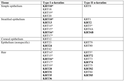
Table 2.1. Type I and Type II α-keratin expression in humans ...16
Table 2.2. Expression of α- and β-keratins during embryonic chicken development ...28
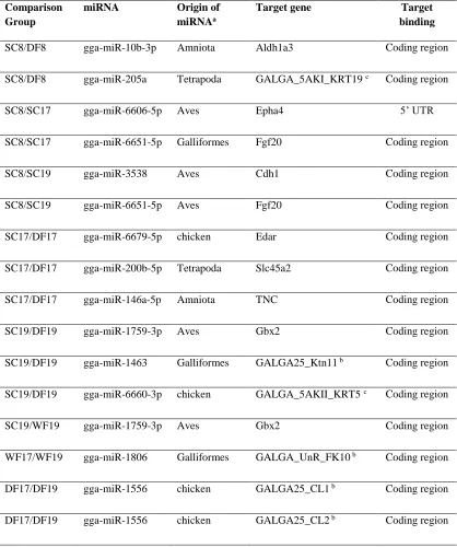
Table 3.1. Differentially expressed miRNAs and their differentially expressed miRNA genes that are feather-related ...58
ix
LIST OF FIGURES
Figure 2.1. Molecular phylogeny and proposed genomic orientation of Type I α-keratins
...21
Figure 2.2. Molecular phylogeny and proposed genomic orientation of Type II α-keratins ...22
Figure 2.3. Genomic orientation of β-keratins in birds ...23
Figure 2.4. Expression of feather β-keratins during embryonic feather development...30
Figure 2.5. Dynamic evolution of β-keratins in the archosaur lineage ...37
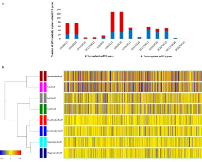
Figure 3.1. Summary of differentially expressed miRNA gene numbers in comparison groups and the hierarchical clustering dendrogram of the transcriptomes ...54
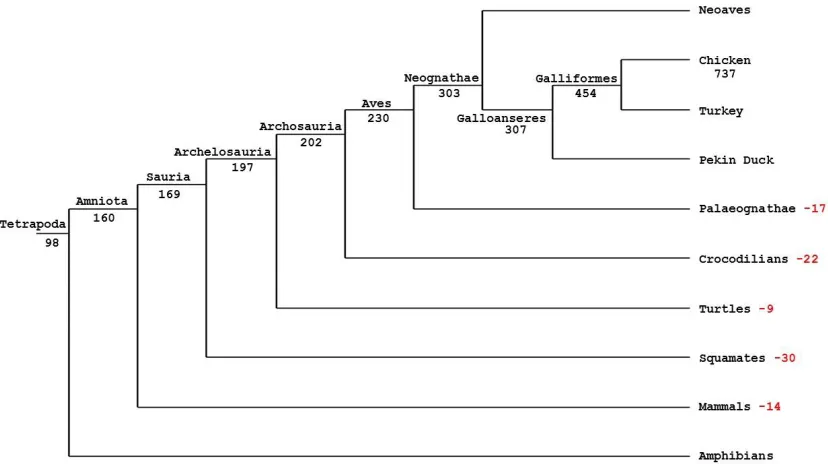
Figure 3.2. Probable birth of chicken miRNA genes across the tetrapod phylogeny ...60
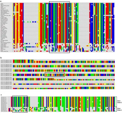
Figure 3.3. Conservation of the miRNA target sites ...66
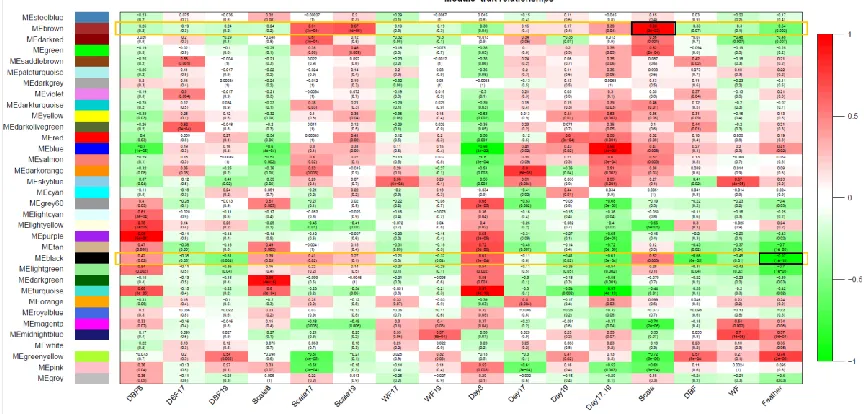
Figure 4.1. Heatmap of Module-Trait relationships for the co-expression network ...81
Figure 4.2. Gene scatter plot of correlation for Module Membership (MM) and Gene significance (GS) ...86
Figure 4.3. Output of MSET enrichment analysis for the customized chicken epidermal development-related gene database within the chicken embryonic epidermal development expression data ...88
1
CHAPTER 1
2
A key event in support of the colonization of land by vertebrates was the evolution of a
protective epidermis and its diversification into novel appendages (Sawyer et al., 1986;
Alibardi and Sawyer, 2002; Greenwold and Sawyer, 2010; Strasser et al., 2014). These
adaptations enabled successful interactions between the organisms and their new
environments, such as thermoregulation, protection against excessive water loss and
mechanical trauma as well as locomotion (Chuong and Homberger, 2003). The epidermis
is a multi-layered epithelium, made up of keratinocytes, which are renewed by the
proliferation of stem cells in the basal layer of the stratified epithelium. The evolution of
the vertebrate epidermis and its appendages, such as claws, scales, beaks and feathers,
was accompanied by molecular innovations of the structural components within the
keratinocytes, as well as innovations of gene regulation via the developmental pathways
during epidermal differentiation (Lowe et al., 2014). The genic and regulatory
complement had been present in nonavian dinosaurs and contributed to the evolution of
the emerging of pinnate feathers in dinosaurs (Lowe et al., 2014).
Alpha (α)-keratins are the major structural proteins found in the epidermis of all
vertebrates, while beta (β)-keratins are only found in epidermis of reptiles and birds (Bell
and Thathachari, 1963; Baden and Maderson, 1970; Haake et al., 1984; Rogers, 1985;
Sawyer et al., 1986). The α-keratins are a type of intermediate filament, and form
obligatory heterodimers of Type I (acidic) and Type II (basic) α-keratins (Berieter-Hahn
et al., 1986; Greenwold et al., 2014). Beta (β)-keratins form a filament-matrix structure
with the filament being 2-3nm in diameter, and containing a β-sheet-rich 34 amino acid
residue domain, which is a common feature of all β-keratins (Sawyer et al., 2000; Fraser
3
In birds, the β-keratins consist of four subfamilies (feather, claw, scale and keratinocyte),
which are expressed in varying degrees in different epidermal appendages (Bereiter-Hahn
et al., 1986; Rice et al., 2013; Smoak and Sawyer, 1983; Carver and Sawyer, 1988;
Shames et al., 1989; Carver and Sawyer, 1989; Knapp et al., 1993). Both the α- and
β-keratins have undergone gene duplication and functional diversification during vertebrate
evolution (Vandebergh and Bossuyt, 2012; Greenwold and Sawyer, 2010; Greenwold and
Sawyer, 2013; Greenwold et al., 2014; Li et al., 2013).
The sequencing of the chicken and zebra finch genomes (Hillier et al., 2004;
Warren et al., 2010) demonstrated that the β-keratins make up a large gene family in
these avian species. Numerous studies have characterized the β-keratin genes (Huth,
2008; Glenn et al., 2008; Greenwold and Sawyer, 2010; Greenwold and Sawyer, 2011;
Greenwold and Sawyer, 2013; Greenwold et al., 2014). The comparison of the β-keratin
genes in the chicken and zebra finch also demonstrated that they have similar genomic
organization (Greenwold and Sawyer, 2010).
The expression of β-keratins in avian epidermal appendages, including scale,
claw, feather, egg tooth, spur and beak, and their development have been characterized
using electron microscopy, 2-dimentional protein gels and immuno-cytology (Sawyer and
Abbott, 1972; Sawyer and Fallon, 1983; Shames et al., 1988; Lin et al., 2006). In scales,
epidermal placodes begin to form by day 9 of incubation and by day 12 the scale ridge
forms (Sawyer, 1972 a, b.; Sawyer and Abbott, 1972; Bereiter-Hahn et al., 1986).
Expression of β-keratins in the developing scutate scale begins around day 14 of
incubation in the embryonic epidermis and continues in the Beta Stratum of the adult
4
which lacks scutate scales, β-keratin expression begins at 14 days of incubation, in the
embryonic epidermis, but ceases by 17 days because the embryonic epidermis is lost at
hatching (Shames and Sawyer, 1986). Since scales do not form in the mutant, a Beta
Stratum, with its scale type β-keratins, does not develop (Shames and Sawyer, 1986).
Although chicken feathers start to develop as early as day 6 of incubation, the
expression of β-keratin does not occur until around day 12.5 (Lucas and Stettenheim,
1972; Sengel, 1976; Haake et al., 1984). Beta (β)-keratin expression in feathers is
continuous through hatching (Barnes, 1994; Shames et al., 1988). Presland et al., (1989a)
found that at least five specific feather β-keratins are expressed in 14 day embryonic
chickens. Glenn et. al., (2008) examined the evolutionary relationships among copies of
feather beta keratin genes from several different orders of birds, and Greenwold and
Sawyer (2010) used the newly sequenced genomes of the chicken and zebra finch to
determine the genomic organization and molecular phylogenies of the beta keratin
multi-gene family in these two birds.
Luo et al. (2012) compared early and late feathering in the day 1 post-hatched
chicken by applying microarray technology. They found that at least 14 differentially
expressed genes were significantly related to either α- or β-keratins. Therefore, in
Chapter 2, I characterized the differentially expressed epidermal structural genes during
the chicken embryonic development. We used a 44K chicken microarray, which was
customized by Bao and Greenwold (Greenwold et al., 2014), to investigate the expression
of α- and β-keratin genes during the embryonic development of scales and feathers in the
chicken. With the availability of the newly sequenced genomes of 48 phylogenetically
5
involved with sequencing the genomes of 48 birds, we investigated the evolution of the
avian epidermal structural genes (α- and β- keratins) by examining the gene copy number
variation, the construction of molecular phylogenies, and the determination of the
genomic orientation of the β-keratins (Greenwold, et al., 2014 – Bao is the co-first
author).
MicroRNAs (miRNAs) have been drawing more attention from biologists since
they were found to widely regulate biological processes (Betel et al., 2008). With their
flexible targeting mechanism, I reasoned that the miRNAs may play a role in the
development of the avian feather and scale. In the Chapter 3, I combined miRNA target
prediction tools with high-throughput microarray expression data of miRNA genes and
mRNA genes to identify miRNA-mRNA duplexes involved in the development of
chicken epidermal appendages which have evolved to perform multiple important
functions in birds. Furthermore, I have taken advantage of 60 genomes across Tetrapoda
including the 48 bird genomes (Zhang et al. 2014) to investigate the evolution of chicken
miRNA genes.
In chapter 4, I used a novel bioinformatic approach to examine gene networks in
scale and feather development. Gene co-expression network analysis is a system biology
approach to identify modules which cluster highly correlated genes (Langfelder et al.,
2008). Weighted Gene Co-Expression Network Analysis (WGCNA) algorithm
introduces topological overlap matrix to construct a more robust scale free network
(Langfelder et al., 2008). In gene co-expression network, each gene corresponds to a
node and the two genes are connected by an edge if their expression values are highly
6
have only limited annotations based on the prediction tools. By utilizing the customized
44K chicken microarray expression data, I was able to construct gene co-expression
networks to identify gene modules highly correlated with chicken epidermal traits
(feather or scale). In addition to the traditional Gene Ontology (GO) enrichment analysis,
I applied the novel Modular Single-Set Enrichment Test (MSET) (Eisinger et al., 2013)
and Medical Subject Headings (MeSH)-informed enrichment analysis (Lu, 2011; Morota
et al., 2016) to demonstrate the validation of the WGCNA module construction by
introducing external traits. Further enrichment analysis on the module hub genes with
high connection with nodes also suggests the importance of biological role for the hub
genes.
For many years, reptilian scales and avian scales have been considered to be
homologous structures, as have the reptilian scales and avian feathers. In fact, studies
have shown that the developmental signaling molecules (Wnt, Sonic Hedgehog) show
similar patterns of expression during the early stages of scale development in the alligator
and chicken, as well as in the early developmental stages of feathers (Harris et al. 2002;
Musser et al., 2013). Most recently, it has been demonstrated that the anatomical
“epidermal placode” characterizes the first step in the formation of reptilian scales, avian
scale and feathers and the hair of mammals (Prum, 1999). Various scenarios have been
suggested for the evolution of feathers from reptilian or avian scales (Prum, 1999; Prum
and Brush, 2003; Sawyer and Knapp, 2003; Sawyer et al., 2005b). In the 1970s, it was
first discovered that the scutate scales of the chicken developed from epidermal placodes
similar to feathers (Sawyer, 1972 a, b). In addition, it was found that the scale epidermis
7
hatching (Sawyer et al., 1974 a, b). This embryonic epidermis is made up of independent
cell populations originating from the basally located stem cell population. The first cell
population generated is known as the periderm, which is found in all amniotes, and is
characterized by unique peridermal granules that contain scaffoldin of the Epidermal
Differentiation Complex (EDC) common to most vertebrates (Mlitz et al., 2014). The
second cell population produced is known as the subperiderm, which has been
extensively characterized in reptiles and birds (Sawyer and Knapp, 2003; Sawyer et. al,
2005b; Alibardi and Toni, 2008; Strasser et al., 2015). Using antibodies and in situ
hybridization probes to the feather specific beta-keratins (Sawyer et al., 2005a), the
Histidine-Rich Protein (HRP) of the EDC (Barnes and Sawyer, 1995; Alibardi et al.,
2016), and Cysteine-rich Protein (EDCRP) of the EDC (Strasser et al., 2015), it was
discovered that they are expressed in both the subperiderm of the embryonic epidermis
and the barbules and barbs of the embryonic feather (Sawyer et al., 2003a; b; Sawyer et
al., 2005a; Alibardi et al., 2006; Strasser et al., 2015; Alibardi et al., 2016). Unlike the
independent subperidermal cell population of the apteric skin (general body skin) and
epidermis of scales, which is lost at hatching, the subperidermal cells give rise to the
barbules and barbs of the first downy feathers (Sawyer and Knapp, 2003; Sawyer et al.,
2003a; Sawyer et al., 2005a; Strasser et al., 2015; Alibardi et al., 2016). These results
strongly suggest that the evolution of the subperidermal population of cells, which
originated in the ancestor of the archosaurians (crocodiles, dinosaurs and birds), gave rise
to the barbs and barbules of the first feathers. This implies that the first feathers may have
evolved directly from the epidermis without an intermediate scale step. In this study, the
8
embryogenesis provides the possible gene collaboration contributing to the distinct
9
CHAPTER 2
DYNAMIC EVOLUTION OF THE ALPHA (α) AND BETA (β)
KERATINS HAS ACCOMPANIED INTEGUMENT DIVERSIFICATION
AND THE ADAPTATION OF BIRDS INTO NOVEL LIFESTYLES
Greenwold, M. J., Bao, W., Jarvis, E. D., Hu, H., Li, C., Gilbert, M. T. P., Zhang, G., Sawyer, R. H. (2014). BMC Evolutionary Biology, 14 (1), 249
10 ABSTRACT
Background
Vertebrate skin appendages are constructed of keratins produced by multigene families.
Alpha (α) keratins are found in all vertebrates, while beta (β) keratins are found
exclusively in reptiles and birds. We have studied the molecular evolution of these gene
families in the genomes of 48 phylogenetically diverse birds and their expression in the
scales and feathers of the chicken.
Results
We found that the total number of α-keratins is lower in birds than mammals and
non-avian reptiles, yet two α-keratin genes (KRT42 and KRT75) have expanded in birds. The
β-keratins, however, demonstrate a dynamic evolution associated with avian lifestyle.
The avian specific feather keratins comprise a large majority of the total number of
β-keratins, but independently derived lineages of aquatic and predatory birds have smaller
proportions of feather β-keratin genes and larger proportions of keratinocyte β-keratin
genes. Additionally, birds of prey have a larger proportion of claw β-keratins. Analysis of
α- and β-keratin expression during development of chicken scales and feathers
demonstrates that while α-keratins are expressed in these tissues, the number and
magnitude of expressed β-keratin genes far exceeds that of α-keratins.
Conclusions
These results support the view that the number of α- and β-keratin genes expressed, the
proportion of the keratin subfamily genes expressed and the diversification of the
β-keratin genes have been important for the evolution of the feather and the adaptation of
11
BACKGROUND
The integument of amniotes has evolved from a basic cornified epidermis for protection
against the environment and the retention of water into an elaborate covering with
epidermal structures used additionally for sexual display, camouflage, locomotion, and
thermoregulation (Gill, 1995). The claws, scales, beaks and feathers of reptiles and birds
are formed from the products of two multigene families, alpha (α) and beta (β) keratins
(Bell and Thathachari, 1963; Baden and Maderson, 1970; Haake et al., 1984; Rogers,
1985; Bereiter-Hahn et al., 1986). Alpha keratins, a subtype of intermediate filaments
found in the epithelia of all vertebrates, have expanded and functionally diversified in
amniotes through gene duplication (Vandebergh and Bossuyt, 2012). The β-keratins are
found exclusively in reptiles and birds and have also expanded and diversified especially
in the avian and chelonian lineages (Greenwold and Sawyer, 2010; Greenwold and
Sawyer, 2013; Li et al., 2013).
The Type I (acidic) and Type II (basic/neutral) α-keratins form obligatory
heterodimers (Lee and Baden, 1976; Hatzfeld and Franke, 1985) that make up the
structural basis of the cornified epidermis and the epidermal appendages in mammals,
such as wool, hair, claws, horns and hooves (Bereiter-Hahn et al., 1986; Vandebergh and
Bossuyt, 2012; Fuchs and Marchuk, 1983; Rice et al., 2012). In birds, epidermal
α-keratins make up the stratum corneum of the general epidermis and epidermal
appendages such as the reticulate scale (O’Guin and Sawyer, 1982; Sawyer et al., 1986).
They are present in varying degrees along with the β-keratins in the avian scutate scales,
claws, beaks, spurs, and lingual nails (Bereiter-Hahn et al., 1986; Rice et al., 2012;
12
Sawyer, 1989; Knapp et al., 1993). Although α-keratins are expressed in the early stages
of feather development and in the cells of the rachis (Ng et al., 2012), the β-keratins make
up 90% of the barbs and barbules of the mature feather (Haake et al., 1984; Walker and
Rogers, 1976a and 1976b; Powell and Rogers, 1976; Alibardi and Sawyer, 2006;
Alibardi, 2013; Kowata et al., 2014). In other words, the dynamic duplication and
diversification of the β-keratin genes are thought to have contributed to the emergence of
a novel epidermal appendage, the feather, which characterizes over 10,000 species of
birds (Greenwold and Sawyer, 2010; Greenwold and Sawyer, 2013; Li et al., 2013).
The avian β-keratins were originally grouped into four subfamilies (claw, feather,
feather-like, and scale β-keratins) based on expression profiles and sequence
heterogeneity (Presland et al., 1989a and 1989b; Whitbread et al., 1991). More recently,
an avian β-keratin isolated from cultured keratinocytes has been reported
(Vanhoutteghem et al., 2004) and it is phylogenetically distinct from other β-keratin
subfamilies (Greenwold and Sawyer, 2010; Greenwold and Sawyer, 2013;
Vanhoutteghem et al., 2004; Dalla Valle et al., 2009a and 2009b; Greenwold and Sawyer,
2011). This keratinocyte β-keratin is also found in crocodilians, but not in the squamates
examined to date (Greenwold and Sawyer, 2013). An additional β-keratin gene, BKJ,
which is similar to feather-like β-keratins, has been identified on a unique locus and
annotated as β-keratin from jun-transformed cells (Greenwold and Sawyer, 2010;
Greenwold and Sawyer, 2013; Hartl and Bister, 1995). Thus, recent studies have
regrouped the β-keratins into four different, but overlapping phylogenetically distinct
13
like and BKJ genes are basal genes within the feather β-keratin clade (Greenwold and
Sawyer, 2013).
The Type I and II α-keratins are found on two unlinked genomic loci. In
mammals, the Type I α-keratin locus is separated by a small cluster of keratin associated
proteins (KAPs). However this Type I mammalian locus still shows a high level of
synteny with the green anole lizard, chicken and zebra finch which lacks the KAPs
(Vandebergh and Bossuyt, 2012). In birds, the Type I cluster is found on
microchromosome 27 ((Vandebergh and Bossuyt, 2012; Hesse et al., 2004; Zimek and
Weber, 2005). The Type II α-keratin cluster has been localized to linkage groups in the
chicken and zebra finch genomes where they also show a high level of synteny with
mammals and the green anole lizard (Vandebergh and Bossuyt, 2012). One Type I gene
variant is found on the Type II cluster suggesting a common genomic locus of origin for
the α-keratins in amniotes (Vandebergh and Bossuyt, 2012).
All four β-keratin subfamilies (claw, feather, scale, and keratinocyte β-keratins)
have been localized to a single locus in both the chicken and zebra finch;
microchromosome 25. However, several other unlinked loci contain feather β-keratins
(Greenwold and Sawyer, 2010). Furthermore, the β-keratins from the green anole lizard
are found on a single locus (Li et al., 2013; Greenwold and Sawyer, 2011; Alföldi et al.,
2011) and nearly half of the western painted turtle β-keratins are found on a single locus
that is syntenic to microchromosome 25 of the chicken and zebra finch suggesting a
common ancestral locus for β-keratins (Li et al., 2013).
Here we have taken advantage of the sequencing of 48 bird genomes (Zhang et
14
investigate the evolutionary landscape of α- and β-keratins in the avian clade using copy
number, molecular phylogenies, genomic orientation and transcriptome data. Our copy
number data indicates that both α- and β-keratins have evolved in a dynamic manner with
gene number contractions and expansions over the course of avian evolution leading to
modern birds. Comparative transcriptome analyses demonstrate that 26 α-keratins and
102 β-keratins are differentially expressed in chicken scales and feathers during
embryonic development. All four β-keratin subfamilies are highly expressed in
developing scales, whereas the feather and keratinocyte β-keratins are highly expressed in
the developing feather. The scales and feathers of birds have played important roles in the
diversification of birds and their adaptation to multiple ecological niches. The dynamic
evolution of the α- and β-keratins in the avian lineage accompanied these adaptations
with the avian specific feather keratins making up to 85% of the total number of
β-keratins, becoming the major structural component of the avian feather.
RESULTS
Genome searches of α-keratins show lineage specific gene losses and gains
We searched the genomes of 48 avian species that span the avian phylogeny, 2
crocodilians and 2 turtles (Zhang et al., 2014; Jarvis et al., 2014; St John et al., 2012;
Wang et al., 2013) for α-keratins and made use of the α-keratin copy number estimates
for the green anole, human, opossum, house mouse and platypus from Vandebergh and
Bossuyt (2012) to test the hypothesis that there are no differences in copy number
between birds and mammals and non-avian reptiles. We found that the total number of
bird α-keratins, 26-38 (x̄=31.271), is different from mammals and non-avian reptiles (fold
15
Furthermore, birds have a lower number of Type I α-keratins (range 10-18; x̄=14.292)
than mammals and non-avian reptiles (range 13-35; x̄ =23.22; fold change=0.616). The
number of bird Type II α-keratins (range 11-22; x̄=16.979) was also lower than that of
mammals and non-avian reptiles (range 14-27; x̄ =21; fold change=0.809).
The differences in copy number of Type I and II α-keratins among vertebrate
groups suggest a dynamic gain or loss of this gene family. To test this hypothesis, we
annotated all of the bird, crocodilian, turtle, human and green anole α-keratins to
determine which α-keratin genes may have been lost in the avian lineage (Additional file
2). We applied the same nomenclature of α-keratins as the one based on
human/mammalian genes (Schweizer et al., 2006). We considered a gene to be lost in the
avian lineage if it is present in human and at least one reptile and not found in any of the
48 bird genomes or avian expressed sequence tag (EST) libraries. Concurrently, we also
identified the number of genes lost in the crocodilian and turtle lineages. Based on our
annotations, we classified all avian and reptilian α-keratins into 17 Type I and 16 Type II
genes (Table 2.1; tissue specificity in humans from Schweizer et al. (2006) and Moll et
al. (2008). We found 8 Type I (see Table 2.1) and 6 Type II (see Table 2.1) α-keratins
missing in birds. Interestingly, turtles have a relatively low number of Type I and II
α-keratins and appear to have lost KRT10, KRT13, KRT31, KRT35 and KRT36 Type I
genes and KRT1, KRT6A, KRT74, KRT79, KRT82 and KRT85 Type II genes.
Crocodilians have also lost four of the same Type II genes, apparently independently (or
possibly through incomplete lineage sorting of the common ancestor of turtles, birds and
crocodiles) and lost the Type I gene KRT24. The cochleal or otokeratin Type II α-keratin
16
have originated early in the reptilian lineage, as all reptiles and birds have at least one
cochleal gene while none are found in humans.
Table 2.1. Type I and II α-keratin expression in humans
Tissue Type I α-keratins Type II α-keratins
Simple epithelium KRT10*
KRT18 KRT19* KRT20
KRT8
Stratified epithelium KRT10*
KRT13 KRT14* KRT15* KRT16* KRT17* KRT1 KRT2 KRT5* KRT6A KRT6B
Corneal epithelium KRT12
Epithelium (nonspecific) KRT23
KRT24
KRT42
KRT79 KRT80
Hair KRT14*
KRT15* KRT16* KRT17* KRT19* KRT28 KRT31 KRT35 KRT36 KRT5* KRT72 KRT73 KRT74 KRT75 KRT82 KRT84 KRT85
This table listed Type I and II α-keratin expression in humans and it is recreated from Schweizer et al. (2006) and Moll et al. (2008). Gene names in bold text have been lost in birds. *Indicates genes that are expressed in two of the tissues listed in the table.
While several α-keratin genes are absent in the avian lineage, at least one Type I
and one Type II variant have expanded. KRT42, a Type I α-keratin found in epithelia of
mammals has a mean copy number of 4.042 genes in birds and 1.143 genes in human and
non-avian reptiles (fold change=3.54). KRT75, a Type II α-keratin gene associated with a
feather rachis anomaly (Ng et al., 2012), is higher in birds (x̄ =8.333) relative to humans
or non-avian reptiles (x̄ =4.714; fold change=1.76). At least two copies of KRT42 and
17
The KRT75 expansion in birds led us to investigate whether an increase in copy
number is related to mutations in KRT75 genes such as the mutation that causes the
chicken frizzle feather phenotype (Ng et al., 2012). The associated mutation is at the exon
5/intron 5 junction that induces a cryptic splice site found within exon 5 of KRT75
resulting in a 69 base pair deletion. This cryptic splice site has a similar nucleotide motif
(GTGAAG) as that of the normal splice site at the exon5/intron5 junction. A total of 286
α-keratins were annotated as KRT75 genes for the 48 bird genomes, with most species
having 5 or more variants (Additional file 2). Of these 286, we found that only 12 KRT75
genes have the GTGAAG motif from 7 species (wild turkey, chicken, medium ground
finch, zebra finch, American crow, chimney swift and domestic pigeon). Unexpectedly,
these 7 species all had below the mean KRT75 copy number (8.333). These data suggest
that the frizzle feather phenotype should be rare among birds. Indeed, only chickens,
pigeons, geese and canaries have been described as having feather characteristics similar
to the frizzle phenotype (Landauer and Dunn, 1930). This is consistent with our finding
that the pigeon has the cryptic splice site as well as the two finches, which are in the same
Family as canaries.
Avian adaptations to novel lifestyles was accompanied by β-keratin gene family
dynamics
From all 48 bird genomes combined, we found 1623 complete β-keratin genes with both
start and stop codons and without unknown sequence (-NNN-) or frame shift mutations.
We also found 1084 incomplete avian β-keratins (Additional file 2). This analysis
showed extreme variation in copy number for birds with the barn owl having only 6
18
[Zhang et al., 2014]). Consistent with earlier studies (Greenwold and Sawyer, 2013; Li et
al., 2013), the American alligator and green sea turtle have 20 and 26 β-keratins,
respectively. While, the barn owl and zebra finch represent the minimum and maximum
number of total β-keratins, respectively, we found that the mean number of β-keratins in
birds was 33.81. We identified 4 statistical outliers (zebra finch: 149, chicken: 133,
pigeon: 81 and budgerigar: 71) that have a value greater than or equal to the third quartile
plus 1.5 times the IQR (interquartile range), which are birds with the highest number of
β-keratin genes (Additional file 1; [Zhang et al., 2014]). These drastic copy number
differences may relate to the quality of the genome build or to other factors such as
domestication, since they belong to 4 of 5 domesticated species among the 48 birds.
Annotation of the β-keratins was performed using the Greenwold and Sawyer
(2010) dataset. The feather-like β-keratins and β-keratins from jun-transformed cells
(BKJ) have been shown to group with feather β-keratins in previous phylogenetic
analyses (Greenwold and Sawyer, 2013), therefore genes resulting in a best hit to those
genes were annotated as feather β-keratins. We found that feather β-keratins comprised
up to 85% of the total number of β-keratins for birds. Using Levene’s test we can reject
the null hypothesis of equal variance among claw (W=23.442, p-value<0.001), scale
(W=23.107, p-value<0.001), keratinocyte (W=21.744, p-value<0.001) and total number
of β-keratins, but not for feather β-keratins and the total number of β-keratins (W=0.479,
p-value=0.491), indicating that the variance of feather β-keratin copy number for birds
can be used as an indicator of the variance in the total β-keratin copy number for these 48
19
In order to ascertain if β-keratin copy number differences in birds correlate to
species phylogenetic relatedness or lifestyle (aquatic and semi-aquatic and predatory;
Additional file 1 [Zhang et al., 2014]; Additional file 3 [Jarvis et al. 2014]), we calculated
the proportion of each of the four β-keratin subfamilies (Additional file 2) to the total
number of β-keratins for each lifestyle using standard and phylogenetic ANOVA. We
found that the proportion of feather β-keratins to the total number of β-keratins is
significantly lower for aquatic and semi-aquatic birds (see Additional file 1; [Zhang et al.,
2014]) than for land birds (F1,46=7.84; standard ANOVA p=0.007; phylogenetic
ANOVA p=0.029), while the proportion of keratinocyte β-keratins is significantly higher
for aquatic and semi-aquatic birds than land birds (F1,46=10.79; standard ANOVA
p=0.002; phylogenetic ANOVA p=0.013). This includes aquatic and semi-aquatic species
that are considered to have been independently derived according to the genome scale
phylogeny in our companion study (Jarvis et al. (2014); Additional file 3) However, the
species phylogeny (Additional file 3; [Jarvis et al., 2014]) indicates that the eagles do not
group with the other aquatic birds, we therefore removed them from the aquatic bird list
and found that only the higher proportion of keratinocyte β-keratins remained statistically
significant (F1,46=6.84; standard ANOVA p=0.012; phylogenetic ANOVA p=0.043)
indicating more strongly that the change in β-keratin gene numbers could be associated
with an aquatic lifestyle. We next considered birds with a predatory lifestyle (see
Additional file 1; [Zhang et al., 2014]) and found that the proportion of claw β-keratins
(F1,46=6.75; standard ANOVA p=0.0126; phylogenetic ANOVA p=0.033) and
keratinocyte β-keratins (F1,46=5.77; standard ANOVA p=0.02; phylogenetic ANOVA
20
of feather β-keratins (F1,46=7.81; standard ANOVA p=0.008; phylogenetic ANOVA
p=0.022) is significantly lower for predatory birds. Like the aquatic species, this finding
occurs for independent lineages of predatory birds. We did not find any significant
differences in copy number for the four β-keratin subfamilies between the major stem
lineages of birds: Paleognathae vs. Neognathae or Paleognathae and Galloanserae vs.
Neoaves; see also Jarvis et al. (2014). Together these data indicate that dynamic changes
in the proportion of β-keratin subfamilies have occurred as birds have adapted to novel
lifestyles.
The α- and β-keratin multigene families have similar patterns of sequence divergence
following gene duplication
To further our understanding of the evolution of the α- and β-keratin multigene families
in birds we performed phylogenetic analyses and examined their genomic orientation
(Figure 2.1- 2.3, Additional data file 4; [Zhang et al., 2014]). By examining these two
types of data we were able to elucidate the gene duplication history of multigene families
and gain a deeper understanding of their genomic origin.
The phylogenetic analyses of the Type I and II α-keratins (Figure 2.1A and 2.2A)
demonstrate that they can be separated into 3 main clades (Clade A, B and C) with Clade
A being composed of a single basal gene. The remaining two clades (Clade B and C) of
the Type I and Type II α-keratins are composed of multiple phylogenetically significant
sub-clades of different gene variants. The α-keratins genes, KRT75 and 42 are distributed
21
α-22
23
Figure 2.2. Molecular phylogeny and proposed genomic orientation of Type II
α-keratins. Part A is the maximum likelihood phylogeny of Type II α-keratins from human, green anole lizard, green sea turtle, American alligator and the 48 birds. Annotation of Type II α-keratins is based upon avian gene annotations. All clades are statistically significant. Genes labeled as non-avian include genes from human, green anole lizard, green sea turtle and/or American alligator. Part B is the proposed genomic orientation of Type II α-keratins in birds. While this whole region was not found on a single continuous genomic scaffold for some birds, the genomic alignment of scaffolds/contigs with at least 2 different gene variants resulted in this proposed consensus gene orientation of birds. Annotations are based upon Part A. The direction of the arrow is indicative of the DNA strand. The one Type I α-keratin, KRT18, shown in the consensus genomic orientation was only found in 7 species of birds, but was included in this figure based on the present data and previous studies (Vandebergh and Bossuyt, 2012; Hesse et al., 2004; Zimek and Weber, 2005).
Figure 2.3. Genomic orientation of β-keratins in birds. This figure is a genomic alignment of β-keratins in birds containing a genomic locus with at least two β-keratin subfamilies. For the chicken and zebra finch this locus is microchromosome 25. Although feather β-keratins can be found on many genomic loci other than the one shown here (Greenwold and Sawyer, 2010), we focused on this locus as it has members from all of the β-keratin clades. The annotations are based on our β-keratin phylogeny (Additional file 4; [Zhang et al., 2014]). The breaks in the line for each species are indicative of different genomic scaffolds. The direction of the arrow is indicative of the DNA strand. The arrows with solid colors are complete genes and those with white centers are
24
genes in different sub-clades are generally found interposed between other α-keratin
genes on their respective α-keratin loci (Figure 2.1B and 2.2B). These phylogenies
clearly demonstrate that KRT42 and KRT75 genes have expanded and their duplication
history is marked by non-tandem duplication and subsequent sequence divergence. Our
phylogenetic and genomic orientation data further support the idea that Type I and II
α-keratins have evolved through gene duplications in a concerted fashion (Blumenberg,
1988).
For the β-keratin phylogeny, the green anole forms a clade and therefore was
selected as the outgroup (Additional file 4; [Zhang et al., 2014]). Furthermore, a clade of
keratinocyte β-keratins composed of bird, turtle and crocodilian genes forms the basal
sister clade (Clade 1 keratinocyte β-keratins, Additional file 4; [Zhang et al., 2014]). The
remaining bird, turtle and crocodilian β-keratins form a second major clade that is
composed of another keratinocyte β-keratin clade (Clade 2 Keratinocyte β-keratins), two
claw β-keratin clades, one scale β-keratin clade and an avian specific feather β-keratin
clade. While all of the scale β-keratins annotated in Additional file 4 [Zhang et al., 2014]
group together, only a portion of them form a phylogenetically significant clade with
strong bootstrap support. Similar to the α-keratin genes, KRT42 and KRT75, the
β-keratin subfamilies, β-keratinocyte and claw β-β-keratins, have duplicated in a non-tandem
pattern (Figure 2.3) and form multiple phylogenetically significant clades (Additional file
4; [Zhang et al., 2014]). Collectively, these results indicate that the transposition of
duplicated genes on the same locus in a non-tandem fashion is adequate to induce a
25
Generally our gene trees (Figure 2.1A, 2.2A and Additional data file 4; [Zhang et
al., 2014]) did not follow the genome scale phylogeny of the 48 bird species in Jarvis et
al. (2014) (Additional file 3). This is not surprising because in that study no single gene
tree they analyzed was identical to the species tree. Due to incomplete lineage sorting,
most genes differed from the species tree by 20% of the branches, and our findings above
indicate large scale convergence of keratins among aquatic and predatory birds with
relationships different from the species tree. For each gene phylogeny (Figure 2.1A, 2.2A
and Additional data file 4; [Zhang et al., 2014]), however, we found that sub-clades of
closely related species frequently grouped together such as those of the Palaeognathae
(tinamou and ostrich), Galloanseres (chicken, turkey and duck) and some species of the
Psittacopasserimorphae (songbirds and parrots).
The phylogenetic analyses of Type I and II α-keratins (Figure 2.1A and 2.2A)
support the interpretation that birds have lost 14 α-keratin gene variants (see Table 2.1).
We found that the gene variants that are missing in birds form statistically significant
clades in human and non-avian reptiles. Additionally we found that our phylogenetic
analyses resulted in slightly different copy number counts for the α-keratin genes and
β-keratin subfamilies from the annotations detailed above (see Additional file 2). However,
the statistical significance of the comparisons between the β-keratin subfamilies and
lifestyles largely remained valid with the phylogeny data (data not shown). The exception
is with the keratinocyte β-keratin comparison between aquatic and land birds
(F1,46=5.76; standard ANOVA p=0.021; phylogenetic ANOVA p=0.057) when the
26
Previous studies (Vandebergh and Bossuyt, 2012; Hesse et al., 2004; Zimek and
Weber, 2005) have shown that one Type I α-keratin gene (KRT18) is found on the Type
II α-keratin locus for fish, amphibians, mammals and the green anole lizard indicating
that the α-keratins evolved from a single locus. While Vandebergh and Bossuyt (2012)
found a Type I α-keratin on chromosome unknown of the chicken, they found no direct
evidence that Type I and II α-keratins are linked in birds. In fact the gene they found on
chicken chromosome unknown was not found during our genome searches. However, we
did find that 8 of the bird species (chimney swift, common cuckoo, little egret, peregrine
falcon, crested ibis, brown mesite, white-throated tinamou and common ostrich), the 2
crocodilian species and the western painted turtle had one Type I α-keratin on a Type II
locus (Figure 2B). Furthermore, these genes were annotated as KRT18. The green sea
turtle and green anole lizard both have 2 KRT18 genes, which are found on 2 different
loci.
Differential expression of the α- and β-keratin genes in chicken epidermal tissue during
embryogenesis
We performed transcriptome analyses of scale, dorsal feather and wing feather tissues
during chicken development using a customized version of the chicken 44K Agilent
microarray (Li et al., 2008). We customized the microarray chip by adding all 27
α-keratins and 102 of the 133 chicken β-α-keratins. The number of unique 60-mer
oligonucleotides of β-keratins was constrained due to the highly repetitive nature of
feather β-keratins and thus we were only able to produce unique oligonucleotides for 68
27
Tissue samples from the chicken scutate scale, dorsal feather and wing feather
were from embryonic day 17 and 19 and scutate scale and dorsal feather at day 8.
Although feather morphogenesis begins as early as day 6.5 to 7, the cellular
differentiation of barbs and barbules and the accumulation of β-keratin does not begin
until ~ day 12 of embryogenesis (Bell and Thathachari, 1963; Haake et al., 1984; Lucas
and Stettenheim, 1972; Sengel, 1976). Scutate scale morphogenesis does not begin until
day 9.5 of embryogenesis, and β-keratin accumulation is not detected until 15-16 days of
development (Sawyer et al., 1986; Sawyer, 1972; Sawyer and Fallon, 1983; Shames and
Sawyer, 1986; Shames and Sawyer, 1987). Thus, we selected day 8 for the initial
sampling of the scale and feather tissues (Sawyer, 1972).
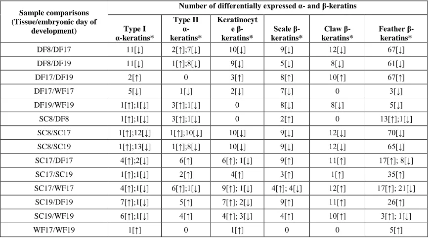
Comparison of day 8 scutate scale and day 8 dorsal feather tissues showed that 6
α-keratins and 18 β-keratins were differentially expressed, but the fold change values
were 5 or below (Table 2.2 and Additional file 5). Comparisons of day 8 and day 17 had
the largest number of differentially expressed α- and β-keratins. In comparing day 8
dorsal feather to day 17 dorsal feather we found 20 up-regulated and 2 down-regulated
α-keratins in the day 17 dorsal feather. Also 98 β-α-keratins were up-regulated in day 17
dorsal feather, which showed up to an 112,000 fold change for a keratinocyte β-keratin
and a 5,000 fold change for a feather β-keratin. We found 23 α-keratins that were
up-regulated and 2 α-keratins that were down-up-regulated in day 17 scutate scale while 101
β¬-keratins were up-regulated in day 17 scale (Table 2.2). Day 8 and 19 comparisons of
the dorsal feather and scutate scale had slightly lower numbers of α- and β-keratin
28
Table 2.2. Expression of α- and β-keratins during embryonic chicken development.
Sample comparisons (Tissue/embryonic day of
development)
Number of differentially expressed α- and β-keratins
Type I α-keratins* Type II α-keratins* Keratinocyt e β-keratins* Scale β-keratins* Claw β-keratins* Feather β-keratins*
DF8/DF17 11[↓] 2[↑];7[↓] 10[↓] 9[↓] 12[↓] 67[↓] DF8/DF19 11[↓] 1[↑];8[↓] 9[↓] 5[↓] 8[↓] 61[↓]
DF17/DF19 2[↑] 0 3[↑] 8[↑] 10[↑] 67[↑]
DF17/WF17 5[↓] 1[↓] 2[↓] 7[↓] 0 3[↓]
DF19/WF19 1[↑];1[↓] 3[↑];1[↓] 0 8[↓] 8[↓] 5[↓] SC8/DF8 1[↑];1[↓] 3[↑];1[↓] 0 2[↑] 0 13[↑];1[↓] SC8/SC17 1[↑];12[↓] 1[↑];10[↓] 10[↓] 9[↓] 12[↓] 70[↓] SC8/SC19 1[↑];13[↓] 1[↑];8[↓] 10[↓] 9[↓] 12[↓] 65[↓] SC17/DF17 4[↑];2[↓] 6[↑] 6[↑]; 1[↓] 9[↑] 11[↑] 17[↑]; 8[↓] SC17/SC19 1[↑];1[↓] 2[↑] 4[↑] 3[↑] 1[↑] 35[↑] SC17/WF17 4[↑];1[↓] 6[↑];1[↓] 9[↑]; 1[↓] 4[↑]; 4[↓] 12[↑] 17[↑]; 21[↓] SC19/DF19 7[↑];1[↓] 5[↑] 7[↑]; 2[↓] 9[↑] 11[↑] 26[↑] SC19/WF19 6[↑];1[↓] 4[↑] 4[↑]; 3[↓] 4[↑] 10[↑] 3[↑]; 1[↓]
WF17/WF19 1[↑] 0 1[↑] 0 0 5[↑]
This table listed the expression of α- and β-keratins during embryonic chicken
development. Number of differentially expressed α- and β-keratins during embryonic chicken development for 14 sample comparisons with a p-value cutoff of 0.05 and a fold change cutoff of 2.0. DF, dorsal feather; WF, wing feather; SC, scale. *Direction of selection is indicated in brackets (↑: Up-regulated; ↓: Down-regulated) after each copy number and refers to the first sample for each comparison.
Although the scale β-keratins were annotated based upon their expression in scale
tissue (Presland et al., 1989b), it appears that the claw β-keratins are expressed at the
highest level in scale tissue. In the scutate scale comparisons of day 8 vs. 17 and day 8 vs.
19, 7 out of the 10 highest fold changes (up-regulated genes) in day 17 and 19 scutate
scale are claw β-keratins. Additionally, the day 17 and 19 scutate scale inter-tissue
comparisons (dorsal and wing feather) showed that 9 of the highest fold changes
(up-regulated genes) in the scutate scale are claw β-keratins indicating that claw β-keratins
have an important role in the composition of epidermal appendages, such as scales, in
29
Four sample comparisons had genes from the feather β-keratin subfamily that
were up and down regulated (Table 2.2). The day 8 comparison of the scutate scale and
dorsal feather indicates that a single feather β-keratin from microchromosome 27 is
up-regulated in the day 8 dorsal feather while all of the down-up-regulated feather β-keratins are
on different loci (chromosome 1, 2, microchromosome 25, and chromosome unknown).
Furthermore, feather β-keratins on microchromosome 27 are up-regulated in day 17
scutate scale in comparisons of the scutate scale day 17 and dorsal and wing feather day
17. Additionally, comparisons between the scutate scale and wing feather during day 19
of embryogenesis show an up-regulation of microchromosome 27 feather β-keratins and
down-regulation of feather β-keratins on other loci in the day 19 scutate scale tissue.
These data indicate that feather β-keratins on microchromosome 27 are regulated
differently from feather β-keratins on other loci (chromosome 1, 2, 6, 10, and unknown
and microchromosome 25).
We found that the basal BKJ genes of the feather β-keratin clade (Additional file
4; [Zhang et al., 2014]) are expressed at a higher level in the dorsal and wing feather
when compared to the scutate scale at day 17 and 19 (Additional file 5). Although BKJ
genes are expressed in higher levels in the feather, they are also expressed in the scutate
scales as evidenced by the down-regulation in the scutate scale comparisons between day
8 and day 17 and 19. The feather-like β-keratins are found in multiple comparisons
indicating they are expressed in both feather and scutate scale tissue. Interestingly, the
only three feather β-keratins expressed in dorsal and wing feather comparisons at day 17
are the feather-like β-keratins suggesting that they have an important role in feather
30
on chromosome 25, the BKJ genes are found on chromosome 6 and are not linked to any
other β-keratins indicating that intra and inter-locus differential expression occurs among
the feather β-keratin clade.
Feather β-keratins in the chicken genome are found on multiple loci (Additional
file 4; [Zhang et al., 2014]) (Greenwold and Sawyer, 2010). Based on our sample
comparisons in this study we were able to determine which feather β-keratins from which
chicken genomic loci (GALGA, chromosomes) were being expressed in the dorsal and
wing feathers during embryonic development. The genomic loci of feather β-keratins
being expressed in day 17 dorsal (down) feathers and day 17 wing feathers are
summarized in Figure 2.4. The feather β-keratins expressed in the day 17 down feathers
are located on GALGA 1, 6, 10, 25 and 27. In addition, the feather-like genes on
GALGA 25 were expressed as was feather β-keratin on GALGA unknown. The feather
β-keratins expressed in the day 17 wing feathers are located on GALGA 6, 10, and 25. In
addition, the feather-like genes on GALGA 25 were expressed as was feather β-keratin
31
Figure 2.4. Expression of feather β-keratins during embryonic feather development. This figure summarizes the present data on the expression of feather β-keratin by
chromosomal location in embryonic (day 17 down and wing feathers) feathers using data from the present study. For each feather type (Day 17 down, Day 17 Wing), the chicken (GALGA) chromosome number of the feather β-keratins expressed is listed. The feathers of day 17 dorsal skin express feather β-keratins located on GALGA chromosome 1, 6, 10, 25 (both feather and feather-like β-keratins) and chromosome unknown. Wing feathers, at day 17, express feather β-keratins from GALGA chromosome 6, 10, 25 (both feather and feather-like β-keratins) and chromosome unknown.
The only comparison showing both up and down-regulated scale β-keratins is the
day 17 scutate scale versus wing feather, which indicates that scale genes annotated as 1,
2, 3 and 5 are up-regulated while scale 7, 8, 9 and 10 are down-regulated. These scale
β-keratins are all found on the same locus (GALGA 25) and their number describes their
orientation in a 5’ to 3’ direction. Alternatively, only one keratinocyte β-keratin
(GALGA25_Ktn6) is consistently differentially expressed from the other keratinocyte
β-keratins on GALGA 25. These results indicate that while β-β-keratins from all subfamilies
are being expressed in these tissues, intra-locus differential expression of GALGA 25
β-keratins and inter-locus differential expression of feather β-β-keratins may contribute to the
structural complexity of these and other avian epidermal appendages.
DISCUSSION
This study made use of the newly published genomes of 45 birds in addition to the 3
previously published bird genomes (chicken, zebra finch and turkey) to investigate the
multigene families of α- and β-keratins (Zhang et al., 2014). Incomplete or low coverage
genomes can lead to an underestimate of gene family copy numbers. For α-keratins, little
variation is seen among our copy number estimates (Additional file 1; [Zhang et al.,
2014]). Furthermore, based on the α-keratin annotation, we find consistent copy number
α-32
keratins, the β-keratins have a much larger variation in copy number estimates
(Additional file 1; [Zhang et al., 2014]). While both of these gene families are tandemly
arrayed on at least 2 genomic loci strong differences exist in the variation of the copy
number estimates among the 48 birds. The newly sequenced bird genomes are separated
into two coverage groups; low (<50X coverage) and high (>50X coverage) [Zhang et al.,
2014]. The coverage in these two groups vary, but if the β-keratin copy number is related
to genome coverage the copy number of β-keratins should correlate with fold coverage
and contig and scaffold N50. However, for each fold coverage group (low and high) we
do not find a statistically significant correlation between β-keratin copy number and fold
coverage, contig N50 or scaffold N50. While, we do not discount the likelihood that
some of the bird species in this study have unsequenced β-keratins, we believe that the
relative variation in β-keratin copy number among birds is appropriately represented in
this study.
Alpha (α)-keratins
The α-keratin nomenclature used in this study is based upon mammals and more
specifically humans (Schweizer et al., 2006). While mammals have shown the largest
expansion of α-keratins among amniotes (Vandebergh and Bossuyt, 2012), we find that
there is avian specific gene loss and gain of keratins. The expansion of specific
α-keratin gene variants (KRT42 and KRT75) in birds may not be the result of gene
duplication of a single “parent” gene, but instead the duplication of several different gene
variants resulting in novel keratins of avian origin. If some of these genes are novel
α-keratins as the phylogeny and genomic alignment data indicate, then the current α-keratin
33
found in birds. Therefore, we suggest that the KRT42 genes be annotated as KRT42a and
b and the KRT75 genes be annotated as KRT75a-e to reflect their phylogenetic
relationship and genomic orientation.
Our discovery of the KRT42 and KRT75 expansion in the avian lineage and Ng et
al. (2012) discovery that KRT75 is important in feather rachis development indicates that
the duplication of KRT42 and KRT75 α-keratins may be the result of concerted evolution
and that together they form the α-keratin heterodimer in feathers. Furthermore, it is likely
that these duplicated genes contributed to the evolution of feathers as did the feather
β-keratins (Ng et al., 2014).
Beta (β)-Keratins
The fact that the extreme statistical outliers from the average number of ~34 β-keratins
across bird species are all species that have undergone various degrees of domestication
(zebra finch, chicken, pigeon and budgerigar), indicate that there could be an association
between these observations. In support of this relationship, both the Peking duck (46) and
turkey (46), the remaining domesticated species among the birds, have an above average
number of β-keratins. Given that domestication may increase recombination rate (Burt
and Bell, 1987; Ross-Ibarra, 2004) the extreme variation in β-keratin copy numbers
among birds may be partially linked to higher recombination rates on β-keratin loci and
the domestication of these species. The differential expression of feather β-keratins is
related to their genomic locus (Ng et al., 2014), signifying that expansion of feather
β-keratins, through unequal crossing over events on specific loci, may be induced by
artificial selection.
34
Numerous studies have examined the biochemical and molecular make up of embryonic
and adult feathers, as well as their component parts (Sawyer et al., 1986; Walker and
Rogers, 1976a and 1976b; Alibardi and Sawyer, 2006; Alibardi, 2013; Kowata et al.,
2014; Dalla Valle et al., 2009a and 2009b; Dalla Valle et al., 2010;Shames and Sawyer,
1987; Ng et al., 2014;Kemp and Rogers, 1972; Walker and Bridgen, 1976; Kemp, 1975;
Gregg and Rogers, 1986; Shames et al., 1988). For example Kemp (1975) suggested that
there were 25-35 different feather keratin mRNA molecules in the embryonic feather and
a total of 100-240 keratin genes in the chicken genome. The present study supports the
view that a high number of α- and β-keratins are expressed during the embryonic
development of scutate scales and feathers in the chicken (Table 2.2). These results are
further supported by a recent study by Ng et al. (2014), which found that 90% of
α-keratin and over 95% of β-α-keratin genes in the chicken are differentially expressed during
post-hatching feather genesis. However, the number of β-keratins that can be extracted
from the cornified tissues of scales and feathers and detected on 2-dimensional gels is
considerably smaller (Rice et al., 2012; Knapp et al., 1993; Shames and Sawyer, 1986;
Gregg and Rogers, 1986; Shames et al., 1988; Knapp et al., 1991), suggesting that
messenger RNAs are being inactivated, perhaps by microRNAs (Zhang et al., 2013).
Recently, Kowata et al. (2014) found that the feather β-keratin on chromosome 7
of the chicken (GALGA 7) is expressed in the cells that form the barbules of pennaceous
feathers but is not expressed in the barbules of plumulaceous feathers. In the present
study, we did not find any differential expression of the GALGA 7 feather β-keratin in
embryonic feathers supporting the results of Kowata et al. (2014). However, we did find
35
β-keratins on other loci and that GALGA 27 feather β-keratins are generally up-regulated
in scale tissue. Previous studies have demonstrated that the ancestral locus of β-keratins is
homologous to GALGA 25 of the chicken (Greenwold and Sawyer, 2010; Greenwold
and Sawyer, 2013; Li et al., 2013), suggesting that feather β-keratins diversified to other
genomic loci through duplication and translocation. Recently, Ng et al. (2014) examined
which genomic loci (chromosome) are utilized for the expression of feather β-keratins in
post-hatched contour and flight feathers. While it is clear that feather β-keratin located on
GALGA 7 is only expressed in the barbules and possibly the hooklets of pennaceous
feathers, feather β-keratins from multiple loci are expressed in the ramus, rachis, and
calamus of post-hatched feathers. Overall these data suggest that as the avian epidermis
evolved to produce novel structures (such as pennaceous feathers) it took advantage of
the diversity of feather β-keratins that evolved on different loci.
Evolution of birds into novel ecological niches
Bird diversification is marked by evolution into novel habitats and ways of life such as
predatory and aquatic lifestyles. Birds of prey are identified by their powerful beaks and
claws. In the case of the claw, studies indicate that the morphology of the claw of birds of
prey (also referred to as a talon) differs from non-raptorial birds and between different
Orders of birds of prey (Csermely and Rossi, 2006; Fowler et al., 2009) (however see
Birn-Jeffery et al., 2012). In addition to being expressed in the claw (Whitbread et al.,
1991), claw β-keratins are also expressed in the beak of the chicken (Greenwold and
Sawyer, 2010; Wu et al., 2004). In this study we found that the birds of prey have a
36
indicate that they have played an important role in the evolution of these unique
epidermal appendages.
The feathers of aquatic birds have been shown to have a higher hydrophobicity
than the feathers of terrestrial birds (Rijke, 1970). This may be important for thermal
regulation especially for birds in adverse climates, such as penguins. Our analysis of
β-keratin copy number variation among birds has shown that the proportion of β-keratinocyte
β-keratins is higher and the proportion of feather β-keratins is lower for aquatic birds
compared to terrestrial birds. Also, we found that at least 98 of the 133 chicken β-keratin
genes are transcribed during the formation of feathers in the chicken. While feather
β-keratins are annotated based upon the tissue in which their amino acid sequence was first
determined (O’Donnell and Inglis, 1974), it has been shown that there are actually
multiple β-keratin gene variants (subfamilies) expressed during embryonic development
of feathers (Greenwold and Sawyer, 2013; Presland et al., 1989a and 1989b).
Collectively, this indicates that the proportion of gene variants is important as birds have
adapted to their lifestyles (aquatic, terrestrial, predatory) and that their feather, claw and
beak structure may have been modified by the dynamic expansion and contraction of
specific β-keratin gene variants.
CONCLUSION
The number of α-keratin genes is reduced in the avian lineage, and while still important
for feather development, for example during rachis morphogenesis (Ng et al., 2012), their
low abundance in the barbs and barbules of feathers demonstrates that they have a
reduced role in establishing the composition of mature feathers. On the other hand, the
37
lineages resulting in novel epidermal appendages (Greenwold and Sawyer, 2010;
Greenwold and Sawyer, 2013; Li et al., 2013). Members of all β-keratin subfamilies are
expressed during the development of scutate scales and feathers with feather β-keratins
becoming specialized in their expression profiles in the diverse assortment of feathers
found in present day birds (Kowata et al., 2014; Ng et al., 2014) (Figure 2.4). The early
evolution of β-keratins in the archosaurian lineage is marked by lineage specific
expansions, but differences in the proportion of claw, feather, and keratinocyte β-keratin
genes in modern birds may be attributed to their ecological niche (Figure 2.5). Our
overall findings suggest that the number of keratins and the relative proportion of
β-keratins in each subfamily influenced the composition of avian skin appendages and
therefore their structural properties. Clearly, the evolution of feathers in the lineage
leading to modern birds has been shaped by the dynamic evolution of α- and β-keratins.
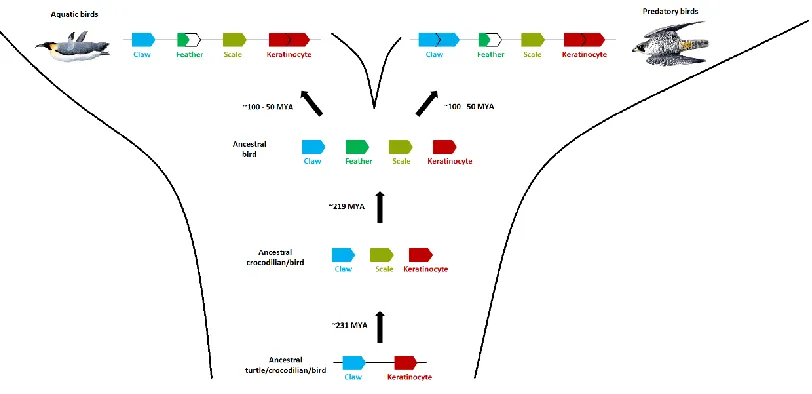
Figure 2.5. Dynamic evolution of β-keratins in the archosaur lineage. This figure
38
emerged since the divergence of turtles from crocodilians and birds. The origin of the feather β-keratins occurs after the divergence of crocodilians and birds. The order of the β-keratins subfamilies is based on our genomic data from the 48 birds and green sea turtle. The top row illustrates the dynamic changes of the proportions of β-keratin subfamilies in modern birds with aquatic and predatory lifestyles. Both aquatic and predatory birds have a larger proportion of keratinocyte β-keratins and smaller proportion of feather β-keratins in their genomes. Additionally, predatory birds have a larger
proportion of claw β-keratins. The divergence times are in millions of years ago (MYA). The divergence estimates of the turtle – crocodilian/bird split and the crocodilian – bird split are from Shedlock and Edwards (2009). The divergence time estimates for birds is from Jarvis et al. (2014) and is the range starting with the divergence of the
Palaeognathae and Neognathae (~100 MYA) and the subsequent divergences of most ordinal groups by ~50 MYA. Jon Fjeldså produced the images of the birds (emperor penguin on the left and peregrine falcon on the right).
METHODS
Genome searches
The genome build information and statistics for the birds used in this study are detailed in
Zhang et al. (2014). Additional file 1is reprinted with permission from Zhang et al.
(2014) and lists the English names for all species used in this study, while scientific Latin
species names are listed in Additional file 2.
Alpha keratin sequences for the chicken, green anole and human were
downloaded via NCBI and their accession numbers and names are listed in Additional
file 6. The copy number and position of these sequences coincide with the results
reported in Vandebergh and Bossuyt (2012). Genome searches were conducted using
standalone BLAT (v35) fast sequence search command line tool (Kent, 2002) and
GeneWise (v2.2.0) was used as a homology based predictor of gene structure (Birney et
al., 2004) with chicken and green anole α-keratin sequences downloaded via NCBI
(Additional file 6). Type I and II α-keratin searches were conducted separately. BLAT
hits with a Match score, the number of matches minus the mismatches, of ≥ 250 were