ABSTRACT
HASLAUER, CARLA MARIA. Design and Validation of Three-Dimensional Scaffolds for Bone Tissue Engineering Applications Using Human Adipose-Derived Adult Stem Cells. (Under the direction of Elizabeth G. Loboa).
Functional bone tissue engineering has arisen to address the need for implantable bone tissue in cases of trauma or disease. Some challenges to
generating functional bone tissue have included the need to identify biocompatible substrates with appropriate mechanical properties, as well as a suitable cell source. Subsequently, human adipose-derived adult stem cells (hASCs) have been used in an assortment of tissue engineering applications in recent years. They are attractive cells for use in clinical applications due to their multi-lineage potential, relative
abundance, and ease of harvest relative to other cell lines. The pluripotency of hASCs isolated from adipose tissue and used in the various studies described here has been tested by inducing both osteogenic and adipogenic differentiation
and fabricated for bone tissue engineering applications. Initial cell viability was examined over a 4 week time period for hASCs seeded on the novel scaffolds. Titanium implants are commonly used in medical procedures such as hip replacements. There is a need however, to design patient specific titanium
scaffolds. An important factor in scaffold acceptance by the body is osseointegration, or bone ingrowth. Micromotion between the implant and surrounding tissue can hinder osseointegration, and ultimately lead to implant failure. Using computed tomography (CT) data, micromotion can be minimized by designing patient-specific implants. Implants from these designs can then be fabricated by electron beam melting (EBM) using titanium powder, built layer by layer to the appropriate specifications. In this study, hASCs were used to validate the effects of EBM processing when compared to commercially available medical-grade titanium implants. Results suggest EBM fabricated porous scaffolds promote hASC proliferation and do not adversely affect biocompatibility.
Natural polymers have also been investigated for bone tissue engineering applications. This study describes the design of electrospun fibers, which are on a similar size scale to the native collagen fibrils found in human bone tissue.
that of hASCs seeded on uncoated PCL scaffolds. The results indicated the collagen coating increased cell spreading and osteogenic differentiation of hASCs seeded on the sheath-core electrospun scaffolds.
Nanoporous fibers are also desirable for bone tissue applications, as the micropores should enhance nutrient delivery and waste removal. In the final study, melt spun islands-in-the-sea fibers were extruded with a polylactic acid (PLA) sea matrix and islands comprised of EastONE, a water-dispersible sulfopolyester. The EastONE polymer was also added to the sea matrix in various concentrations,
yielding micropores within the PLA sea after washing. These fibers were then knitted into three-dimensional fabrics, and the physical properties characterized.
Design and Validation of Three-Dimensional Scaffolds for Bone Tissue Engineering Applications Using Human Adipose-Derived Adult Stem Cells
by
Carla Maria Haslauer
A dissertation submitted to the Graduate Faculty of North Carolina State University
in partial fulfillment of the requirements for the degree of
Doctor of Philosophy Biomedical Engineering
Raleigh, North Carolina 2010
APPROVED BY:
___________________________ ______________________________ Dr. Elizabeth G. Loboa Dr. Behnam Pourdeyhimi
Committee Chair
___________________________ ________________________________ Dr. Nancy Monteiro-Riviere Dr. Ola Harrysson
DEDICATION
BIOGRAPHY
Carla Maria Haslauer was born on September 27, 1983 in New Orleans, Louisiana to David John Haslauer and Maria Viviano Haslauer. Carla is the eldest of four girls, whose younger sisters Monica Alyssa, Alexandra Marisa, and Erica Arianna
Haslauer. Carla attended St. Christopher the Martyr Elementary School in Metairie, Louisiana and in May 2001 graduated from St. Mary‟s Dominican High School, New Orleans. She attended Louisiana State University, Baton Rouge where she received a Bachelor of Science degree in Biological and Agricultural Engineering in May 2005. While at LSU, she performed undergraduate research under the direction of Dr. William Todd Monroe and Dr. David D. Pollock. Carla moved to North Carolina in June 2005 to begin doctoral studies in the Joint Department of Biomedical
ACKNOWLEDGMENTS
I would like to thank my family for their unconditional support throughout this journey. This would not have been possible without my mom lending a listening ear or my dad going through the effort of boiling crawfish for my friends and me every time I returned home.
I would like to thank LSU for providing me with a strong undergraduate education upon which I could build my graduate studies. I greatly appreciate the guidance of Dr. Todd Monroe and Dr. Marybeth Lima during my undergraduate tenure, and their continued help and support throughout my graduate career. I am also indebted to Dr. Julianne Forman Audiffred for mentoring me as an
undergraduate, then becoming a great resource as a colleague while we pursued our doctoral degrees.
I would like to thank the members of the Cell Mechanics Lab, particularly graduate students Josie Bodle, Adisri Charoenpanich, and Seth McCullen. I am grateful for the cell culture assistance provided by undergraduates Cara Buchanan and Elizabeth Kirk, and Jenny Puetzer‟s help to keep the lab functioning. Special thanks go to Dr. Ruwan Sumanasighe for his guidance with the melt-spun porous fiber experiments. I would also like to thank Dr. Wayne Pfeiler not only for his help in the lab, but also for being a great friend and my lunch buddy for four years!
I would like to thank Rachael Turner for being such a great friend and
colleague, always providing the impetus to keep trekking along. Thanks also to Dr. Susan Bernacki for providing the much-needed cell culture training, and Ajit Moghe and Jessica Springer for providing two of the novel substrates described in this dissertation. I would also like to thank Dr. Jason Osborne and Matthew Avery for their statistical assistance. Also, thanks to Nancy McKinney for her tireless efforts in coordinating the BME graduate program across both Universities.
I am grateful for the funding provided by the Nonwovens Cooperative Research Center (NCRC), and the aid and support of Sue Pegram, Steve Sharp, and Wendy Cox. I would like to thank Hills Inc. and Eastman Chemical for
TABLE OF CONTENTS
LIST OF TABLES ... xii
LIST OF FIGURES ... xiii
NOMENCLATURE ...xx
PREFACE ... 1
1. Introduction ... 3
1.1 Background ... 3
1.1.1 Three-dimensional Bone Tissue Engineering Scaffolds ... 3
1.1.2 Use and Healing Potential of Human Adipose-Derived Adult Stem Cells ... 6
1.1.3 Bone-graft Substitutes ... 8
1.2 Project Overview and Objectives ... 10
2. Evaluation of Human Adipose-Derived Adult Stem Cell Response to Custom Titanium Implant Fabrication Methods ... 11
2.1 Introduction ... 12
2.2 Materials and Methods ... 17
2.2.1 Scaffolds Preparation ... 17
2.2.4 Cell Proliferation ... 21
2.2.5 Cell Viability ... 21
2.2.6 Cytokine Analysis ... 22
2.3 Results ... 24
2.3.1 Cell Proliferation ... 24
2.3.2 Cell Viability ... 25
2.3.3 Release of Cytokines IL-6 and IL-8 ... 26
2.4 Discussion ... 30
2.5 Summary ... 34
3. Three-dimensional Collagen-PCL Sheath-Core Bicomponent Electrospun Scaffolds Increase Osteogenic Differentiation and Calcium Deposition of Human Adipose-Derived Adult Stem Cells ... 35
3.1 Introduction ... 36
3.2 Materials and Methods ... 43
3.2.1 Materials ... 43
3.2.2. Fabrication of PCL Scaffolds ... 43
3.2.3. Fabrication of Sheath-Core Nanofibrous Scaffolds ... 44
3.2.5 hASC Isolation and Expansion ... 46
3.2.6 Cell Seeding ... 47
3.2.7 hASC Proliferation ... 47
3.2.8 Cell Viability ... 48
3.2.9 Calcium Quantification... 49
3.2.10 Statistical Analysis ... 50
3.3 Results ... 51
3.3.1. Scaffold Morphology ... 51
3.3.2 Cellular Interactions ... 54
3.4 Discussion ... 60
3.4.1 Scaffold Fabrication ... 61
3.4.2 Cellular Interactions ... 63
3.5 Summary ... 66
4. Initial Design Iterations of Porous Melt-spun Fibers ... 68
4.1 Introduction ... 69
4.2 Experimental Approach ... 72
4.2.3 Examination of Fiber Cross-Section ... 77
4.2.4 Differential Scanning Calorimetry ... 80
4.2.5 Viscosity Average Molecular Weight ... 81
4.2.6 Extrusion of Solid Monofilaments ... 82
4.2.7 Viscosity ... 85
4.2.8 Melt-Spinning Fibers ... 85
4.2.9 SEM Images ... 86
4.2.10 Pore Size Measurement of Multifilament Fibers ... 87
4.2.11 Creation of Hollow Monofilaments ... 88
4.2.12 Extrusion of Hollow Fibers ... 90
4.3 Summary ... 92
5. Characterization of Porous Islands-In-The-Sea Biodegradable Scaffolds for Bone Tissue Engineering Applications ... 94
5.1 Introduction ... 95
5.2 Materials and Methods ... 99
5.2.1 Fiber Extrusion and Scaffold Fabrication ... 99
5.2.2 Fabric Washing ... 100
5.2.4 Focused Ion Beam Imaging ... 101
5.2.5 BET Surface Area Analysis ... 102
5.2.6 Transverse Wicking ... 103
5.2.7 Sterilization and Cell Seeding ... 104
5.2.8 Human ASC Viability ... 105
5.3 Results ... 106
5.3.1 Weight Loss ... 106
5.3.2 FIB Imaging ... 107
5.3.3 BET Surface Area Analysis ... 108
5.3.4 Transverse Wicking ... 109
5.3.5 Viability ... 111
5.4 Discussion ... 114
5.5 Summary ... 120
6. Conclusions and Recommendations for Future Research ... 121
6.1 Conclusions ... 121
6.2 Future Research ... 123
LIST OF TABLES
Table 4.1. Polymer composition and spinning speeds of each fiber spool. ... 86
Table 4.2. Polymer composition and spinning speeds of each fiber spool. ... 88
Table 5.1. Table reflecting the polymer compositions for the islands and sea
LIST OF FIGURES
Figure 2.1. Photographs of titanium discs. A) polished commercially-produced (control), B) polished EBM-produced, C) unpolished EBM-produced, and D)
unpolished, porous EBM-produced ... 17 Figure 2.2. Images of hASCs after induction down the adipogenic (left) and
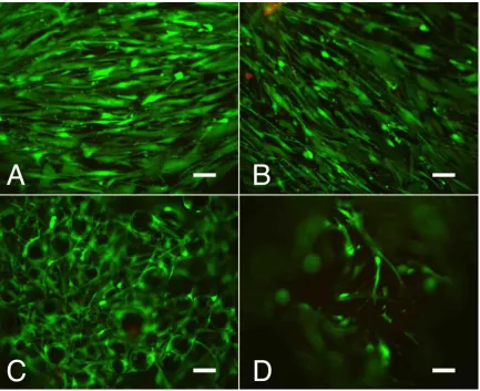
osteogenic (right) pathways. Red stain indicates presence of oil droplets (left) and calcium deposits (right). ... 20 Figure 2.2. Percent reduction of alamarBlue by hASCs on four titanium discs. p<.05 is considered statistically significant. ... 24 Figure 2.3. Representative fluorescence microscopy (10X objective, scale
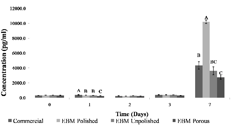
bar=100µm) Live/Dead images of hASCs seeded on titanium discs after 8 days of culture (live= green, red= dead). A) polished commercially-produced (control), B) polished EBM-produced, C) unpolished EBM-produced, and D) unpolished, porous EBM-produced ... 26 Figure 2.4. Mean IL-6 release (± SEM), normalized to proliferation data, by hASCs on titanium implants. Histogram with different letters (A-C) denote mean values that are statistically different at p<.05. If no letters are present, then no significant
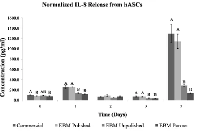
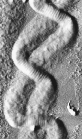
Figure 2.5. Mean IL-8 release (± SEM), normalized to proliferation data, by hASCs on titanium implants. Histogram with different letters (A and B) denote mean values that are statistically different at p<.05. If no letters are present, then no significant difference was noted. ... 29 Figure 3.1. Image of two entangled collagen fibrils displaying a two-ply rope
structure. Width of entangled structure is approximately 0.6µm. (Adapted from [66]) ... 37 Figure 3.2. SEM image of an electrospun PLA fibrous mat. (Scale bar = 20 µm) ... 39
Figure 3.3. Schematic of the co-axial electrospinning system. (Adapted from: [80])45
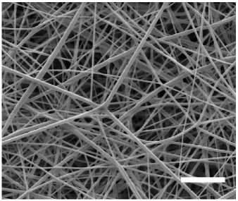
Figure 3.4. SEM image of the PCL scaffold structure. Average fiber diameter: 280
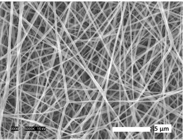
51 nm. (Adapted from: [80]) ... 52 Figure 3.5. SEM image of the collagen-PCL sheath-core scaffold structure.
Average fiber diameter: 442 45nm. (Adapted from: [80]) ... 53
Figure 3.8. Representative images (10X) of cells seeded on collagen-coated and uncoated PCL scaffolds showing live (green) and dead (red) cells. (A) collagen-coated after day 1; (B) uncollagen-coated PCL after day 1; (C) collagen-collagen-coated after day 2; (D) uncoated PCL after day 2; (E) collagen-coated after day 3; (F) uncoated PCL after day 3 (scale bar = 100µm). (Adapted from: [80]) ... 57 Figure 3.9. Representative confocal microscopy images (10X) of cells seeded on collagen-coated and uncoated PCL scaffolds with calcein AM and ethidium
Figure 4.1. Microscopic images of PLA microfilms before (top) and after (bottom) washing. ... 73 Figure 4.2. Thermo Haake Minicompounder used to extrude solid monofilaments of varying polymer concentrations. ... 74 Figure 4.3. SEM images of unwashed (A, C, E, G, and I) and washed (B, D, F, H and J) composite fibers acquired at a magnification of 2500X. (Adapted from: [94]) 75 Figure 4.4. SEM image of washed 10%EastONE/ 90% PLA fiber showing
image of mapped area, B) Contour map with intensity gradients (Adapted from: [94]) ... 80 Figure 4.9. Fuji Filter Melt Spinning Tester MST-CII. ... 83
Figure 4.10. SEM image of washed 20%EastONE/80% PLA fiber prior to
compounding. ... 83 Figure 4.11. SEM image of washed 15%EastONE/85% PLA fiber (left) and 100% PLA fiber (right) at 5000X. ... 84 Figure 4.12. Plot of viscosity vs. shear rate. ... 85
Figure 4.13. SEM images at 8000X of washed (left – A, C, E, G, I, K) vs. unwashed
cap layer is then deposited with a 500pA Ga+ beam (b). Mass removal with a 20nA Ga+ ion beam (c) and subsequent polishing with 5nA, 1nA, 500pA and 100pA Ga+ beams give rise to the final cross-section (d). (Adapted from: [97]) ... 102 Figure 5.2. Percent weight loss of the four fabric samples. Bars reflect standard deviation of the measured mean for each sample. Different letters denote
significance <.05. ... 106 Figure 5.3. Micrographs of a biaxial cross-section of an unwashed 80/20
PLA/EastONE fiber showing the distribution of the EastONE islands within the sea matrix: a) longitudinal view, b) biaxial view, and c) radial view. The smooth regions correspond to the EastONE islands and the rough regions correspond to the
EastONE/PLA sea matrix. (Adapted from: [97]) ... 107 Figure 5.4. Micrographs of a biaxial cross section of an 80/20 PLA/EastONE fiber after washing. A biaxial cross section of the washed fiber is displayed in (a). A portion of the fiber is further magnified (region denoted by the dotted line) in the subsequent micrographs (b and c). (Adapted from: [97]) ... 108 Figure 5.5. Fiber surface area (m2/g) for each of the four samples measured using the BET method. Error bars reflect standard deviation. ... 109 Figure 5.6. Photographs of fabrics following washing and drying, and the addition of 10 µL of dye. Fabrics were imaged after 30 seconds. A) PLA/PLA; B)
Figure 5.7. Average transverse wicking diameter for each fabric type. Error bars reflect standard deviation from the mean. ... 111 Figure 5.8. Live/ Dead images after 1 day in culture of hASCs seeded on: ... 112
A) PLA/PLA; B) PLA/EastONE (scale bar = 100µm) ... 112 Figure 5.9. Live/ Dead images after 21 days in culture of hASCs seeded on: ... 112
A) PLA/PLA; B) PLA/EastONE; (scale bar = 100µm) ... 112 Figure 5.10. Live/ Dead images after 28 days in culture of hASCs seeded on: .... 113
NOMENCLATURE ANOVA Analysis of Variance
Au-Pd Gold-Palladium
BET Brunauer, Emmett and Teller CCS Cleaning Cross Section
CT Computed Tomography
DSC Differential Scanning Calorimetry EBM Electron Beam Melting
ECM Extracellular Matrix FBS Fetal bovine Serum FIB Focused Ion Beam
FTIR Fourier Transform Infrared Spectrophotometer HFIP 1,1,1,2,2,2-hexafluoroisopropanol
hASCs Human adipose-derived adult stem cells hMSCs Human Mesenchymal Stem Cells
HCl Hydrochloric Acid
ISE Induced Secondary Electron MCT Mercury Cadmium Telluride MEM Minimal Essential Medium NaCl Sodium Chloride
PCL Poly -ε- caprolactone PLA Polylactic acid
PREFACE
The work presented in this dissertation involved collaborative research projects with many investigators. The research presented in Chapter 2 has been accepted for publication in Medical Engineering and Physics with co-first author Jessica Springer and co-authors Ola Harrysson, Elizabeth Loboa, Nancy Monteiro-Riviere, and Denis Marcellin-Little. This work was funded by two grants from the North Carolina State University College of Veterinary Medicine (Marcellin-Little and Loboa, 1/2008 - 3/2009). Titanium scaffolds were fabricated by Jessica Springer. The work presented in Chapter 3 has been submitted for publication to Journal of Biomaterials Science: Polymer Edition with first author Ajit K. Moghe and co-authors Buhpender Gupta, Jason Osborne, and Elizabeth Loboa. This work was funded by a grant from the National Textile Center (Electrospun Core-Sheath Fibers for Soft Tissue Engineering- Gupta 5/2005-3/2009). Electrospun scaffolds were fabricated by Ajit Moghe. Some work presented in Chapter 4 has been submitted for publication to the Journal of Applied Polymer Science with co-authors Ruwan
1. Introduction 1.1 Background
1.1.1 Three-dimensional Bone Tissue Engineering Scaffolds
Nearly 800,000 surgical procedures involving bone occur annually in the United States alone, making it the most commonly replaced tissue of the body [1]. Replacement bone tissue is typically obtained from the patient, with an autograft, or from a donor source, termed allograft. Problems with autografts, which are typically taken from the iliac crest, include donor site morbidity, limited quantity, and
increased operative time [1, 2]. Allografts on the other hand, have the potential for contamination or other complications including infections, fracture, or a host immune response [1]. Tissue engineering techniques which incorporate autologous cells are believed to provide a better grafting solution by easing the problems of supply limitation/ donor scarcity of bone, as well as immune rejection or transfer of pathogens [3]. Tissue engineering scaffolds should be biocompatible and
biodegradable, as well as possess proper mechanical properties and encourage tissue development at the graft site while maintaining cell viability [3].
improve nutrient delivery to, and waste removal from, cells in the center of the three-dimensional (3D) structure, it is desirable to incorporate a macroarchitecture into the scaffold. This could be achieved through the addition of channels and pores, both random and controlled. Improvements in nutrient delivery and waste removal are expected to reduce the likelihood of developing a necrotic core, as cells which have migrated into the scaffold center will still have access to vital nutrients.
In addition to nutrient delivery, three-dimensional bone tissue engineering scaffolds must be designed to maintain their structural integrity once implanted, while allowing cells to proliferate and secrete extracellular matrix proteins [5]. As the scaffolds gradually degrade, space for new tissue growth and matrix deposition is created until the tissue has been fully replaced [5]. Previous studies have indicated that for bone tissue engineering applications, osteoblasts prefer pores 200-400µm in diameter [6]. Osteoblasts can then migrate into the porous structure and excrete extracellular proteins to replace the degrading scaffold. The combination of cellular proliferation and extracellular matrix accumulation should fill any voids left by the degrading scaffold material.
differing degradation kinetics of synthetic polymers as needed for a particular application. The rate of degradation can be tailored for specific applications by altering crystallinity and molecular weight, or through the addition of other polymers [3]. For example, it has been shown that polycaprolactone (PCL), another nontoxic biodegradable polymer, can take several years to degrade in vivo [7, 8]. PLA, with its hydrophobic methyl groups, can take more than 5 years to degrade when comprised of L-lactic repeating units or as little as 1 year for amorphous PLA [5, 8]. PCL
degrades similarly to PLA, with hydrolytic cleavage of ester bonds as well as weight loss of oligometric species diffusing from the bulk. PCL degradation is considered relatively slow, especially in comparison to some natural polymers, with complete removal from the body requiring three years [9]. Composite scaffolds of various synthetic and/or natural polymers have been considered for tissue engineering applications to tailor scaffold degradation rates. Beyond controlling degradation rates, composite scaffolds also have the additional advantage of combining
properties to suit the mechanical and physiological demands of the surrounding host tissue [5]. The combination of naturally occurring polymers, such as collagen, with synthetic polymers has the potential to provide both cellular attachment sites as well as mechanical strength.
seen by cells seeded on other types of collagen matrices (type II, III, or V collagen) [10]. The surface chemistry of most synthetic polymers is not familiar to cells, whose typical extracellular matrix consist of collagen, laminin, elastin, and fibronectin, among other proteins [11]. Since type I collagen is a major protein constituent of the ECM, it is easily recognized by cells and has been used as a scaffolding material, with other synthetic polymers, for bone tissue engineering applications.
1.1.2 Use and Healing Potential of Human Adipose-Derived Adult Stem Cells
Adipose tissue develops both pre- and postnatally from the mesodermal layer of the embryo [12, 13]. Human adipose-derived adult stem cells (hASCs) typically have a cell doubling time of 2-4 days which is dependent on factors such as culture medium and passage number [14, 15]. Stem cells, including ASCs, are
characterized by their ability to self-renew as well as differentiate along multiple lineage pathways. In order to be used in regenerative medicine applications, stem cells should be found in abundant quantities, involve a minimally invasive procedure for harvesting, be capable of differentiation along multiple pathways, and safely and effectively transplanted to either an autologous or allogenic host [16].
cytokines and growth factors to stimulate recovery in a paracrine manner. The hASCs may also recruit endogenous stem cells to the site and promote their lineage appropriate differentiation. Autologous hASCs offer a variety of advantages, from histocompatibility and infectious perspectives to providing regulatory aid. While autologous cells are preferable when feasible, it is not always possible for a patient to provide his own therapeutic cell product. Initial studies indicate that passaged ASCs exhibit a reduced expression of histocompatibility antigens and no longer exhibit a lymphocyte reaction, and have been shown to actually suppress
immunoreactions [18-20]. These results suggest that ASCSs implanted in vivo may not elicit a cytotoxic T-cell response, and subsequently suggest transplanted ASCs would not elicit a robust immune response [16]. However, in order to improve the feasibility of culturing hASCs in vitro prior to implantation, there also exists a need for serum free culture medium to avoid exposure to bovine spongiform
encephalopathy or other xenogenic infections [16].
cells are also categorized by their potency. In many bone tissue engineering applications, multipotent adult stem cells are induced down an osteogenic lineage through the addition of soluble factors to the medium, electrical stimulation, fluid shear stress, or applied tension. While osteoblasts only have a life span of approximately forty days in vivo, they are important in that they give rise to the matrix of new bone tissue as well as the osteocytes and bone lining cells that comprise bone tissue [21]. The ability to differentiate into bone forming cells, combined with decreased disease transmission risks and increased immune compatibility, is an important consideration when designing bone-graft substitutes.
1.1.3 Bone-graft Substitutes
sizes [22, 24]. Although the tissue is carefully screened and matched for immune purposes, there is always the risk that the tissue has been infected or will be rejected by the host patient. To further address patient needs, tissue engineering has emerged as a method of gaining insight into tissue development as well as a resource for replacement surgeries.
Tissue engineering interests in recent years have focused on the development of viable, implantable tissue constructs, including bone-graft substitutes. For several years now, adult stem cells have been harvested and isolated from patients and coaxed into a differentiated state by various chemical and mechanical processes [25-30]. Human adipose-derived adult stem cells have been shown to differentiate and proliferate in both monolayer culture conditions and on various polymer scaffolds [29, 31-33]. The relative abundance of adipose tissue, combined with a significantly reduced risk of infection or immune rejection, makes hASCs a great potential cell source for bone-graft substitutes.
location may not exhibit the same performance in a different location [24]. Therefore, the site-specific effectiveness, as well as the efficacy of the graft, can only be clearly determined in human trials. The work here describes the initial in vitro validation of potential bone-graft substitutes. Additional in vivo studies will be required to assess properties such as capillary formation and examine the interface between the grafts and native tissue, as well as note any immune response.
1.2 Project Overview and Objectives
2. Evaluation of Human Adipose-Derived Adult Stem Cell Response to Custom Titanium Implant Fabrication Methods
2.1 Introduction
The most prominent role of bone is to provide skeletal integrity and allow locomotion [35]. However, it may sometimes be necessary to replace bone, such as in cases of disease or trauma. Titanium is one such replacement option, and is commonly used because it is not degradable, thereby maintaining its material properties over time [35]. Titanium is also a popular implant choice because it is biocompatible and possesses high strength and favorable fatigue behavior, and can resist corrosion from body fluids.
Integration at the bone-implant interface, termed osseointegration, improves stability and proper load transfer of implants [36-39]. A variety of factors can
necrosis of surrounding tissue due to release of unreacted methyl methacrylate monomer [45]. It can also result in the formation of cement particles, which evoke an inflammatory response from the surrounding tissue, further increasing destruction of the bone around the implant [45]. To avoid the necessity of bone cement, while still reducing interfacial micromotion, custom-designed, patient-specific implants have been developed.
The fabrication of a custom implants using conventional fabrication methods (machining, casting) has major limitations because of the unique and complex
geometry of bones. Additionally, fabrication of both solid and porous portions on the implant surface is often a time and labor intensive process, increasing the cost of production [46]. Other possible fabrication methods for titanium implants include laser sintering by an EOSINT M-270 system (EOS, Krailling, Germany) or using Laser Engineered Net Shaping technology developed by Sandia National
Laboratories. The EBM fabrication method described here similarly reduces
production costs of multi-piece assembly by allowing the structure to be generated in one step, with the added advantage of being more energy efficient than the laser based models.
textures imparted by processes such as mechanical grit-blasting or chemical etching [42]. Researchers have compared a variety of other titanium surfaces treatments as well, such as sintered porous-surfaced and plasma-sprayed implants. The results indicated greater matrix mineralization and tissue integration occurred on porous-surfaced implants, and these implants subsequently exhibited greater interfacial stiffness and attachment strength [42]. These results suggested that porous surfaces could improve early implant stability, thereby accelerating the body‟s healing
response [42]. Research has similarly suggested that surface texture has a
significant impact on direct bone fixation [47]. It was found that roughened surfaces exhibited greater bone apposition, as opposed to fibrous tissue encasement for smooth implant surfaces, leading to greater interface stiffness and shear strength [47]. It has also been shown that bone ingrowth into these porous metal surfaces is dependent on the micromotion between the bone and implant, surface porosity, and the presence of gaps between the native bone and the implant surface [48].
Custom-designed implants are uniquely poised to reduce gaps and possess appropriate porosity, thereby reducing the chances of implant failure.
decreases [49]. The incorporation of pores will also affect the strength and fatigue resistance, thereby reducing the stress shielding effects [49]. Their results
suggested a porous titanium material would possess a lower material modulus, and that the stiffness could be oriented in specific directions by designing elongated pores [49]. Porous titanium structures have also been shown to facilitate bone tissue ingrowth, improving the anchorage at the bone/ implant interface [50]. Animal
studies of rapid prototyped porous titanium implants revealed bone inside the pores and between the struts of the implants [50]. This ingrowth provided biological
fixation of the implant to the skeleton [50]. These results suggest that implants could be improved by incorporating the patient‟s own bone marrow cells in a rapid
prototyped, porous titanium implant [50].
In this study, commercially produced solid, polished Ti6Al4V discs (control group) were compared to 3 types of EBM-produced Ti6Al4V discs (experimental groups) with different surface morphologies. Human adipose-derived adult stem cell (hASC) viability, proliferation, and release of proinflammatory cytokines IL-6 and IL-8 were compared between the experimental EBM samples and the commercial
2.2 Materials and Methods 2.2.1 Scaffolds Preparation
Four groups of titanium scaffolds (Figure 2.1), 10mm diameter and 6mm height, were produced: A) polished commercially-produced (control), B) polished produced, C) unpolished produced, and D) unpolished, porous EBM-produced. Triplicate scaffolds (n=3) of each group were analyzed for hASC proliferation, viability, and cytokine production.
Commercially-produced, high strength titanium (Grade 5) was purchased from McMaster-Carr Supply Company (Santa Fe Springs, CA) and machined to the final dimensions (10mm diameter, 6 mm height) for 6 discs (Group A) using a computer numerically controlled (CNC) lathe (MAG Cincinnati, Hebron, KY). For the solid, polished experimental samples (Group B), a solid rod of the same diameter (5/16”) was fabricated via EBM using Ti-6Al-4V powder, and further machined in a manner identical to the control scaffolds. Two additional groups of six discs were fabricated via EBM: solid, unpolished discs (Group C) and porous discs (Group D).
2.2.2 Human Adipose-Derived Adult Stem Cell (hASC) Isolation
Excess human adipose tissue was obtained from an elective abdominoplasty surgery with donor consent from a Caucasian female (50-years-old) in accordance with protocols approved by the Institutional Review Board of the University of North Carolina at Chapel Hill (04-1622). Briefly, the hASCs were isolated from the adipose tissue using a density and differential adhesion based assay modified from Zuk et al. and as reported previously by the Cell Mechanics Laboratory [32, 51-54]. The tissue was rinsed with an equal volume of phosphate buffered saline (PBS) and 100
units/ml Penicillin/ 100 µg/ml Streptomycin (Mediatech, Inc., Herndon, VA) to remove excess blood. The tissue was then minced with a scalpel and combined with
0.075% Collagenase I (Worthington Biochemical Corp., Lakewood, NJ), 10,000 I.U. Penicillin / 10,000 µg/mL Streptomycin and alpha-modified minimal essential
medium and seeded into tissue culture flasks (one 75 cm2 flask per 5 grams initial tissue) in 25 mL growth medium. The culture flasks were washed with PBS to remove non-adherent cells after 24 hours, and fresh growth medium was added. Isolated hASCs were then characterized by their ability to differentiate down osteogenic and adipogenic pathways. Human ASC cultures were passaged and cryopreserved at 70-80% confluency, then thawed and re-seeded at a density of 100,000 cells per 75 cm2 tissue culture flask. Cells used in this study were third passage cells following isolation.
Figure 2.2. Images of hASCs after induction down the adipogenic (left) and
osteogenic (right) pathways. Red stain indicates presence of oil droplets (left) and calcium deposits (right).
2.2.3 Cell Seeding on Scaffolds
density of 20,000 hASCs/ 20 µL was used for each scaffold, and cells were allowed to adhere for 30 mins before 2mL growth medium was added to each well.
2.2.4 Cell Proliferation
Human ASC viability and proliferation was determined with an alamarBlue assay (AbD Serotec, Raleigh, NC) at 0, 24, 48, 72 h, and 1 week post-seeding. AlamarBlue was added to each well five hours before the sampling time point at a volume of 10% of the culture medium. After incubation of the scaffolds in the alamarBlue solution, 200µL samples were taken in triplicate and the absorbance read at 570 and 600 nm using a Tecan GENios microplate reader (Tecan, Switzerland). Triplicate scaffolds (n=3) for each of the four groups were assayed. The percent reduction of alamarBlue for cells growing on the EBM porous, EBM polished and EBM unpolished scaffolds was compared to reduction by cells on the commercial samples. Greater alamarBlue reduction was indicative of greater hASC proliferation. Statistical analysis was performed with a repeated measures design, using PROC MIXED in SAS (SAS 9.1 for Windows; SAS Institute, Cary, NC). A value of p<0.05 was considered significant.
2.2.5 Cell Viability
washed twice in PBS and incubated in a calcein AM / ethidium homodimer-1 solution for 15 min in the dark. A fluorescence microscope (Leica Microsystems Inc.,
Bannockburn, IL) was used to image the cells on the scaffolds with a 10x objective. SimplePCI image analysis software (Compix Inc. Imaging systems, Cranberry Township, PA) was used to acquire the images.
2.2.6 Cytokine Analysis
Culture media was extracted at days 0, 1, 2, 3, and 7 post-seeding to analyze cytokine production. Extracted media was frozen at -20°C so all samples could be analyzed simultaneously. Interleukin-6 and -8 (IL-6, IL-8) from the media were simultaneously quantitated utilizing the Bio-Plex suspension array system (Bio-Rad Laboratories, Hercules, CA) and 50µL of culture medium from each well. Beads (5.6m) conjugated to a capture antibody specific to IL-6 and IL-8 were incubated in a 96-well filter plate with each sample (3 replicates per sample). The beads were then incubated with a fluorescent-labeled reporter molecule that specifically binds the analyte. The contents of each well were analyzed in the Bio-Plex array reader (Luminex xMAP Technology, Luminex Corporation, Austin, TX), with IL-6 and IL-8 quantitated relative to the standard curve specific to that cytokine.
2.3 Results
2.3.1 Cell Proliferation
Significantly greater alamarBlue reduction was noted for the porous samples on days 3 and 7 in comparison to the commercial samples, indicating greater cell proliferation on the porous EBM samples (Group D) relative to the commercially produced polished samples (Group A) (Figure 2.2). There was also significantly greater alamarBlue reduction on the EBM unpolished samples than the commercial samples on day 7 (Figure 2.2).
Figure 2.2. Percent reduction of alamarBlue by hASCs on four titanium discs. p<.05
0 5 10 15 20 25 30 35 40 45 50
0 1 2 3 7
2.3.2 Cell Viability
Figure 2.3. Representative fluorescence microscopy (10X objective, scale
bar=100µm) Live/Dead images of hASCs seeded on titanium discs after 8 days of culture (live= green, red= dead). A) polished commercially-produced (control), B) polished EBM-produced, C) unpolished EBM-produced, and D) unpolished, porous EBM-produced
2.3.3 Release of Cytokines IL-6 and IL-8
IL-6 release (10,210 pg/mL) after day 7, followed by the commercial (Group A), EBM unpolished (Group C) and then the EBM porous (Group D) scaffolds (Figure 2.4). The limit of detection for IL-6 was 1.1pg/mL.
Figure 2.4. Mean IL-6 release (± SEM), normalized to proliferation data, by hASCs on titanium implants. Histogram with different letters (A-C) denote mean values that are statistically different at p<.05. If no letters are present, then no significant
2.4 Discussion
Custom-designed, patient-specific implant fabrication is possible with EBM, however, the biological response of human adult-derived stem cells to
EBM-processed titanium structures has not previously been reported. Titanium implants have been fabricated using a variety of other methods, including rapid prototyping, and porous coatings have been applied by a variety of means such as plasma-spraying, machining, shot-blasting and acid etching [34, 50]. This was a preliminary study to explore the biocompatibility of implants fabricated via EBM, enabling
Pore size has previously been shown to be an important factor in bone ingrowth. Researchers have noted that higher porosity and pore size result in
greater bone ingrowth in the in vivo environment [34]. Others have designed porous titanium implants comprised of meshes with 60% porosity and pore sizes of 800 and 1200 µm [50]. Results indicated bone ingrowth occurred on scaffolds of both pore sizes, with no significant difference noted between the two pore sizes [50]. In this study, the porous discs were comprised of 76% porosity and pores approximately 640 µm in diameter. Results from this study similarly indicated that increased porosity aided cell viability and proliferation, suggesting EBM processed titanium would be a good candidate for orthopedic applications as the porous structure yielded greater hASC proliferation at earlier time points.
This difference was likely due to an increased surface area of the unpolished samples compared to the polished samples. These results were supported by previous observations which noted that the attachment and proliferation of
anchorage-dependent bone forming cells are enhanced by surface roughness [56]. The Live/Dead staining images obtained in this study confirmed the results of the proliferation study, indicating that viable cells were present on all scaffold types throughout the experimental period and that few dead cells were present. Images obtained from the EBM porous scaffold also showed viable cells on various layers of the scaffold (only one layer was reported here). Similarly, the three-dimensional nature of the EBM unpolished sample resulted in dark areas correlating to Ti6A14V surface peaks. Viable hASCs were therefore observed on all implant types, with few dead cells. This suggests that cells were capable of adhering and proliferating along the EBM implants, and should thereby improve tissue adherence.
[59]. Therefore, it has been suggested that IL-6 plays the most significant role during the initial inflammatory response by triggering cytokine release [59]. Additionally, since IL-6 is produced by almost all cells, it is not uncommon to note elevated levels in pathological conditions [59]. Both IL-6 and IL-8 are believed to be
pro-inflammatory, and are involved in both systemic and local inflammatory responses [59]. IL-8 is described as a chemokine, and is now also known as CXCL8 [60]. Chemokines act on leukocytes rolling along the blood vessel wall causing the leukocyte to bind, travel through the vessel wall, and migrate to an infection site following the concentration gradient of the chemokine [60]. At each time point, the quantity of IL-6 and IL-8 released by hASCs on EBM unpolished and EBM porous scaffolds was equal to, or significantly lower than, the quantity released by cells on the commercial samples. These results suggest EBM processing does not adversely affect the properties of the titanium, as it does not elicit an increased immunogenic response in comparison to the commercially available titanium implants.
Findings from this in vitro study indicate that EBM structures can successfully host living cells. Furthermore we have shown that EBM-fabricated porous titanium structures generally result in reduced cytokine production and greater cell
proliferation than commercially-produced titanium scaffolds, suggesting that it would be a good candidate for orthopedic applications and potentially promote cell
2.5 Summary
3. Three-dimensional Collagen-PCL Sheath-Core Bicomponent Electrospun Scaffolds Increase Osteogenic Differentiation and Calcium Deposition of Human Adipose-Derived Adult Stem Cells
In the previous chapter, four surface coatings for titanium implants were assessed for cell viability and proliferation. Results from that study showed a porous mesh design created using EBM supported significantly increased cell proliferation in comparison to commercial-grade titanium discs. These EBM porous scaffolds did not show an increase in cytokine production compared to commercially available scaffolds, suggesting similar biocompatibility. These scaffolds were designed to facilitate bone ingrowth and reduce micromotion for weight-bearing implants. In non-weight-bearing injuries where less tissue is required, it may be preferable to
incorporate a biodegradable implant. In this chapter, sheath-core electrospun scaffolds were designed for such bone tissue engineering applications.
3.1 Introduction
Tissue engineering scaffolds need to mimic the structure and function of the native extracellular matrix (ECM) by providing both mechanical support and sites for cell interaction [61]. Various strategies have been developed to improve
cell-substrate interactions on polymer scaffolds [62-65]. The ideal scaffold requires a porous network which is biocompatible and biodegradable with controlled
degradation kinetics, while providing appropriate mechanical strength.
Figure 3.1. Image of two entangled collagen fibrils displaying a two-ply rope
structure. Width of entangled structure is approximately 0.6µm. (Adapted from [66])
field causes the droplet emerging from the capillary end to undergo deformation into a conical shape, commonly called the „Taylor cone‟ [74]. This shape is assumed as the surface tension of the solution, the electric charges developed in the solution, and the electrostatic force acting on the droplet, interact. As the strength of the applied potential is increased to the point at which the electrostatic force has
.
Figure 3.2. SEM image of an electrospun PLA fibrous mat. (Scale bar = 20 µm)
Various biodegradable polymers of both synthetic and natural origin have been used to prepare nanofibrous scaffolds via the electrospinning technique [61, 67, 76]. The primary reason for using natural polymers has been their inherent
biocompatibility established with the presence of specific cell recognition sites that are capable of binding cells [67, 76]. Alternatively, wide interest in synthetic
cell recognition sites. Accordingly, when used alone, neither material provides an ideal base for long-term development of tissues. Hybridization of synthetic and natural polymers could, therefore, provide an effective approach to utilize the unique properties of the materials while overcoming the drawbacks. Although the blending of materials can improve cell growth on the scaffold, a more appropriate approach could be a differentially biodegradable, bicomponent, nanofiber with a natural polymer sheath and synthetic polymer core [79]. The efficacy of using the polymers in this way has been demonstrated with human dermal fibroblasts by Zhang et al [62]. They created sheath-core nanofibers using collagen Type I from calf skin
(sheath) and polycaprolactone (PCL) (core). They showed that the density of human dermal fibroblast increased linearly from 19.5% on day 2 to 31.8% by day 6 as compared to the initial cell density [62]. This increase was shown to be significantly higher than that on the pure PCL nanofiber scaffolds. However, no longer term behavior of the fibroblast cells was examined.
Researchers have also examined the use of blended PCL/collagen nanofibers with various compositional ratios (PCL/collagen = 1/3, 1, and 3 by weight). Lee et al. examined pre-osteoblastic MC3T3-E1 cell viability on collagen/PCL (50 wt%
collagen) and pure PCL scaffolds and reported significantly increased cell viability on the collagen/PCL scaffolds at 3h, 3 and 7 days relative to pure PCL. Gene
reported a significant upregulation of these genes on the collagen/PCL scaffolds, relative to pure PCL, with upregulation most increased on days 1 and 3 [65].
These prior studies examined cells cultured on similar PCL and collagen I polymers at early time points, but none have looked at the effects of a collagen sheath on human cell osteodifferentiation over a longer, e.g. 14 day, period. Further, no previous studies have examined the effects of such sheath-core configurations on hASCs. Therefore, in the present study we investigated the potential of bicomponent, sheath-core, collagen-PCL fibers for bone tissue
engineering using hASCs. After seeding, the hASCs were allowed to proliferate on two biocompatible scaffolds, and then were induced down the osteogenic lineage through the addition of soluble osteogenic inductive factors (dexamethasone, β-glycerolphosphate, and ascorbic acid) to the complete growth medium. The combined effects of scaffold architecture and surface chemistry, imparted by both the electrospinning process and a collagen I coating, on PCL nanofibers were analyzed for their effects on hASC viability, proliferation, and osteogenic
differentiation. We examined cell viability and proliferation over 14 days (analysis at time points 1, 2, 3, 7, and 14 days), and osteogenic differentiation after 14 days of culture in osteogenic medium. The goal of this study was to determine the ability of this three-dimensional scaffold to increase osteogenic differentiation of hASCs, thus verifying its potential to serve as a bone tissue engineering scaffold. We
3.2 Materials and Methods 3.2.1 Materials
Polycaprolactone (PCL) (MW 80 KDa) was obtained from Sigma-Aldrich (St. Louis, MO, USA). Collagen (Type I, Lyophilized) extracted from calf skin was obtained from MP Biomedical (Solon, OH, USA). To prepare pure PCL scaffolds, a mixture of glacial acetic acid (ACS Reagent grade, Fisher Scientific,Pittsburgh, PA, USA), and pyridine (ACS Reagent grade, Sigma-aldrich, St. Louis, MO, USA), was used as a solvent for the polymer. 1,1,1,2,2,2-hexafluoroisopropanol (HFIP)
obtained from Alfa-Aesar (Ward Hill, MA, USA) was used as a solvent for both collagen I and PCL to produce sheath-core bicomponent nanofiber scaffolds.
3.2.2. Fabrication of PCL Scaffolds
New Era Pump Systems, Inc., Farmingdale, NY, USA). An aluminum plate of 15 cm diameter was used as a collector. The distance between the end of the capillary and the point of collection was kept constant at 15 cm. A high voltage power supply (ES- 30P, Gamma High Voltage Research, Ormond Beach, FL, USA) was connected between the metal capillary and the collector plate and the voltage was maintained at 11 kV.
3.2.3. Fabrication of Sheath-Core Nanofibrous Scaffolds
To create the 4% (wt/vol) collagen solution, 40 mg collagen was dissolved in 1 mL HFIP. Similarly, 12% (wt/vol) PCL solution was prepared by dissolving 1.2 grams of PCL in 10 mL of HFIP. The polymer-solvent mixtures were stirred gently (200 rpm) for four hours in order to obtain homogeneous solutions.
A co-axial electrospinning approach was used to produce the hybrid
the two materials. A high voltage supply (Gamma High Voltage Research) was connected between the co-axial capillary and the aluminum collector plate. The distance between the end of the capillary and the point of collection was kept constant at 15 cm. The applied voltage and the flow rates of the solutions were adjusted as needed to achieve the maximum stability of the compound Taylor cone. The voltage was maintained at 9.5 kV and the flow rates for the collagen sheath and PCL core solutions were maintained at 0.3 mL/ hr and 0.5 mL/ hr, respectively.
Figure 3.3. Schematic of the co-axial electrospinning system. (Adapted from: [80])
3.2.4. Characterization of Fiber Morphology
microscopy (SEM) (JEOL JSM- 5900 LV) to analyze fiber morphology. Fiber diameter and distribution were assessed using NIH Image J software. The image pixels were calibrated using the pixel length of a magnification bar in nanometers generated on the SEM image. To measure the fiber diameter, a line was drawn on the fiber perpendicular to its longitudinal axis. The length of the line was
automatically converted into nanometers by the software, producing a fiber diameter value at that location. One hundred readings were taken from each image to
calculate the average value of the fiber diameter for each electrospun scaffold and to determine the standard deviation.
Sheath-core morphology within the fibers was confirmed by a freeze fracturing technique [79]. The fiber mats were immersed in liquid nitrogen for approximately 10 seconds. While still in the liquid nitrogen bath, the scaffold fibers were fractured at various locations using sharp tweezers to induce differential fibrillation between the collagen sheath and the PCL core. The fractured fibers were then viewed using SEM (FESEM- JEOL 6400F).
3.2.5 hASC Isolation and Expansion
were isolated from the tissue using a differential adhesion and density based assay modified from Zuk et al. and as reported previously by the Cell Mechanics
Laboratory [32, 51-54]. Following the steps described previously in Chapter 2 Section 2.2, adipose derived adult stem cells were isolated, characterized, and cryopreserved. Human ASCs were then thawed and re-seeded at a density of 100,000 cells per 75 cm2 flask prior to experimental use. Cells used in this study were first passage cells following isolation.
3.2.6 Cell Seeding
Each 1cm2 electrospun scaffold was sterilized by ultraviolet irradiation overnight prior to cell seeding, and subsequently pre-wetted with PBS. The PBS was then aspirated, and 20µL of a cell suspension with a concentration of 1,000 cells/µL media was added to each scaffold and allowed to adhere for 5 minutes before flooding the well with growth medium.
3.2.7 hASC Proliferation
10% volume of alamarBlue was added to each well. Following the 4h incubation period, the medium was sampled in triplicate and the absorbance read at 570 and 600 nm using a Tecan GENios microplate reader (Tecan, Switzerland). Proliferation was determined by calculating the reduction of alamarBlue dye per the
manufacturer‟s protocol for the treated (collagen-coated PCL bicomponent) and control (monocomponent PCL) hASC-seeded scaffolds in the wells. Greater
alamarBlue reduction was indicative of greater cell proliferation and number. Three scaffolds of each type, for all three cell donors, were analyzed at each time point (18 replicate scaffolds).
3.2.8 Cell Viability
Cell viability was assessed at days 1, 2, 3, 7, and 14 after seeding using the Live/Dead ® Viability/ Cytotoxicity Kit (Molecular Probes, Eugene, OR). Each scaffold was washed twice in PBS and incubated in a calcein AM / ethidium
3.2.9 Calcium Quantification
Human ASCs were seeded on the scaffolds and grown in complete growth medium for one week. Cells were then cultured in osteogenic medium (growth medium supplemented with 0.1uM dexamethasone, 50uM ascorbic acid, and 10mM β-glycerolphosphate) and calcium was quantified after 1 and 2 wks. Extent of osteogenic differentiation was determined by quantifying calcium deposition per scaffold. Calcium deposits were quantified using the Calcium LiquiColor assay (Stanbio, Boerne, TX). Three scaffolds of each type (PCL alone and collagen/PCL sheath-core), for all three cell donors, were analyzed in triplicate at each time point (18 replicate scaffolds).
To perform the Calcium LiquiColor assay, the medium was aspirated from each well, and scaffolds were carefully rinsed two times in PBS. Next, 0.5N HCl (Fisher, Fair Lawn, NJ) was added to each scaffold, then the scaffolds were vortexed and frozen at -20°C. All samples were later thawed and placed on an orbital shaker at 4°C overnight to extract the calcium. Per the manufacturer‟s protocol, the working solution was added to each sample, from which the
3.2.10 Statistical Analysis
To investigate the possible effect of scaffold type and time on hASC
proliferation, a mixed effects model appropriate to the repeated measured design was used. In the design, 18 replicate scaffolds were used, three for each
combination of the three cell donors and two scaffold types, and repeated
measurements were made at five time points on each scaffold. This model involved fixed effects for scaffold type and time and their interaction, and random effects for scaffold, nested within the combination of cell donor and scaffold type. To
3.3 Results
3.3.1. Scaffold Morphology
PCL monocomponent scaffolds possessed a uniform fiber structure with narrow fiber diameter distribution (Figure 3.4). The average fiber diameter was 280 51 nm and the average thickness of the PCL monocomponent scaffold was
Figure 3.5. SEM image of the collagen-PCL sheath-core scaffold structure. Average fiber diameter: 442 45nm. (Adapted from: [80])
3.3.2 Cellular Interactions
Proliferation of hASCs on both the bicomponent collagen/PCL sheath-core and monocomponent PCL scaffolds was assessed at days 1, 2, 3, 7, and 14 by calculating the reduction of alamarBlue (Figure 3.7). Data indicated a trend of increased cell number on collagen coated PCL scaffolds compared to PCL controls for all time points, although the differences were not significant. When considering all three donor cell lines together, there is no evidence of a scaffold type effect,
regardless of whether the time point was considered as a fixed effect or an average effect.
Figure 3.7. Relative absorbance units of alamarBlue dye reduced by hASCS seeded on the bicomponent and monocomponent scaffolds. Standard error bars reflect variability in the estimate of the mean. No statistical significance was noted between groups at any time. (Adapted from:[80])
Similarly, live/dead images revealed viable cells on both scaffold types (Figure 3.8 and 9). At 24 hrs, however, hASCs appeared well spread on collagen coated PCL scaffolds (Figure 3.8A), and, in comparison, appeared to have a clumped morphology on PCL alone (Figure 3.8B). By 1wk this distinction
Figure 3.9. Representative confocal microscopy images (10X) of cells seeded on collagen-coated and uncoated PCL scaffolds with calcein AM and ethidium
Calcium content from each of the three hASC lines was determined on three scaffolds of each type after 14 days of culture in osteogenic medium (Figure 3.10). Significantly higher calcium deposition was found on collagen-PCL bicomponent scaffolds as compared to pure PCL controls. On average, a four to five-fold higher calcium deposition was observed for each hASC line on the bicomponent collagen-PCL scaffolds.
3.4 Discussion
One of the greatest challenges in tissue engineering is the design and development of biomimetic scaffolds. Several factors contribute to the overall feasibility of a particular structure serving as a cell scaffold, including its porosity, fiber diameter, and the presence of cell adhesion molecules. Nanofibrous structures produced by electrospinning techniques are capable of addressing each of these important areas. The results from this study are significant in that they show that further optimization of the nanofibrous structures for bone tissue engineering can be achieved through the addition of a collagen coating.
3.4.1 Scaffold Fabrication
PCL scaffolds were fabricated using a conventional single-fluid
electrospinning technique. A novel solvent system consisting of acetic acid and pyridine was used to dissolve PCL as we have previously described [81]. Briefly, the solvent system involved an acid-base reaction to produce weak salt complexes that served to increase the conductivity of the polymer solution in order to form small diameter fibers with narrow fiber diameter distribution.
To produce sheath-core nanofiber scaffolds, the co-axial electrospinning technique was used. The process of co-axial electrospinning is conceptually similar to that of the single jet electrospinning approach [82, 83]. When the polymer
solutions are charged using high voltage, the charge accumulation occurs
trajectory (see Figure 3.3), and thins out greatly [82, 85]. The two solvents evaporate, and nanofibers consisting of a sheath-core morphology are collected.
Figure 3.11. Schematic illustration of compound Taylor cone formation. A) Surface charges on the sheath solution, B) viscous drag exerted on the core by the deformed sheath droplet, C) Sheath-core compound Taylor cone formed due to continuous viscous drag (Adapted from: [80])
It is expected that as long as the compound cone is stable, the core will be uniformly incorporated into the sheath and the sheath-core morphology will develop. Many factors govern the stability of the compound cone, which include the interfacial tension between the sheath and the core solutions, and the applied voltage [79]. In order to minimize the interfacial tension between the collagen and the PCL solutions, a common solvent (HFIP) was used for the two polymers. Further, by maintaining the voltage relatively constant (~ 9.5 kV), a stable compound cone was obtained that led to highly uniform fibers (Figure 3.5).
behavior of collagen-PCL sheath-core nanofibers in PBS, and found that for a structure similar to that used in the current study, the diameter of the bicomponent fiber decreases by approximately 13% within the first hour of PBS treatment due to the degradation of collagen from the sheath [86]. The rate of collagen mass
degradation decreases logarithmically with time, resulting in a similar trend for the reduction in diameter. We would expect that the bicomponent fiber in this study would exhibit similar behavior. Therefore, it was also expected that the fiber diameters for the two types of scaffolds in this study would be comparable after seeding with hASCs and the results obtained were not due to differences in fiber diameters of the two scaffolds.
3.4.2 Cellular Interactions
From the results of the alamarBlue assay (Figure 3.7), no significant
difference in cell proliferation was observed on either scaffold at any of the five time points when averaging over all three hASC lines. There appeared to be an initial increase in cell proliferation at days 1, 2, and 3 for hASCs on each scaffold type. By day 7, hASCs were confluent (as confirmed by live/dead images) and experienced a reduced proliferation rate. Results from day 7 and 14 therefore reflect a decrease in metabolic activity as compared to day 3, but not a decrease in overall cell number. Since both scaffolds were fabricated with biocompatible polymers, and were on the size scale similar to that of collagen fibrils found in native tissue, it was expected that viable cells would proliferate on, and within, the two biocompatible polymer
scaffolds.
The addition of a collagen sheath on the PCL core resulted in greatly increased calcium deposition for all three hASC lines after two weeks of culture in osteogenic medium. Previous work has shown that human mesenchymal stem cells plated on two-dimensional surfaces coated with collagen I exhibit the greatest
osteogenic differentiation in comparison to cell growth on an uncoated substrate [88]. Though it is thought that a variety of ECM proteins may support cell attachment to the matrix, the most significant role in osteogenic differentiation appears to involve the interaction of the COL I receptor α1β1 integrin [88]. It has been shown that
differentiation, the collagen matrix also enhances cell sensitivity to osteogenic differentiation medium [88].
A similar effect was observed in this study for hASCs seeded on collagen-PCL sheath-core nanofibers arranged as a three-dimensional bone tissue scaffold. Less than one microgram calcium was measured on either scaffold type after one week of culture in osteogenic medium (data not shown). However, after two weeks in culture there was a significant increase in calcium deposition on the collagen coated scaffolds (four to five-fold), indicating greater hASC differentiation down the osteogenic lineage. Since alamarBlue data reflected a similar number of cells present on each scaffold type, it appears that the increased calcium deposition was an effect of increased osteogenic differentiation and not due to the presence of a greater quantity of hASCs.
3.5 Summary
The objective of this study was to determine if the addition of a collagen type I sheath to the surface of poly(-caprolactone) (PCL) nanofibers enhances the viability and osteogenic differentiation of human adipose-derived adult stem cells (hASCs). This is the first study to examine the differentiation behavior of hASCs on collagen-PCL sheath-core bicomponent nanofiber scaffolds. Scaffolds were developed using a co-axial electrospinning technique. Use of sheath-core configuration ensured a uniform coating of collagen on the surface of the PCL nanofibers throughout the three-dimensional scaffolds. PCL nanofiber scaffolds were prepared using a
conventional electrospinning technique and served as controls. Human ASCs were seeded at a density of 20,000 cells/cm2 on 1 cm2 electrospun nanofiber (PCL
controls or collagen-PCL sheath-core) sheets. Fluorescent confocal images and cell proliferation data confirmed the presence of viable cells after two weeks in culture on all scaffolds, indicating cell attachment and spreading occurred on both matrices. Initial observations, however, revealed greater cell spreading on the bicomponent collagen-PCL scaffolds at earlier time points. The hASCs were induced to
differentiation after two weeks of culture in osteogenic medium. This is the first study to look at collagen coated sheath-core composite fibers on hASC osteogenic
differentiation. While the collagen I coating did not enhance proliferation, it
4. Initial Design Iterations of Porous Melt-spun Fibers
In the previous chapter, effects of a type I collagen coating on a PCL electrospun fiber were described. The surface coating appeared to enhance cell sensitivity to osteogenic supplements. In addition to osteogenic growth factors, nutrient delivery and waste removal also play important roles in cell viability and differentiation. This chapter describes the initial design phases of a nanoporous fiber. The goal of these fibers was to aid the development of a three-dimensional scaffold with enhanced fluid flow properties. These enhanced diffusion properties were expected to increase the delivery of osteogenic supplements to hASCs throughout the construct. In this chapter, the polymers, geometry, and physical properties were analyzed and modified to generate a biocompatible fiber with
interconnected micropores. This chapter reflects the chronological progression of the nanoporous fiber design, which will be further discussed in Chapter 5 for its
4.1 Introduction
Nanoporous fibers are desirable for a variety of applications, including surgical drapes, microfiltration, water desalination, gas separation, and tissue engineering scaffolds. Nanoporous fabrics have been generated using a variety of techniques including gas foaming, solvent casting, particulate leaching and thermally induced phase separation. Other methods of producing porous structures, such as those described previously in Chapters 2 and 3, include electrospinning and rapid prototyping. Emulsion freeze-drying and three-dimensional (3D) printing techniques have also been used to generate porous structures [1]. Porous fabrics are
particularly interesting for bone tissue engineering applications, as they possess increased diffusional properties which aid the delivery of nutrients to cells throughout the structure.
Of the 6.2 million fractures which occur annually in the United States,
bone forming cells or even mesenchymal stem cells from surrounding muscle tissue [89]. A biodegradable scaffold possessing a porous network should encourage cell migration from the surrounding tissue, as well as deliver nutrients, excreted
hormones and cytokines to the cells found within the construct.
Biodegradable materials have been used in many medical applications, such as in cases of cartilage, meniscal, or bone repair, as well as fracture fixation and drug delivery [91, 92]. Polymer degradation in vivo is influenced by a variety of factors, including the site and size of the implant, molecular weight, presence of impurities, degradation mechanism (e.g. hydrolysis or enzymatic), applied stress to the implant and the patient‟s age [91]. Poly(lactic) acid (PLA) is a commonly used biodegradable polymer, whose degradation results in acidic by-products, which can further accelerate the degradation process due to the relatively slow diffusion of by-products from the implant center. This slow rate of diffusion creates an acidic environment, autocatalyzing the reaction, leading to increased resorption of the central material [91]. This can cause the mechanical strength of the implant to decline well before fracture union or tissue healing has occurred [91]. Therefore, the degradation kinetics of the PLA scaffold should be modified based upon factors such as implant size and location to achieve an optimal tissue ingrowth/ implant


![Figure 3.3. Schematic of the co-axial electrospinning system. (Adapted from: [80])](https://thumb-us.123doks.com/thumbv2/123dok_us/1326967.1165602/69.612.94.523.263.507/figure-schematic-axial-electrospinning-adapted.webp)
![Figure 3.6. SEM images of freeze-fractured nanofibers (Sheath-Collagen; Core- PCL). (Adapted from: [80])](https://thumb-us.123doks.com/thumbv2/123dok_us/1326967.1165602/77.612.135.500.70.358/figure-images-freeze-fractured-nanofibers-sheath-collagen-adapted.webp)
