correspondence
Not all
TIAa
are equal
To the Editor: The study of Rothrock et all concerning the clinical and angiographic correlation of crescendo transient ischemic attacks (TIAs) is a timely one. More detail is required about clinical and pathophysiologic subtypes of TIA, particularly in view of the modest results of the recent UK aspirin trial2 and the implications for the ongoing trials of carotid endarterectomy in both the US and Europe. We have recently drawn attention to the characteristic features of crescendo capsular TIAs,3J termed the “capsular warning syndrome”: low incidence of angiographic abnormalities but high incidence of early stroke. This form of crescendo TIA is a clear pathophysiologic subgroup of some importance.
A more logical and practical approach to future trials of medical or surgical therapy in patients with TIA may be to study such discrete pathophysiologic subsets in smaller numbers.
Geoffrey A. Donnun, MD Peter F. Bladin, MD Melbourne, Awtralia Reply from the Authors: Drs. Donnan and Bladin imply that TIA should not be considered a diagnosis in and of itaelf, and that “blanket” application of any therapy, old or new, to the TIA popula- tion at large is likely to yield statistically disappointing results. We agree completely.
As
for patients with presumed lacunar TIAB and normal arteriograms, we, too, have noted a high incidence of early stroke but a relatively good, long-term functional o ~ t c o m e . ~John F. Rothrock, MD San Diego, CA
References
1. Rothrock JF, Lyden PD, Yee J, Wiederholt WC. ‘Crescendo’ transient kche- mic attacks: clinical and angiographic correlations. Neurology
2. UK-TIAstudy group. United Kingdom transient iachaemic attack (UK-TIA)
3. Donnan GA, T m BM, Bladin PF. A pmpective study of lac- infarction
4. Donnan GA, Bladin PF. Capsular warning syndrome: repetitive hemiplegie
5. Rothmck JF. Lvden PD. Yee J. Wiederholt WC. Unstable carotid arterv 1988,38:198-201.
aspirin trial: interim results. Br Med J [Clin Res] 1988,296316-320.
using computerized tomography. Neurology 1982;32:49-56.
preceding capsular stroke. h t r a c t . Stroke 1987;18:296.
syndrome: clinical management. risks, end outcome. Vascular Surge6 19872 1:248-256.
Carnitine palmitoyltransferase I
and
I1
To the Editor: Zien and Engell.2 published experiments favoring the existence of a bifunctional carnitine palmitoyltransferase (CPT), cat- alyzing both CPT I and 11. In their opinion, the absence of latent CPT activity in intact mitochondria proves that CPT I1 does not exist. However, their forward and backward assays are unable to distinguish between CPT I and 11. Moreover, latency of citrate synthase does not imply that CPT is also latent.
Arguments in favor of the existence of CPT I and I1 in muscle mitochondria and their independent meaeurement were formulated in an earlier letter to this journal.3 CPT I and I1 were ale0 measured independently in mitochondria from liver and heart. Hepatic CPT I showed a partial latency; the other activities were We detected two patients with CPT I1 deficiency, each of whom had an asymptom- atic sister with CPT I1 deficiency in leukocytes. The activity of CPT I was normal. In one other male patient we founda combined CPT I and I1 deficiency.5
We postulated that in patients with CPT I1 deficiency long-chain acylcarnitine, formed by the action of CPT I, protects the muscles from neutral lipid storage during fasting.‘ In most reported patients with CPT deficiency, CPT I and I1 were not measured independently. The absence of lipid accumulation, then, implies that they suffer from CPT I1 deficiency.
Additional evidence on the existence of separate CPT I and I1 was
Figure. Fatty acid catabolism by mitochondria. PCS
-
palmitoyl- CoA synthetase, CPT = carnitine palmitoyltransfeme, ATP =ATP synthetase. The catalytic site of CPT I is on the inner face of the outer mitochondria1 membrane, while the malonyl-CoA binding site of the regulatory subunit is on the outer face of the membrane.6 Redrawn from reference 7.
recently presented by Murthy and Pande,E who showed by fractiona- tion study of liver mitochondria that CPT I is locatedon the inner face of the outer membrane (figure).
Hans R. Scholte, PhD
Rotterdam, The Netherlands
Frans G.I. Jennekens, MD
Utrecht, The Netherlands
Reply from the Authors: CPT catalyzes the reversible conversion of palmitoyl-CoA to palmitoylcarnitine using L-carnitine.8 According to the laws of thermodynamics and mass action, the direction of the reaction (ie, forward, producing palmitoylcarnitine, or backward, pro- ducing palmitoyl-CoA) is solely dependent on the actual concentre- tions of the substrates and products. Regardless of whether two forms of CPT exist (and if two forms do exist, whether these are different proteins or the same protein differently deployed on mitochondrial membranes), any
CPT
amay always meaeurea only the portion of CPT that has access to the substrate under the assay conditions. Neither the so-called “forward” assay, nor the so-called “backward” aasay, nor the “isotope-exchange” assay can distinguish betweenCPT I and CPT 11. This applies not only to our assay, but alao to the studies of Scholte et al.3.S.7 The notion proposed by Scholte and coworkers7 that the forward assay measures only CPT I is incorrect. Scholte and Jennekens misinterpret our data and conclusions1e2: We did not postulate the existence of a bifunctional enzyme or an absence of CPT I1 in skeletal muscle. Rather, we showed that the earlier experimental approaches that imply the existence of CPT I and I1 in liver mitochondria do not distinguish between CPT I and I1 in skeletal muscle mitochondria.’ From this, it follows that selective CPT
I1
deficiency of muscle mitochondria cannot be a serious consid- eration until the existence and distribution of two forms of C P T in normal skeletal muscle mitochondria has been clearly established.’ Further, it needs to be emphasized that the latency of citrate synthaee is a generally accepted measure of the intactneae of mitochondria.The recent report by Murthy and Pandes supports the existence of two topographically separate forma of CPT in liver mitochondria. These workers have not demonstrated two eeparate forms of CPT in skeletal muscle mitochondria. Our data’ are consistent with two forma of CPT in liver mitochondria, but fail to show two forms of CPT in muscle mitochondria.
The problems of the data and conclusions provided by Scholte e t a133.’ had been previously d i a c d by Layzefl in his reply to another letter from Scholte and Jennekens to Neurology in 198L3 Lazyer pointed out that the notion of selective CPT I1 deficiency was tenuous because it was based on studies of homogenatea of previously frozen and thawed muscle. Leyzer’s reply was
also noteworthy in that he
correctly predicted the existence of an abnormally regulated CPT with altered kinetic properties as the cauae of CPT “deficiency.”Stephun Zierz,
MD
Bonn, West Germany
Andrew G. Engel, MD Rochester, M N
References
1. Zien S, Engel AG. Are there two forma of carnitine palmitoyltransferase in
muscle? Neurology 1987;371785-1790.
2. Zierz S, Engel AG. Regulatory properties of a mutant carnitine pal- mitoyltransferaae in human skeletal muscle. Eur J Biochem 1985;149207-214.
3. Scholte H R , Jennekens FGI. Measurement8 of carnitine pal- mitoyltransferase I and 11. Letter. Neurology 1981;31:361.
4. Scholte HR, HUlamann WC, Luyt-Houwen E M , Stinis JT, Jennekens FGI. Carnitine palmitoyltransferase deficiencies. Biochem SOC Trans 1985;13643-645.
5. Scholte HR, Luyt-Houwen IEM, Blom W, et al. Defects in mitoehondrial beta oxidation. Ann NY Acad Sci 1986;488:511-512.
6. Murthy MSR. Pande SV. Malonyl-CoA binding site and the overt camitine palmitoyltransferase activity reside on the opposite sides of the outer mito- chondrial membrane. P m Natl Acad Sci USA 1987;84:378-382.
7. Scholte HR, Meijer AEFH, Van Wijngaarden GK, Leenden, KL. Familial carnitine deficiency: a fatal ease and subclinical state in a sister. J Neuml Sci 1979;4287-101.
8. Bieber LL. Carnitine. Ann Rev Biochem 1988.57261-283.
9. Layzer RB. Measurements of camitine palmitoyltransferase I and 11. Reply to letter. Neurology 1981;31:361-362.
Pathologic diagnosis of Alzheimer’s
disease
To the Editor: This letter concerns the article by M.C. Tierney et a l . I In their report, Tierney et al appropriately concluded that there is a need for universally accepted pathologic criteria for the diagnoeis of Alzheimer’s disease (AD). This conclusion is b a d on their com- parieon of nine different diagnostic criteria for AD that were, in part,
based on the quantitation of neurofibrillary tangles (NFT) and plaques (PL). Not surprisingly, these criteria d8ered with respect to their accuracy, sensitivity, and specificity.
Nevertheless, we would like to draw attention to a posaible source
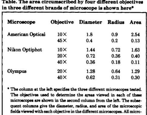
of interobserver error in the implementation of the criteria for the diagnosis of AD used by Tierney et al that required the quantitation of NFT and/or PL per X25 microscopic field. Objectives and oculars attached to different brands of microscopes may circumscribe dif- ferent viewing areas (ie, microscopic fields) despite being assigned the same magnification factor (aee table). Thus, the quantitation of “I’
and PL should be reported as the number of each per mmz rather than per magnification factor (ie, X 25). As shown in the table, the number of items counted with a 20 X objective will differ considerably depend- ing upon whether or not such counts are performed with a Nikon Optiphot (0.4 mm* viewing area) versus an Olympus (1.29 mmz view- ing area) microscope. This potential eource of error was considered in the NIA criteria for
AD*
and should be considered by others who propose to contrast the pathologic criteria for AD with respect to accuracy, sensitivity, and specificity.J.Q. Trojanowski, MD, PhD
M.L.
Schmidt, PhDPhiladelphia, PA
Table. The area circumacribed by four different objectiveo in three different brands of micrcwcope Ie shown here.
Microeoope Objective Diameter Radiue Area
American Optical 1OX 1.8 0.9 2.54
45
x
0.4 0.2 0.13Nikon Optiphot 10
x
1.44 0.72 1.63 20x
0.72 0.36 0.40 40 X 0.36 0.18 0.11O b P U 20
x
1.28 0.64 1.2940X 0.62 0.31 0.30
The column at the left specifies the three Merent microseopee hted.
The objective8 UMd to determine the apaa viewed in each of thsse
micmcojm am o h m in the Becond column from the left. The nubse- quent columns give the diameter, radius, and ane of the microampic fields viewed with each objective in the Merent microscopes. AU micro-
ecopea were equipped with 1OX oeulrs. T h e units for diameter and
radius were mm, and thcvs for men were mm? Note that objective8 with the came magni8cation fsctom on Merent micmcopm c-ribe Merant m.
Reply from the Authors: Dre. Trojanowski and Schmidt are of course quite correct in asserting that a X 25 microscopic field will vary according to the instrument used, and that therefore report3 of plaque and tangle frequency would be better based on some absolute unit such as 1 mmz.
In our study a Leitz Orthoplan microscope with a X 25 planapo- chromat objective was used, with a field diameter of 0.76 mm and aren
of 0.45 mm2. T o convert to a 1 mmz standard, our criteria of “one or more neurofib~illary tangles and one or more neuritic plaques per field” ought therefore to read “two or more neurofibrillary tangles and two or more neuritic plaques per mmz.” In other respects, our study appears to fulfill the procedural conditions laid down in the NIA criteria for AD.2
The letter from Drs. Trojanowski and Schmidt further underlines the need for universally accepted criteria for the pathologic diagnoeis of Alzheimer’s disease which we noted in our article.
Anthony J . Lewis, MD Mary C. Tierney, PhD Rory H . Fisher, MD Maria L. Zorzitto, MD W . Gary Snow, PhD David W . Reid, PhD
Paula Nieuwstraten, MD Toronto, Ontario. Canada
Referencee
1. TierneyMC.FisherRH.LewisAl,etal.TheNINCDS-ADRDA WorkGroup criteria for the clinical diagnosis of probable Alzheimer’s diaeaae: a clinicopathologic study of 57 caaes. Neurology 1988;38:359-364.
2. Khachaturian ZS. Diagnosis of Alzheimer’s disease. Arch Neurol 1985;421097-1105.
Optic neuritis and
MS
To the Editor: The recent article by Rizzo and Leeeel11 is in- formative but contains some unwarranted conclusions. The authors depend upon clinical criteria as well as the life-table analysis to arrive a t their conclusions. Previous clinical investigations have yielded few clues to indicate which patients with optic neuritis (ON) would de- velop clinical
MS.
Their cohort of white patients living in NewEngland raises issues of uncertainty as to the comparability of sub- jects and overrepreeentation of some geographic “hot epots,” and it neglects aspects of genetic factors. I
ale0
believe this study is seriously limited in its conclusions regarding augmented risk without the bene- fit of MRI data. The following personal study of 16 cases of ON, studied with MRI, suggests that this diagnostic tool offers potential predictive accuracy as to who may develop MS.Sixteen consecutive patients were diagnosed with ON. Four pa- tients (2 women, 2 men) displayed multiple high-signal intensity lesions in the periventricular matter on epin-echo sequences, consis- tent with disseminated plaque formation. AU these lesiom were clini- cally silent. No lesions were diecovered in the optic nerves. Within 6 months of diagnosis of ON, the four patients developed clinical MS. Of interest is the finding that 214 had positive family histories of several members with MS. CSF abnormalities were present in less than 50% of the entire series. One of the men with a positive family history was Hispanic.
MRI imaging appears to be the most sensitive indicator of de- myelination in clinical medicine. Because MS is such a common problem, with variability of expression, it is not surprising that Jacobs et al2 did not we any overt symptom of MS for a 2-year period after
onset of ON, whereas Rizzo and Lessell found over 50% conversions during the first 2 years. If a large series of cases with ON were studied prospectively with MRI, a spectrum of clinical expression would become apparent. Perhaps the number of demyelinative plaques seen on MRI is the key factor related to prognosis. Additionally, future protocols could address the issue of responsiveness to corticosteroida and prognosis.
Familial clusters of MS have been reported as occurring 5% of the time. McAlpine et al3 have calculated that first-degree relatives are at least 5 to 15 times at greater risk for developing
MS
than the general population. In view of the observation that 214 individuals had posi- tive family histories and became symptomatic within 6 months, it would seem reasonable to consider MRI as a useful noninvasive screening procedure for prognosis among family members. The issue of genetic aspects was not mentioned in the authors’ series.In conclusion, a long-term prospective study is needed with MRI and family screening data in order to accurately understand the close relationship between ON and MS.
Michael I . Weintraub,
MD
Croton-on-H&n, N YReply f r o m the Authors: Dr. Weintraub accurately statea that it would be unwarranted to indicate that our conclusions necessarily applied to
all populations. That is why we included a statement in
both the summary (first line) and in the text (first line, laat paragraph) that our patients were white and living in New England.The importance of genetic factors in the etiology of MS remains controversial. None of our patienta had a first-order relative with MS a t the time of their epiaode of ON, and thus the family history could not have helped predict which patient would develop MS.
Dr. Weintraub is troubled that we depended upon clinical criteria to diagnose MS and considers our study flawed by the lack of MFU
data. Of course, MRI was not available when the patients entered the study, so the issue of using MRI to predict which of our subjects would develop MS is moot. However, we reject the notion that MRI
has
been established as a means of unequivocally diagnosing MS. The articles by Paty et a14 and by Kurtzke5 in the same issue of Neurology addreea this topic directly. Paty prospectively comparea clinical eval- uation, evoked potentiah, oligoclonal banding, MRI, and CT in the diagnosis of MS and concludes that the “clinical diagnosis of MS. .
.,
even though only 95% accurate, must remain the gold standard.” Kurtzke concludes “Until we have a pathognomonic laboratory test. ifwe wish not to erase 120 yeare of clinical experience and start de moo
to characterize this illneas as a clinical entity, then I believe we must adhere to the clinical phenomena.” We agree.
Joseph Rizzo,
MD
Simmons Lessell,
MD
Boston, M AReferenoes
1. Rizzo JF, Lessell S. Risk of developing multiple sclerosis after uncompli- cated optic neuritis: a long-term prospective study. Neurology
2. Jacobs L. Kinkel PR, Kinkel WR. Silent brain leeions in patienta with isolated idiopathic optic neuritin. A clinical and nuclear magnetic reaonnnce imaging study. Arch Neurol1986,43:452-455.
3. McAlpine D, Lumsden CE, Acheson ED. Multiple sclerosis: a reappraisal. Edinburgh: Churchill-Livingatone, 1972.
4. Paty DW, Oger JJF, Kastrukoff LF. et al. MRI in the diagnosis of MS: a prospective study with comparison of clinical evaluation, evoked poten- tials, oligoclonal banding, and CT. Neurology 1988;38:180-185. 5. Kurtzke J F . Multiple sclerosis: what’s in a name? Neurology
1988;38185-190.
1988.38309-316.
Ptosis
and
Hering’s
law
To
the Editor: I read with interest the article entitled, “Unilateral ptosis and Hering’s law”l and wish to offer one comment. Dr. Lepore studied 21 patients with unilateral ptoais and found only one patient with contralateral superior lid retraction. Hering’s law states that there is bilateral equal and simultaneous innervation of synergistic extraocular muscles. By applying this law to the levator muscles. we would expect more patients in this study to have contralateral lid retraction.With respect to the analysis of lid and ocular motility, it is impor- tant to identify the fixatingeye. For example, a paretic left sixth nerve
will c a w a greater (secondary) deviation when the left eye fixates as
oppoeed to the right eye. Similarly, it matters whether the ptotic or fellow eye is fixating when comparing lid fissures. While there was “no change in ptoeis behind the occluder”’ (eye with ptosis is covered, fellow eye is fixating), no mention is made of ptosia behind the occluder with the fellow eye coveredand the ptotic eye fixating. Was it the failure to specify the fixating eye that led to the finding of only one in 21 patients with unilateral ptoeis who demonstrated lid retraction of the contralateral eye?
In addition, other exceptions to Hering’s law include d h i a t e d vertical deviations, unilateral nystagmus, convergence, and Bell’s phenomenon.
Mark S. Dresner,
MD
Bronx, N YReply from the Author: Dr. Dresner’s speculation that the pres- ence of lid retraction of the fellow eye in patients with unilateral ptosis may be underestimated ifthe ptotic eye is not allowed to fixate was not confirmed by my series of patients. As part of my “routine aesessment of ocular and lid motility,”’ alternate cover testing elicited no change of lid position of the fellow eye, ie, lid elevation with occlusion of the nonptotic eye or lid descent with occlusion of the ptotic eye was not observed. Of intereet, the one myasthenic patient who had unilateral lid retraction in my series did not demonstrate changing lid position on alternate cover testing. In contrast, Gay et al2 found that occlusion of the ptotic eye would lessen the lid retraction of the fellow eye in three patients and, although his finding doeen’t directly a d h Dr. Dreener’s contention that lid retraction will increase in the fellow eye when the ptotic eye fixates, it supports the premise that Hering’s law governs levator function.
Gay’s observations notwithstanding, Hering’s law is clinically manifest in very few c a m of unilateral ptoaie. Kansu aand Subutay3 recently observed upper lid retraction in only four of 150 patients with myasthenia gravis, and even questioned the assumption that Hering’s law accounts for the lid retraction in these few cases.
Dr. Dresner’s thoughtful question as well a~ his additional listing of ocular motility “culprits” that violate Hering’s law is greatly appre- ciated.
References
1. Lepore FE. Unilateral ptosis and Hering’s law. Neurology 1988,38319-322. 2. Gay AJ, Salmon ML, Windsor CE. Hering’s law, the levators. and their
3. Kansu T, Subutay N. Lid retraction in myasthenia gmvis. J Clin N e w relationship in dieease states. Arch Ophthalmol 1967;77:157-160.
Ophthalmol 198T7: 145- 148.
Thalamic aphasia
To the Editor: Tuszinski and Petito’ reported an interesting patient with aphasic speech disturbances due to a discrete left thalamic infarct proved a t autopsy. However, it is a pity that an incomplete review of the literature led them to believe that this was the first case of aphasia due to thalamic infarct verified a t autopsy. To our knowl- edge, at least three cases with clinical-pathologic correlation have been published pre~iously.*-~ All corresponded to a unilateral left thalamic infarct in the paramedian territory; the dorsomedial nucleus and the intralaminar formation were severely involved; the ventral lateral nucleus was only partially involved in two of them?.‘ and the pulvinar was spared. It can be argued that in the first two cases,23 other infarcts were also present; however, none was relevant to the speech disorder, except in Molnhr‘s patient, in whom alexia was present, and was probably related to infarction of the lingual and fusiform gym on the left side. In the third case, which we reported in 1986,4 the left thalamic infarct was isolated. Speech disturbances included reduced fluency, frequent pauses, decreased volume of the voice, verbal and phonemic paraphasias, mildly impaired comprehen- sion and spared repetition. Other neuropsychological disturbances included right-sided motor neglect and anterograde amnesia. After reviewing the cases of MolniW and Davous et a1,3 we suggested that involvement of the dorsomedial nucleus might be critical because of its connections with Broca’s area, Wernicke’s area, and the gyrus supramarginalis, whereas involvement of other nuclei such as the ventral lateral nucleus and pulvinar might be of less importance. Since we published our case, we have been able to further study the cerebral cortex of this patient using techniques for degenerated fibers.’ This study showed marked axonal degeneration in the premotor cortex, including Broca’s area, somatosensory cortex, Wernicke’s area, and gyrus supramarginalis on the left side. These data in mind, the case of Tuszinski and Petito may be considered a further confirmation that in thalamic infarction the dorsomedial nucleus is a critical structure for speech dysfunction, which might be related to a thalamic-cortical disconnection phenomenon, as P E T studies have sugge~ted.~
J. Bogowslavsky, MD J . Miklossy, MD J.P. Deruaz, M D F. Regli, MD Luusanne, Switzerland
Reply from the Authors: We thank Dr. Bogowlavsky et al for bringing to our attention their own case of thalamic aphasia as well as
two additional cases from the non-English literature. They review their previous hypothesis that thalamic aphasia is related to destruc- tion of the dorsomedial nucleus, and correctly point out that the anitomic location of the thalamic infarct in our patient further con- firms this hypothesis.
Clinical pathologic studies of thalamic aphasia, uncomplicated by other clinical symptoms and signs and by extrathalamic infarcts, are infrequent. Our particular case was useful in documenting the exis- tence of aphasia due to a discrete left thalamic infarct since, at the time of the patient’s presentation, symptomatology other than speech disturbances were absent. In contrast, other cases are complicated by multiple clinical signs and symptoms and infarcts at the time the patients present with aphasia.
Mark
H.
Twzynski, MD Son Diego, CACarol K . Petito, MD
New York. N Y
References
1. Tuszinski MH, Petito CK. Ischemic thalamic aphasia with pathologic con- firmation. Neurology 1988;38:800-802.
2. MolnAr L. Die lokal diagnostiscbe Bedeutung der vertikalen Blicklllhmung.
BeitrUge zur Symptomatologie und Faaeranatomie des meaodiencephalen Ubergangagebietes. Arch Psychiat Nervenkr 1958-59;198523-534. 3. Davow P, Bianco C. Duval-Lota AM, de Recondo J, Vedrenne C, Rondot P.
Aphasie par infarctw thalamique p a r a m k l i i gauche. Observation anatomo- clinique. Rev Neurol (Paria) 19&1,140:711-719.
4. Bogowlavsky J. Miklosey J, D e m z J P , Regli F, Assal G. Unilateral left paramedian infarction of thalamus and midbrain: a clinicopethological study. J N e w 1 Neuwurg Psychiatry 1986;49:686-694.
5. Baron JC, D’Antona R, Serdaru M, Pantano P, Boueser MG, Samson Y. Hypombtaboliime cortical a p h lQion thalamique chez I’homme: btude par la tomogmphie 1 positons. Rev Neurol (Paris) 1986;142:465-474.
Correction
The authors have indicated two corrections in the arti-
cle “Central pontine myelinolysis in severely burned
patients: Relationship
to serum hyperosmolality”
(McKee et al, 1988;381211-1217), which appeared in
the August issue. The formula on page
1212for calculat-
ing
serum
osmolality should have been:
Serum osmolality = 2([Na]
+
[K])+
glucose (mg/dI)/l8+
BUN (mg/d)/2.8.Also, on page 1214
inthe second paragraph, the sixth
sentence should read
“The gray matter
ofthe pons was
abnormal
in seven
patients” [emphasis added].
I
I