Examples of Weak, If Not Absent, Form-Function Relations in the
Vertebrate Heart
Bjarke Jensen and Theodoor H. Smit
University of Amsterdam, Amsterdam UMC, Department of Medical Biology, Amsterdam Cardiovascular Sciences, Meibergdreef 15, 1105AZ, Amsterdam, The Netherlands.
Corresponding author: [email protected]
Abstract: That form and function relates, is the maxim to anatomy and physiology. Yet form-function relations can be difficult to establish. Human subjects with excessive trabeculated myocardium in the left ventricle, for example, are diagnosed with non-compaction cardiomyopathy, but the extent of trabeculations may be without relation to ejection fraction. Rather than rejecting a relation between form and function, we may ask whether the salient function is assessed; is there a relation to electrical propagation, mean arterial blood pressure, propensity to form blood clots, or all? And how should extent of trabeculated muscle be assessed? While reviewing literature on trabeculated muscle, we applied Tinbergen’s four types of causation - how does it work, why does it work, how is it made, and why did it evolve - to better parse what is meant by form and function. The paper is structured around cases that highlight advantages and pitfalls of applying Tinbergen’s questions. It further uses the evolution of lunglessness in amphibians to argue lung reduction can impact on chamber septation, and it considers the evolution of an arterial outflow in fishes to argue that reductions in energy consumption may drive structural changes with little consequences to function. Concerning trabeculations, we argue they relate to pump function in the embryo in the couple of weeks before the onset of coronary circulation. In fetal and postnatal stages, a spectrum of trabeculated-to-compact myocardium makes no difference to cardiac function and in this period form and function may appear unrelated.
Key words: evolution; development; physiology; structure
INTRODUCTION
Four weeks into human embryonic development, a single vessel connects the forming pulmonary vasculature to the left atrium [1]. 11 weeks later, pulmonary venous tissue has been incorporated to the left atrium and 4 separate pulmonary veins now open to the left atrium [2]. This ontogenetic acquisition of veno-atrial connections varies between individuals and abnormal connections, for instance 3 or 5, occur in approximately 1 out of 4 people [3]. However, it is inconsequential to a person’s health whether there are 3, 4, or 5 pulmonary veins connecting to the left atrium [3], suggesting there is no relation between the normal variation in number of pulmonary veins, i.e. the form, and its function. Streaming of blood in the left atrium will of course be impacted on by the number of veins giving blood to the cavity, but is this not an unimportant functional relation if there are no consequences to whole organ and body performance?
The example of the number of pulmonary veins illustrates some of the pitfalls of establishing relation between form and function. First, at what level of biological organization should form-function relations be assessed; tissue, chamber, organ, organism, etc.? Second, which function to assess? Any structure of the body impacts on (parts of) the body and it is in principle possible to established a consequence to the presence of the structure. But consequence is different from adaptation in evolutionary biology. Adaptation is a trait that has been selected for by natural selection and thus relates directly or indirectly to the reproductive success of the organism [4]. Third, are we seeking proximal answers, such as how are left atrial blood streams affected by the number of pulmonary veins, or ultimate answers, such as why is the number of pulmonary veins variable?
When attempting to establish causality, Nobel laureate Niko Tinbergen suggested the application of 4 types of questions which are derivatives of the 4 categories of causes of Aristotle (given in parenthesis):
Mechanism (material cause), how does it work? Example: The heart pumps blood. Function (finale cause), why does it work?
Example: to drive perfusion of blood in the tissues? Ontogeny (formal cause), how is it made?
Example: the propulsion of blood through the tissues compensates for the increased diffusion distance between tissue and environment associated with greater body sizes.
August Krogh, also a Nobel laureate, proposed that for “a large number of problems there will be some animal of choice or a few such animals on which it can be most conveniently studied” [5]. For instance, concerning the biology around blood pressure, would one not want to study giraffes, the animal with the highest known systemic blood pressures [6]? In comparing different animal species, we may encounter evolutionary differences in mechanisms and functions, that, if understood correctly, can make us understand the efficient cause, why something became successful. As always, it is important that a functional advantage is not assumed beforehand. For example, Gould and Lewontin [4] emphasize that the question of “what did the Tyrannosaurus rex use its tiny front limbs for?” will likely receive an unsatisfactory and unfalsifiable answer. In contrast, we can reasonably answer “how did the Tyrannosaurus rex get its tiny front limbs” because the fossil record shows a conspicuous reduction of the front limbs concomitant with increments in the size of the hind limbs and head [4]. Viewed such, the reduction in the front limbs allows the prioritization of (energy to) the hind limbs and head. Similarly, we are not inclined to ponder the use of the tiny limbs of ancestral snakes or the pair of a claws next to the cloaca of pythons [7] because extant snakes abundantly show that life can be successful without limbs. It is rarely considered for the heart, however, whether there are vestigial features without function [8].
Confusion and poor reasoning, then, may arise from asking the wrong questions and ‘why’ questions can do us a disfavor by implying purpose. Or, as it is stated in the quote attributed to Ernst Wilhelm van Brücke “teleology is the mistress that the biologist cannot live without, but is too ashamed to be seen with in public”. Below, we will focus on trabeculated ventricular muscle because it exhibits more than one function – which is the salient one? - it shows ontogenetic changes – at what stages is it important? – and it shows phylogenetic changes – why is there a reduction of trabeculated muscle in the independent evolution of endothermy in mammals and birds?
CASES
The crocodilian heart has a full ventricular septum which distinguishes it from the hearts of all other ectothermic vertebrates (fishes, amphibians, and reptiles) (Figure 1A-B) [9, 10, 11]. The ventricular septum has a membranous part and a larger myocardial part which, like all cardiac muscle, propagates the electrical impulse and contracts upon electrical activation [11, 12]. Besides these two functions, the ventricular septum has at least 3 additional functional consequences on organ level. First, the blood pressure of the right ventricle can be substantially lower than the blood pressure of the left ventricle (Figure 1C) [13, 14, 15]. This allows for low blood pressures in the lung circulation, which in turn allows for a thinner blood-gas barrier at the respiratory epithelium [16]. Second, left-to-right shunting, the re-entry of pulmonary venous blood to the pulmonary circulation, which occurs in non-crocodylian reptiles, is anatomically impossible (Figure 1D) [11, 17]. The absence of shunting improves the efficacy of oxygen transport [18, 19]. Third, electrical activation spreads from the ventricular septum rather than from the base to the apex as in non-crocodylians (Figure 1E) [12, 20, 21].
Consequences of the ventricular septum can be established as above, but it is surprisingly difficult to ascertain its functional advantage besides the basic properties of myocardium (electrical propagation and contraction): The specialized manner of ventricular electrical activation does not shorten ventricular activation time as it does in mammals and birds [12]; oxygen consumption-dependent behaviors are not limited by the level of shunting in crocodylians and reptiles [19, 22, 23]; the rates of oxygen consumption in crocodylians are not higher than in lizards despite the thin blood-gas barrier [24, 25]. On the level of the organism, therefore, an advantage to the ventricular septum is not evident. It has then been proposed that the specializations of the crocodilian heart, including the ventricular septum, may have been selected for much earlier in evolution at a time, presumably, when crocodylians would have had much more active behaviors [26]. Although this conjecture is difficult to test, it does emphasize the possibility that the conditions are extinct in which the character provided an advantage to reproductive success.
Ontogeny (formal cause): when is form and function related?
We can postulate that for any structure, there is one or several stages in ontogeny when the form-function relation is the strongest {Richardson, 1999 #20238}. The term ‘immature’ implies this. In embryonic chicken, electrical propagation develops prior to cardiac contraction [31] and heart formation and pumping commences days before circulation of plasma/blood is necessary for development, as shown in embryos with ligated outflow tract [32]. Cessation of cardiac pumping by genetic perturbations in developing zebrafish and fruit flies have similarly shown that many early features of embryogenesis are not reliant of cardiac pumping [33, 34]. At least in embryogenesis, then, structures may develop before they provide a functional advantage to the organism.
Form unrelated to function: no final cause? Trabeculation of the human left ventricle
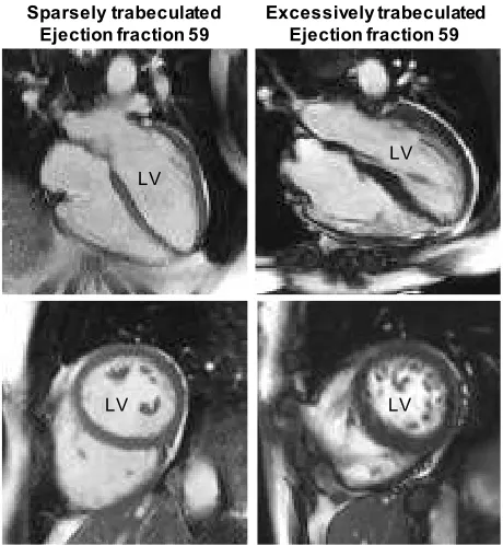
The human left ventricle is composed of a compact wall with a network of trabecular myocardium on its luminal side (Figure 3). Most prominent of the trabecular myocardium are the papillary muscles which anchor the atrioventricular [48]. The non-papillary trabecular network can be very meagre or extensive, and may make up between near-zero % to some 25% of the left ventricular mass (Figure 3) [49]. Ventricular wall architecture can be measured as the ratio of trabecular-to-compact wall and analysis of thousands of cohort participants have shown the ratio has a log-normal distribution in the population [43]. Surprisingly, the ratio is not related to functional measures like ejection fraction and blood pressure [50, 51], or so poorly it is deemed clinically irrelevant [43]. Even in cases of excessive trabeculation, which can be diagnosed as left ventricular non-compaction cardiomyopathy [52, 53], there is no, or only very poor, relation between the extent of trabecular myocardium and function [49, 51]. Further, adverse outcomes like sudden cardiac death also appear unrelated to the extent of trabecular myocardium, but instead to ventricular dilation and fibrosis [43, 50, 54]. It therefore appears that there is no functional relation to the trabecular-to-compact wall architecture on the level of organ and individual in adult human [55]. This conjecture is supported by the meta-analytical finding that systemic blood pressure is similar across mammal phylogeny and body size [56], despite a substantial variation in the extent of trabeculations [57].
Figure 3. The architecture of the human left ventricle (LV) appears to have no impact on function. Normal ejection fractions (stroke volume/end-diastolic volume) occur in ‘Sparsely trabeculated’ and ‘Excessively
Sparsely trabeculated Ejection fraction 59
Excessively trabeculated Ejection fraction 59
LV LV
trabeculated’ ventricles alike. Top row shows so-called 4-chamber views, the bottom row shows transverse views of the two ventricles. Note the much more numerous and extensive trabeculations in the images on the right. Images from published data [41].
Relation of form and function in evolution (efficient cause)? Ventricular trabeculations in vertebrates
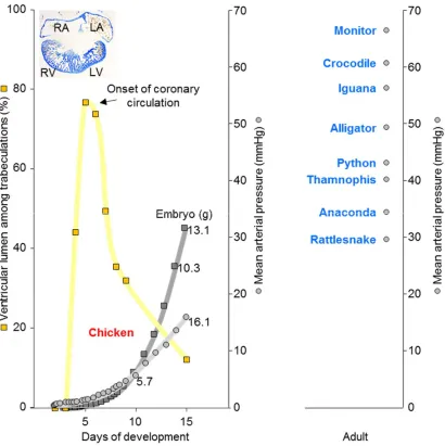
The inverse relation of trabeculated wall architecture and blood pressure between 6 and 15 days of development in chicken shown in Figure 2, suggests that coronary circulation favors compact wall growth and causes a decrement in the proportion of trabeculated myocardium. This could be the case in the ontogeny of mammals and birds. Coronary vasculature is found in most ectotherms and can be found within the trabeculated myocardium of the extensively trabeculated ventricles [58, 59, 60]. This comparative analysis suggests that coronary vascularization is a necessary condition for the prioritization of the compact architecture over the trabeculated architecture, but not a sufficient condition. In fact, the adaptive value of compact architecture over the trabeculated architecture is not clear, i.e. the functional advantage (final cause) and explanation for its evolution (efficient cause).
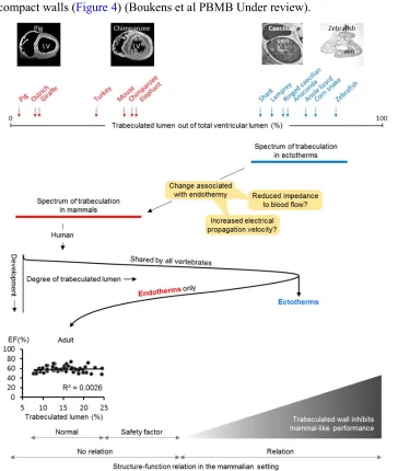
that characterizes endotherms (Figure 4) [65]. Higher heart rates allow for greater cardiac outputs and therefore higher blood pressure, thus making the compact wall architecture linked to blood pressure via heart rates. Also, the faster chamber activation of ectotherms is not explained by higher body temperatures and conduction systems only, suggesting a role for compact walls (Figure 4) (Boukens et al PBMB Under review).
ectotherm-like, appear incompatible with life in endotherms [66]. This suggest that the extent of trabeculated myocardium in endotherms will have a normal range, a safety factor range where excessive extent of trabeculated myocardium is not pathological, and an ectotherm-like range which will be detrimental to pump-function, possibly because of impairment of filling and emptying. Partly based on and adapted from [41].
Can a reduction in energy expenditure have a structural consequence? The outflow tract of the fish heart
In the evolution of teleost fish there was a change to the arterial pole, whereby the myocardial outflow tract, the conus arteriosus, disappeared and a pear-shaped arterial outlet, the bulbus arteriosus, took its place (Figure 5) [67]. It is debated whether the bulbus of teleost fish should be considered a modification of an older structure or an evolutionary novelty, but the bulbus is evidently much more developed in teleosts than in non-teleosts [68, 69]. A recent study shows in embryonic teleost fish that the myocardial outflow tract undergoes reprogramming to an arterial phenotype mediated by elnb [70]. Despite the advances in understanding the phylogenetic appearance, the ontogenetic change, and the mechanism behind it, the functional advantage of an arterial bulbus over a myocardial conus remains elusive. Both the bulbus and conus work as a pressure and flow reservoir that ensures blood flow in the diastolic interval between ventricular ejections [71, 72].
that the adaptive value of the bulbus is a low energy demand. If so, the form and function of the bulbus are then secondary consequences.
Figure 5. Arterial pole of fishes. The heart (158g) of a 304cm long Bigeye thresher (Alopias superciliosus), a shark, had a myocardial outflow tract (Conus, ventral view) that comprised 8.9% of the cardiac mass. In the insert, the conus has been isolated, cut longitudinally, and folded out, exposing its 3 rows of valve leaflets (1-3, 1st to 3rd row of valve leaflets). The heart of an approximately 50kg Swordfish (Xiphias gladius), a teleost fish, had an arterial outflow tract (Bulbus, view from the right). In the insert, the interior is exposed of the left half of the bulbus showing its thick arterial wall (1, only 1 row of valve leaflets). The categorization of metabolism as being high or low is based on values for mammals [74, 75].
Lungs, then heart
Can loss of the lungs impact on cardiac septation?
bridge of mesenchyme between the developing atria and the pharyngeal mesoderm in which the lungs develop (Jensen et al In press Anat Rec). The dorsal mesocardium projects into the atrial lumen as the dorsal mesenchymal protrusion, which is necessary for the closure of the primary foramen between the left and right atrial cavity [83, 84]. Accordingly, a poorly developed dorsal mesenchymal protrusion leads to atrial septal defects [85]. Further, the pharyngeal mesoderm also contributes cells to the arterial pole of the heart [86]. These observations allow for the conjecture that the reduced cardiac septation of lungless salamanders is a direct consequence of the reduced development of the pharyngeal mesoderm [82], rather than an adaptation related to intracardiac flow patterns.
SYNTHESIS
We have attempted to apply the four causes of Aristotle that Tinbergen adapted to biology. Our motivation came from the confusion and conflicting views concerning the role of trabeculated muscle in vertebrate hearts. ‘Function’ is a concept that often implies purpose, and teleology may exacerbate the confusion. The application of the four causes, we hope, has revealed that function can be interpreted in several ways. In part, this is because function may be assessed under conditions that are not conducive to test a particular form-function relation. While our cases are based mostly in evolutionary biology, we would argue that there is relevance for medicine: Left ventricular non-compaction cardiomyopathy is characterized as a distinct form of cardiomyopathy by the American Heart Association and is diagnosed when trabeculated myocardium is excessive [52, 53]. (Non-compaction cardiomyopathy has also been reported for a domestic cat [87]). Yet in patients and healthy subjects, left ventricular function does not correlate with the extent of trabeculated myocardium and the diagnostic criteria have very poor sensitivity [50, 51, 54, 88]. These observations suggest that form and function are not related in human when we consider trabeculated myocardium. As argued above, however, when we consider all life stages, a credible argument can be made that function is tightly related to trabeculated muscle in the embryonic heart before coronary circulation is established. Therefore, the extent of trabeculated muscle is related to function, but only in certain life stages. If this conjecture is true, it may be futile to use and develop morphometrics to identify true LVNC cases from the general population. Functional assessment remains the crucial readout.
ask whether there could be salient functions that have not been characterized? Secondly, which of the known functions should be assessed in order to identify the adaptive value of a structure, or should all functions be assessed? In this context, it is interesting to note that considerable effort has been spend on showing the adaptive value of right-to-left shunting in crocodylians, without a clear-cut adaptive value being shown [19].
In most instances, we can rightly assume a structural change to the heart relates to a functional change. However, as argued for the case of lunglessness in amphibians, it is entirely conceivable that a primary adaptation, lunglessness, induces a reduction of cardiac septation (in the atria and the outflow tract) because of developmental changes to the mesoderm that gives rise to both lungs and cells of the septae. That is, if cardiac changes are seen in isolation, we may miss the primary adaptation. It also follows, that it may be futile to assess the functional advantage of the reduction of the septae, because the advantage may not be there anymore. Similar concerns for establishing form-function relations can be expressed when trying to understand the significance of the myocardial to arterial identity change of the outflow tract of fishes. Possibly, the primary adaptation relates to energy consumption, with form-function relations being secondary.
References
1 Sizarov A, Anderson RH, Christoffels VM, et al. Three-dimensional and molecular
analysis of the venous pole of the developing human heart. Circulation 2010;122:798-807.
2 Webb S, Kanani M, Anderson RH, et al. Development of the human pulmonary vein
and its incorporation in the morphologically left atrium. Cardiol Young 2001;11:632-42.
3 Kato R, Lickfett L, Meininger G, et al. Pulmonary vein anatomy in patients undergoing
catheter ablation of atrial fibrillation: lessons learned by use of magnetic resonance
imaging. Circulation 2003;107:2004-10.
4 Gould SJ, Lewontin RC. The spandrels of San Marco and the Panglossian paradigm: a
critique of the adaptationist programme. ProcRSocLond 1979:581-98.
5 Krogh A. The progress of physiology. AmJPhysiol 1929:243-51.
6 Smerup M, Damkjaer M, Brondum E, et al. The thick left ventricular wall of the
giraffe heart normalises wall tension, but limits stroke volume and cardiac output. J Exp Biol
2016;219:457-63.
7 Martill DM, Tischlinger H, Longrich NR. EVOLUTION. A four-legged snake from the
Early Cretaceous of Gondwana. Science 2015;349:416-9.
8 Jensen B, Vesterskov S, Boukens BJ, et al. Morpho-functional characterization of the
systemic venous pole of the reptile heart. Sci Rep 2017;7:6644.
9 Greil A. Beitrage zur vergelichenden anatomie und entwicklungsgeschichte des
herzens und des trauncus arteriosus der wirbelthiere. MorphJahrbuch 1903;31:123-310.
10 Webb GJW. Comparative cardiac anatomy of the reptilia III. The heart of crocodilians
and an hypothesis on the completion of the interventricular septum of crocodilians and
birds. JMorph 1979;161(2):221-40.
11 Cook AC, Tran VH, Spicer DE, et al. Sequential segmental analysis of the crocodilian
heart. J Anat 2017.
12 Jensen B, Boukens BJ, Crossley DA, et al. Specialized impulse conduction pathway in
the alligator heart. Elife 2018;7.
13 White FN. Central vascular shunts and their control in reptiles. FedProc
1970;29:1149-53.
14 Grigg GC, Johansen K. Cardiovascular Dynamics in Crocodylus-Porosus Breathing Air
and During Voluntary Aerobic Dives. Journal Of Comparative Physiology B-Biochemical
Systemic And Environmental Physiology 1987;157:381-92.
15 Axelsson M. The crocodilian heart; more controlled than we thought? Experimental
Physiology 2001;86:785-9.
16 Perry SF. Gas exchange strategy in the Nile crocodile: a morphometric study.
JCompPhysiolB 1990;159:761-9.
17 Webb GJW. Comparative cardiac anatomy of the reptilia. III. The heart of
crocodilians and an hypothesis on the completion of the interventricular septum of
crocodilians and birds. Journal of Morphology 1979;161:221-40.
18 Axelsson M, Franklin CE. From Anatomy to Angioscopy: 164 Years of Crocodilian
Cardiovascular Research, Recent Advances, and Speculations. Comparative Biochemistry and
Physiology Part A: Physiology 1997;118:51-62.
19 Hicks JW, Wang T. The Functional Significance of the Reptilian Heart: New Insights
into an Old Question. In: Sedmera D, Wang T, eds. Ontogeny and Phylogeny of the
20 Christian E, Grigg GC. Electrical activation of the ventricular myocardium of the crocodile Crocodylus johnstoni: a combined microscopic and electrophysiological study. Comp BiochemPhysiol A MolIntegrPhysiol 1999;123:17-23.
21 Jensen B, Boukens BJ, Postma AV, et al. Identifying the evolutionary building blocks
of the cardiac conduction system. PLoS One 2012;7:e44231.
22 Eme J, Gwalthney J, Owerkowicz T, et al. Turning crocodilian hearts into bird hearts:
growth rates are similar for alligators with and without right-to-left cardiac shunt. J Exp Biol
2010;213:2673-80.
23 Leite CAC, Taylor EW, Wang T, et al. Ablation of the ability to control the right-to-left
cardiac shunt does not affect oxygen consumption, specific dynamic action or growth in
rattlesnakes, Crotalus durissus. The Journal of Experimental Biology 2013;216:1881-9.
24 Gleeson TT, Mitchell GS, Bennett AF. Cardiovascular responses to graded activity in
the lizards Varanus and Iguana. AmJPhysiol 1980;8(1):R174-R9.
25 Farmer CG, Carrier DR. Ventilation and gas exchange during treadmill locomotion in
the American alligator (Alligator mississippiensis). JExpBiol 2000;203 Pt 11:1671-8.
26 Seymour RS, nett-Stamper CL, Johnston SD, et al. Evidence for endothermic
ancestors of crocodiles at the stem of archosaur evolution. Physiol BiochemZool
2004;77:1051-67.
27 Jensen B, Larsen CK, Nielsen JM, et al. Change of cardiac function, but not form, in
postprandial pythons. Comp BiochemPhysiol A MolIntegrPhysiol 2011;160:35-42.
28 Jensen B, Wang T. Hemodynamic Consequences of Cardiac Malformations in Two
Juvenile Ball Pythons (Python regius). Journal of Zoo and Wildlife Medicine: American
Association of Zoo Veterinarians 2009:752-6.
29 Jensen B, Nielsen JM, Axelsson M, et al. How the python heart separates pulmonary
and systemic blood pressures and blood flows. J Exp Biol 2010;213:1611-7.
30 Brücke E. Beiträge zur vergleichenden Anatomie und Physiologie des
Gefäss-Systemes. Denkschriften der kaiserliche Akademie der Wissenschaften -
Mathematisch-Naturwissenschaftliche Classe 1852;3:335-67.
31 Hirota A, Sakai T, Fujii S, et al. Initial development of conduction pattern of
spontaneous action potential in early embryonic precontractile chick heart. Dev Biol
1983;99:517-23.
32 Burggren WW. What is the purpose of the embryonic heart beat? or how facts can
ultimately prevail over physiological dogma. Physiological And Biochemical Zoology
2004;77:333-45.
33 Stainier DY, Weinstein BM, Detrich HW, 3rd, et al. Cloche, an early acting zebrafish
gene, is required by both the endothelial and hematopoietic lineages. Development
1995;121:3141-50.
34 Bodmer R. The gene tinman is required for specification of the heart and visceral
muscles in Drosophila. Development 1993;118:719-29.
35 Sedmera D. Function and form in the developing cardiovascular system.
CardiovascRes 2011;91:252-9.
36 Menendez-Montes I, Escobar B, Palacios B, et al. Myocardial VHL-HIF Signaling
Controls an Embryonic Metabolic Switch Essential for Cardiac Maturation. Dev Cell
2016;39:724-39.
37 Grego-Bessa J, Luna-Zurita L, del Monte G, et al. Notch signaling is essential for
38 Sedmera D, Reckova M, DeAlmeida A, et al. Spatiotemporal pattern of commitment to slowed proliferation in the embryonic mouse heart indicates progressive differentiation
of the cardiac conduction system. Anat Rec A DiscovMolCell EvolBiol 2003;274:773-7.
39 Sizarov A, Ya J, de Boer BA, et al. Formation of the building plan of the human heart:
morphogenesis, growth, and differentiation. Circulation 2011;123:1125-35.
40 de Boer BA, van den Berg G, de Boer PA, et al. Growth of the developing mouse
heart: An interactive qualitative and quantitative 3D atlas. Dev Biol 2012;368:203-13.
41 Jensen B, Agger P, de Boer BA, et al. The hypertrabeculated (noncompacted) left
ventricle is different from the ventricle of embryos and ectothermic vertebrates. Biochim
Biophys Acta 2016;1863:1696-706.
42 Gati S, Papadakis M, Papamichael ND, et al. Reversible de novo left ventricular
trabeculations in pregnant women: implications for the diagnosis of left ventricular
noncompaction in low-risk populations. Circulation 2014;130:475-83.
43 Anderson RH, Jensen B, Mohun TJ, et al. Key Questions Relating to Left Ventricular
Noncompaction Cardiomyopathy: Is the Emperor Still Wearing Any Clothes? Can J Cardiol
2017.
44 de Bakker BS, de Jong KH, Hagoort J, et al. An interactive three-dimensional digital
atlas and quantitative database of human development. Science 2016;354.
45 Van Mierop LH, Bertuch CJ, Jr. Development of arterial blood pressure in the chick
embryo. AmJ Physiol 1967;212:43-8.
46 Enok S, Slay C, Abe AS, et al. Intraspecific scaling of arterial blood pressure in the
Burmese python. J Exp Biol 2014;217:2232-4.
47 Jensen B, Moorman AF, Wang T. Structure and function of the hearts of lizards and
snakes. BiolRevCambPhilosSoc 2014;89:302-36.
48 Spicer DE, Bridgeman JM, Brown NA, et al. The anatomy and development of the
cardiac valves. Cardiol Young 2014;24:1008-22.
49 Amzulescu MS, Rousseau MF, Ahn SA, et al. Prognostic Impact of Hypertrabeculation
and Noncompaction Phenotype in Dilated Cardiomyopathy: A CMR Study. JACCCardiovasc
Imaging 2015.
50 Zemrak F, Ahlman MA, Captur G, et al. The Relationship of Left Ventricular
Trabeculation to Ventricular Function and Structure Over a 9.5-Year Follow-UpThe MESA
Study. Journal of the American College of Cardiology 2014;64:1971-80.
51 Weir-McCall JR, Yeap PM, Papagiorcopulo C, et al. Left Ventricular Noncompaction:
Anatomical Phenotype or Distinct Cardiomyopathy? J Am Coll Cardiol 2016;68:2157-65.
52 Finsterer J, Stollberger C, Towbin JA. Left ventricular noncompaction
cardiomyopathy: cardiac, neuromuscular, and genetic factors. Nat Rev Cardiol 2017.
53 Towbin JA, Jefferies JL. Cardiomyopathies Due to Left Ventricular Noncompaction,
Mitochondrial and Storage Diseases, and Inborn Errors of Metabolism. Circ Res
2017;121:838-54.
54 Andreini D, Pontone G, Bogaert J, et al. Long-Term Prognostic Value of Cardiac
Magnetic Resonance in Left Ventricle Noncompaction: A Prospective Multicenter Study. J
Am Coll Cardiol 2016;68:2166-81.
55 Aung N, Zemrak F, Petersen SE. Left Ventricular Noncompaction, or Is It? J Am Coll
Cardiol 2016;68:2182-4.
56 Poulsen CB, Wang T, Assersen K, et al. Does mean arterial blood pressure scale with
body mass in mammals? Effects of measurement of blood pressure. Acta Physiol (Oxf)
57 Rowlatt U. Comparative Anatomy of the Heart of Mammals. Zoological Journal Of The Linnean Society 1990;98:73-110.
58 Jensen B, Nyengaard J, Pedersen M, et al. Anatomy of the python heart. Anatomical
Science International 2010;85:194-203.
59 Kohmoto T, Argenziano M, Yamamoto N, et al. Assessment of Transmyocardial
Perfusion in Alligator Hearts. Circulation 1997;95:1585-91.
60 Cox GK, Kennedy GE, Farrell AP. Morphological arrangement of the coronary
vasculature in a shark (Squalus sucklei) and a teleost (Oncorhynchus mykiss). J Morphol
2016;277:896-905.
61 Benninghoff A. Über die Beziehungen des Reizleitungssystems und der
papillarmuskeln zu den Konturfasern des Herzschlauches. Anat Anz 1923;57:185-208.
62 Burggren W, Farrell A, Lillywhite H. Vertebrate Cardiovascular Systems.
Comprehensive Physiology: John Wiley & Sons, Inc. 2010:215-308.
63 Jensen B, Wang T, Christoffels VM, et al. Evolution and development of the building
plan of the vertebrate heart. BiochimBiophysActa 2013;1833:783-94.
64 Johansen K, Martin AW. Circulation in a giant earthworm, Glossoscolex giaganteus I.
Contractile processes and pressure gradients in the large blood vessels. JExpBiol
1965;43:333-47.
65 Burggren WW, Christoffels VM, Crossley DA, et al. Comparative cardiovascular
physiology: future trends, opportunities and challenges. Acta Physiologica 2014;210:257-76.
66 Rhee S, Chung JI, King DA, et al. Endothelial deletion of Ino80 disrupts coronary
angiogenesis and causes congenital heart disease. Nat Commun 2018;9:368.
67 Icardo JM. Heart Morphology and Anatomy. Fish Physiology 2017;36:1-54.
68 Grimes AC, Kirby ML. The outflow tract of the heart in fishes: anatomy, genes and
evolution. J Fish Biol 2009;74:983-1036.
69 Lorenzale M, Lopez-Unzu MA, Rodriguez C, et al. The anatomical components of the
cardiac outflow tract of chondrichthyans and actinopterygians. Biol Rev Camb Philos Soc
2018.
70 Moriyama Y, Ito F, Takeda H, et al. Evolution of the fish heart by
sub/neofunctionalization of an elastin gene. Nat Commun 2016;7:10397.
71 Satchell GH. Physiology and form of fish circulation. Cambridge University Press
1991.
72 Farrell AP, Smith F. Cardiac Form, Function and Physiology. Fish Physiology
2017;36:155-264.
73 Gregory JA, Graham JB, Cech JJ, Jr., et al. Pericardial and pericardioperitoneal canal
relationships to cardiac function in the white sturgeon (Acipenser transmontanus). Comp
BiochemPhysiol A MolIntegrPhysiol 2004;138:203-13.
74 Mootha VK, Arai AE, Balaban RS. Maximum oxidative phosphorylation capacity of the
mammalian heart. Am J Physiol 1997;272:H769-75.
75 Tsai AG, Friesenecker B, Mazzoni MC, et al. Microvascular and tissue oxygen
gradients in the rat mesentery. Proc Natl Acad Sci U S A 1998;95:6590-5.
76 Seth H, Axelsson M, Grans A. The peptide hormone cholecystokinin modulates the
tonus and compliance of the bulbus arteriosus and pre-branchial vessels of the rainbow
trout (Oncorhynchus mykiss). Comp Biochem Physiol A Mol Integr Physiol 2014;178:18-23.
77 Hillman SS, Hedrick MS. A meta-analysis of in vivo vertebrate cardiac performance:
implications for cardiovascular support in the evolution of endothermy. J ExpBiol
78 Clark TD, Sandblom E, Hinch SG, et al. Simultaneous biologging of heart rate and acceleration, and their relationships with energy expenditure in free-swimming sockeye
salmon (Oncorhynchus nerka). J Comp Physiol B 2010;180:673-84.
79 Kaeriyama M, Nakamura M, Yamaguchi M, et al. Feeding ecology of sockeye and
pink salmon in the Gulf of Alaska. N Pac Anadr Fish Comm Bull 2000;2:55-63.
80 Wilkinson M, Nussbaum RA. Comparative morphology and evolution of the lungless
caecilian Atretochoana eiselti (Taylor) (Amphibia: Gymnophiona: Typhlonectidae). Biological
Journal Of The Linnean Society 1997;62:39-109.
81 Wake DB, Hanken J. Direct development in the lungless salamanders: what are the
consequences for developmental biology, evolution and phylogenesis? Int J Dev Biol
1996;40:859-69.
82 Lewis ZR, Hanken J. Convergent evolutionary reduction of atrial septation in lungless
salamanders. J Anat 2017;230:16-29.
83 Briggs LE, Kakarla J, Wessels A. The pathogenesis of atrial and atrioventricular septal
defects with special emphasis on the role of the dorsal mesenchymal protrusion. Differentiation 2012;84:117-30.
84 Mommersteeg MT, Soufan AT, de Lange FJ, et al. Two distinct pools of mesenchyme
contribute to the development of the atrial septum. Circ Res 2006;99:351-3.
85 Xie L, Hoffmann AD, Burnicka-Turek O, et al. Tbx5-hedgehog molecular networks are
essential in the second heart field for atrial septation. Dev Cell 2012;23:280-91.
86 van den Berg G, Abu-Issa R, de Boer BA, et al. A caudal proliferating growth center
contributes to both poles of the forming heart tube. CircRes 2009;104:179-88.
87 Kittleson MD, Fox PR, Basso C, et al. Naturally Occurring Biventricular
Noncompaction in an Adult Domestic Cat. J Vet Intern Med 2017;31:527-31.
88 Ivanov A, Dabiesingh DS, Bhumireddy GP, et al. Prevalence and Prognostic
Significance of Left Ventricular Noncompaction in Patients Referred for Cardiac Magnetic