1
Association Between Serum Selenium Concentrations and Levels of Proinflammatory
and Profibrotic Cytokines - Interleukin-6 and Growth Differentiation Factor-15, in
Patients with Alcoholic Liver Cirrhosis
Andrzej Prystupa1, Paweł Kiciński2, Dorota Luchowska-Kocot3†, Anna Błażewicz4†, Jarosław Niedziałek5, Grzegorz Mizerski2, Mariusz Jojczuk6, Andrzej Ochal6, Jarosław J. Sak7,*, Wojciech Załuska8
1 Department of Internal Medicine, Medical University of Lublin, Staszica 16, 20-081 Lublin,
Poland, email: [email protected]; [email protected]
2 Department of Family Medicine, Medical University of Lublin, Staszica 11, 20-081 Lublin,
Poland, email: [email protected]
3 Department of Medical Chemistry, Medical University of Lublin, 20-093 Lublin, Chodźki 4a,
Poland, email: [email protected]
4 Department of Analytical Chemistry, Medical University of Lublin, Chodźki 4a (Collegium
Pharmaceuticum), 20-093 Lublin, Poland, email: [email protected]
5 Individual Medical Practice, Lublin, Ludwika Hirszfelda 5/11, 20-092 Lublin, Poland, email:
6Department of Trauma Surgery and Emergency Medicine, Medical University of Lublin,
Staszica 16, 20-081 Lublin, Poland, e-mail: [email protected]; [email protected]
7 Department of Ethics and Human Philosophy, Medical University of Lublin, Staszica 4/6
(Col-legium Maximum), 20-059 Lublin, Poland, email: [email protected]
8 Department of Nephrology, Medical University of Lublin, Jaczewskiego 8, 20-954 Lublin,
Poland, email: [email protected] * Correspondence: [email protected]
2
Abstract: According to some authors, the serum selenium level is strongly associated with the severity of liver diseases including liver cirrhosis. The aim of the study was to determine the relationship between the concentration of selenium and pro-inflammatory and profibrotic cytokines – interleukin-6 (IL-6) and growth differentiation factor 15 (GDF-15) in patients with alcoholic liver cirrhosis. The parameters studied were determined in serum of 99 alcoholic liver cirrhosis patients divided based on the severity of disease according to the Child-Turcotte-Pugh criteria. In patients with liver cirrhosis, the serum selenium concentration was statistically lower whereas serum IL-6 and GDF-15 concentrations were higher than those in the control group. Moreover, the concentration of selenium negatively correlated with the levels of GDF-15 and IL-6. The above results may indicate a role of selenium deficiency in the pathogenesis and progression of alcoholic liver disease.
3
1. Introduction
Alcohol remains a major cause of liver disease worldwide. Alcohol liver cirrhosis includes medical conditions ranging from simple steatosis to decompensatedcirrhosis[1] and is the twelfth cause of deaths in the United States with an age–adjusted death rate of 9.6 per 100,000 population[2]. The precise molecular pathways of the initiation and progression of alcohol-induced liver tissue injury are not fully understood, but some authors suggest that alcohol toxicity to organs is connected with the generation of oxidative and non-oxidative ethanol metabolites and the translocation of gut-derived endotoxin into the bloodstream. These processes lead to cellular injury and stimulation of inflammatory responses releasing a great variety of cytokines.Continuation of alcohol abuse progresses the injury through impairment of tissue regeneration and extracellular matrix turnover, leading to fibrogenesis and cirrhosis[3]. Selenium, an essential trace mineral for humans, is delivered to the body mainly with food and water. Selenium has antioxidant properties and is a cofactor of many enzymes, including glutathione peroxidase. Currently, this element is considered to be an anticancer agent that prevents the processes of cell proliferation and tumor growth [4]. According to some authors, the serum selenium level is strongly associated with the severity of liver damage[5,6]. However, it should be emphasized that both the excess and deficiency of selenium exert adverse effects The aim of the study was to determine the relationship between the concentration of selenium and pro-inflammatory and profibrotic cytokines – interleukin-6 (IL-6) and growth differentiation factor 15 (GDF-15) in patients with alcoholic liver cirrhosis.
2. Experimental Section
2.1. Patients
4 The subjects with viral and autoimmune diseases were excluded from the study. Liver cirrhosis was diagnosed based on clinical features, history of heavy alcohol consumption, laboratory tests and abdominal ultrasonography. The patients with alcoholic hepatitis were excluded. The degree of liver cirrhosis was evaluated according to the Child-Turcotte-Pugh criteria (Child-Pugh score)[7]. Based on them patients were assigned to one of three groups: 1st – P-Ch A – 29
with stage A , 2nd – P-Ch B – 26 with stage B and 3rd – P-Ch C – 34 with stage C of liver
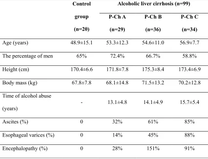
cirrhosis. The control group consisted of 20 healthy individuals without liver disease who did not abuse alcohol. None of the patients and healthy participants received mineral supplements. Both clinical assessment and laboratory tests were used to exclude the underlying liver diseases in the control group. There were no significant age- or gender-related differences in the subgroups (Table 1). Detailed demographic, clinical and biochemical characteristics of patients are presented in Table 1 and 2. The study protocol was approved by the Bioethics Committee at the Medical University of Lublin, Poland (agreement number KE-0254/349/2015). All subjects gave their written informed consent for participation in the study.
2.2. Human serum samples
The studied material consisted of 119 samples of human serum. Samples were taken from healthy individuals and patients with alcoholic liver cirrhosis and analyzed in the same way. They were transported and stored in polypropylene containers. The digestion was carried out in the NovaWAVE Microwave Tunnel Digestion System (SCP Science, Canada) using Teflon®
vessels. The microwave – assisted sample preparation was conducted in a closed system. Each time an acidic digestion with 65 % nitric acid water solution was applied (1mL of HNO3 : 9 mL
of deionized H2O). The optimized conditions of the mineralization procedure had been
5
2.3. Instrumentation and Reagents
After mineralization, each human serum sample was analyzed at least in triplicate using a high-resolution atomic absorption spectrometer. The measurements were performed with the ContrAA700 high-resolution continuum source graphite tube AAS instrument (Analytik Jena AG, Jena, Germany). A transversely heated graphite furnace was used as an atomizer. The parameters were set as follows: wavelengths - 196.0267 nm, pyrolysis temperature - 9500C,
atomization temperature- 19000C, and 5 uL Pd(NO3)2 (0.1%Pd) as a modifier. The
concentration of a stock solution (100 µg/L Se in 1 % HNO3) was prepared.
The method accuracy was verified by the use of Seronorm™ Trace Elements Serum L-2 (Billingstad, Norway) human serum certified reference material. The average recovery for five separate determinations was 97.8 %. The limit of detection (3σ) was estimated to be 2.00 µg/L. All reagents used were of at least analytical grade. Water with a resistivity of 18.2 MΩ cm was deionized in a Milli-Q system (Millipore, Bedford, MA, USA); 65 % nitric acid solution and other stock solutions were purchased from Sigma- Aldrich, Germany.
6 The serum growth differentiation factor 15 was determined applying Human GDF-15 ELISA sandwich enzyme immunoassay (BioVendor, Czech Republic) for the quantitative measurement of human GDF-15/MIC-1 (growth differentiation factor 15/macrophage inhibitory cytokine 1). The standards and samples were incubated in microtitrate wells pre-coated with polyclonal anti-human GDF-15 antibody. After incubation, biotin labeled polyclonal anti-human GDH was added. The further procedure (addition of streptavidin-conjugated to HRP and substrate solution, etc.) was analogous to the human IL-6 assays.
2.4. Statistical analysis
STATISTICA 12 PL was used for data analysis. Continuous variables were expressed as mean ± standard deviation (SD). Before calculations, variables were checked for normality using the Shapiro-Wilk test; the Brown-Forsythe test was applied to test equality of variances. To compare continuous variables between two groups (the control and the study group), the Mann-Whitney test was used; for more than two groups, the Kruskal-Wallis rank test was used, a nonparametric equivalent of ANOVA. The Dunn test was applied for detailed identification of statistically different groups. Correlations among variables were tested using Spearman’s rank correlation. Qualitative variables were shown as indicators of structures and compared using the χ2 test. For all tests, p< 0.05 was considered as statistically significant.
3. Results
3.1. Serum selenium concentration in patients with alcoholic liver cirrhosis compared to the
control group.
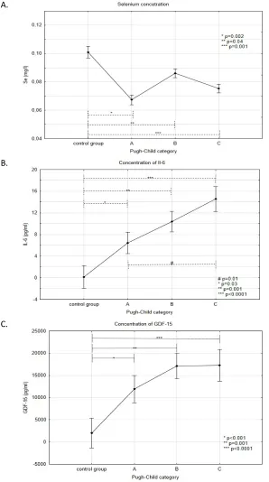
7 between the control group and groups with various stages of liver cirrhosis (p values were respectively 0.002, 0.04 and 0.001 for the control group vs. A, B and C Child-Pugh). However, no statistically significant differences in the concentrations of Se were demonstrated comparing particular groups of liver cirrhosis.
3.2. Levels of interleukin-6 in patients with alcoholic liver cirrhosis
The highest concentration of interleukin-6 was observed in patients with decompensated cir-rhosis (14.55±12.51 pg/ml in stage C and 10.38 ± 9.96 pg/ml in stage B) whereas the lowest one in the control group (0.12 ± 0.41 pg/ml). Multiple comparison tests revealed significant differences between the control group as compared to the groups with various stages of severity of liver cirrhosis – A, B and C (p-value: 0.03, 0.001 and <0.0001, respectively). In addition, a statistically significant difference between patients with stage A and C according to Child-Pugh was observed (p = 0.01) (see: Fig. 1.B.).
3.3. Levels of GDF-15 in patients with alcoholic liver cirrhosis
The highest concentration of GDF-15 (17.86±7.2) was noticed in stage C patients. Moreover, its concentration decreased with the severity of disease ( stage B - 16.53±8.01 and stage A – 12.49±7.93). The lowest value of GDF-15 concentration was observed in the control group (1.98 ± 0.88). Multiple comparison tests demonstrated significant differences in the levels of this parameter comparing the control group versus groups with various stages of disease severity (p-values for comparisons with phases A, B and C were <0.001; 0.001 and <0.0001, respectively). However, there were no significant intergroup differences in the concentrations of GDF-15 in patients with different stages of cirrhosis (see Fig.1.C.).
3.4.Correlations among serum selenium concentration and GDF-15 and IL-6
8 interleukin-6 (r=0.64; p<0.0001) (Table 3). However, there were no significant correlations between serum interleukin-1 or GDF-15 and gender, age as well as duration of alcohol abuse. Moreover, there was no correlation between age and the concentration of GDF-15 (p=0.46) as well as of Il-6 (p=0.27). Otherwise, a weak negative correlation was observed between age and the concentration of selenium (r=-0.26; p=0.01).
Furthermore, no significant differences were found in the concentrations of GDF-15 (p=0.87), Il-6 (p=0.3) and selenium (p=0.63) between male and female patients.
4. Discussion
9 Proinflammatory cytokines, such as interleukin-1 and -6 or tumor necrosis factor α (TNF-α), are involved in the pathogenesis of hepatic cirrhosis. They are produced by the Kupffer cells and play a role in sustaining the inflammatory process associated with fibrosis, intensification of necrosis and apoptosis of hepatocytes[13,14]. The reduced concentration of selenium in patients with liver cirrhosis may be related to the pathophysiological processes corresponding to the progression of disease. Himoto et al. found that low concentrations of selenium in patients with hepatitis C-related cirrhosis was associated with increased insulin resistance and negatively correlated with the severity of fibrosis[15].
Several literature reports stressed the hepatoprotective effect of selenium. In a rat model, the administration of probiotics containing selenium was demonstrated to inhibit liver fibrosis induced by carbon tetrachloride, which, according to the authors, was likely to be associated with reduced oxidative stress, inflammation and stellate cells apoptosis[16,17]. Furthermore, hepatoprotective impact of selenium on toxic thioacetamide-induced liver injury was reported in a rat model[18]. In another study, selenium supplementation was found to decrease liver fibrosis by inhibiting the expression of NFκB and TGF-β[19]. Mertens et al.,who studied cell cultures, showed that under conditions mimicking sepsis, decreased levels of zinc and selenium were associated with intensified oxidative stress and elevated levels of interleukin-6[20]. Pei et al. demonstrated that sodium selenite inhibited lipopolysaccharide (LPS) induced expression of VEGF, TGF beta, and interleukin-6 in cultured human cells PC3[21]. Moreover, according to Shilo et al.,selenium supplementation was associated with increased hepatic expression of manganese superoxide dismutase – a key antioxidant enzyme – as well as decreased production of IL-6 by Kupffer cells in animals treated with LPS[22].
10 of supplementation were equally effective in patients with liver cirrhosis and selenium deficiency. The authors reported considerably better results after selenate supplementation, as compared to selenomethionine[23].
The findings presented by Tseng et al. are of interest. In their study, the concentration of interleukin-6 negatively correlated with the concentration of selenium in the group of elderly patients[24]. Not all the studies published, however, provided consistent results. Ansar et al. demonstrated that exposure of rats to acute (not chronic) toxic effects of mercury chloride, and, in addition, selenium supplementation, resulted in higher concentrations of TNF, 6 and IL-[25]. Furthermore, Daeian et al. did not show the influence of selenium supplementation on the concentration of proinflammatory cytokines such as TNF, IL-1 beta and IL-6 in patients after autologous stem cell transplantation[26].
11 To our best knowledge, our study findings are the first to demonstrate a negative correlation between the concentration of selenium and profibrotic and proinflammatory cytokines, i.e. IL-6 and GDF-15, in patients with alcoholic liver cirrhosis.
5. Conclusions
In our study, decreased serum selenium concentrations and a negative correlation between serum Se and concentrations of interleukin-6 and GDF-15 were noticed in patients with alcoholic liver cirrhosis, which may indicate a role of selenium deficiency in the pathogenesis and progression of alcoholic liver disease.
Acknowledgments: This study was supported by grants from Medical Univeristy of Lublin, Poland. Grant of the Medical University of Lublin No. DS 507.
Author Contributions: Andrzej Prystupa conceptualized the design and the data analysis, con-ducted the experimental procedure, interpreted the data and drafted and finalized the manu-script. Dorota Luchowska-Kocot and Anna Błażewicz contributed to the design of the analysis, conducted the laboratory and statistical analysis and contributed to the draft manuscript. Paweł
Kiciński, Grzegorz Mizerski, Mariusz Jojczuk, Andrzej Ochal, Jarosław Niedziałek and Jarosław J. Sak contributed to the design of the analysis and interpreted the data and contributed to the draft manuscript. Wojciech Załuska contributed to the draft manuscript. All authors read and approved the final manuscript.
12
References
1. O’Shea, R.; Dasarthy, S.; McCullough, A. Alcoholic liver disease. Hepatol. 2010, 51,
307–328
2. World Health Organization. The Global Status Report on Alcohol. World Health Organization, Department of Substance Abuse, Geneva, 2000.
3. Seth, D.; D'Souza El-Guindy, N.B.; Apte, M; Mari, M.; Dooley, S.; Neuman, M.; Haber, P.S. Alcohol, signaling, and ECM turnover. Alcohol Clin. Exp. Res. 2010, 34, 4–18.
4. Brenneisen, P.; Steinbrenner, H.; Sies, H. Selenium, oxidative stress, and health aspects. Mol Aspects Med. 2005, 26, 256-267.
5. Petrovski, B.É.; Pataki, V.; Jenei, T.; Adány, R.; Vokó, Z. Selenium levels in men with liver disease in Hungary. J Trace Elem Med Biol. 2012, 26, 31-35.
6. Rua, R.M.; Ojeda, M.L.; Nogales, F.; Rubio, J.M.; Romero-Gómez, M.; Funuyet, J.; Murillo, ML.; Carreras, O. Serum selenium levels and oxidative balance as differential markers in hepatic damage caused by alcohol. Life Sci. 2014, 94, 158-163.
7. Pugh, R.N.; Murray-Lyon, I.M.; Dawson, J.L.; Pietroni, M.C.; Williams, R.
Transection of the oesophagus for bleeding oesophageal varices.. Br. J. Surg. 1973, 60, 646-649.
8. Błażewicz, A.; Klatka, M.; Astel, A.; Korona-Glowniak, I.; Dolliver, W.; Szwerc, W.;
Kocjan, R. Serum and urinary selenium levels in obese children: a cross-sectional study.
J. Trace Elem. Med. Biol. 2015, 29, 116-122.
13 10. Nangliya, V.; Sharma, A.; Yadav, D.; Sunder, S.; Nijhawan, S.; Mishra, S. Study of
trace elements in liver cirrhosis patients and their role in prognosis of disease. Biol. Trace Elem. Res. 2015, 165, 35-40.
11. Kazi, T.G.; Kolachi, N.F.; Afridi, H.I.; Kazi, N.G.; Sirajuddin Naeemullah Arain, SS. Effects of mineral supplementation on liver cirrhotic/cancer male patients. Biol. Trace Elem. Res. 2012, 150, 81-90.
12. Martínez-Peinado, M.; Nogueras-López, F.; Arcos-Cebrián, A.; Agil, A.; Navarro-Alarcón, M. Serum selenium levels in cirrhotic patients are not influenced by the disease severity index. Nutr. Res. 2010, 30, 574-578..
13. H.Tilg, A.; Kaser, A.; Moschen, R. How to modulate inflammatory cytokines in liver diseases. Liver Int. 2006, 26, 1029–1039.
14. Prystupa, A;. Kiciński, P.; Sak, J.; Boguszewska-Czubara, A.; Toruń-Jurkowska, A.; Załuska, W. Proinflammatory Cytokines (IL-1α, IL-6) and Hepatocyte Growth Factor in Patients with Alcoholic Liver Cirrhosis. Gastroenterol. Res. Pract. 2015, 2015, 532615.
15. Himoto, T.; Yoneyama, H.; Kurokohchi, K.; Inukai, M.; Masugata, H.; Goda, F.; Haba, R.; Watababe, S.; Kubota, S.; Senda, S.; Masaki, T. Selenium deficiency is associated with insulin resistance in patients with hepatitis C virus-related chronic liver disease. Nutr. Res. 2011, 31, 829-835.
16. Liu, Y.; Liu, Q.; Ye, G.; Khan, A.; Liu, J.; Gan, F.; Zhang, X.; Kumbhar, S.; Huang, K.
Protective effects of Selenium-enriched probiotics on carbon tetrachloride-induced liver fibrosis in rats. J. Agric. Food Chem. 2015, 63, 242-249.
14 18. Fatima, S.N.; Mahboob, T. Role of selenium in protection of liver cirrhosis. Pak. J.
Pharm. Sci. 2013, 26, 1097-1102.
19. He, Y.T.; Liu, D.W.; Ding, L.Y.; Li, Q.; Xiao, Y.H.; Therapeutic effects and molecular mechanisms of anti-fibrosis herbs and selenium on rats with hepatic fibrosis. World. J. Gastroenterol. 2004, 10, 703-706.
20. Mertens, K.; Lowes, D.A.; Webster, N.R.; Talib, J.; Hall, L.; Davies, M.J.; Beattie, J.H.; Galley, H.F. Low zinc and selenium concentrations in sepsis are associated with oxidative damage and inflammation. Br. J. Anaesth. 2015, 114, 990-999.
21. Pei, Z.; Li, H.; Guo, Y.; Jin, Y.; Lin, D. Sodium selenite inhibits the expression of VEGF, TGFbeta(1) and IL-6 induced by LPS in human PC3 cells via TLR4-NF-(K)B signaling blockage. Int. Immunopharmacol. 2010, 10, 50-56.
22. Shilo, S.; Pardo, M.; Aharoni-Simon, M.; Glibter, S.; Tirosh, O. Selenium supplementation increases liver MnSOD expression: molecular mechanism for hepato-protection. J. Inorg. Biochem. 2008, 102, 110-118.
23. Burk, R.F.; Hill, K.E.; Motley, A.K.; Byrne, D.W.; Norsworthy, BK. Selenium deficiency occurs in some patients with moderate-to-severe cirrhosis and can be corrected by administration of selenate but not selenomethionine: a randomized controlled trial. Am. J. Clin. Nutr. 2015, 102, 1126-1133.
24. Tseng, C.K.; Ho, C.T.; Hsu, H.S.; Lin, C.H.; Li, C.I.; Li, T.C.; Liu, C.S.; Lin, C.C.; Lin,
W.Y. Selenium is inversely associated with interleukin-6 in the elderly. J. Nutr. Health Aging 2013, 17, 280-284.
25. Ansar, S. Effect of Selenium on the Levels of Cytokines and Trace Elements in Toxin-Mediated Oxidative Stress in Male Rats. Biol. Trace Elem. Res. 2016, 169, 129-133. 26. Daeian, N.; Radfar, M.; Jahangard-Rafsanjani, Z.; Hadjibabaie, M.; Ghavamzadeh, A.
15 transplantation: effects on pro-inflammatory cytokines levels. Daru 2014, 17, 22-51. doi: 10.1186/2008-2231-22-51
27. Liu, X.; Chi, X.; Gong, Q.; Gao, L.; Niu, Y.; Chi, X.; Cheng, M.; Si, Y.; Wang, M.; Zhong, J.; Niu, J.; Yang, W. Association of Serum Level of Growth Differentiation Factor 15 with Liver Cirrhosis and Hepatocellular Carcinoma. PLoS ONE 2015, 10, e0127518.
16
Table 1. Demographic and clinical characteristics of the study and control groups (mean ± SD)
Control
group
(n=20)
Alcoholic liver cirrhosis (n=99)
P-Ch A
(n=29)
P-Ch B
(n=36)
P-Ch C
(n=34)
Age (years) 48.9±15.1 53.3±12.3 54.6±11.0 56.9±7.7
The percentage of men 65% 72.4% 66.7% 58.8%
Height (cm) 170.4±6.6 171.8±7.8 175.3±8.4 173.4±6.9
Body mass (kg) 67.8±7.8 68.1±14.8 71.5±13.2 70.2±12.8
Time of alcohol abuse
(years) - 13.1±4.8 14.1±4.9 15.7±5.4
Ascites (%) 0 32% 61% 85%
Esophageal varices (%) 0 14% 45% 88%
17
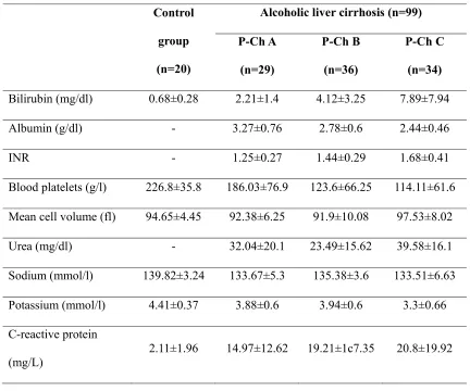
Table 2. Biochemical characteristics of the study and control groups (mean ± SD)
Control
group
(n=20)
Alcoholic liver cirrhosis (n=99)
P-Ch A
(n=29)
P-Ch B
(n=36)
P-Ch C
(n=34)
Bilirubin (mg/dl) 0.68±0.28 2.21±1.4 4.12±3.25 7.89±7.94
Albumin (g/dl) - 3.27±0.76 2.78±0.6 2.44±0.46
INR - 1.25±0.27 1.44±0.29 1.68±0.41
Blood platelets (g/l) 226.8±35.8 186.03±76.9 123.6±66.25 114.11±61.6
Mean cell volume (fl) 94.65±4.45 92.38±6.25 91.9±10.08 97.53±8.02
Urea (mg/dl) - 32.04±20.1 23.49±15.62 39.58±16.1
Sodium (mmol/l) 139.82±3.24 133.67±5.3 135.38±3.6 133.51±6.63
Potassium (mmol/l) 4.41±0.37 3.88±0.6 3.94±0.6 3.3±0.66
C-reactive protein
18
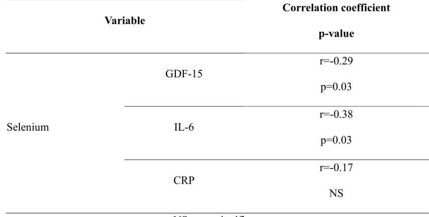
Table 3. Correlation between serum concentrations of selenium and growth differentiation factor-15, interleukin-6 and C-reactive protein
Variable
Correlation coefficient
p-value
Selenium
GDF-15 r=-0.29
p=0.03
IL-6 r=-0.38
p=0.03
CRP
r=-0.17 NS
19
Fig. 1. Concentrations of selenium (A), IL-5 (B) and GDF-15 (C) in patients with alcoholic liver cirrhosis and in the control group. Values expressed as the mean ± standard deviation.
A.
B.
C.