Representations in the Olfactory
System of the Locust and Fly
Thesis by
M
aria
P
apadopoulou
In Partial Fulfillment of the Requirements
for the Degree of
Doctor of Philosophy
California Institute of Technology
Pasadena, California
2010
© 2010
Acknowledgements
I feel forever grateful to many people at Caltech, friends and family for helping me navigate through this long process. They made it a very valuable and often very enjoyable learning experience.
First, I would like to thank my advisor, Gilles Laurent for having me in his lab and giving me the opportunity to be part of the very exciting research he has started. I especially want to thank him for giving me such an interesting project and the freedom to develop it. I feel very privileged to have been part of the lab that he has created. The people who made up the lab were very supportive and instrumental in my scientific training and thinking as a scientist.
I would also like to thank my committee members Erin Schuman, Thanos Siapas, Mark Konishi, and David Anderson for their useful feedback, patience through all my committee presentations and for comments on my thesis. I would particularly like to thank Thanos Siapas, whose advice I have sought outside of those meetings and whose encouragement during some hard personal times at the end of my Ph.D. was extremely helpful.
demonstrating how fun science can be and for teaching me to enjoy every moment in and out of the lab. In addition to being a great collaborator, I am thankful for providing me access to his setup, a step that was critical for performing some of the work described in this thesis. For the fly work, I am especially grateful to Vivek Jayaraman, Glenn Turner and Greg Jefferis, without whom the fly project would
have never started. In particular, I am thankful to Greg for sharing the dGGN image of the single MARCM clone of the GH146 line. Vivek has always been very encouraging, enjoyable to discuss science with and has helped me keep a healthy perspective throughout graduate school while also teaching me the importance of assumptions. Whether hunting snails, aplysia or flies, they have all been very enjoyable scientific collaborations. I am also thankful to Glenn for always being very generous with his time and advice and for demonstrating the real meaning of experimental thoroughness and skating. I would like to thank Anusha Narayan for being an incredible friend, colleague and collaborator on many non-scientific projects through the years and also for making life so much more interesting, unpredictable and fun. Together with Cindy they made the basement of the BI feel like home. I would also like to thank Cindy Chiu for demonstrating the perfect rig setup, for gps, valuable culinary advice and for sharing the BI basement. I would also like to thank Ofer Mazor for many fun and educational experiences associated with TAing the electrophysiology class during my first year in graduate school. Together with Stijn, he made learning physiology extremely enjoyable.
Murthy for help with fly work and Kai for welcoming self improvement comments. Mikko for the importance of temperature regulation in Greek / Finish rig room
sharing situations.
Outside the lab I am thankful to Anne Hergarden and Jasper Simon who together with Vivek Jayaraman constituted our fly journal club, where we spent hours over lunch having animated discussions on papers, techniques and our research projects. We certainly found many uses for the paper tablecloth in Citrus Bistro.
I would like to thank the Siapas Lab and in particular Eugene Lubenov and Casimir Wierzynski for very useful feedback during my defense preparations, and in general for very inspiring science.
Melanie Pribisko Yen for her friendship and help on many projects, including the time she synthesized a compound for me that induces aggression in a particular ant species. This experience really brought home to me how privileged I was to be at Caltech.
I would also like to thank a support group of friends whom I met on my first day at Caltech. We started graduate school together and have been friends ever since: Lily Kharevych, Sean Gordon, Rogier Braakaman, Ivan Bermejo, Aaron Noel, Waheb Bishara and Daniel Chung. While all in different divisions, we shared
common goals and they provided the crucial mix of encouragement and distraction necessary to survive graduate school. Over the years our group grew to include other friends/family: Eric, Marta, Raquel, Giselle, and Roshan whose company I
have enjoyed greatly throughout the years.
Abstract
The giant GABAergic neuron (GGN) is a single, paired, non-spiking neuron that arborizes extensively in the mushroom body (MB) of the locust (Leitch and Laurent, 1996), where it overlaps with the dendrites and the axons of Kenyon cells (KCs). KCs are the intrinsic neurons of the MB and are thought to be important for olfactory learning and memory(Davis, 2004). We are interested in understanding the function of GGN in olfactory processing: in particular, its pattern of arborization makes it an attractive candidate for controlling or modulating KC responses to odors, with potential implications for learning and recall. Physiological recordings of KCs in the locust show that these neurons respond sparsely to odors, in marked contrast to their excitatory inputs from the antennal lobe (projection neurons or PNs) (Laurent and Naraghi, 1994; Perez-Orive et al., 2002; Stopfer et al., 2003; Mazor and Laurent, 2005). Inhibition appears to be critical to control KC response threshold, probability and duration during odor stimulation (Perez-Orive et al., 2002). We show that there exists a feedback loop whereby KCs provide excitatory input to GGN , which in return provides inhibitory control of KC excitability. We further demonstrate that manipulating GGN during olfactory stimulation affects
neurons that receive input from KCs in theβ-lobe of the MB (β-LNs). We show that GGN can suppress KC activity to such an extent as to eliminate all spiking in the down-stream neurons. With these experiments in the locust, we show that GGN controls the gain of PN-to-KC information transfer and normalizes the output of the KC-population, much reducing its dependence on input strength.
With experiments inDrosophila Melanogasterwe try to extend the generality of GGN as a solution for gain control in the MB. Specifically, we carry out intracel-lular recordings from a Drosophila neuron discovered by Greg Jefferis, which has
Contents
Acknowledgements iv
Abstract viii
Table of Contents x
List of Figures xii
1 Chapter 1: Introduction 1
1.1 Olfactory system 1
1.1.1 Characteristics of Olfaction 1
1.1.2 Olfactory Receptor & Olfactory Receptor Neurons 3
1.1.3 ORN postsynaptic targets 5
1.1.4 Kenyon Cells 11
1.2 Mushroom Body 14
1.2.1 Anatomy 15
1.2.2 MB’s role in olfactory learning and memory 20
1.2.3 Physiology 22
2 A Single GABA-ergic Neuron Subserving Normalization for Sparse
En-coding in an Olfactory Network 24
2.1 Introduction 24
2.2 Results 27
2.2.1 KC input to GGN 29
2.2.2 GGN output onto KCs 37
2.2.3 Manipulation of GGN state during odor stimulation 39
2.3 Discussion 47
2.4 Methods 49
2.4.1 Preparation and stimuli 49
2.4.3 Intracellular recordings 50 2.4.4 Local Field Potential Recordings 51
2.4.5 Electrical stimulation 52
2.4.6 Immunocytochemistry 52
3 DrosophilaGGN 54
3.1 Introduction 54
3.2 Results & Discussion 56
3.2.1 What makes this neuron a homologue of the locust GGN? 59
3.2.2 Generating a specific GGN line 65
3.3 Future experiments: 66
3.3.1 Closing the feedback loop; GGN output on KCs during odor 66 3.3.2 Behavioral assessment of GGN’s gain control in olfaction 66 3.4 Comparison with recently published result 68
3.5 Methods 72
3.5.1 Fly lines 72
3.5.2 Fly preparation 72
3.5.3 Odor delivery 73
3.5.4 Electrophysiology 73
3.5.5 Immunohistochemistry 74
4 Additional GGN Features 75
4.1 Introduction 75
4.2 Results & Discussion 76
4.2.1 Facilitation 78
4.2.2 Lack of Direct Antennal lobe input in GGN 81 4.2.3 Assessing KC output throughβlobe LFP recordings 82
4.2.4 GGN Ablation 84
4.3 Methods 88
4.3.1 Preparation and stimuli 88
4.3.2 Odor Delivery 89
4.3.3 GGN Intracellular recordings 89
4.3.4 Local Field Potential Recordings 89
4.3.5 KC Electrical stimulation 90
4.3.6 AL Electrical stimulation 90
5 Conclusions & Future directions 91
5.1 Future directions: 93
5.1.1 Locust 94
List of Figures
1.1 Olfactory Bulb and Antennal Lobe Circuitry 6
1.2 Locust Olfactory Anatomy 9
1.3 Mushroom Body Anatomy 17
2.1 GGN Morphology 28
2.2 GGN & LFP Different Odor Concentrations 29
2.3 GGN Response to Different Odors 31
2.4 Excitatory Input to GGN 32
2.5 DC shift 34
2.6 Source of Inhibitory Input to GGN 35
2.7 GGN output onto KCs 39
2.8 Summary data for multiple experiments. 41 2.9 Effect of manipulating GGN activity on KC activity during odor
stim-ulation 43
2.10 Effect of manipulating GGN activity on KC activity during odor
stim-ulation 45
3.1 Drosophila olfactory system 57
3.2 Anatomy or Presumed GGN in drosophila 58
3.3 Drosophila GGN is GABAergic 60
3.4 Drosophila GGN Odor Response 62
3.5 Drosophila GGN Odor Response to Increased Odor Concentration 63 3.6 Output Synapses in GH146-Gal4/UAS SpH flies 64
3.7 Additional GABAergic labeled GH146-Gal4 cells 71 4.1 Kinetics of GGN’s response to KC & Odor Stimulation in 3 locations
along GGN. 77
4.2 Facilitation of the KC to GGN synapse 78
4.3 Facilitation dependence on IPI. 80
4.4 GGN Response to AL stimulation. 82
4.5 βlobe LFP Increases with an Increase in Odor Concentration. 83
4.6 Enhancing GGN activity during Odor decreases LFP power in the &
βlobe 85
C
hapter
1
Chapter 1: Introduction
1.1 Olfactory system
1.1.1 Characteristics of Olfaction
Sensory systems have evolved to detect and process information about the external world with the task of extracting useful relationships for the organism’s survival and procreation. The sense of olfaction in particular is integral in food and mate localization, as well as forming associations predictive of food (reward) and danger (punishment).
mixtures is not perceived as more complex than the odor of single chemicals.(Keller and Vosshall, 2004).
Humans have the ability to generalize odors and to categorize them perceptually as fruity, citrus etc (Bitterman, 1983; Dravnieks, 1982). Bees have also been shown to generalize in behavioral assays. After pairing of an odor (unconditioned stimulus) with sucrose (conditioned stimulus), bees start to extend their proboscis to this odor alone in the absence of reward. They are able to identify the odor precisely, and they are also able to generalize to structurally similar odors (Smith, 1989). Therefore the olfactory system has evolved to extract both types of information with presumably the same machinery (Laurent, 2002).
Evolution appears to have found some optimal solutions for solving the prob-lems discussed as demonstrated by how well the organization of the olfactory system is preserved across phyla and species (Strausfeld et al., 1998). I will briefly re-view the general organization and then expand in the following sections to explore in more detail the anatomy and the transformations that occur in each successive layer.
in insects, an analogous area associated with multimodal processing, learning and memory is called the mushroom body(MB).
Because of this organization, i.e., one layer (OB or AL) separating high associ-ation areas from the periphery, olfaction is considered a shallow sensory system. It appears, then, that the formatting of odor information in the second layer is sufficient before it targets learning and memory areas.
In summary olfaction is integral to the survival and procreation of many organ-isms, it is a synthetic and shallow sense, and the olfactory system has evolved to solve both identification and generalization problems. We next explore how this is achieved, starting with odor binding to the olfactory receptors.
1.1.2 Olfactory Receptor & Olfactory Receptor Neurons
Considerable advancement in olfaction research was made with the discovery and cloning by Axel and Buck of the first members of the odorant receptors proteins (Buck and Axel, 1991; Buck, 1996). Odorant receptors (ORs) are a large and diverse gene family of G-protein coupled receptors (∼ 1000 genes in rodents, ∼ 350 in humans) (Wilson and Mainen, 2006; Keller and Vosshall, 2004)). There are many (est. 100-1000) unique olfactory receptors. Insects express a phylogenetically distinct family of olfactory receptors (Sato et al., 2008; Kaupp, 2010). In vertebrates these receptors are located in the cilia of the nose, while in insects along the length of the antennae (Mombaerts, 1999; Vosshall et al., 1999; Wilson and Mainen, 2006).
lesions show that rats can still discriminate odors, pointing to a distributed acti-vation of a combinatorial nature (Laurent, 1999). It appears therefore that an odor is encoded in the pattern of ORNs it activates (Hallem and Carlson, 2006). Fur-thermore, flies that express only one OR are still capable of odor discrimination, which has been interpreted to suggest that odors elicit different temporal patterns
for a given receptor and that the temporal dynamics of the response could be an important coding variable(DasGupta and Waddell, 2008). In the locust AL, ORN responses to odor show temporal patterning that is both odor and ORN dependent. (Raman et al., 2010). The tuning of ORNs varies in breadth; there exist broadly tuned receptors as well as narrowly tuned ones, and many in between1 (Su et
al., 2009). As the concentration of an odor increases it leads to the recruitment of additional ORNs (Su et al., 2009).
While the focus so far has been on theactivationof ORs by odor molecules, it is also possible for an odor toinhibitan ORN (decrease over baseline firing); this has been argued to be mediated via odor "antagonism" (Su et al., 2009). The duration of the ORN response is dependent on the receptor it expresses, as shown by recording of an ORN where the receptor expressed can be controlled genetically(Hallem and Carlson, 2004; Su et al., 2009).
Each ORN typically expresses one OR, which confers its sensitivity (Mombaerts et al., 1996; Vosshall et al., 2000). While olfactory receptor neurons are dispersed along the epithelium (rodents) and antennae (insects), the second layer appears to be more organized. ORNs target a glomerulus based on the OR they express; that is,
1It is worth mentioning that in addition to generalist channels there are some ORNs that respond
only ORNs with the same OR target the same glomerulus (Mombaerts et al., 1996; Vosshall et al., 2000). ORNs form excitatory synapses onto glutamatergic M/T cells
in the OB. In insects ORNs synapse onto PNs, which are predominantly cholinergic.
1.1.3 ORN postsynaptic targets: Projection Neurons-Mitral
/
Tufted Cells
& Local Interneurons
In addition to receiving input from ORNs, processes of M/T or PNs also
con-tact cells locally within the OB and AL. Lateral connections within the OB as well as the AL can modify the input from the ORNs. In the OB, ORNs also synapse with periglomerular cells (PG), which are inhibitory, GABAergic, and, within a short range, extend to other glomeruli. PGs can synapse back onto the ORN axons to inhibit release, as well as onto M/T cells. M/T cells can excite each
other laterally through spill over of NT from the apical tuft, but they can also inhibit each other via an intervening PG. M/T cells in addition activate
gran-ule cells that are axonless GABAergic cells residing in the external plexiform layer that in turn through dendrodendritic interactions inhibit M/T cells. The
ef-fects of these interactions can be long range as M/T cells extend dendrites 10-12
glomerular diameters, contacting many granule cells (Wilson and Mainen, 2006; Laurent et al., 2001)
2005). The majority of LNs are inhibitory, releasing GABA, but some are cholinergic, mediating lateral excitation (Shang et al., 2007; Olsen et al., 2007). The presence of this lateral excitation explains partly why PNs are more broadly tuned than their presynaptic ORNs (Wilson et al., 2004).
Figure 1.1.Olfactory Bulb and Antennal LobeCircuitry. Excitatory neurons are shown in or-ange and inhibitory neurons in blue. (A) Olfactory receptor neurons (ORNs) in the olfactory epithelium that express different olfactory receptors project axons to separate glomeruli
(dashed outlines) in the olfactory bulb where they synapse on mitral and tufted (M/T) cells,
whose apical dendrite is usually localized to a single glomerulus. Juxtaglomerular cells (blue) contribute to intraglomerular inhibition. In the glomerulus, ORNs form synapses on juxtaglomerular cell dendrites, which in turn inhibit ORN axon terminals. Reciprocal synapses are also found between juxtaglomerular cell and M/T cell dendrites.
Recipro-cal synapses are formed between the dendrites of granule cells and M/T cells. M/T cells
excite granule cells, which respond by inhibiting M/T cells. Due to the lateral spread of
M/T secondary dendrites, granule cells contact multiple M/T cells associated with diff
er-ent glomeruli, and thus can mediate both intra- and interglomerular inhibition. (B) In Drosophila, ORNs expressing the same olfactory receptors in the antenna or maxillary palp synapse on projection neurons in a single glomerulus, analogous to the olfactory bulb. GABA-releasing local neurons (LNs) presynaptically inhibit ORN axon terminals in mul-tiple glomeruli, mediating interglomerular inhibition. Excitatory cholinergic LNs mediate interglomerular excitation. Figure from (Su et al., 2009)
and their presynaptic ORNs have been shown to arise partly because of the high convergence ratio of ORNs to PN and because the connection between ORNs and PNs is strong. Specifically, the convergence ratios of ORN to M/T or PNs
are 5000:1 for rodents and ∼ 100:1 in some insects . (Laurent, 1999; Wilson and Mainen, 2006). Such a high convergence ratio could serve multiple functions. By averaging it reduces noise and enhances the signal received by a M/T or PN. In
particular, the spread of ORNs across the nasal epithelium and the antenna could serve to eliminate local fluctuations, thereby enhancing the signal and eliminating noise(Laurent, 1999).It could serve to also enhance the dynamic range of M/T
provided that the thresholds for ORNs are different (Linster and Smith, 1999).
gain control in the antennal lobe
There is a nonlinear amplification of ORN input to PNs, extending the dynamic range of PNs by amplifying weaker ORN responses more than stronger ORN re-sponses (Olsen and Wilson, 2008). In this way, PNs can separate odors faster and better than ORNs(Bhandawat et al., 2007). Several mechanisms contribute to this. Firstly, reliability across the ORN-to-PN synapse, coupled with the high conver-gence ratios, ensures reliable PNs responses to weak ORN inputs (Bhandawat et al., 2007)). Secondly, the presence of short term depression between the ORN-PN synapse, ensures that gain of the synapse is turned low for strong inputs ensuring that PN responses remain within the dynamic range(Olsen and Wilson, 2008).
inhibition scales with total ORN input, there still exist differences across glomeruli
with respect to total inhibition (Olsen and Wilson, 2008).
projection neurons
PN and MC responses have are distributed in time and space. That is, about 50% of all PNs in the AL respond to any odor, and their responses are often multi-phasic, containing bouts of excitation and inhibition lasting hundreds of milliseconds, generally outlasting the stimulus itself. These response patterns, are odor and PN specific (Wehr and Laurent, 1996; Wehr and Laurent, 1999; Laurent, 2000; Perez-Orive et al., 2002; Stopfer et al., 2003; Mazor and Laurent, 2005; Broome et al., 2006).
potentials (Wehr and Laurent, 1996).
Figure 1.2.Locust Olfactory Anatomy. AL, antennal lobe; LH, lateral horn; MB, mushroom body; OL, optic lobe; agt, antennal-glomerular tract; an, antennal nerve; gl, glomerulus; on, ocellar nerve; p, pedunculus;βLN,β-lobe neuron; KC, Kenyon cell; LN, local neuron;
ORN, olfactory receptor neuron; PN, projection neuron; d, dorsal; l, lateral; mid, midline. Adapted from Laurent and Naraghi (1994); MacLeod and Laurent (1996).
Oscillations and slow patterning have also been described in the olfactory bulb of mammal (Adrian, 1942; Adrian, 1950; Kauer, 1974; Hamilton and Kauer, 1989; Laurent et al., 2001) . Here we focus on results in locusts and in particular on studies that attempt to understand these phenomena and their contributions to olfactory coding. We also refer to some relevant results in zebrafish that further this understanding.
Experiments in the locust have shown that in order to understand how odors are encoded, it is important to consider the PN population activity as a whole and to extract information in a way that is consistent with how downstream cells decode it. In one such experiment, it was shown that when trying to determine how odor concentration is encoded in the AL by PNs, recording the response of a single PN to a range of odor concentrations does not provide much information. For example, one PN might increase its activity in response to a particular range of odor concentrations and then it might become inhibited at higher concentrations in a way that is not consistent across PNs (Stopfer et al., 2003). By recording simul-taneously from 10-20 PNs and pooling across experiments, certain features emerge that characterize how these neurons encode an increase in odor concentration. As the concentration of an odor increases so does the extent of synchrony among PNs (Stopfer et al., 2003). Recordings from downstream neurons, the Kenyon Cells (KCs), have shown that the temporal integration of these neurons is effectively limited to
a single LFP cycle or ∼ 50ms (Perez-Orive et al., 2002). If the activity of the PN population is represented as a vector, with dimensionality equal to the number of neurons, then the evolution of this vector, followed at 50ms time-steps, consists of a trajectory through PN activity space (Stopfer et al., 2003; Mazor and Laurent, 2005; Broome et al., 2006). Such trajectories reveal a family of curves for different
concen-trations that are distinguishable from each other, but still close together compared to sets of trajectories representing different odor families (Stopfer et al., 2003).
Collecting simultaneously recorded data and analyzing them as discussed has provided additional insights into how PNs encode odors. By examining odor pulses of different durations, it has been shown that these trajectories throuhg PN
the onset and offset of the stimulus, rather than during the steady state (Mazor and
Laurent, 2005). Recordings from the KCs downstream of the PNs have shown that these cells respond the least during steady state (Mazor and Laurent, 2005). Steady-state measures of activity thus seem less appropriate to understand the neural code in this system (Mazor and Laurent, 2005). Experiments that examine PN responses to two odors separated by different intervals show that for some delays, the state
reached by PNs is different from the states representing individual components
or from the mixture (Broome et al., 2006). These results suggest that the evolution of PN responses is very much history dependent. All these results point to time as a very relevant feature in olfactory coding. This is likely made possible by the relatively slow evolution of the odor stimulus, in comparison to visual or auditory stimuli.
One way in which time is thought to be used, is to extract/separate different
features of the odor over time. Interaction within the AL and the OB leads to decorrelation; that is, responses across the population of PNs or M/Ts become more
dissimilar over time (Laurent, 2002) . Through this processes, different aspects of
the odor can be extracted at different time-points, which can then be passed on
to the MB and processed for learning (Laurent, 2002). Evidence for decorrelation comes from recordings of M/T in zebrafish, which demonstrate that early responses
1.1.4 Kenyon Cells
how are pn responses transformed in the next stage?
PNs carry olfactory information to the next layers of processing: to the mushroom body and to the lateral horn (LH). They send widespread projections through-out the calyx of the MB, where they contact Kenyon Cell dendrites (Jortner et al., 2007). Recordings from KCs have revealed extraordinary response transformations across the two layers; while PN odor responses are dense and exhibit prolonged slow patterning, KCs respond to odors rarely, and when they do, they only fire on average ∼ 2.3 spikes/sec (Laurent and Naraghi, 1994; Perez-Orive et al., 2002;
Stopfer et al., 2003; Perez-Orive et al., 2004). Kenyon cells responses are quite sparse, whether considering lifetime or population sparseness (Perez-Orive et al., 2002). Such sparseness is particularly attractive for a population of neurons in-volved in olfactory learning and memory. Several theoretical considerations point to the benefits of sparseness as a coding principle in terms of balancing storage capacity with minimizing overlap across representations and therefore the number of synaptic modifications required, as well as facilitating memory recall (Marr, 1969; Kanerva, 1988; Laurent, 2002; Olshausen and Field, 2004; Barlow, 1972; Field, 1994). It was recently discovered that KCs receive input from about 50% of all PNs. While this finding suggests that the number of potential patterns that can be encoded is very large, it also makes the KC sparseness all the more striking (Jortner et al., 2007).
convergence of PNs onto LHIs, LHI output is oscillatory and responsive to all odors. Due to propagation and synaptic delays, the inhibition from LHIs is phase-lagged to the excitatory PN input by 180 degrees. As such it serves to reduce the KC integration window to∼half an oscillation cycle (Perez-Orive et al., 2002). Ad-ditionally, voltage-gated channels in KCs amplify the effect of EPSPs that arrive in
close temporal proximity and sharpen the EPSP. This also contributes to reducing the effective integration window, and making KCs highly responsive to coincident
input (Perez-Orive et al., 2002)2.
Considering these existing mechanisms, cell numbers and connectivity, Jortner
et al. demonstrate that the response probability of a KC to a variety of inputs should change dramatically by adding only a small number of additional PNs (Jortner et al., 2007). This is not consistent, however, with experimental results. As mentioned previously, PNs respond to increasing odor concentration by an increase in synchrony. Since they act as coincidence detectors, KCs are very sensitive to synchrony among their inputs (Perez-Orive et al., 2002). And rather than a very steep and uniform increase in KC activity as a function of odor concentration, the recordings reveal that there are KCs that are concentration invariant, others that only respond to a limited range of odor concentrations and not at all to higher concentrations and so on(Stopfer et al., 2003). These data point to the existence of additional mechanisms to ensure KC sparseness. The main focus of this thesis, detailed in Chapter 2, consists of a form of inhibition that acts through a feedback loop to scale the activity of each KC by the total KC activity, thus dynamically 2Recent recordings from neurons in the piriform cortex, downstream from mitral cells of the
maintaining a sparse representation among KCs.
Consistent with the observation that the KC integration window is limited to half of one oscillation cycle, recordings from PNs and KCs under the same exper-imental conditions show that KCs decode PN output in a piecewise fashion, one LFP cycle at a time. As mentioned above, in experiments were two odors are pre-sented with different temporal delays, for some delays, the PN population response
(represented as an evolving trajectory) differs from either of the components or the
mixture (Broome et al., 2006). Consistent with piecewise decoding from KCs, there exist KCs that selectively respond to a particular temporal delay and therefore to the PN ensemble that was activated but not to any of the components or the mixture(Broome et al., 2006).
In summary, KC responses are sparse and synthetic; KCs receive input from a large fraction of PNs, which they decode piecewise at each LFP cycle. KCs have a high threshold for activation, but when a large enough fraction of their inputs is activated they respond during the corresponding cycle with a rare action potential.
1.2 Mushroom Body
The mushroom body (MB) is a paired neuropil structure found in annelids and in all arthropod groups except crustaceans (Strausfeld et al., 1998). The mushroom bodies differ in size between taxa, as well as between different castes of a single
species of social insect (Strausfeld et al., 1998). The MB was first Identified in 1850 by Felix Dujardin, who was the first to observe that there was a correlation between the size of the MB and the social complexity in different species of bees. That lead
Lesion, genetic manipulation and behavioral experiments all point towards a role for the mushroom body in olfactory learning and memory(Heisenberg et al., 1985; de Belle and Heisenberg, 1994; Dubnau et al., 2001; Krashes et al., 2007; McGuire et al., 2001). Physiological recordings from the intrinsic cells in the MB have shown their responses to odors to be sparse (Laurent and Naraghi, 1994; Perez-Orive et al., 2002; Turner et al., 2008), a feature that could be beneficial for memory storage and recall (Laurent, 2002). Furthermore, the synapses between these cells and a group of extrinsic MB neurons, have been shown to be plastic and affected by the presence of the neuromodulator octopamine ((Cassenaer and
Laurent, 2007) (Cassenaer & Laurent in preparation).The MB has additionally been implicated in sleep and decision making (Joiner et al., 2006; Zhang et al., 2007b). Here we focus on olfactory learning in insects species where most of these studies have taken place, review the anatomy and some of the data from the studies pointing to an important role for this area in olfactory learning and memory.
1.2.1 Anatomy
There is one MB per hemisphere, each being a mirror image of the other. The MB is a neuropil structure resembling a mushroom in shape, occupying a big volume of the brain. Kenyon cells (KCs) are the intrinsic cells of the mushroom body. Their number per MB varies between 2,500 indrosophila to 200.000 in the cockroach (in the locust there are 50,000, while in the bee 170,000). In bees KCs constitute 35% of all the neurons in the bee brain (340,000 KCs out of 960,000) , while indrosophila5% (5,000 out of 100,000)
such it contains KC dendrites and axons of postynaptic neurons to these KCs (ie PNs, LHIs etc). The KC cell bodies sit above and in some case also besides the calyces. A typical KC cell extends a neurite from the soma that gives rise to a dendrite and an axon. While the dendrite targets the calyx of the MB, the axon runs through the pedunculus to reach the lobes. The pedunculus is a bundle of such tightly packed parallel KCs axons that run through right before they bifurcate to send axons ending in a vertical and medial lobe (termedαandβlobes respectively). In the lobes, KCs make output synapses with extrinsic cells of the MB.
These general anatomical characteristics can differ depending on the species.
Some insects lack calyces, while social insects like the bees and ants have two calyces per MB (Strausfeld et al., 1998). Similarly for the lobes, there exists some variation; In some insects , of whichdrosophilais an example, in addition to a vertical lobe that indrosophila it is comprised of the α/ α’ lobes and the medial β, β’ lobes, there is a third lobe called theγ.
Drosophila KCs can be classified in three morphological distinct classes, based on the lobes they target ,ie α/ β, α’ and β’ or γ KCs. Furthermore there exists a correlation between these three classes and their birth order as well as their soma position. Specifically, there are four neuron precursor cells (neuroblasts) that give rise to KCs and as more KCs are born the older ones are pushed in the periphery, forming concentric circles with the later-born KCs in the center. Theγneurons are born first followed by the theα’ andβ’. neurons and last are theα/βneurons (Lee et al., 1999)(Lee and Luo 99). As such in the pedunculus one can observe concentric circles with theα/β neurons are in the center of the bundle and the γneurons in the periphery.
and soma position. Namely, KCs with small somata that are born later and arborize in the the anterior calyx where as KCs with large somata (born earlier) arborize in the posterior calyx (Fahrbach, 2006). Similarly this organization is maintained with the small somata forming the core of the bundle, while the ones with the larger somata are in the periphery (Fahrbach, 2006).
It is important to note that even though KCs, might target their dendrites to certain zones in the calyx, it is not necessary that they’ll always contact the same postynaptic cells and as result respond to a stimulus in the same way across animals. A recent study in fact has shown that indrosophila, KC responses are not sterotyped (Murthy et al., 2008). This in contrast to PNs that reliably target the same glomerulus to contact the same ORNs and produce very similar responses across animals (Jefferis et al., 2001; Masse et al., 2009). Some insights for the differences in the
presence or absence of calyx or the number of calyces, come from looking at the input in these species (see below).
input
In most insect species the input to the calyx appears to be strictly olfactory (lo-custs) 3, while in hymenoptera social insects a large component of the input is
visual(Strausfeld et al., 1998; Fahrbach, 2006). In bees there is evidence of gustatory input as well(Campbell and Turner, 2010; Fahrbach, 2006). Lastly MB has been shown to receive neuromodulatory input, that is thought to mediate reward and punishment (Keene and Waddell, 2005).
The importance of MB in olfaction is highlighted in that species that tend to 3There is evidence of synaptic contacts formed in the pedunculus. It is therefore important to
be anosmic - or have limited need for feeding as adults have smaller MB and lack calyces in most cases (Strausfeld et al., 1998; Fahrbach, 2006) (Figure 1.3) Ultrastruc-ture studies have shown cholinergic and GABAergic input in the MB calyx (Leitch and Laurent, 1996; Fahrbach, 2006). As mentioned in olfactory system section of the introduction, projection neurons in the antennal lobe in insects, receive odor information from olfactory receptor neurons that after processing and formatting they relay to the MB and lateral Horn(LH). In the LH, PNs contact the GABAergic LH internerneurons (LHIs), which in turn send projections back to the MB to in-hibit KCs. The role of this feedforward inin-hibition -as shown in the locust- is to limit the integration window of KCs, contributing to the sparsening of KC responses (Perez-Orive et al., 2002).
Social hymenoptera appear to be an exception to most other insects; in addition to olfactory input, their calyces receive substantial visual input. Medular output from the optic lobe carried through the anterior optic tracy enters enters the MB calyces bilaterally. Olfactory and visual information are segregated in the calyces, targeting different zones within the calyx. In bees olfactory information converges
on the lip of the calyx, while visual information is in the collar of the calyx. (Gronen-berg, 2001). In addition, there is also a projection from an area of the subeasophageal ganglia that receives sensory afferents from the proboscis and itself targets yet a
different area of the MB calyces that does not overlap with visual or olfactory input
(Fahrbach, 2006). Therefore the calyces of the bees are characterized as multimodal. Finally, mechanosensory and gustatory input to a small accessory calyx that is sep-arate from the main calyx has been reported for hemimetabolous insects, like the crickets (Fahrbach, 2006)
modulators that serve as signals for punishment and reward respectively. Dopamin-ergic and octopaminDopamin-ergic cells have been shown to innervate the MB>Activation of
these neurons through genetic manipulation, has been used successfully in place or reward or punishment in order to condition an animal to an odor(Schroll et al., 2006; Claridge-Chang et al., 2009; Keene and Waddell, 2005).
output
MB extrinsic neurons connect to the ipsilateral or contralateral MB lobes, where they synapse with KC axons. Such neurons have been identified in the bee, the coachroach, flies and locusts. In most cases the morphology is interesting, but it is not clear what the neuron’s function might be. In drosophila genetic mutations restricted to some of the extrinsic MB neurons (ex DPM) have been shown to lead to defects in olfactory and memory tasks (Yu et al., 2005; Keene et al., 2006; Krashes et al., 2007). In locusts, recent electrophysiological results provide insights as to how olfactory learning could be accomplished in the presence of a neuromodulator. We examine these results in more detail in the next sections.
1.2.2 MB’s role in olfactory learning and memory
Interestingly, the MB appears to be quite plastic. Many studies have shown cor-relations between behavioral complexity and MB size. In bees, for example, the volume of the MB correlates with the amount of foraging; researchers have shown that worker bees with more foraging experience had larger MB volume when com-pared with aged matched controls (Withers et al., 1993). This effect is robust having
staying in the hive) and thus somehow drives the growth of synapses that leads to the enlargement of the MB neuropil. While this increase in MB volume correlates with the behavioral improvement assessed experimentally, no direct causality has ever been demonstrated. Similar experiments have been conducted on other social insects with similar results (Fahrbach, 2006).
The first evidence implicating the MB in a role for olfactory learning came from chemical ablation study. Heisenberg and deBelle’s study, used hydroxy urea to kill MB neurons and demonstrate the inability of fly’s to learn associations between odors and an electric shock. Specifically, the authors took advantage of the fact that the KC neuroblast is active at a particular time, and by exposing animals to HU during that time the experimenters could almost selectively abolish KCs. The flies were born without MB and while they did not show any abnormalities in locomotion or ability to detect odors, it appeared that they could not associate an odor with an aversive stimulus. The flies appeared unable to use the odor as a predictor for punishment and as such they did not avoid it in a T-maze assay, as normal controls did (de Belle and Heisenberg, 1994).
At the molecular level the cAMP pathway appears to be particularly important for olfactory learning and memory, given that mutants of genes involved in this pathway show deficits in learning/memory olfactory tasks. Some examples include:
a)theduncemutant that affects a cAMP-specific phosphodiasterase; b)theamnesiac
gene that encodes the putative neuropeptide which is similar to pituitary adenylyl cyclase-activating peptide c) mutants affecting protein Kinase A (PKA) and CREB
(a transcription factor activated by PKA) in the MB have been shown to affect
deficits in the T-maze assay.
Restoring expression of some of these genes in the MB and/or some extrinsic
MB neurons, has shown to rescue the behavioral deficit observed in these mutants (Davis, 2005) . Interestingly some of these genes have mammalian analogs, that when mutated lead to impairments in spatial memory (Davis, 2005).
These mutant studies have been successful at demonstrating that the mushroom body is involved in olfactory learning and memory. In order to understand,though, how associations between odors and rewards take place, memories are stored etc, one needs to first understand how odors are encoded in the mushroom body and study the neuronal changes that occur during learning. That provides a framework for understanding the effects of manipulations, genetic or otherwise.
1.2.3 Physiology
Consistent with the KCs involvement in olfactory learning and memory, recordings from these cells in locusts and flies have shown KCs to produce sparse responses to odor (Laurent and Naraghi, 1994; Perez-Orive et al., 2002; Stopfer et al., 2003; Turner et al., 2008); they respond to odors rarely, and when they do their response is made of one or two spikes (Laurent and Naraghi, 1994; Perez-Orive et al., 2002; Stopfer et al., 2003; Turner et al., 2008). From a computational point of view, this sparseness facilitates memory storage, recall and modification of memories cite Barlow:1972p3016,Field94,Laurent02,Olshausen04. For a memory to be formed or erased a minimal number of synapses need to be changed. In addition the overlap of odor representations is minimized, such that affecting one synapse holding a
Recently a population of MB extrinsic cells that receive input from KCs in the
βlobe (βLNs) (Figure 1.2) has been characterized in the locust using electrophysi-ological recordings (Cassenaer and Laurent, 2007). It was found that the synapses between KCs andβLNs are governed by spike-time-dependent-plasticity (STDP) (Cassenaer and Laurent, 2007). One of the roles of STDP in this system is the home-ostatic control ofβLN phase (Cassenaer and Laurent, 2007). Recent results reveled an additional one; when octopamine is injected in theβlobe, it can combine with STDP to selectively change the synapses between KCs and β LNs that STDP has just tagged (Cassenaer and Laurent, in preparation). This is the first experimental demonstrationin vivoof how STDP could interact with a neuromodulator to selec-tively affect only those neurons that were responsive to the stimulus (Cassenaer
C
hapter
2
A Single GABA-ergic Neuron
Subserving Normalization for Sparse
Encoding in an Olfactory Network
2.1 Introduction
relevant representation. Finally, such a scheme is energy efficient (Lennie, 2003;
Olshausen and Field, 2004).
The importance of sparse representations is highlighted by their presence across different sensory modalities. In the visual system of primates, neurons in area V1
exhibit sparse responses when stimulated with image sequences resembling those that occur during natural vision (Vinje and Gallant, 2000). In the primary auditory cortex of rats, neurons can produce a single spike in response to a sound that is highly reliable across trials (DeWeese et al., 2003). In songbirds, neurons in the nucleus HVC (higher vocal center) respond reliably with one spike at a particular time during the song (Hahnloser et al., 2002). In the olfactory system of insects, Kenyon cells (KCs) respond to odors rarely and briefly (Perez-Orive et al., 2002; Turner et al., 2008). KCs are the intrinsic cells of the mushroom body, an area of the insect brain involved in olfactory learning and memory (Heisenberg et al., 1985; Tully and Quinn, 1985; Davis, 1993). The sparseness of the KC population is attractive from a computational perspective, since these cells are thought to be involved in learning and memory, and their sparse tuning allows for easy handling and storage of memories and limits the overlap across memories (Laurent, 2002). Recently a form of Hebbian learning, spike time dependent plasticity (STDP) was discovered to exist between KCs and their downstream neurons, β-lobe neurons (β-LNs) (Cassenaer and Laurent, 2007).
In this study we examine the role of gain control in maintaining the sparseness of KCs as well as the functional implications for the downstream decoders of KCs, the
and Zipser, 1988; Salinas and Sejnowski, 2001). It is particularly prominent in sensory systems where the input can vary over several orders of magnitude while the output of neurons encoding the stimuli is limited. Based on previous studies in the lab, I will argue that gain control is necessary at the synapse between PNs and KCs and that a cell we have identified in the locust serves this role. Given gain control’s generality across sensory systems, we believe this study to be relevant to sensory coding in general.
The organization of the olfactory system is shallow and it is conserved across phyla (Shepherd, 1994). Briefly, odor detection is carried out by olfactory sensory neurons in the antenna that send axons in the antennal lobe to contact projection neurons (PNs) and local interneurons. PNs transmit information to the next layer; specifically, they send excitatory projections along a tract and contact dendrites of KCs in the mushroom body (MB) and lateral horn interneurons (LHIs) in the lateral horn. LHIs in turn send inhibitory projections to KCs. This feedforward inhibition, together with intrinsic synaptic properties, restricts the KC integration window, making KC act as coincidence detectors (Perez-Orive et al., 2002). Theoretical results (Nowotny, unpublished results) show that in order for KC responses to remain sparse, given the high PN-to-KC connectivity (50%,(Jortner et al., 2007)), there is a clear need for gain control.
rate. In particular, simple calculations show that in the absence of gain control, the relationship between KC population response probability and the number of coincident PN spikes is extremely steep (Jortner et al., 2007). Small deviations of just a few PNs should lead to order of magnitude changes in KC population firing probability, which is not consistent with experimental results (Stopfer et al., 2003). Instead, individual KCs’ responses can be selective to a small range of concentrations, or even invariant to concentration, suggesting the existence of gain control.
2.2 Results
We hypothesized that gain control could be provided by a paired giant GABAergic neuron (GGN) that has extensive arborizations throughout the MB (Fig 2.1). This neuron was first described in an electron microscopy study of GABAergic neurons in the locust (Leitch and Laurent, 1996). Intracellular recordings in the locust show GGN to be a non-spiking neuron that responds to all odors tested with graded potential (Figure 2.3). Furthermore, as the concentration of an odor is increased over 6 orders of magnitude, so does the GGN membrane potential, as shown in Figure 2.2i & ii. One way this increase in GGN membrane depolarization can be quantified is by computing the integral of the area under GGN for the duration of the odor response (Fig 2.2vi). Neurotransmitter release in non-spiking cells in the locust has been shown to correlate well with membrane depolarization (Burrows et al., 1982). Taken together these observations suggest that GGN provides increased inhibition as odor concentration is increased (Fig 2.2).
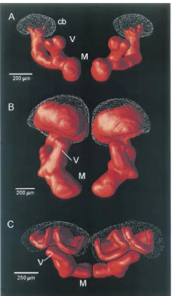
Figure 2.1.GGN Morphology. Intracellular GGN fill with 5% Biocytin. i) An image stack of GGN showing its extensive arborizations in the MB calyx andαlobe, as well as in the lateral horn. Cell body located in the lateral horn. ii)Zoom of an image showing GGN’s processes in the calyx (top) and theαlobe (bottom).
trials, 2.2iv: LFP power, (Stopfer et al., 2003)). The cumulative increase in LFP power during the odor response correlated well with the corresponding increase in GGN activation (measured as the cumulative integral under GGN membrane potential) at the singe-trial level for this experiment and for 5 experiments (n=364
trial pairs over all experiments and concentrations, linear fit, r=0.93, Fig 2.2vi). Both
measures were computed until GGN membrane potential returned to baseline.) Furthermore, the LFP and GGN Vm signals also covary as a function of time: there is a high correlation between the envelope of the LFP for a given trial and the GGN membrane potential for the same trial during odor but not during baseline (Fig 2.3ii, 225 pairs except for air and paraffin oil controls). In other words, we observe that
A closer look at GGN’s anatomy (Fig 2.1i&ii), reveals punctate processes in the calyx of the MB, consistent with output, and finer hair-like projections in the α
lobe , consistent with input. GGN’s anatomy, taken together with the fact that KCs receive input in the calyx and output to the lobes, suggests that GGN could be providing KCs with feedback inhibition.
2.2.1 KC input to GGN
To examine whether KCs indeed provide input to GGN, we recorded intracellularly, simultaneously from GGN and a KC. We evoked spikes in the KC via current injection and recorded the average postsynaptic effect onto GGN. An example of
such a spike-triggered average (STA) resulting from 139 event as well as the average from 11 such experiments are shown in Figure 2.4Ai and ii respectively. The data in these experiments (in particular the sharp onset of the EPSP, the delay accounted for by presynaptic spike propagation and the reproducible time-course) demonstrate that KCs are mono-synaptically connected to GGN. Average unitary EPSPs were 0.88 ± 0.50 mV (n = 11 KCs), with some nearing 2mV on average. Extracellular
KC stimulation also evokes a depolarization in GGN (Fig 2.4Bi) consistent with
Figure 2.2 (on the next page). GGN & LFP Different Odor Concentrations Intracellular
GGN response and LFP recorded in response to six different concentrations of octanol
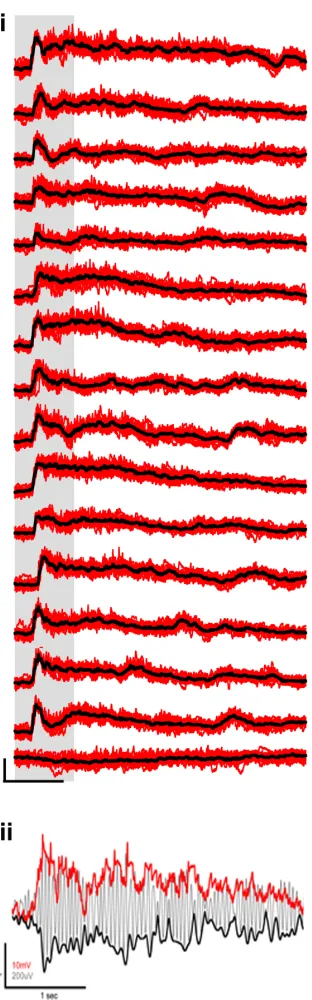
Figure 2.3.GGN Response to Different Odors. i) GGN’s response to 15 odors and control.
Single Trials (red) of GGN’s response to 15 odors and paraffin oil control (last one) and
the excitatory connection between KCs and GGN. In addition, KC extracellular stimulation allowed us to assess the effect on GGN of activating multiple KCs
simultaneously. Increasing stimulation strength (5-140uA), thereby presumably activating more KCs, led to an increase in PSP amplitude on GGN with a peak between 15 and 20 mV (Fig 2.4Bii). This effect was observed whether we measured
the peak amplitude of the PSP (Fig 2.4Ci), the maximum slope (Fig 2.4Cii) or the area under the curve. This effect was measured across 6 experiments for 8 different
stimulation channels. As such, it appears that activating more KCs increases the extent of depolarization in GGN.
Lastly, we asked whether input from KCs alone is sufficient to account for
the sustained depolarization observed in GGN’s odor response. As described in the introduction, olfactory stimulation gives rise to oscillatory population activ-ity in the antennal lobe, which propagates to the MB, evoking sparse volleys of approximately simultaneous KC spikes. Such a profile of KC activity can be read-ily mimicked with extracellular stimulating electrodes, allowing us to address the question we posed. We observe that stimulation of KCs at a frequency similar to the odor-evoked oscillations reproduces the observed DC shift, suggesting that excitatory input from KCs onto GGN can account for GGN’s excitatory response during odor (Fig 2.5).
Figure 2.4(on the next page). Excitatory Input to GGN. A. KC-GGN STA. Ai: example of a KC-GGN STA (black, average of 139 raw Vm events in grey). Aii: Average (black) of 11 such average STAs, each from a different KC (grey). B. GGN PSP in response to
extracellular KC stimulation Bi: Raw (grey) and average(blue) PSP in GGN following KC stimulation (60uA). Bii: Increasing stimulation strength over a large range of stimulation strengths (5-140uA, including 60uA from Bi) results in increased PSP size recorded in GGN (each trace is an average of 20 trials);C.Quantifying the effect of increasing KC stimulation
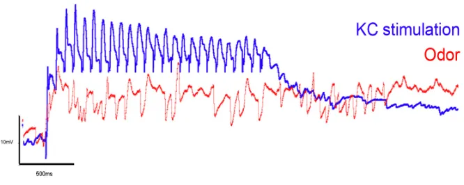
Figure 2.5.DC Shift. GGN response to a train of KC stimulation at a frequency similar to the dominant frequency recorded in the LFP during odor stimulation (25 pulses at 25Hz), overlaid with an example of the odor response for the same recording.
The PSP onto GGN evoked by KC stimulation above a certain threshold is not a pure EPSP; rather it appears that the excitatory effect is followed by a delayed
inhibitory effect, suggesting that KCs might provide input to another cell, which
fashion, the size of the IPSP on this inhibitory neuron increases with increased GGN depolarization (Fig 2.6v).
Thus far, we have identified KCs as the source of the excitatory input to GGN and the inhibitory cell as providing the prominent inhibition to GGN. Furthermore, GGN forms a feedback loop with this inhibitory cell (Fig 2.6vi), thus providing an additional layer of control. Next, we sought to investigate whether GGN outputs onto KCs so as to complete the feedback loop.
2.2.2 GGN output onto KCs
Since GGN is GABAergic, we wanted to verify that its effect is inhibitory; and
if so, to determine whether it exerts its effect presynaptically onto PN terminals
and/or postsynaptically onto KC dendrites. If it has a direct postsynaptic effect
on KCs, we should be able to affect spiking in KCs even in the absence of PN
excitation. To test this we performed dual intracellular recordings; we used current injection of 70-300 pA to evoke 3-4 spikes in a KC and sought to eliminate them in alternating trials by depolarizing current injection into GGN (schematic Fig 2.7B). Since the electrotonic nature of this nonspiking neuron is unknown, and the effect
of current injection at one location on distant release sites is impossible to predict without empirical data, we first performed simultaneous intracellular recordings at two distinct locations along the GGN dendrite (Fig 2.7A). The highest current injected (19.5nA) in the first GGN location appeared to evoke a depolarization in the second location that was comparable in amplitude to the odor response in the second location (Fig 2.7A). Furthermore, intermediate current values (1.5, 5.5, 13.5, 15.5, 17.5nA) produced intermediate depolarizations1in GGN (Fig 2.7A).
We tested the effect of GGN depolarization on the suprathreshold activation of
individual KCs by direct current injection. A KC was impaled in its soma, and a current pulse (150pA, 200 ms) was injected to evoke ∼5 action potentials on average. Figure 2.7B, shows these trials with one KC. Following each pulse, we combined the depolarizing pulse into the KC with the depolarization of GGN, by 1In the ideal case, we would have been able to measure the voltage deviation at the current
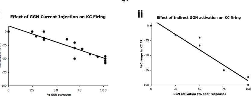
current injection (3.5, 11.5 or 19.5nA, same pulse duration as KC) through a second microelectrode placed in a dendrite of GGN. An injection of 19.5 nA was known to evoke a depolarization of 10mV at a distant site, commensurate with an odor evoked GGN response (see above). The average effect for injecting 19.5 nA was a ∼55% reduction (n=8 KCs). As shown in Figure 2.8i, the extent of the effect on KC
firing is dependent on the amount of current injected into GGN.
Having shown that KC stimulation can depolarize GGN, and that injecting cur-rent in GGN can eliminate curcur-rent-evoked KC spikes, we next tested a variant of this experiment. In this case, the depolarization of GGN was caused not directly by current injection, but by synaptic drive from KCs. Indeed, if GGN adaptively regu-lates KC population output, the extent of its own depolarization should depend on KC activity. We thus kept one intracellular electrode in one KC, to be used as our assay for the effect of GGN depolarization. We kept a second electrode in a GGN
dendrite to assess its membrane polarization. Finally, we added a pair of extracel-lular electrodes in the KC soma region, far from the KC impaled for intracelextracel-lular recording, to stimulate a new set of KCs and thereby indirectly depolarize GGN (Fig 2.7C). As expected based on the previous results, we saw a reduction of KC firing also with this manipulation (n=8, average effect∼85% reduction, Fig 2.7C).
We also found that the decrease in intracellularly recorded KC firing rate was de-pendent on the strength of extracellular stimulation of the other KCs (Fig 2.8ii). Simultaneous intracellular recordings of GGN, showed that the extent of its depo-larization correlates well with KC stimulation strength and also with the extent of the decrease in KC firing rate (Fig 2.8ii). This manipulation was more effective
in eliminating KC spikes compared to direct current injection into GGN (∼85% vs
input to GGN might evoke release more readily than artificial current injection at an arbitrary location within the neuron.
To summarize, we have demonstrated that the degree to which KC output is reduced by GGN activation is related, in a positive way, to the degree to which KCs are excited. Also, we have shown that GGN’s actions are consistent with a normalization or adaptive control of the KC population output. The gain of this loop, however, is unknown and how compressed the KC output range is remains unknown.
2.2.3 Manipulation of GGN state during odor stimulation
To gain insight into its function, we injected current into GGN during odor pre-sentation while monitoring input and/or output from KCs (we use input to refer
to subthreshold effects and output to mean KC action potentials). By imposing an
artificial de- or hyper-polarization of GGN by direct current injection, we perturbed the KC-GGN feedback loop and assessed the effect on KC activity.
Figure 2.7(on the next page).GGN output onto KCs.A.Intracellular recording of GGN in two different locations. Ai) Schematic summarizing the experiment. We injected current
pulses of different amplitude in one GGN location and recorded the effect in a second GGN
location (Vm at second location shown in blue in Aii). Odor response at second location (red) overlaid for comparison.B.Injection of depolarizing current into GGN can eliminate current-induced KC spikes Bi) Diagram explaining the manipulation; Dual intracellular recordings of GGN and a KC. ( Bii) Two current pulses (per trial) in an intracellularly recorded KC evoke spikes (raster and Vm of last trial shown); one pulse is paired with current injection in GGN. Current Injected in GGN from left to right: 3.5nA,11.5nA & 19.5nA. Star: spikes were clipped by 20mV.C.KC stimulation can eliminate current-induced KC spikes (through indirect depolarization of GGN). Ci) Diagram explaining the manipulation; Dual intracellular recordings of GGN and a KC recording the effect of exciting a different set
of KCs with extracellular stimulation. Cii) Two current pulses (per trial) in an intracellularly recorded KC evoke spikes; one pulse is paired with a train of stimulation of a different set
Figure 2.8.Summary data for multiple experiments. i) Scatter plot of 5 KCs & a linear fit of percent reduction in KC firing as function of increasing GGN activation. The KC reduction is computed with respect to the control KC pulse included in the same trial. To compute % GGN activation, the current injection values are rescaled based on their effect on GGN
Vm, as assessed in dual GGN recordings, and normalized to the highest value (see suppl Fig3). ii) Scatter plot of 3 KCs & a linear fit of percent reduction in KC firing as a function of increasing KC stimulation and thereby GGN activation.The KC reduction is computed with respect to the control KC pulse included in the same trial. Stimulation strength is translated to 0, 25, 50, 75 or 100% of a high odor response in the same location.
Injecting depolarizing current into GGN during the odor strongly affected
sub-threshold KC activity. In experiments where positive current was injected in GGN during odor stimulation (alternating trials with and without GGN current injec-tion) LFP power in the 10-30Hz band was reduced to 15% of the odor control (Fig 2.9A). The opposite effect, i.e an increase in LFP power (10-30Hz band, 125%
of control) was observed when GGN activity was reduced by negative current in-jection (Fig 2.9Aiii&iv). Although the origin of the LFP oscillations in the 10-30Hz band is in the AL, and arises from interactions between PNs and LNs, the actual sig-nal measured in the MB is thought to represent synaptic currents flowing into KCs. As shown in Figure 2.7, GGN, in addition to PNs, also provides input to the KCs. It appears, therefore, that by injecting depolarizing current into GGN we reduce the effectiveness of PN input into KCs. Intracellular recordings of single KCs (Fig2.9B)
in the 10-30Hz band in the KC membrane potential is also reduced to about 15% of control when depolarizing current was injected into GGN. An example of such a recorded KC is shown in Figure 2.9B. Conversely, reducing GGN activity during odor leads to an increase in the power of the KC membrane potential by 130% (Fig 2.9Biii & Biv). The effect observed in both LFP and subthreshold KC activity
could be mediated not only by altering KC membrane potential, but also by chang-ing KC input conductance and thus, time constant and reactiveness to synaptic driveÑmore generally, KC integrative properties. Such action would be nonlinear, and be akin to a shunt on PN drive onto KCs. To ensure that our results derived from current injections into GGN do not have an artifactual component, we carried out control measurements following the GGN recordings, where current of either polarity was injected 50-100 microns outside GGN. These manipulations do not give rise to the changes we see when current is injected into GGN (Fig 2.10B).
How is KC output affected?
Given these changes in KC subthreshold activity, we wondered to what extent KC spiking would be affected. Because only a small fraction of KCs respond to an
odor with action potentials, we have relatively few recordings where we can assess the effect of manipulating GGN activity during odor-evoked KC firing. Moreover,
the total KC population is quite large (50,000/MB), and it would be desirable to sample a reasonably large fraction of the KC population to conclusively assess the effect of GGN on KC output.
population of neurons could further reveal the relative importance of GGN in this circuit.β-LNs have a low baseline firing rate, but respond quite vigorously during the odor (Cassenaer and Laurent, 2007). We find that manipulating GGN activity during odor greatly impacts β-lobe neuron (β-LNs) firing. In particular, injection of depolarizing current into GGN could led to a complete shutdown of theβ-LN’s firing during odor in 6 out of 8βLNs recorded (Fig 2.10A & B, and a 96% and 82% reduction in 2/8). GGN exerts its effect on β-lobe neurons indirectly by acting on KCs, since positive current injection in GGN is incapable of eliminating any cur-rent induced β-lobe neuron spikes in the absence of odor (n=3 cells). Conversely,
injecting hyperpolarizing current into GGN during odor in the same cell had the opposite effect, namely increase its firing rate to 140% of control.
Figure 2.9(on the next page).Effect of manipulating GGN activity on KC activity during
odor stimulation:A.Effect of manipulating GGN activity during odor on LFP power: Ai &
Aii) Enhancing GGN activity by positive current injection (Iggn), leads to a decrease in the LFP power. Ai) 2 example traces (bandpassed 10-30Hz) recorded consecutively in response to odor and positive Iggn (blue) or odor alone (red). Aii) Power in the 10-30Hz band for each of the two different conditions (20 interleaved trials, light blue & pink) and their respective
average (red & blue). Aiii & Aiv) Reducing GGN activity by negative current injection (Iggn) leads to an increase in LFP power. Ai) 2 Example traces (bandpassed 10-30Hz) recorded consecutively in response to odor and negative Iggn (green) or odor alone (red). Aii) Power in the 10-30Hz band for each of the two different conditions (20 interleaved trials, light green
& pink) and their respective average (red & green).B.Effect of manipulating GGN activity
during odor on KC subthreshold membrane potential oscillations: Bi & Bii) Enhancing GGN activity by positive current injection (Iggn), leads to a decrease in the power of KC subthreshold membrane oscillations. Bi) 2 Example traces recorded consecutively in response to odor and positive Iggn (blue) or odor alone (red). Bii) Corresponding power in the 10-30Hz band for each of the two different conditions (20 interleaved trials, light
blue & pink) and their respective average (red & blue). Biii & Biv) Reducing GGN activity by negative current injection (Iggn) leads to an Increase in the power of KC subthreshold membrane oscillations Bi) 2 Example traces recorded consecutively in response to odor and negative Iggn (green) or odor alone (red). Bii) Corresponding power in the 10-30Hz band for each of the two different conditions (20 interleaved trials, light green & pink) and their
The effect of all GGN manipulation experiments during odor on LFP and KC
power, and onβ-LN firing are summarized in Figure 2.10B. The % values shown are with respect to averaged preceding & following control trials.
Lastly, I quantified the effect on LFP power and bLN firing as a function of
the amount of current injected into GGN (Fig 2.10C). Given GGN’s graded odor response we reasoned that injecting depolarizing current into GGN at intermediate levels during the odor should result in an intermediate effect on KC activity. Testing
this prediction with 3 current injection values in GGN we observe corresponding changes in LFP power, confirming this hypothesis (Fig 2.10Ci, n=5 experiments).
We also observed this graded effect in an experiment where we quantified the effect
of a wider range of current injected into GGN on both LFP power andβ-LN firing. These results are consistent with GGN’s effect on current-induced KC firing in the
absence of odor stimulation (Fig 2.7 B & C).
Figure 2.10(on the next page).Effect of manipulating GGN activity on KC activity during
odor stimulation:A. Effect of manipulating GGN activity during odor on β-lobe neuron
firing Ai-Av) Raster plots of the sameβ-LN (Vm of last trial also shown, spikes are clipped.) in response to odor alone (Ai, Aiii, Av) or odor and positive Iggn (Aii) or negative Iggn (Aiv). Trials are shown in the order in which they were recorded. Avi) Corresponding smoothed PSTHs+/- SE (β-LNs spike times convolved with a 50ms Gaussian).B.Summary data for multiple experiments: effect of positive (blue) or negative (green) Iggn during
odor for LFP, KCs and β-lobe neurons. Experimental control: Effect on LFP for positive
(light grey) or negative (dark grey) current injection 50-100uM outside GGN.In all cases, values shown are expressed as% of responses recorded to odor alone.C.Effect of increasing
amplitude of positive Iggn on LFP power & β-LN firing. Ei) Summary data for multiple
experiments: 3 different amplitudes of positive Iggn on LFP power (n=5 exp) plotted here
as % of interleaved control odor trials. Eii) One experiment examining the effect of multiple
current injection values in GGN and their effect on simultaneously recorded LFP power
2.3 Discussion
In this study I describe how gain control is employed to maintain sparseness of a population of neurons involved in olfactory memory. The source of this gain control is a single neuron (per hemisphere) implementing a feedback loop to provide each KC with inhibition that is proportional to the total activity of the KC population. I show that as I activate more KCs, GGN becomes more depolarized and in turn provides more inhibition back onto KCs. I also describe an additional feedback loop between GGN and another inhibitory cell that itself also receives input from KCs. This extra feedback loop highlights the level of control in the system and the importance of regulating the network not only during odor, but also during baseline. The high firing rate of this neuron during baseline suggests that is important to limit neurotransmitter release by GGN in the absence of odor stimulation.
To gain insight into how GGN controls the gain of the KCs during sensory stimulation, I perturbed the feedback loop that exists between KCs and GGN by manipulating the extent of GGN activation beyond the level evoked by the stimulus alone. Injecting depolarizing current into GGN during odor stimulation dramatically reduced the subthreshold oscillations observed in KCs, as assessed both at the single-cell level and with the global LFP measurement. Conversely, injecting hyperpolarizing current into GGN had the opposite effect The effect of
GGN is more considerable still when measured on the output of the MB. By injection of depolarizing current into GGN we can effectively and reversibly shut down
essentially the entire output of the MB. Given the KCs’ involvement in learning and memory, we hypothesize that the graded manipulation of GGN would measurably impair learning or memory recall at the behavioral level.
so does the synchrony among PNs, which would dramatically increase KC firing if it were not kept in check.
By increasing KC input resistance, through shunting of PN excitatory input, GGN contributes to keeping KC responses within a certain dynamic range that is suitable for downstream neurons and in this way provides gain control. Shunting inhibition has been proposed as a mechanism underlying divisive scaling of neu-ronal activity that can serve as an implementation of gain control (Carandini and Heeger, 1994). Several studies have examined the conditions under which this could happen. Modeling and electrophysiological studies have shown that when the ex-citatory input is tonic, in the absence of noise, shunting inhibition has a subtractive rather than divisive effect on neuronal activity (Holt and Koch, 1997). Under
condi-tions where there is high variability in the synaptic input (Mitchell and Silver, 2003; Prescott and Koninck, 2003) or there is synaptic noise arising from strong balanced excitation and inhibition (Chance et al., 2002), shunting inhibition provides divisive scaling. If the inhibition is implemented as feedback, it results in divisive scaling of neuronal activity, regardless of the noise in the system (Sutherland et al., 2009).
We cannot exclude that GGN also exerts its effect via presynaptic inhibition2of
PNs in the calyx, as we don’t have at present a method f