AN EVALUATION OF LANGUAGE IN BRAIN TUMOUR PATIENTS USING A NEW COGNITIVE-MOTIVATED TESTING PROTOCOL
by
Josh Faulkner
A thesis
submitted to the Victoria University of Wellington in fulfillment of the
requirements for the degree of Doctor of Philosophy in Psychology
Victoria University of Wellington 2015
In patients undergoing tumour resection surgery, assessment of language is vital, given its crucial role in everyday social functioning. However, despite the unique neuropathological mechanisms in tumours, current literature presents variable results regarding language capabilities in this population. In this thesis we have developed a new neuropsychological test battery, the Brief Language Assessment for Surgical Tumours (BLAST), to specifically evaluate language in brain tumour patients. The BLAST adopts a core skills approach, which identifies and examines 11 core cognitive skills that have been derived based on current cognitive and psycholinguistic theories, and are required for everyday language processing. In this study, we administered the BLAST to a cohort of 40 undifferentiated tumour surgery patients, both pre and postoperatively. Also tested were 60 healthy controls categorised into three age groups (18-29, 30-50 and
51+years). We examined various aspects of overall test performance in order to evaluate: 1) the overall sensitivity of the test battery at detecting abnormalities in this population; 2) selectivity: the relative incidence of impairments across the various subtests; and 3) their sensitivity to change following surgery. We also explored the effects of lesion localisation and other lesion characteristics (malignancy, oedema and volume) on test performance. Following this, we then used participants' test performance to create operationalised measures of our 11 core cognitive skills, and evaluated these measures in a similar way to the basic test scores. Finally, we
used Voxel-Based Lesion Symptom Mapping to determine the specific anatomical predictors for each core cognitive skill score. When investigating overall task performance, we found that 94% of preoperative patients and 90% of postoperative patients were impaired in at least one task within the BLAST. Also, 65% and 68% of patients had impaired scores on at least one core skill preoperatively and postoperatively respectively. It was also found that the core skills measures
were effective at discriminating amongst different neurological profiles. Specifically, patients with a left posterior tumour had significantly lower scores than other groups on measures of accessing semantic knowledge,lexical selection and phonological encoding, either pre or
postoperatively, or both. Conversely, patients with a left frontal tumour had significantly lower scores on measures of articulatory motor planning and verb retrieval. Our Voxel-Lesion-Symptom-Mapping analysis corroborated these findings. Lesions within the left superior temporal lobe significantly predicted lows scores in accessing semantic knowledge, lexical selection and phonological encoding. Conversely, lesions within the left inferior, as well as the
superior posterior frontal lobe, significantly predicted low scores on goal-driven response selection, articulatory-motor planning and verb retrieval.
We conclude that a core skills approach may be a more effective means of assessing language in tumour populations than more conventional tools that emphasise overall task performance. Such derived measures are sensitive to impairments in this population, and are less likely to be
confounded by nonlinguistic impairments that can impact significantly on overall task scores. They may also be useful in guiding postoperative rehabilitation. Further, the scores derived here are associated with quite specific neural substrates, making them potentially useful in guiding surgery and reducing postoperative linguistic deficits. Finally, we conclude that the investigation of tumour populations can also provide unique theoretical insights into language processing and its neural underpinnings in its own right.
Acknowledgements
I have been truly privileged to be involved in this research project, and there are a number of people I would like to thank. Firstly, I would like to thank every participant for their generous time and the effort they spent taking part in this research. Thank you in particular to the participants with a brain tumour and their families. I have been so amazed by these patients’ willingness to be involved in my research during an extremely difficult time. I have learnt so much from working with this group both at a research, clinical, and personal level.
I would also like to give a special thank you to my primary supervisor Dr Carolyn
Wilshire. I cannot thank her enough for her continual support, expertise, knowledge, and sense of humor. Without her, this project would be nonexistent. I am so incredibly grateful to have had the opportunity to work with her, and the skills I have learnt I will use for the rest of my career. I would also like to thank neurosurgeons Mr Andrew Parker and Mr Aliashkevich, neurosurgical administrator Leigh Storm, and Clinical Neuropsychologist Kay Cunningham for their
involvement in this project. Further, thank you to my research assistant Bridget Burmester. Thank you to Victoria University for their financial support by a Victoria University PhD Scholarship (2012-2015) and a travel grant (FSRG 2013), and to Neurological Foundation of New Zealand for their financial support by a Small Project Grant.
Outside of the University, I have been so fortunate to have the support of my family and friends. I would like to thank in particular my wonderful parents, Ian Faulkner and Anita
Faulkner, who have supported me throughout my studies. Thank you also to my sister, Francesca Faulkner, for her support and sense of humour. Lastly, I am so thankful to my partner, Chelsea Leadbetter, for her abundant love and endless support – practically, financially, and emotionally. I could not have gone through this journey without you.
Contents
Abstract ... i
Acknowledgments... iii
Contents...iv
List of Tables ... iv
List of Figures ...ix
Chapter 1: Introduction and Literature Review... 1
Chapter 2: Introduction to the Current Study... 42
Chapter 3: Method... 58
Chapter 4: Overall Task Performance ... 90
Chapter 5: Examination of Core Cognitive Skills... 102
Chapter 6:Voxel-Based Lesion Symptom Mapping Analysis... 126
Chapter 7: General Discussion ... 146
Appendices: List of Appendix Tables ... 168
List of Appendix Figures... 170
Appendix A: Brain Tumour Patients’ Case Descriptions... 172
Appendix B: Information Sheet for Healthy Controls... 194
Appendix C: Neurological Status Questionnaire... 197
Appendix D: Information and Consent Form for Brain Tumour Patients... 198
Appendix E: List of BLAST Stimuli... 203
List of Tables
Table 1.1………. 4
The WHO grading of central nervous system tumours
Table 1.2………...……… 34
A summary of the core cognitive skills identified from cognitive theories of language and a description of the language profile in patients with a deficit to each skill
Table 1.3………... 37
A summary of the cortical regions associated with each cognitive skill assessed by the BLAST
Table 2.1………... 45
The eight tests selected for inclusion in BLAST, and where relevant, the variables that were manipulated within each.
Table 2.2………... 54
Task profiles used to operationalise the core cognitive skills assessed by the BLAST
Table 2.3………... 56
Predictions derived for specific brain regions that significantly predict performance in each core cognitive skill.
Table 3.1………..……. 59
The number of patients in each anatomical group pre and postoperatively based on presurgical radiology reports
Table 3.2……….…………... 64
The demographic and clinical information of each patient who completed the BLAST
Table 3.3……….……….. 68
Table 3.4………... 70
Types of errors coded for in the picture-naming task
Table 3.5………... 72
The average Selection strength and frequency values for the two versions of the verb generation task
Table 3.6………... 75
The average frequency and syllable length for the two versions of the picture word verification task
Table 3.7………... 78
Mean log frequency (Kucera, 1967) and imageability for the four blocks in the first section of the reading test
Table 3.8………... 85
Key variables manipulated in each of the tests within the protocol, and, where relevant, mean values for these variables
Table 4.1… ……….. 91
Tumour patients’ overall scores on each task within the BLAST as a percentage both pre and postoperatively
Table 4.2………. 97
The percentage of patients impaired on each task in the BLAST by tumour type both pre and postoperatively
Table 4.3……….. 98
The percentage of patients impaired on each task in the BLAST by tumour oedema presence both pre- and postoperatively
Table 4.4………. 99
Correlations coefficient calculated for the relationship between tumour volume and overall task performance both pre and postoperatively
Table 5.1………...…….. 103
Summary of the task profiles associated with each of the 11 core cognitive skills assessed by the BLAST
Table 5.2………. 105
Formulae used to calculate each cognitive skill using the key performance measures outlined in table 5.1
Table 5.3………. 107
The scores derived for each cognitive skill for each patient who completed the BLAST both pre- and postoperatively
Table 5.4………. 119
The mean T scores for each cognitive skill for high and low malignant tumours, and
meningioma’s both pre and postoperatively
Table 5.5………. 120
Mean T Scores of the core cognitive skills assessed by the BLAST by oedema presence both pre- and postoperatively
Table 5.6………. 121
Correlations between tumour volume and cognitive skill performance both pre and postoperatively
Table 6.1………. 131
The percentage of gyri with significant power to detect an effect at the FDR p<. 01 and FDR p<. 05
Table 6.2………. 133
The percentage of significant voxels at each significant brain region at the FDR p<. 05 threshold for preoperative performance
Table 6.3………. 137
The percentage of significant voxels at each significant brain region at the FDR p<. 01 and FDR p<. 05
Table 6.4………. 139
The percentage of gyri with significant voxels at the FDR P<. 05 threshold for postoperative performance in accessing semantic knowledge, phonological encoding, goal driven response selection and articulatory-motor planning
Table 7.1………. 156
Key behavioural and VLSM findings from the anatomical localization analysis of the core cognitive skills both pre- and postoperatively
List of Figures
Figure 1.1 The two-stage theory of single word production (adpated from Dell et al., 1997)…. 17
Figure 1.2 The four key cognitive skills involved in single word production using picture naming
as a framework (Wilshire, 2014)……..………...…………... 21
Figure 2.1 The set of 11 core language skills derived from current cognitive theories of language that are assessed in the BLAST. ………..……...………... 43
Figure 3.1 Lesion overlay map for individuals in the left frontal group …...….……..………... 59
Figure 3.2 Lesion overlay map for individuals in the left posterior group…...……....………… 60
Figure 3.3 Lesion overlay map for individuals in the right frontal group…...….…...….……… 60
Figure 3.4 Lesion overlay map for individuals in the right posterior group…...…...…..….…... 61
Figure 3.5 Item presentation on a laptop computer for the picture naming task….……...…... 69
Figure 3.6 Item presentation in the verb generation task…...….…....………... 73
Figure 3.7 Item presentation in the picture word verification task...…...…...………… 76
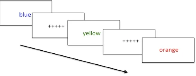
Figure 3.8 Item presentation in the Stroop task………...…....…...………… 81
Figure 4.1 The proportion of patients who were significantly different to their appropriate control group based on the number of tasks within BLAST that were completed both pre and postoperative………. 93
Figure 4.2 The proportion of patients significantly different to their appropriate control group for each task within BLAST both pre and postoperatively...……….……….. 94
Figure 4.3 The percentage of patients impaired in the most sensitive tasks within BLAST by tumour localization both pre- and postoperatively...………...……….. 96
Figure 5.1 The numbers of cognitive skills within BLAST that patients were significantly
different to their respective controls groups on, both pre and postoperatively…… 111
Figure 5.2 The percentage of patients impaired on each cognitive skills assessed by the BLAST
both pre and postoperatively …...……….………... 112
Figure 5.3 The percentage of patients significantly different to there respective control group on each cognitive skill both pre and postoperatively ………….………... 114
Figure 5.4 The mean T Score for each core cognitive skill for the control group and the right anterior and right posterior group both pre and postoperatively. Error bars represent standard error of the mean. …...………...………... 117
Figure 5.5 The mean T Score for each core cognitive skill for the control group and the left anterior and left posterior group both pre and postoperatively. Error bars represent standard error of the mean…...……….………... 118
Figure 6.1. Overall preoperative lesion overlap map for brain tumour patients……….... 130
Figure 6.2. Power map for preoperative brain tumour patients showing axial slices on a
standardised template………...……….... 131
Figure 6.3 VLSM analysis for preoperative performance on accessing semantic knowledge,
lexical selection, goal driven response selection, verb retrieval and articulatory
motor planning………..………..………... 134
Figure 6.4 Overall postoperative lesion overlap map for brain tumour patients………...…... 136
Figure 6.5 Power map for postoperative brain tumour patients……….……….... 137
Figure 6.6 VLSM analysis for postoperative performance on accessing semantic knowledge,
Chapter 1: Introduction and Literature Review
Each year, doctors in the United States diagnose approximately 17,000 new primary brain tumours, and 100,000 secondary brain tumours (Porter, McCarthy, Freels, Kim & Davies, 2010). According to Cancer New Zealand, approximately 100-120 people are diagnosed with a specific malignant brain tumour each year (“Brain Cancer: Glioblastoma multiforme (GBM),”2013). Overthe past few decades, there have been significant improvements in outcomes for patients with brain tumours. The proliferation of more effective treatment procedures, such as the use of new chemotherapy agents and surgical interventions (e.g., awake craniotomy), is increasing life expectancy in this population. With the development of these new techniques, surgeons and clinicians have been able to shift their attention from acute management towards the
consideration of quality of life issues, and more specifically how it can be maximised. One important goal in this respect has been the preservation of language function during surgical intervention, since the ability to communicate is such an essential component of everyday life.
Effective language assessment is central to this goal. First, effective preoperative
language assessment can identify candidates for awake craniotomy, a surgical technique that has been found to be more effective than standard resective surgery at reducing postoperative
language deficits (Ali, Fadel, & Abouldahab, 2009; De Benedictis, Moritz-Gasser & Duffau, 2010; Duffau, 2007; Gupta et al., 2007; Peruzzi, Bergese, Viloria, Puente, Abdel-Rasoul & Chiocca, 2011; Sacko et al., 2011). Second, preoperative assessment can also help assist in the selection of language tasks for intraoperative testing during awake craniotomy. This can help the neurosurgeon identify cortical tissue that is essential to language for each patient, and thereby help preserve language function (De Witte & Mariën, 2013). Third, the detection of language impairments by postoperative assessment can guide more effective rehabilitation (Davie,
Hutcheson, Barringer, Weinberg, & Lewin, 2009). However, before these goals can be met, it is imperative that we examine in detail the language capabilities of brain tumour patients generally. This thesis aims to carry out such an endeavour. The following section will give a brief overview of the characteristics and etiologies of a brain tumour. Following this, we then explore the
current literature pertaining to the implications of a brain tumour on language functioning. Brain Tumours: A Brief Overview
A brain tumour is a solid abnormal mass of tissue within the brain or the central spinal canal. It comes in various shapes, locations and sizes, and exhibits many different types of growth patterns (Ricard, Idbaih, Ducray, Lahutte, Hoang-Xuan, & Delattre, 2012). Brain tumours are created by abnormal and uncontrolled cell division. They can develop within the brain itself (e.g., glial cells: astrocytomas, oligodendroglioma, ependymomas), but also in lymphatic tissue (e.g., cerebral lymphoma), in blood vessels (e.g., hemangioblastoma), in the cranial nerves (e.g., schwannoma), in the brain meninges (e.g., meningioma), the skull (e.g., chondrosarcomas pituitary gland (e.g., pituitary adenoma), or pineal gland (e.g., pineocytoma). Tumours that grow in this manner are called primary brain tumours. The most common type of primary brain tumours is gliomas (50.4%), followed by meningiomas (20.8%) and then pituitary adenomas (15%) (Park, Kim, Sade & Lee, 2009). Brain tumours may also spread from cancers primarily located outside the central nervous system; these are called secondary or metastatic
tumours. The most common source of origin for metastatic tumours derives from carcinomas of
the breast, lung, and malignant melanoma. Metastatic tumours occur more frequently than primary brain tumours (4:1) (Marsh, 2009).
Currently, no one knows exactly what causes brain tumours, and there have only been a few risk factors identified. For example, children who receive radiation to the head have a higher
risk of later developing a brain tumour (for review see Pettorini, Park, Caldarelli, & Massimi, 2008), as do people who have rare genetic conditions (Behin, Hoang-Xuan, Carpentier, Delattre, 2003). However these cases only represent a fraction of those who are diagnosed with a primary brain tumour each year.
Brain tumours can result in a range of neurological symptoms, and these can be divided into three main categories. The first set of symptoms derives from increased intracranial pressure. Clinically, this translates into headaches, vomiting, altered state of consciousness, dilation of the pupil, and papilledema (swelling of the optic disc, located at the back of the eye). The second results directly from damage to the brain by either compression or infiltration of the tumour. Any type of focal neurological symptom may occur, such as motor, cognitive, and behavioural
impairment, and/or personality or emotional changes. The third and final category is irritation. This includes such symptoms as abnormal fatigue, weariness, absences, and tremors (see especially Arber, Faithfull, Plaskota, Lucas & de Vries, 2010; Cahill, LoBiondo-Wood, Bergstrom, & Armstrong, 2012; Davies & Clarke, 2004; Forsyth & Posner, 1993; Gutin & Posner 2000; Omuro, Leite, Mokhtari & Delattre, 2006; Squires, 1989;). Importantly, this also includes epileptic seizures, which are the most common neurological symptom that will motivate a brain tumour patient to seek medical attention. For example, Taphoorn and Klein (2004) reported that 80% of brain tumour patients had a seizure prior to diagnosis.
The neuroanatomical locations, as well as the rate of growth and invasiveness of a brain tumour, are key determiners of this symptomology (DeAngelis, 2001). Tumour growth and invasiveness are directly related to the histological features of a tumour (Bosman, Carneiro, Hruban & Theise, 2010). Broadly speaking, a tumour can either be cancerous (malignant) or non-cancerous (benign). More specifically, the World Health Organization (WHO) has
developed a malignancy scale, from grade I-IV, to quantify the histological features of brain tumours (Kleihues & Sobin, 2000). The specific histologic features used for each grade are presented in Table 1.1. Patients with a high-grade brain tumour have a much poorer prognosis than lower grade tumours. A high-grade tumour carries a prognosis of 6-18 months, depending on age and disability at presentation. A low-grade brain tumour can carry a prognosis of many years, but will ultimately transform to a higher grade if there is incomplete surgical resection of the tumour (Marsh, 2009).
Table 1.1
The WHO grading of Central Nervous System tumours WHO grade
I
Lesions with low proliferative potential, a frequently discrete nature, and the possibility of cure following surgical resection alone
WHO grade II
Lesions that are generally infiltrating and low in mitotic activity but recur
WHO grade III
Lesions with histologic evidence of malignancy, generally in the form of mitotic activity, clearly expressed infiltrative capabilities, and anaplasia
WHO grade IV
Lesions that are mitotically active, necrosis-prone, and generally associated with rapid preoperative and postoperative evolution of disease.
Brain Tumours and Language
There is consensus in the literature that a brain tumour can have a profound impact on cognitive functioning (see especially Klein et al., 2002; Meyers, Hess, Yung & Levin, 2000; Murray et al., 2000; Scheibel, Meyers & Levin, 1990; Talacchi, Santini, Savazzi & Gerosa, 2011; Taphoorn & Klein, 2004; Taylor et al., 1998). One such aspect of cognitive functioning
within this domain is language. It has been difficult to establish the exact prevalence of linguistic dysfunction in this population, and this is due to a number of pitfalls evident throughout the literature. Specifically, these research investigations that have set out to investigate language dysfunction in brain tumour patients differ greatly as to the characteristics of the patient sample, and type of surgical intervention used. Such studies investigate language function in awake craniotomy patients only,a procedure that may be especially indicated for when a tumour is in “classical” language areas (e.g., Broca’s and Wernicke’s area). Language dysfunction is likely to be more prevalent in this kind of sample than in an undifferentiated sample. Others investigate language function only in left hemisphere patients, and often only in cases where the tumour is in close proximity to the traditional language areas. Consequently, it is difficult to draw any
definitive conclusions across these investigations, and very little is therefore known about language capability in tumour patients more generally.
Furthermore, there is huge variation in the testing protocols used throughout these investigations. Measures used range from informal self-report measures (e.g., Thomas et al., 1995) to formal aphasia assessments (e.g., Duffau, Peggy Gatignol, Mandonnet, Capelle & Taillandier, 2008; Whittle et al. 1998) through to specific neuropsychological protocols (e.g., Bello et al., 2007; Sanai, Mirzadeh, & Berger, 2008). Caution must therefore be used in
interpreting linguistic deficit rates in this population, and one must always consider the sample of brain tumour patients used, as well as the sensitivity of the linguistic assessment methodology.
With this in mind, from the growing body of research exploring linguistic deficits in brain tumour patients, it is clear that language dysfunction occurs in this population. In
preoperative samples, estimates of the prevalence of language deficits range from 37% to 63% (Bello et al., 2007; Haglund, Berger, Shamseldin, Lettich, & Ojemann, 1994; Recht, McCarthy,
O’Donnell, Cohen & Drachmann, 1989; Sanai, Mirzadeh, & Berger, 2008; Tandon & Mahapatra, 1993; Thomas, O’Connor & Ashley, 1995; Whittle, Pringle & Taylor, 1998;). For example, Thomas, O’Connor and Ashley (1995) found that in an undifferentiated sample of 116 patients with a high-grade glioma, 37% had a “speech deficit” at presentation, according to the report of the patients themselves and/or their primary caregivers. Whittle, Pringle and Taylor (1998) found that in 40 left hemisphere brain tumour patients about to undergo tumour resection surgery, 62.5% were classified as dysphasic according to the Western Aphasic Battery (WAB) (Kertesz, 1982). Finally, Sanai, Mirzadeh, and Berger (2008) also found that of 250 left hemisphere glioma patients, 36.4% had a language deficit preoperatively. This was identified by impairment on at least one of the following tasks: counting, object naming, single word reading, sentence repetition, and writing words and sentences.
These patterns of linguistic dysfunction occur as a result of the infiltration, displacement, and compression of both gray and white matter (subcortical nuclei) within the cerebral cortex. Furthermore, it is well know that language abilities rely on the integrity of neural networks whose cortical nodes are frequently located within more than one lobe of the left hemisphere (sometimes both hemispheres) and are connected by white matter pathways (for review see Friederici, 2014). A brain tumour can also impact on language by causing disruption in the connectivity of white matter pathways due to deviation, infiltration, edematous and destruction (Jellison, Field, Medow, Lazar, Salamat & Alexander, 2004).
Second, language dysfunction in brain tumour patients may occur as a result of the surgery itself. This may occur due to the resection of brain tissue essential for language function (known as eloquent cortex), or from postoperative complications such as swelling and
difficult to draw conclusive evidence about the effects of neurosurgery on language processing. Some research has found a low incidence of language deficits caused by surgery in postoperative brain tumour patients. For example, Duffau and colleagues (2008) assessed a series of 115 left hemisphere patients with grade II gliomas on the Boston Diagnostic Aphasia Examination (Goodglass, Kaplan & Barresi, 2001) both before and after awake craniotomy surgery, and observed new language impairments following surgery in only 2% of patients (Duffau, Peggy Gatignol, Mandonnet, Capelle, & Taillandier, 2008). McGirt and colleagues (2009) conducted a comprehensive retrospective analysis of 306 undifferentiated patients who had undergone
resection of glioblastoma. They found that 5% of patients developed an acquired language deficit postoperatively (the language assessment protocol used in this investigation was not explicitly stated) (McGirt, Mukherjee, Chaichana, Than, Weingart, & Quinones-Hinojosa, 2009).
However, several other studies have suggested that language impairments induced as a result of surgery may be more common. For example, Bello and colleagues (2007) conducted a comprehensive language examination in 88 left hemisphere glioma patients who had undergone intraoperative mapping of subcortical language tracts during an awake craniotomy. The
participants performed a range of language tasks, including picture naming, famous face naming, action naming, word repetition (real and nonsense words), picture-word matching, and
letter/category fluency (e.g., “name as many items as you can think of that start with letter ‘F’/ belong to the category ‘animals’). In patients whose intraoperative mapping revealed positive subcortical language sites (N=52), evaluation of language three days after surgery showed new deficits or worsening of existing language deficits in 67.3% of patients (Bello et al., 2007). In addition, Ilmberger and colleagues (2008) conducted a prospective longitudinal study to evaluate language in 149 patients with a tumour in close proximity to or within language areas (therefore
the location of the tumour was confined to the left hemisphere). To achieve this, the Aachen Aphasia Test (AAT: Huber, Poeck, & Willmes, 1983) was used. This standardized battery consists of five subtests: 1) the token test, which involved pointing to and manipulating
geometric forms in response to a command; 2) repetition of phonemes, words and sentences; 3) written language, consisting of reading and writing single words; 4) naming, involving naming visually presented objects, colours and scenes; and 5) comprehension, using the picture word matching task. Patients were classified as having a language deficit whenever they showed at least mild disturbance in one of the subtests above. Using this classification, it was found that within 21 days after surgery 32% of patients without preoperative deficits had a new language deficit (Ilmberger, Ruge, Kreth, Briegel, Reulen, & Tonn, 2008). Thus, brain tumour patients maybe particularly susceptible to new or worsening of existing language function shortly post-surgery (for a review of similar findings see Finch & Copland, 2014).
Within this body of literature, a limited pool of research investigations have suggested
that the linguistic profiles of brain tumour patients are distinctly different from those observed in post-stroke aphasia. First, it has been found that language impairments in brain tumour patients are more likely to appear as mild deficits in common aphasic testing protocols. In contrast, language impairments evoked by a stroke are more likely to be severe and appear as more globalised deficits in common aphasic testing. For example, Anderson, Damasio and Tranel (1990) compared a sample of 17 brain tumour participants (eight with a left hemisphere tumour, nine with a right hemisphere) with an equal sized sample of unilateral stroke patients. Each stroke patient was anatomically matched to one of the tumour patients on the basis of lesion location and size. It was specified that lesions in stroke cases could be either as large or smaller than the lesion in the matched tumour cases. The Multilingual Aphasia Battery (Benton, 1969)
was administered as well as the Boston Diagnostic Aphasia Examination Reading Sentences and Paragraphs subtest (Goodglass & Kaplan, 1983). Despite the close matching of the two groups, there were major differences in each group’s performance. Of the left hemisphere cases, all of those in the stroke group had more severe language deficits than did those in the tumour group, despite the fact that the average lesion size was larger in the brain tumour group. The left hemisphere stroke subjects showed greater impairment than the tumour group in all subtests of the Multilingual Aphasia Examination (Benton, 1969), as well as on the Boston Aphasia
Examination Reading subtest. Furthermore, Davie, Hutcheson, Barringer, Weinberg, and Lewin (2009) used the Western Aphasia Battery (Kertesz, 1982) to evaluate the language performance of 65 patients who had recently undergone malignant tumour resection. They found that anomic aphasia was the most common type of aphasia in this group (48% of patients), whereas global aphasia was the least common (3% of patients). This markedly contrasts with the profile of aphasia in stroke patients, where there are higher rates of global aphasia (20-40%) and lower rates of anomic aphasia (9-28%) (for similar findings of aphasia profile in stroke, see Kauhanen et al., 2000; Kertesz & Sheppard, 1981; Pashek & Holland, 1988; Pedersen et al., 2004).
There also appears to be differences in the patterns of language recovery observed between brain tumour patients postoperatively and in post-stroke aphasia (Shafi & Carozza, 2012). In post-stroke aphasia, some degree of recovery typically occurs spontaneously within eight to 12 weeks, and peaks after one year with only minimal improvements thereafter (see Berthier, 2005). In brain tumour patients, studies have shown that the majority of patients who experience a decline in language function immediately post surgery, will experience considerable recovery of function within three months of surgery (Finch & Copland, 2014; Wu et al., 2011). This pattern has been attributed to a number of factors that are more salient in brain tumour
compared to stroke. These include resolution of postsurgical oedema, transient retraction injury, initial displacement of neural structures, and neuroplastic mechanisms (Bello et al., 2007). However, although such a pattern has been argued within the literature, it must be noted here that some studies do present a far less promising picture of language recovery in this population. For example, Papagno, Casarotti, Comi, Gallucci, Riva, and Bello, (2012) found that at three months post surgery, a significant proportion of left hemisphere brain tumour patients were still impaired on certain language tasks. Specifically, 48% of left temporal patients who had a low-grade glioma were impaired in a naming famous people task1, and 40% of left frontal patients were impaired in a letter fluency task. In addition, Ilmberger et al., (2008) used a battery of language tasks previously described and found that in a sample of 153 awake craniotomy patients, 17.6% of patients had a persistent postoperative language disturbance seven months post surgery.
The emergence of evidence that indicates that language profiles in tumour patients are different to those in stroke patients is perhaps not surprising, given the contrasting pathological mechanisms between the two neurological entities. One such difference is that unlike stroke, which generally has an acute onset, tumour growth progresses gradually, allowing for the possibility of cortical reorganisation (for discussion see Miceli, Capasso, Monti, Santini & Talacchi, 2012). Indeed, there is evidence that Broca’s aphasia, which is commonly observed in post-stroke aphasia following damage to Broca’s area and surrounding regions, is rarely
observed when a tumour develops in that region (Plaza, Gatignol, Leroy & Duffau, 2009). This observation might well be attributable to the greater opportunities for neuroplasticity phenomena to occur in tumour patients (Duffau, 2007). A second difference is that whereas vascular damage results directly in neural cell necrosis, tumours grow by infiltrating nonneural cells (e.g., glial
1 The famous person to be named belonged to one of four professional categories (artists/scientist, athletes, actors, politicians) and are graded for the period of his/her fame.
cells or meningeal tissue), and only begin to impact on neural function when there is significant displacement and compression of neural tissue. For that reason, tumour growth may be
considerably advanced before any functional impairment is observed (Miceli et al., 2012). A third difference between vascular and tumour damage concerns the distribution of the lesion. The cerebral regions most vulnerable to stroke (particularly ischemia) are those that lie within the region supplied by the occluded artery/arteriole, and consequently some regions are consistently more vulnerable than others. The cerebral regions impacted by a tumour, on the other hand, can be extremely variable, particularly when the tumour involves interstitial tissue.
Therefore, it is safe to say that the language profiles observed in subjects with brain tumours are likely to differ significantly and substantially from those observed in patients with aphasia of vascular origin. Consequently, classic test batteries (e.g. the Boston Diagnostic Aphasia Examination (BDAE) (Goodglass, Kaplan & Barresi, 2001), or the Western Aphasia Battery (WAB) (Kertesz, 1982)) that have been developed primarily for the classification of “classical” aphasic syndromes induced by a cerebrovascular accident (Broca’s, Wernicke’s, conduction, transcortical etc.), may not be optimal for the detection of language deficits in brain tumour patients. Indeed, this is a view that has been espoused previously by a number of
researchers (De Witte & Mariën, 2013; Meyers & Brown, 2006; Miceli et al. 2012; Påhlson, Ek, Ahlström & Smits, 2003; Talacchi, Santini, Savazzi & Gerosa, 2010).
One recent study serves as a rare example of a neuropsychological assessment tailored specifically to tumour patients. The recent Milano-Biocca Battery is designed to investigate the performance of tumour patients in three cognitive domains: language, memory and executive function (Papagno, Casarotti, Comi, Gallucci, Riva, & Bello, 2012). The entire test battery has been administered to 226 tumour patients both pre- and postoperatively, and at three months post
surgery. To investigate language, the following tasks were administered: letter and category fluency, naming famous persons from photographs, object naming, picture-word matching, action naming, naming by description, and real word, nonword and sentence repetition. Although only preliminary results have been published, Papagno and colleagues (2012) found that the following language tasks were the most sensitive at detecting language impairments before surgery and at three-month follow up: naming famous people, action naming, object naming and category and letter fluency. In addition, patients were categorised into four groups based on the anatomical location of the tumour – left frontal, left temporal, right frontal, and right temporal. It was found that tumour localisation was a strong predictor of performance in most of the
language tasks administered. Specifically, patients in the left temporal group performed more poorly than all other groups in naming people and objects, and this was true of all the testing points. They were also worse in category fluency at three months post surgery. In contrast, patients with a left frontal lesion performed more poorly than all other groups in the letter fluency task, and this was true both preoperatively and at three months post surgery (Papagno et al., 2012).
However, comprehensive neuropsychological batteries of this kind take time to
administer (up to two hours for the Milano-Bicocca), and even then, the number of test results that are directly pertinent to language function is relatively small. Here we focus on just one cognitive domain – language. This will enable us to test that domain more extensively, and in doing so, perhaps identify those measures that are most sensitive at detecting impairment in this population. It is interesting to note that in the original study of the Milano-Biocca Battery, only five of the 11 language tasks in the original battery were sufficiently sensitive at detecting
more exploratory assessment of language, which aims to assess as many aspects of language function as possible, as sensitively as possible, may be a useful first step in the development of shorter, more carefully tailored assessment protocols for use with a brain tumour population.
Current cognitive theories of language may provide a particularly useful starting point for such an endeavour, because such theories delineate the various core cognitive operations that are essential for performing particular language behaviours. Assessments can then be developed which target each of these “core skills”. This approach offers a systematic, theory-driven method for deciding what tasks should be included in the assessment. Also, if applied effectively, this method may help to maximise the range of skills examined (thereby providing better breadth and sensitivity), and is likely to offer greater power to discriminate amongst different language profiles based on the neurological profile of the patient. The following section reviews cognitive and neuropsychological research that is relevant to this objective. It is then followed by a review of research that investigates the neural structures associated with each core skill, with a particular emphasis on lesion studies.
Cognitive Theories of Language
Current cognitive theories of language posit that key language behaviours, such as producing words and sentences, understanding spoken words and sentences, and reading, can be decomposed into several more elementary cognitive skills. Indeed, impairments to each
cognitive skill have been found to be associated with a unique neurological and linguistic profile (see Table 1.2 for a summary of these skills and their associated neurological profiles). Before beginning this review, we acknowledge that within these models there is considerable debate in the literature as to the exact cognitive mechanisms associated with certain skills. These debates go beyond the aims of this investigation. We have therefore chosen to examine only those
cognitive skills for where there is widespread agreement for their existence in the literature. Accessing Semantic Knowledge
If we begin by considering single word production, most psycholinguistic theories start with the simple task of naming a pictured object. There is wide agreement in the literature that at least four key cognitive skills are required for this task (see Figure 1.2). The first cognitive skill, which we will call accessing semantic knowledge, refers to the process of retrieving information
about the semantic category, function, colour, size, etc., of the item to be named (Friedmann, Biran & Dotan, 2013). Many psycholinguistic models suggest that this semantic information is organised into a network consisting of an interconnected matrix of nodes, which correspond to individual features of the target item. For example, in the case of DOG, the attributes has four legs, barks, and chews bones, might all be coded for by different, but interconnected nodes, which collectively form the semantic representation for dog (e.g., Masson, 1991, 1995). Once partial information about an item is activated, activation spreads to these interconnected nodes, making additional information about the item accessible (Collins & Loftus, 1975; Neely 1997).
Evidence to suggest that accessing semantic knowledge is a distinct entity can be drawn from the neuropsychological literature. In a neurodegenerative disorder known as semantic dementia (SD), the most prominent early feature and presenting complaint is a difficulty in “remembering” the names of people, places and things (e.g., Pijnenburg, Gillissen, Jonker, Scheltens, 2004; Thompson, Patterson, & Hodges, 2003). Spontaneous speech retains its normal grammatical structure, but there may be frequent pauses as the speaker struggles to find a
particular word, and some terms may be replaced by commoner, more general terms (e.g., “thing” instead of “kettle”, and “doing” instead of “cooking”). Pronunciation and phonological skills are usually unaffected (Adlam et al., 2006; Ash, Moore, Antani, McCawley, Work, & Grossman,
2006). As the disease progresses, difficulties become evident in tasks involving comprehension. For example, when asked to define a word, people with SD may be able to provide only very general information (e.g., “Ostrich. Can you say that?” “Yeah, ostrich”; “What is a
hippopotamus?” “An animal”) or simply absent (“I think I’ve heard of a hippopotamus, but I can’t say what it is”) (Hodges & Patterson, 2007).
Tasks that are commonly used to examine SD are picture naming, category fluency and picture-word matching. On picture naming, individuals with SD may be particularly prone to
semantic errors, where the target word is replaced by another word from the same semantic
category (e.g., zebra -> “giraffe”) (Garrard, Perry & Hodges, 1997; Hodges, Graham & Patterson, 1995; Jefferies & Lambon Ralph, 2006). On category fluency tasks, there is also a notable
decrease in the number of words that the individuals can generate when given a semantic category; in contrast, letter fluency is relatively spared (Bozeat, Lambon Ralph, Patterson,
Garrard & Hodges, 2000; Graham & Hodges, 1997; Hodges, Patterson, Oxbury & Funnell, 1992; Rascovsky, Salmon, Hansen, Thai, & Galasko, 2007;). This is an unusual pattern, in most brain-damaged patients, letter fluency is disproportionately impaired. Finally, on picture word
matching tasks, SD patients tend to show confusion between semantically related items. They are particularly prone to errors on tasks where they must choose a picture match for a word from amongst a number of alternatives from the same semantic category (see esp. Corbett, Jefferies, Ehsan & Lambon Ralph, 2009).
Lexical Selection
Many theories of single word production also distinguish this general semantic processing stage from a subsequent lexical (or lemma) selection stage. This cognitive skill involves selecting the appropriate word from the mental lexicon that best matches the semantic
concept in mind. In many theories, this stage of processing in conceptualised within a spreading activation framework (e.g., Caramazza 1997; Dell, 1986; Levelt, 1999, Rapp & Goldrick, 2000; Roelofs, 2004; Ruml, Caramazza, Capasso & Miceli, 2005; Schwartz, Dell, Martin, Gahl & Sobel, 2006). For example, according to Dell’s (1986) model, depicted in Figure 1.1, during single word production, semantic nodes that represent aspects of the meaning of an item transmit activation to their associated lexical units. All units of associated words receive some activation (for example, if the target item is a cat, the lexical unit for “dog” will also become activated, because dogs possesses some of the same semantic attributes). However, the word that contains the greatest number of semantic features will generally receive the most activation. The lexical selection step is complete when the most highly activated lexical unit is “selected” for production.
Support for the existence of this lexical selection process, as distinct from semantic access, comes from studies that have demonstrated reduced naming efficiency in normal speakers when two or more words “compete” for selection. For example, in the picture-word interference task, participants must name a picture, which is accompanied by an irrelevant auditory or written word (for example, a picture of a tiger is accompanied by the word “lion”). Specifically, when the irrelevant distractor word is semantically related to the target, and is presented just before or at the same time as the picture, individuals are substantially slower to name the picture than they are when the word is unrelated (Glaser & Düngelhoff, 1984; Roelofs, 1992; Starreveld & La Heij, 1995, 1996). It has been suggested that this delay is caused by
competition for selection between the two concurrently activated lexical items – that is, the name of the target item, and the distractor word itself.
Figure 1.1: The two-stage theory of single word production (adpated from Dell et al. 1997).
One neuropsychological profile in which this lexical selection stage appears to be implicated is classical or pure anomia (Andreetta, Cantagallo & Marini, 2012; Butterworth, 1992; Lambon Ralph, Sage & Roberts, 2000; McNeil, Odell, & Tseng, 1991). The hallmark feature of this disorder is poor picture naming without any accompanying impairment in general semantic knowledge (for example, comprehension may be normal; Franklin, Howard &
Patterson, 1995; Howard, 1995; Gonon, Bruckert & Michel, 1989; Laine, Kujala, Niemi, & Uusipaikka, 1992; Raymer et al., 1997). In picture naming tasks, these patients tend to produce a mix of errors, including many failures to respond and/or circumlocutions (“They live in the sea and they lay eggs on the beach, but I can’t think of the name”). It has been argued that these kinds of errors in particular may be a consequence of a failure to retrieve any lexical item from the mental lexicon (for review, see Dell, Lawler, Harris & Gordon, 2004). For example, case, RBO, who suffered from a ruptured A-V malformation of the left posterior communicating artery, failed to provide any response at all to 40% of the items in a large picture naming test (Miceli, Amitrano, Capasso & Caramazza, 1996). Individuals with this profile may also produce
semantic errors, but in contrast to individuals with SD, they are more likely to recognise their errors as incorrect (Lambon Ralph, Sage & Roberts, 2000). One final hallmark of individuals with this profile is a marked word frequency effect in naming and similar word production tasks: individuals are considerably more prone to errors/omissions on lower frequency words (e.g., case AW: Jacobs, Singer & Miozzo, 2004; case FR: Avila, Lambon Ralph, Parcet, Geffner &
Gonzalez-Darder, 2001). It has been suggested that the lexical representations of high-frequency words have higher resting levels of activation or lower selection thresholds than those of lower frequency words. Therefore, they require less activation to reach the activation level that is critical for selection, so may be less affected by any impairment affecting lexical activation (for review see Nickels, 2002).
Phonological Encoding
Subsequent to lexical selection, cognitive models of single word production generally propose a stage of processing called phonological encoding. This involves retrieving information
about the selected word’s sound form from the mental lexicon (see especially Dell, 1986; Levelt, 1999; Rapp & Goldrick, 2000; Schwartz et al., 2006). This abstract sound information then forms the primary input for articulatory-motor programming. Some models suggest that the phonological encoding process can itself be subdivided into a number of smaller cognitive processes. For example, Levelt (1999) proposed that the relevant phonological segments and their metrical information (for example, the number of syllables and their stress pattern) are retrieved independently and in parallel, and then subsequently combined.
There is a wealth of evidence for the existence of an aphasic disorder that arises due to a selective impairment in the ability to encode phonological information. In conduction aphasia individuals speak fluently and with ease, but produce a number of phonological errors in
spontaneous speech and on a range of single word production tasks (for example, the person may say “pabacco” instead of “tobacco”; see Buchsbaum, et al., 2011). As many as 50% of these individuals’ responses may be phonological errors (Kohn & Goodglass, 1985; see also
Butterworth, 1992; Caplan, Vanier & Baker, 1986; Kohn & Smith, 1993; Pate, Saffran & Martin, 1987; Pradat-Diehl, Tessier, Vallat, Mailhan, Mazevet & Lauriot-Prevost, 2001; Wilshire & McCarthy, 1996). These patients also exhibit a number of other features consistent with a deficit in phonological encoding. For example, they tend show a strong length effect in picture naming – that is, they are less accurate at producing words that contain multiple syllables (Caplan, Vanier, & Baker, 1986; Kohn & Smith, 1993; Pate, Saffran, & Martin 1987; Wilshire &
McCarthy, 1996; Wilshire, 2002). Most models predict that multisyllabic items will place extra demands on phonological encoding, as additional phonemes need to be retrieved and/or inserted into the correct metric frame. In addition, they may also produce phonological errors in auditory word repetition tasks, particularly on longer words (see esp. Caplan, Vanier & Baker, 1986; Caramazza, Basil, Koller & Berndt, 1981; Dell, Schwartz, Martin & Saffran 1997; Strub & Gardner, 1974). One of the critical prerequisites for successful performance in this task is likely to be the ability to encode the phonological information of the word to be repeated.
Articulatory-Motor Programming
According to most theories, the output of the phonological encoding process consists of fairly abstract, syllabified phonological words that are then translated into articulatory-motor programmes. This final processing stage will be referred to as articulatory-motor programming. This process involves constructing a motor plan for the articulatory execution of that utterance (see esp. Romani, Olson, Semenza, Granà, 2002; Romani & Galluzzi, 2005; Indefrey & Levelt, 2004). According to Levelt’s (1999) theory, speakers have access to a repository of syllabic
gestures, termed the ‘mental syllabary’, that contains the articulatory scores for at least the most common syllables in language (Levelt 1992; Levelt & Wheeldon, 1994). As soon as a syllable emerges from the phonological encoding process, the corresponding syllabic articulatory gesture will be selected from the repository (Indefrey & Levelt, 2004; see also Dronkers, 1996; Kerzel & Bekkering, 2000).
There is certainly supporting neuropsychological evidence for the existence of a distinct articulatory-motor programming stage of processing. Apraxia of speech (AOS) has been
described as a disorder of motor-speech programming, which leads to errors in sequencing, timing, coordination, initiation and vocal tract shaping (Darley, Aronson, & Brown, 1975; Kent & Rosenbek, 1983). Hallmark features of patients with AOS are articulatory errors and prosodic abnormalities. Articulatory errors are more common on certain kinds of segments than others – for example, affricates (e.g., ch and j) and fricatives (e.g., s and z) tend to be particularly error-prone, and errors are also more common on consonant clusters rather than singleton consonants (e.g. ‘strict’ will be more difficult than sit) (for review see Ogar, Slama, Dronkers, Amici, & Gorno-Tempini, 2005). Patients with AOS additionally have a markedly reduced rate of speech – considerably lower than that of individuals with aphasia that do not have AOS (Canter, Trost & Burns, 1985).
Figure 1.2: The four key cognitive skills involved in single word production using picture
naming as a framework (adapted from Wilshire, 2014) Sentence-level Planning
So far, we have considered the production of single words, but within language a crucial skill is being able to incorporate these words into a grammatically correct sentence. Cognitive theories of sentence-level production vary considerably in the specific cognitive processes they propose (see especially Dell, 1986; Garrett, 1975; Levelt, 1989; Levelt, 1999; Stemberger, 1985). Some theorists propose a frame-allocation process. That is, the speaker builds an abstract
representation of a “sentence “frame” which specifies the classes of lexical elements that appear, their order, and any necessary grammatical elements. In most models the elements in the frame are defined grammatically (noun, verb, etc.). Words that fulfil these grammatical criteria can then be inserted, resulting in a fully formed ordered ‘plan’ of the sentence (Garret, 1975, 1976, 1982). In some models, selection of the appropriate verb is crucial for the development of an appropriate sentence frame, as the verb specifies important aspects of the frame, such as the
number of arguments (how many direct and indirect objects) the verb can take (e.g. Ahrens, 2003; Levelt 1989, 1999; Shapiro & Levine, 1990, Shapiro, Zurif, & Grimshaw, 1987; Trueswell & Kim, 1998). A rather different view recently espoused is that the sentence “plan” might be more like a proposition – that specifies the main entities, their properties and their relations to, or actions upon one another (e.g. dog-> agent, cat-> patient chase-> action; Chang, Dell and Bock, (2006)). The most salient conceptual element in this proposition wins the competition to initiate sentence planning. Sentence order and grammatical structure is then generated by applying a set of rote-learned ordering rules. In sum, a number of cognitive skills have been identified that are uniquely involved in sentence-level planning. These include grammatical frame insertion, the ability to generate verbs, as well as the development of an internally generated sentence level plan. For the purpose of the current review we will focus on the latter three cognitive processes, as these skills can be examined at the single word level. Assessment of other cognitive processes such as grammatical frame insertion needs to be examined at the sentence level, and this will be assessed in future investigations.
Sentence-level Planning: Verb Production
First and foremost, in order to produce a sentence, a speaker must be able to successfully retrieve all the key lexical content elements, most particularly the main verb (verb retrieval), as it imposes powerful constraints on the structure of the sentence to be produced (for example, it determines whether a direct or indirect object can be included (see especially, Sloan Berndt et al.1997a, 1997b; Webster & Whitworth, 20102). This ability can be disproportionately impaired after brain damage (for a review see Mätzig et al., 2009). Black and Chiat (2003) argue that nouns and verbs differ at both the semantic and the syntactic level. At the semantic level, verbs differ from nouns in their sensory richness and tightness of
conceptual-semantic fit. Moreover, as several recent authors have pointed out, they contribute to the
meaning of a sentence in a different way from nouns, because they specify not just the nature of an action, but also the entire event involving that action, including the participating roles of the entities described in the sentence (Marshall, Chiat, & Pring, 1998). At the syntactic level, verbs vary as to the number of arguments they can take, and these impose significant constraints on the syntactic structure of the sentence. So for example, the verb “fell” requires an agent argument only (e.g. “the clown fell”), whereas “kick” requires a direct and an indirect object (e.g. the horse kicked the jockey”), while “send” requires three arguments: an agent, a theme and a goal, as in “Dean sent the car to the garage” (Agent is assigned to the subject Dean, Theme is assigned to
the direct object the car, and Goal is assigned to the indirect object the garage) (Thompson, Shapiro, Li & Schendel, 1995).
Deficits in verb retrieval have long been observed in a range of aphasic syndromes, and this most likely reflects the complexity of this linguistic skill. However, it has been reported on numerous occasions that patients with nonfluent aphasia (e.g., Broca’s aphasia) have
significantly greater impairments in verb than noun retrieval (for review see Mätzig, Druks, Masterson & Vigliocco, 2009). In nonfluent aphasia, utterances are often reduced to one or two words, and are separated by long pauses, even though single word naming can be relatively well preserved (e.g., McCarthy & Kartsounis, 2000; Schwartz & Hodgson, 2002; Williams & Canter, 1982). In a comprehensive review of the literature, Mätzig and colleagues (2009) explored the dissociation between verb retrieval as measured by a commonly used action-naming task and an object-naming task in 269 aphasia patients. Of the nonfluent aphasics in this sample (N=132), three-quarters showed poorer performance in verb naming than object naming. This was the highest proportion of dissociation when compared with other aphasic subtypes. In addition, 49
aphasics had a dissociation of greater than 30% accuracy in object-naming compared to an action-naming task. Of these, 60% of patients were nonfluent aphasics (Mätzig, Druks,
Masterson & Vigliocco, 2009). Such observations, have led some to argue that deficits in verb retrieval can explain the difficulty in producing cohesive sentences in nonfluent aphasia (see Marshall, Pring & Chiat, 1998). Specifically, it has been found that deficits in single word action naming tasks are associated with further impairments in sentence-level processing. For example, Thompson, Lange, Schneider, and Shapiro (1997) found that a group of 10 patients with
nonfluent aphasia who were disproportionately poor at an action-naming task (when compared to object naming) were also disproportionately poor at producing sentences when they contained verbs with multiple argument structures; this impairment was much less evident in sentences that contained a verb with one argument structure.
Sentence-level Planning: Goal-Driven Response Selection
To produce longer utterances, a speaker must also manage their activation levels so that no item gets selected before it is supposed to. It has been suggested that there is a dedicated mechanism that performs this function, which operates by biasing the flow of activation, either enhancing, or inhibiting lexical representations based on the current production goal (see especially, January, Trueswell & Thompson-Schill, 2009; Schnur, Schwartz, Brecher, &
Hodgson, 2006; Scott & Wilshire, 2010; Speer & Wilshire, 2013; Wilshire & McCarthy, 2002). It has been suggested that such a mechanism may play a crucial role in managing competition between elements planned in the same utterance (see Martin et al., 1999).
As previously mentioned, patients with nonfluent aphasia have immense difficulty producing sentences, despite relatively well preserved single word production. It has been suggested that these patients have a deficit in this control system and therefore have difficulties
managing competition between conflicting representations (Biegler, Crowther, & Martin, 2008; Hamilton & Martin, 2005; January et al., 2009; Schnur et al., 2006; Thompson-Schill,
D’Esposito, Aguirre & Farah, 1997; Wilshire & McCarthy, 2002). Evidence in support of this theory comes from the performance of individuals with nonfluent aphasia on tasks that appear to make heavy competition-resolution demands. For example, several individuals with nonfluent aphasia have demonstrated prolonged naming latencies in the Stroop task, where a colour name is presented and the participant must identify its display colour while ignoring what the word actually says (Hamilton & Martin, 2005; Scott & Wilshire, 2010). This task would appear to involve inhibiting a potentially competing response – the word name – in order to produce the desired response. Such individuals may also exhibit a characteristic pattern of performance in the verb generation task, which involves generating an action that is associated with a given noun (e.g., scissors -> “cut”). Thompson-Schill and colleagues argue that when the noun offers several alternatives (e.g., rope -> “tie”, “knot”, “pull”, “drag”), competition must be resolved between these alternatives before a single response can be selected (Thompson-Schill, D’Esposito, Aguirre, & Farah, 1997; Thompson-Schill, D’Esposito, & Kan, 1999). Using this task, Cameron-Jones (2008) found that nonfluent aphasics exhibited a disproportionate difficulty with multiple-alternative items, when compared to items where one single verb response is dominant (e.g., scissors -> “cut”).
Spontaneous connected speech also differs from simple single word tasks, such as object naming, in that it involves coming up with a message intention, which is then used to “drive” sentence production. This message intention if sufficiently strongly activated and maintained exerts top-down control of the language system, enabling goal-appropriate elements to be selected, and inappropriate elements to be rejected. A difficulty with this aspect of spontaneous
production may also impact on a person’s communicative abilities. One task that arguably involves spontaneously generating a very simple message intention is letter fluency, where the participant must generate as many words as they can think of which start with a particular letter. This task requires the participant to search through their mental lexicon in order to select
appropriate lexical items that adhere to goals of the task (for review see Henry & Crawford, 2004). There is a wealth of evidence to suggest that patients who suffer damage to the left frontal region of the brain show specific impairments in this task, (Baldo & Shimamura, 1998; Rogers et al., 1998; Schwartz & Baldo, 2001; Stuss et al., 1998) when compared to both patients with posterior lesions and controls (Pendleton et al., 1982; Perret, 1974). For example, Robinson, Shallice, Bozzali and Cipolotti (2012) found that letter fluency performance in 47 patients with a frontal lesion primarily due to stroke was significantly worse than 20 patients with a posterior lesion, and also when compared with 35 healthy controls. Additionally, they also found that patients with a left frontal lesion performed significantly more poorly in this task, compared to patients with a right frontal lesion. Additionally Papagno et al., (2012) also found that patients with a left frontal brain tumour were more likely to be impaired on this task than patients with a brain tumour in other anatomical regions.
The two hypothesised processes described here – the management of competition and “top-down” language control – may in fact turn out to be aspects of a single common capacity. For example, the ability to select the correct word for production when several candidates are currently activated may depend crucially on the strength of the “message intention”. A strong propositional message intention may provide a conceptual framework that helps the speaker ensure that each word selected has the desired function. This in turn may help to minimise direct competition between the different elements planned for production in a single sentence.
Therefore, for the purposes of this thesis, we will refrain from taking a position on this argument and instead refer to these various abilities collectively as “goal-driven response generation”. Language Comprehension: Auditory Word Identification
So far we have considered cognitive skills that are critical for language production – both at the level of the single word and the sentence level. We now turn to the question of language comprehension. Cognitive theories of auditory language comprehension suggest that this process can be broken down in similar ways to those depicted in theories of single word production. Firstly, if we consider the simplest example – comprehending a single auditory word – cognitive theories make a clear distinction between the auditory word identification process, whereby the auditory stimulus is associated with a single known word, and the subsequent meaning retrieval process, whereby a semantic description of the stimulus word is generated (Hickok & Poeppel, 2007; Marslen-Wilson & Welsh, 1978; McClelland & Elman, 1986; Norris, McQueen, & Cutler, 2000). Virtually all these theories agree that the former involves identification of the acoustic properties of the speech signal, and the subsequent mapping onto the most appropriate word representation in the mental lexicon, which we will call auditory word identification. Many cognitive theories of auditory word identification argue that spoken words are processed as speech unfolds, and in doing so listeners attempt to map incremental segments of the acoustic signal onto a representation in the mental lexicon. For example, according to the cohort model,
first proposed by Marslen-Wilson and Welsh (1978), the first few phonemes of a spoken word activate a set or cohort of word candidates that are consistent with that input. These candidates then compete with one another for activation. As more acoustic input is analysed, candidates that are no longer consistent with the input drop out of the set. This process continues until only one word candidate matches the input or the best fitting word may be chosen if no single candidate is
a clear winner (e.g. Cole & Jakimik, 1980; Dahan, Magnuson, Tanenhaus, & Hogan, 2001; Marslen-Wilson, 1987; Marslen-Wilson & Tyler, 1980; McClelland, Elman, & Diego 1986; Norris, McQueen, & Cutler, 2000; Taft & Hambly, 1986; Tyler 1984; Vitevitch & Luce, 1999).
Neuropsychological studies have found that patients with Wernicke’s Aphasia (WA), as well as patients with pure word deafness have profound deficits in auditory word identification (e.g. Caplan, Gow & Makris, 1995). WA is an acquired language impairment characterised by severely impaired single word comprehension with fluent and discorded speech (Goodglass & Kaplan, 1983; Goodglass, Kaplan, & Barresi, 2001). A common task used in the literature to examine this deficit is single word repetition, as successful performance in this task relies on the ability to identify the acoustic properties of the to-be-repeated word. It has been extensively reported that patients with WA demonstrate significantly poor performance on this task. Patients with WA have normal articulation; therefore failure on this task cannot be attributed to an inability to articulate the word (for review see Robson, Grube, Lambon Ralph, Griffiths & Sage, 2013). Another task commonly used is phoneme discrimination, in which participants have to determine if two spoken words are identical. The pairs are most commonly made to differ by one phonemic feature (e.g., “cap-tap”). It has been consistently demonstrated that patients with WA have marked difficulties with this task (e.g., Basso, Casati & Vignolo, 1977; Baum, 2002; Blumstein, Baker, & Goodglass ,1977; Miceli, Gainotti, Caltagirone & Masullo, 1980; Tallal & Newcombe, 1978). In one study, Robson and colleagues (2013) had 11 patients with WA complete the phoneme discrimination task. It was found that at a group level these patients were significantly worse on this task when compared to a group of 11 age and hearing-matched controls (Robson, Grube, Lambon Ralph, Griffiths, & Sage, 2013).