1
VIABILITY OF CULTURED PRIMARY HUMAN SKIN CELLS TREATED WITH 1, 6-HEXAMETHYLENE DIISOCYANATE MONOMER AND ITS OLIGOMER
ISOCYANURATE
Kaitlyn Phillips
A technical report submitted to the faculty at the University of North Carolina at Chapel Hill in partial fulfillment of the requirements for the degree of Master of Science in Public Health in the
Department of Environmental Sciences and Engineering in the Gillings School of Global Public Health.
Chapel Hill 2017
Approved by,
Advisor: Leena A. Nylander-French Reader: Rebecca Fry
3
TABLE OF CONTENTS
Abstract……….….……..4
Acknowledgements……….……….……5
List of Tables………...……6
List of Figures………...…….…..7
List of Abbreviations……….……….….8
Introduction……….….9
Specific Aims……….…17
Methods………...18
Results………...……….…………26
Discussion………..………32
4 ABSTRACT
Kaitlyn Phillips
Viability of Cultured Primary Human Skin Cells Treated With
1, 6-Hexamethylene Diisocyanate Monomer and Its Oligomer Isocyanurate (Under the direction of Dr. Leena Nylander-French)
5
ACKNOWLEDGMENTS
This study is supported by the National Institute for Occupational Safety and Health (T42/OH-008673) and the National Institute of Environmental Health Sciences, National Institutes of Health (National Research Service Award T32 ES007126).
I would like to thank my advisor, Dr. Leena Nylander-French, for the wealth of guidance and support she has given me over the past two years. I would like to thank Dr. Jayne Boyer for all the help and instruction she provided me with as well. I am lucky to have such wonderful female role models in my life, and graduate school would have been dramatically different (and worse) without these two.
This research would not have been possible without the contributions of past and present lab members. I would like to thank Laura Taylor for all her help, especially with data analysis. I would also like to acknowledge Rachel Vaughan, Jennifer Thomasen, Linda Gaines, Sheila Flack, and Kenneth Fent for their previous research.
6
LIST OF TABLES
7
LIST OF FIGURES
1. The structures of 1,6-hexamethylene diisocyanate (HDI monomer) (left) and HDI isocyanurate (right) - Page 8
2. Formation of the urethane bond - Page 8
3. A representative rATP standard curve. - Page 25
4. The average LC50 levels for cells exposed to HDI monomer or HDI isocyanurate (n = 4-7 per cell type; see Table 1) - Page 27
5. The average LC50 levels for the different cell types when exposed to HDI isocyanurate (n = 4-7 per cell type; see Table 1) - Page 27
6. Time course of cell death as measured using the CellTox Green cytotoxicity assay performed on primary human keratinocytes, K075 - Page 29
7. Results from the ApoTox Glo Assay performed on primary human keratinocytes, K075 (n=3) - Page 30
8
LIST OF ABBREVIATIONS
ACGIH American Conference of Governmental Industrial Hygienists ANOVA Analysis of variance
DMEM Dulbecco’s Modified Eagle’s Medium DMSO Dimethyl sulfoxide
DNA Deoxyribonucleic acid
FLG; FLG Filaggrin gene; filaggrin protein HDI 1,6-Hexamethylene diisocyanate LC50 50% of the lethal concentration MDI Methylene diphenyl diisocyanate NCO Nitrogen carbon oxygen
NEAA Non-essential amino acids
NIOSH National Institute for Occupational Safety and Health OEL Occupational exposure limit
OSHA Occupational Safety and Health Administration PEL Permissible exposure limit
PPE Personal protective equipment REL Recommended exposure limit RLU Relative light units
STEL Short-term exposure limit TDI Toluene diisocyanate TLV Threshold limit value TWA Time weighted average Th1 Type 1 T helper cell Th2 Type 2 T helper cell
9
INTRODUCTION
Isocyanate Chemical Structures and Characteristics
Isocyanates are a group of small reactive chemicals (with N = C = O functional groups) that are used in polyurethane products such as foams and coatings 1,2. Classification of individual compounds depends on the number of NCO-groups present; diisocyanate monomers such as HDI are comprised of two NCO-groups, and poly-isocyanates such as HDI isocyanurate are
comprised of multiple NCO-groups (Figure 1) 2. Isocyanates react with compounds containing active hydrogen atoms (i.e., polyols or amines) to form polyurethane or other complex polymeric products 2–4. Figure 2 illustrates the formation of the urethane bond.
Figure 1: The structures of 1,6-hexamethylene diisocyanate (HDI) (left) and HDI isocyanurate
(right).
10 Isocyanates in the Workplace
The use and production of isocyanates doubled between 2004 and 2014 and is predicted to continue to increase at an estimated rate of 5% a year 5,6. Isocyanates used in the automotive-repair industry are primarily aliphatic isocyanates, particularly HDI 7. Aromatic isocyanates include toluene diisocyanate (TDI) and methylene diphenyl diisocyanate (MDI) and are mainly used for foam production, elastomers, and coatings8. Isocyanates are also found in specialty glues and paints, so there is a potential for non-occupational exposure in general population 9.
Polyurethane paints commonly used in the automotive industry contain monomeric and polymeric species of HDI 7. Polymeric diisocyanates like HDI isocyanurate are less volatile than smaller diisocyanates like HDI monomer, and may as a result remain on the skin surface longer and potentially elicit stronger toxic, adverse responses than the monomer 10. Exposure to
isocyanates can damage or irritate skin and mucous membranes, or cause allergic sensitization 11. Regulatory Standards
There are fewer regulatory standards for poly-isocyanates than for monomeric
isocyanates, despite the fact that poly-isocyanates are the major source of isocyanate exposure in workplaces 2. Current NIOSH documentation regarding HDI monomer exposure shows a National Institute for Occupational Safety and Health (NIOSH) time-weighted average (TWA) recommended exposure limit (REL) of 0.005 ppm for an 8-h work day, and an Occupational Safety and Health Administration (OSHA) 10-minute ceiling permissible exposure limit (PEL) of 0.020 ppm. This means that an employee can be exposed to 0.020 ppm HDI monomer, and no higher, for a maximum period of 10 minutes during an 8-h work day 12.
11
TWA occupational exposure limit (OEL) of 500 mg/m3 for HDI monomer 13,14. These levels were set in an effort to prevent sensitization, but they are not necessarily sufficient to protect workers who have already been sensitized. Bayer offers some guidelines about HDI poly-isocyanates, but nothing specifically for HDI isocyanurate. Bayer recommends a TWA of 500 µg/m3 for HDI poly-isocyanates during an 8-h work day 15.
Exposure Routes
Exposure may occur via inhalation or skin exposure. Factors such as personal protective equipment (PPE) used, number of exposure periods per day, and duration of exposure are common causes for exposure variability 16.
Inhalation Exposure
12
continues to occur even in work settings where measured respiratory exposures are non-detectable, but where there is an opportunity for skin exposure to occur 18–22.
Skin Exposure
Skin exposure may occur when airborne vapor, aerosols, and/or particles contact the skin via deposition on unprotected skin, or may result from unintended penetration of protective equipment 7,21,23–25. In a recent review, Redlich et al concluded that skin exposure takes place frequently despite the use of PPE 26. There is considerable variation in PPE usage between workers 27. A majority of workers (69%) reported always using gloves, but there was a large amount of variation between type of glove used (i.e., latex or nitrile) 27,28. Fent et al., in a study of automotive spray-painters, observed that skin concentrations of isocyanates were significantly higher in painters who did not wear coveralls or gloves 29. HDI isocyanurate was the primary species measured in the skin, regardless of PPE, with a 95% detection rate. Analyte-specific breathing-zone concentration and paint time were the most significant factors contributing to skin exposure 29.
13
exposure could comprise the bulk of exposure for workers exposed to isocyanates, but skin exposure was not accounted for when the major regulatory limits were developed and set. Health Effects
The NIOSH publication, Preventing Asthma and Death from Diisocyanate Exposure, lists potential health effects of isocyanate exposure as contact dermatitis, skin and respiratory tract irritation, immune sensitization, occupational asthma, and, occasionally, hypersensitivity pneumonitis 33. Occupational asthma is the most common health outcome as a result of isocyanate sensitization, and death from severe asthma as a result of sensitization has been reported 25,33–35. The estimated prevalence of occupational asthma in workers exposed to isocyanate is 1% - 20% 33,36. Contact dermatitis has also been linked to sensitization. Typically, sensitization will take place over months or years; however, as few as one or two exposures can be sufficient. After sensitization has taken place, a very small exposure can be enough to trigger a dramatic and potentially deadly response 11,12,37.
The NCO-group can react with nucleophiles that are commonly in carrier proteins like blood albumin. Because of its abundance in blood, albumin is the main protein carrier of
isocyanates in vivo 38–40. Several other peptides and proteins found in cells, serum, and skin have also been observed to bind with isocyanates 38,39. HDI monomer has been observed to react with glutathione in vitro to form a conjugate 41. The significance of isocyanate reactivity with
proteins is that it can act as a hapten when covalently bound to a protein or macromolecule and, subsequently, elicit an immune response 38,39. Haptenization is important for immune
recognition and the development of allergy 41.
14
the cells over several hours post-exposure 42. They also documented susceptibility of airway epithelial cell proteins to isocyanate conjugation42. Verstraelen et al. investigated alterations in gene expression of in vitro human alveolar epithelial cells after exposure to a variety of
sensitizing chemicals, including HDI monomer43. They observed enhanced gene expression of proteins associated with immune response and function, but not specifically with respiratory sensitization. HDI is a known respiratory sensitizer, but the specific role of airway epithelial cells is unclear 41,44. Airway epithelial cells may play a role in the human respiratory immune response. Human and animal studies indicate potential gene profiles and biological pathways that are activated during exposure, as well as the disease progression of respiratory sensitization, but additional research is required to understand the role epithelial cells play in the respiratory immune response 3,40,41,43–47
Sensitization in Animal Studies
Animal studies have provided evidence that skin exposure can provide a route for respiratory and allergic sensitization 20,26,31,48–51. Studies have shown that skin may be a more effective sensitization route than inhalation exposure 45,49 and result in airway sensitization when followed by a subsequent inhalation exposure 25,45,47,51. Several animal studies have shown that only one or two skin exposures at low isocyanate concentrations could induce sensitization 48,49,51.
Cell Studies
15
contributes to skin barrier function 52. Filaggrin loss of function mutations have been
significantly associated with atopic disease, asthma, and allergies 52,53. Additionally, disruptions such as atopic dermatitis in the skin barrier predispose the skin to being penetrated by external chemicals and pathogens 54. Atopic dermatitis is one of the most common chronic inflammatory skin diseases, and its overall prevalence is increasing 55. It is estimated to affect over 15% of children and between 2-10% of adults in industrialized countries 55. Kabashima et al., when investigating the pathogenesis of atopic dermatitis, observed that while a single hapten exposure provoked a T helper type 1 cell (Th1) response, repeated exposure shifted the response towards T helper type 2 cell (Th2)-dominated responses 54. Barrier dysfunction may predispose the skin to Th2 conditions due to the activation of keratinocytes, which favor a Th2 response 54. Th2
cytokines will decrease filaggrin expression by keratinocytes, suggesting that Th2 conditions will lead to further barrier dysfunction 54. De Benedetto et al. came to a slightly different conclusion 53. They reported that in individuals who already had atopic dermatitis, initial exposure to an allergen induced a Th2 response via the exposed keratinocytes; which was amplified with future exposures 53. Disruptions in the barrier function of the skin may predispose an individual to receiving a higher than average dose from an average exposure. This chain of events, namely exposure, followed by an atopic inflammatory response, and resulting disruptions in the skin barrier leads to the development of a positive feedback loop leading to more disruption and more exposure. This amplified exposure to haptens leads to the development of an immune response, which may result in isocyanate induced asthma.
16
56. Subjects with isocyanate-induced asthma had higher levels of IFN-gamma promoter
17
GOAL AND SPECIFIC AIMS
In this study, my goal was to investigate differences in toxicity and mechanism of cell death caused by HDI monomer and its oligomer, HDI isocyanurate, exposure in cultured normal human skin cells; specifically, keratinocytes, fibroblasts, and melanocytes. Previous studies have shown that HDI monomer, oligomers, and other isocyanates can have a variety of effects on cultured cells, cancer cell lines, or animal models 7,25,38–41,43,48,49,57–59. However, only in a few of the studies cell type-specific toxicity in cultured primary human cells were investigated or the mechanism of cell death identified 40,42,43,50,59.
Cancer cell lines have many mutations that may cause them to respond to insult differently than normal cells 60. As a result, experiments using cancer cell lines are not as relevant to human exposures. Cultured primary human skin cells are more challenging to isolate and have a limited lifespan, but generally respond similarly as the same cells in vivo. It is important to use primary human skin cells when performing toxicology studies because they because they are more relevant to human exposures.
This study was designed to:
1. Quantify LC50 values for HDI monomer and HDI isocyanurate in cultured human skin cells. 2. Investigate the mechanism of cell death in cells exposed to HDI monomer and HDI
18 METHODS
Human Primary Skin Cell Culture Conditions
Primary skin cells were isolated from neonatal foreskin obtained from the University of North Carolina Memorial Hospital, Chapel Hill, NC. Unidentified tissues are considered medical waste and thus were exempt from University of North Carolina Institutional Review Board’s approval (IRB exemption Study #10-1251). Keratinocytes and melanocytes were isolated from the epidermis and fibroblasts from the dermis of four to seven individuals using a method similar to Basic Protocol 1 in “Isolation, Culture, and Transfection of Melanocytes” by Godwin et al 61. Fibroblasts isolated from neonatal foreskin were cultured in Dulbecco’s
Modified Eagle’s Medium (DMEM; Gibco, Grand Island, NY) supplemented with 10% Cosmic calf serum (Hyclone GE Healthcare Life Sciences, Logan Utah), 1X NEAA (non-essential amino acids; Gibco), and Glutamax™ (Gibco). Melanocytes and keratinocytes were cultured in
DermaLife Basal Medium (Lifeline Cell Technology, Frederick, MD) supplemented with LifeFactors DermaLife M or LifeFactors DermaLife K (Lifeline Cell Technology) growth
19
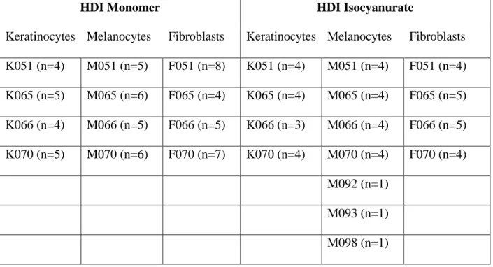
Table 1: The cell types used in the study. The number of experiments performed on a given cell type is indicated by parentheses. The notation for specific cell strain is as follows: the first letter, K, M, or F, corresponds to keratinocytes, melanocytes, and fibroblasts. The three-digit number identifies skin samples.
HDI Monomer HDI Isocyanurate
Keratinocytes Melanocytes Fibroblasts Keratinocytes Melanocytes Fibroblasts K051 (n=4) M051 (n=5) F051 (n=8) K051 (n=4) M051 (n=4) F051 (n=4) K065 (n=5) M065 (n=6) F065 (n=4) K065 (n=4) M065 (n=4) F065 (n=5) K066 (n=4) M066 (n=5) F066 (n=5) K066 (n=3) M066 (n=4) F066 (n=5) K070 (n=5) M070 (n=6) F070 (n=7) K070 (n=4) M070 (n=4) F070 (n=4)
M092 (n=1) M093 (n=1) M098 (n=1)
Isocyanate Treatment of Primary Skin Cells
Logarithmically growing cells (~70% confluent) were plated in black 96-well tissue culture plates (Greiner Bio-one, Germany) at a concentration of approximately 7,000 cells per well for fibroblasts and 12,000 – 15,000 cells per well for keratinocytes and melanocytes. After plating, cells were allowed to re-attach and recover overnight prior to treatment. Cells were rinsed with 200 µL of cell-appropriate basal medium (Gibco or Lifeline Cell Technology) and then exposed to 200 µL of dilutions of either HDI monomer or HDI isocyanurate. The
20
Because of its reactivity to air and water, isocyanate dilutions were prepared immediately before treatment in glass vials with Teflon™ cap liners. The concentration of the stock HDI monomer solution in DMSO was 6 M (1.51 g isocyanate in 0.5 mL DMSO); and 0.1 M for the stock HDI isocyanurate solution (0.2 g isocyanurate in 4 mL DMSO). The stock HDI monomer or HDI isocyanurate solution was then serially diluted into DMSO, producing concentrations ranging from 0.06 M to 6 M HDI monomer in DMSO or 0.001 M to 0.1 M HDI isocyanurate in DMSO. These solutions were then diluted 1:1000 into 2 mL of basal media so that the final DMSO concentration was 0.05%. This produced a working concentration range of 30µM to 3000 µM HDI monomer or 20 µM to 0.5 µM HDI isocyanurate in basal media. Specifically, cells were exposed to (1) 30 µM, 100 µM, 200 µM, 300 µM, 600 µM, 1200 µM, 2400 µM, or 3000 µM of HDI monomer or (2) 0.5 µM, 1 µM, 2 µM, 5 µM, 10 µM, or 20 µM of HDI isocyanurate. A 4-h exposure period was used to simulate a worker’s average daily exposure period. At the end of the 4-h exposure, the exposure media was removed, 200 µL of supplemented growth media was added into each well, and cells were cultured overnight (18 h) to allow for cellular recovery to occur.
Cell Viability
Approximately 18 h after the exposure, cell viability was measured using the Promega CellTiter Glo 2.0 ATP luminescence assay according to manufacturer’s instructions (Promega, Madison, WI) 62. This assay, after adding the single reagent, results in cell lysis and then
21
cells. Because the half-life of the assay reagent is greater than five hours, its stability helps avoid errors that may be present in other methodologies used to measure ATP 62. We chose to use Promega CellTiter Glo 2.0 instead of the common MTT assay for several reasons. The MTT assay uses MTT tetrazolium compound to measure the number of viable cells, and has been a widely-accepted method since the 1980’s. The MTT compound is directly added to cells in culture and incubated for 1 to 4 h. Viable cells will reduce tetrazolium into a precipitate that accumulates inside the cells. A second reagent is added to lyse the cells and solubilize the
precipitate. The absorbance is measured to estimate viable cell number. However, the MTT assay is less sensitive than fluorescent or luminescent methods for measuring viable cell number, and many chemical compounds are known to interfere with this assay 63. The MTT reagent is also cytotoxic, so longer incubation times that would otherwise enhance sensitivity are limited. The detection sensitivity also varies considerably between different cell types and the metabolic activity of the cells. Results from an MTT assay will not produce as accurate a dose-response curve as would results from a more sensitive assay, such as CellTiter Glo.
22
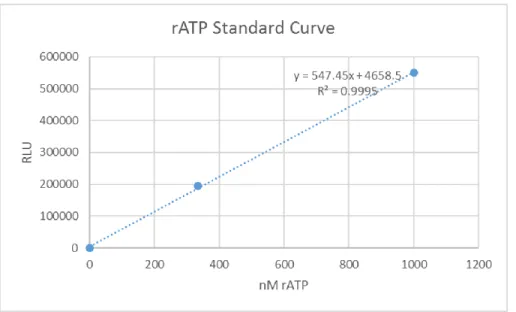
the CellTiter Glo 2.0 luminescence reagent in the 96-well plate. The standard curve included samples of 0, 0.3, 1, 3, 6, and 100 µM rATP. A graph of the linear values (0 – 3 µM rATP) was then produced within Microsoft Excel, and the r-squared value of a linear trend line for the data was determined to ensure the standard curve was linear. All r-squared values were greater than 0.9, with values ranging from 0.95 to 0.99 most common.
The average RLU value from triplicate samples was recorded and background RLU value from the wells containing only basal medium (no cells) were subtracted from all triplicate sample RLU averages. Viability of the isocyanate-treated cells was compared to the 0.05% DMSO control. To calculate the LC50 (concentration that kills 50% of cells), the percent viability was plotted against log-transformed (base 10) exposure concentrations and the 50% decrease in viability was determined from the linear portion of the curve using the linear regression equation of the line. Standard error was determined by calculating the standard deviation for all LC50 values for one cell type, and dividing the standard deviation by the square root of the number of experiments. All statistical analyses were performed using R version 3.3.0. P-values comparing differences in responses to the test chemical between cell types were produced using paired t-tests. We used ANOVA tests to investigate differences in responses between the three cell types to HDI monomer, differences in responses between the three cell types to HDI isocyanurate, and differences in responses between cells used within a specific cell type such as keratinocytes (i.e. comparing K051, K065, K066, and K070 responses to HDI isocyanurate) (see Table 1).
Cell Death
23
complex is stable over time, cell death can be quantified cumulatively 64. Cells were exposed to isocyanates and the fluorescent complex concurrently and the kinetics of cell death followed over time. Cells were plated in a black 96-well plate at a concentration of 15,000 cells
(keratinocytes and melanocytes) per well and left to attach and recover overnight. HDI monomer and HDI isocyanurate dilutions were prepared according the same dilution protocols outlined above, and CellTox Green reagent was diluted into the cell medium at a concentration of 1:1000. Cells lysed with supplied CellTox Green Lysis Solution acted as 100% toxicity control. Cells were rinsed with 200 µL basal medium per well immediately before treatment. Then, 100 µL of solution with both diluted isocyanates (prepared as before) and fluorescent reagent (specific to CellTox Green) was added to each well and fluorescence was measured every 15-30 min for three hours. Plates were read at 485 excitation wave-length in nanometers (ex) /520 emission wavelength in nanometers (em) using a BioTek Cytation 3 microplate reader after shaking for one minute at 700 rpm. Cultures were returned to incubator between the readings.
To investigate the mechanism of cell death, we performed the ApoTox Glo Assay (Promega). This is a 3-in-1 assay that measures both live and dead cells concurrently using fluorescent substrates. Cell viability is measured via a protease detected within intact viable cells. Dead cells are quantified by detection of a dead cell-specific protease activity released into the medium. Apoptosis is measured by quantification of caspase 3/7 activities in lysed cells using luminescent substrates. The resulting luminescent signal is proportional to the amount of caspase activity 65.
24
buffer. This was used immediately or stored at 4°C and used within seven days as per
manufacturer instructions. The Caspase-Glo® 3/7 reagent is the reagent measuring apoptosis. The Caspase-Glo® 3/7 reagent was prepared by transferring the contents of the Caspase-Glo® 3/7 buffer bottle into the amber bottle containing Caspase-Glo® 3/7 substrate. The Caspase-Glo® 3/7 reagent was mixed by swirling or inverting the contents until the substrate is thoroughly
dissolved to form the Caspase-Glo® 3/7 reagent (~20 seconds). The Caspase-Glo® 3/7 was used immediately or stored at 4°C and used within seven days as per manufacturer instructions.
Cells were prepared for the assay by plating at 17,000 cells per well in a 96-well plate. The test chemical concentrations used were 300 µM and 800 µM HDI monomer and 5 µM and 10 µM HDI isocyanurate. These doses were used because they represent the middle and upper range of the LC50 values calculated previously. The necrosis control was 50 µM ionomycin in basal media and the apoptosis control was 10 µM staurosporine in basal media. Cells treated with basal media only and cells treated with basal media plus DMSO were positive controls. Cells were treated for 4 h.
After the treatment period, 20 µL viability/cytotoxicity reagent was added to each well, briefly mixed by orbital shaking (300 – 500 rpm for 30 seconds) and incubated at 37°C for 45 min. Fluorescence was measured at 400Ex/505Em (viability) and 485Ex/520Em (cytotoxicity) with the BioTek Cytation 3 microplate reader.
25 Statistical Analyses
26 RESULTS
Cell Viability
A rATP standard curve was prepared with each Celltiter Glo 2.0 assay performed. This was done to ensure the reagent was performing consistently between experiments, and to
determine the linear range of the assay. The optimal number of cells per well was determined by comparing RLU values to the standard curve to ensure it was in linear range. The standard curve included concentrations of 0, 0.3, 1, 3, 6, and 100 µM rATP. A graph of the linear values (0 – 3 µM rATP) was then produced within Microsoft Excel, and the r-squared value of a linear trend line for the data was determined to ensure the standard curve was linear. All r-squared values were greater than 0.9, with values ranging from 0.95 to 0.99 most common.
27
We used the CellTiter Glo 2.0 results to calculate LC50 values, since this assay had the most sensitive and reproducible dose response curve. In CellTiter Glo 2.0, cells are treated and then allowed to recover in supplemented media for 18 hours before being rinsed and being exposed to the reagent in basal media 62. Then, the reagent in CellTiter Glo 2.0 lyses the remaining living cells and reacts with the ATP that was inside those cells. This could give inaccurate readings if cells had stopped producing ATP but not died. We attempted to minimize this effect by allowing cells to recover for 18 hours between treatment and measurements.
28
Figure 4: The average LC50 levels for cells exposed to HDI monomer or HDI isocyanurate (n =
4-7 per cell type; see Table 1).
Figure 5: The average LC50 levels for the different cell types when exposed to HDI isocyanurate
29
ANOVA was used to compare cell viability between the different cell types exposed to HDI monomer or HDI isocyanurate. A highly significant difference (p<0.001) was observed between the cell types when exposed to either of these compounds. These results support the observed significant difference in LC50 values between the different cell types when exposed to either of these compounds. Melanocytes were the most sensitive cell type to HDI monomer exposure. Keratinocytes were the least sensitive cell type when exposed to HDI isocyanurate while fibroblasts and melanocytes had approximately similar response.
In addition, ANOVA tests were performed to investigate differences between individual donors of keratinocytes, melanocytes, or fibroblasts. However, no significant differences were observed between individual donors for any of the cell types; further confirming our observation that there was no significant difference in toxicity between individual donors’ primary cultured human skin cells (keratinocytes, melanocytes, or fibroblasts).
A second cell viability assay was performed with the ApoTox Glo assay. The viability results were confirmatory of the results observed from the CellTiter Glo 2.0 assay (data not shown).
Cell Death
30
death. The dose-response curve obtained by the CellTox Green Assay shows a trend for dose response but falls off at the highest dose. We interpret these results to indicate that at high doses, the isocyanates inhibit the assay to some degree. This experiment demonstrates that necrotic cell death by isocyanate exposure occurs rapidly and within the first hour of exposure.
Figure 6: Time course of cell death as measured using the CellTox Green cytotoxicity assay performed on primary human keratinocytes, K075.
31
death in isocyanate-treated cells. The experiment conducted with fibroblasts yielded similar results (F070, n = 1) (data not shown).
The ApoTox Glo assay also included a fluorescent cell death assay that measured the activity of a protease released from dead cells. This cytotoxicity assay did not work with our compounds. HDI monomer exposure did not result in toxicity levels that were significantly different than the positive controls of basal media or DMSO in basal media. We can conclude that HDI monomer appears to interfere with the cytotoxicity protease reaction. HDI isocyanurate exposure, interestingly, did produce expected levels of cytotoxicity. These results demonstrate the importance of including proper controls in order to determine if the test compound interferes with proper function of the assay.
32 DISCUSSION
In this study, we investigated differences in toxicity and mechanism of cell death caused by HDI monomer and oligomer exposure in normal human skin cells; specifically, keratinocytes, fibroblasts, and melanocytes. We quantified LC50 values for HDI monomer and HDI
isocyanurate in cultured human skin cells, and further investigated the mechanism of cell death. Assay Comparisons
33
unwinding of the DNA to make it available to the intercalator. Isocyanates are known to
preferentially bind amines on proteins that act as a sink for these compounds in vivo (e.g., serum albumin). The ApoTox Glo kit contains fluorescent reagents for measuring proteases inside of living intact cells as well as proteases released from lysed dead cells. Since isocyanates bind proteins readily, we believe that they inhibited the dead cell protease activity in the ApoTox Glo cytotoxicity assay. The fact that the viability assay in this same kit worked well confirms our hypothesis that the live cell protease was protected from isocyanate binding by the cell membrane. Thus, the type of a viability/cytotoxicity assay used should be thoroughly vetted before use.
We have shown that isocyanates kill cells rapidly by cell necrosis (Fig 6) and confirmed this by the absence of caspase 3/7 activity (Fig 7). Verstraelen et al suggested the method of death within immortalized human bronchial cells was apoptosis, based on gene activity and signaling pathways. There are several factors that could explain this. They used a cancer line of respiratory cells, which may respond differently to isocyanates than primary skin cells. They measured selective gene markers and canonical signaling pathways, including the gene CASP9 and several pathways involving the CASP9 protein, including an antigen presentation pathway and an apoptosis signaling pathway. They theorized that CASP9 may have some biological relevance relating to respiratory sensitization43. We measured caspase 3/7 activities, which are effector caspases at the terminal end of the apoptotic pathway and are more specific for
apoptosis. Additionally and more importantly, they exposed cancer cells to a variety of different chemical combinations of sensitizing, irritant, and non-sensitizing chemicals which in
34
have affected the effective isocyanate dose due to serum proteins acting as a sink for isocyanate binding.
Differences between Isocyanates
In this study, we observed a large difference in cellular toxicity between HDI monomer and HDI isocyanurate exposure. In order to theorize why we observed this difference, we must consider the many complex chemical and physical differences between these compound that could have factored into the results. Reactivity, lipid solubility, and deposition site are all thought to influence health outcomes 67,68. Poly-isocyanates have additional reactive NCO-groups, but the inherent reactivity of the NCO-group alone does not sufficiently describe the observed differences 2,40,42. Bello et al. theorized that properties of the protein that becomes attached to the NCO-group may have an effect on cell permeability, which may contribute to variations in reactivity and toxicity 2.
Different isocyanate monomers and poly-isocyanates cause similar health outcomes, typically immune sensitization and asthma. Immunologic cross-reactivity has been observed between different isocyanates, which suggests commonality in the mechanism of sensitization 69– 71,58. This may be due to the same protein becoming attached to the NCO-group, or simply due to the NCO-group itself 2. Data on health effects relating to poly-isocyanates such as HDI
isocyanurate are more limited, but sufficient to demonstrate that these compounds can cause similar health outcomes as isocyanate monomers. Chemical-induced asthma and hypersensitivity pneumonitis have been observed as a result of poly-isocyanate exposure 20,25,26,34.
35
the allergenic response may be very similar 47,68. However, Pauluhn did not observe a response in animals sensitized with HDI isocyanurate when later exposed to conjugates; but he
acknowledged that this may have been related to inappropriately produced conjugates 47. In this study, we observed much lower LC50 values associated with HDI isocyanurate exposure than HDI monomer. We cannot directly compare the LC50 values calculated here to regulatory standards for exposure, such as TLV. However, we can consider our results in context with the published peer-reviewed scientific literature. HDI isocyanurate may sensitize humans and animals using the same mechanism as HDI monomer, and HDI isocyanurate penetrates the skin faster 10. Taken together, evidence exists that HDI isocyanurate may have a large impact on overall exposure that is not being accounted for in current regulatory standards.
Factors Influencing Susceptibility to Isocyanate Toxicity Structure of the Skin
The structure of the skin itself has significant effects on penetration of the compounds, and ultimately on toxicity. As individual isolated cell types were used in this study, we must consider how the cell types may behave or how the exposure may be modified by the overall structure of the skin.
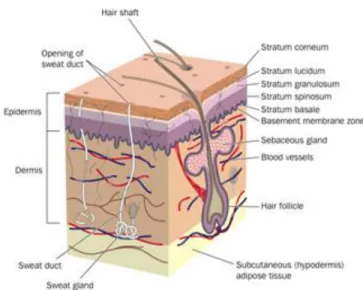
We observed differences in susceptibility for isocyanate toxicity between the cell types we tested. It is well known that different skin cell types communicate with each other, and this influences cell activity. The top layer of the skin is the cornified layer or stratum corneum, made up of dead keratinocytes and lipids, which forms the major protective barrier against water loss and foreign substances (see Figure 8). Below is the viable epidermis, containing mostly
36
fibroblasts, hair follicles, blood vessels, and many other elements embedded in a collagen matrix 25,53,72.
Figure 8: Structure of skin. Courtesy of CliniMed UK 72
There are also differences in skin between different areas of the body. The thickness of the non-hydrated stratum corneum is between 10 and 50 µm in most areas of the body, but it may be up to 10 times as thick on areas like the palms of the hands or soles of the feet 73. The rate of absorption of chemicals through the various areas of the skin generally follows the following order, from the fastest to the slowest: scrotal, forehead, armpit, scalp, back, abdomen, palm, and undersurface of foot 74. Despite large variability in permeability and skin structure, current regulations for skin exposure do not address the differing doses that may be absorbed by different areas of the body.
Cell Types
37
isolated from neonatal foreskins. Keratinocytes are localized in the stratum corneum, the top or most external part of the skin and comprise 90% of the cells within the epidermis 73. In the experiments performed here, keratinocytes were the least susceptible to isocyanate toxicity of tested skin cells types. Because of the location, abundance, and relative resistance to toxicity, keratinocytes are the most important cells used here regarding the development of an immune response. In other studies, keratinocytes were observed to be relatively resilient, potentially because of higher enzymatic activities 75–77. Differing activity levels may play a role in the cell response to xenobiotic exposure and, thus, warrant further investigation. Of the three cell types used here, melanocytes are the least abundant within the skin and were the least resistant to the isocyanate toxicity. Other research suggests that melanocytes may be the most vulnerable skin cell, potentially because of low antioxidant enzyme activity or high cellular proliferation with low relative efficiency of DNA repair processes 78,75,77,76. As isocyanates do not cause cell death through oxidation or DNA damage, these observations are not directly applicable within this study. However, it is interesting that melanocytes appear to be an especially vulnerable cell in many settings.
Skin Penetration
38
readily absorbed through the excised full thickness human skin 10. HDI isocyanurate was
observed to have the shortest absorption time regardless of paint formulation. Compounds tested were neat or in ethyl acetate (EA) solution, because EA is a common solvent used for
isocyanates. EA was observed to enhance skin penetration. Polyurethanes used in occupational settings are frequently mixed with reducers, which could have a similar enhancing effect.
In the second set of experiments described by Thomasen et al, penetration patterns for both slow-drying and fast-drying clearcoat were investigated 10. Both formulations contain a larger proportion of HDI poly-isocyanates than HDI monomer. Slow-drying clearcoat contains significantly more HDI monomer and HDI biuret, another HDI trimer, than fast-drying clearcoat. Evaporative losses were minimized through the use of a plastic lid that covered the skin samples. Slow-drying clearcoat was observed to penetrate the skin more quickly than fast-drying clear coat. These results are supported by Bello et al. 2006, which investigated the residence time of isocyanates on hairless guinea pig skin in vitro and saw similar patterns regarding rapid
penetration and minimal evaporation 31. Observations indicating that HDI isocyanurate is absorbed either just as well or faster than HDI monomer suggests that even a short exposure could result in a considerable body burden 10,29,30. Here, we observed a higher toxicity associated with polymeric HDI. The results observed in this study suggest that even a short exposure to HDI isocyanurate could result in a significant body burden and, thus, a higher cytotoxicity. This has concerning implications for worker health and future development of limit values merits further investigation.
Inter-individual Variability
39
differentiation of keratinocytes, which contributes to the formation of the cornified layer 52. Null mutations in FLG as well as inflammation in the skin can cause changes in FLG expression leading to atopic dermatitis. Atopic dermatitis compromises the skin barrier function and thus increases individual susceptibility to environmental exposures. Individual genetic and epigenetic alterations can influence immune responses. The immune response is modified by keratinocytes, which act as an important first line of defense. DNA methylation may also play a role in
individual differences, and may increase susceptibility to HDI22 . DNA methylation may affect gene expression of proteins involved in isocyanate mass transport, permeation, and metabolism and thereby mediate individual responses 22. These genetic factors can have a significant impact on susceptibility, and our current safety limits may not protect all workers.
In a review published by the World Health Organization, Byford reports that the barrier properties of the skin may be influenced by species of animal, age, sex, and race, anatomical site, skin condition, temperature and blood flow rate, and hydration 73. When considering inter-individual variability, age, sex, and race as well as hydration status are relevant. In this study, we only used cells from male donors due to the availability of foreskin samples. Given that most isocyanate exposed workers are male, the use of cells from male donors in this study is appropriate 80. Age may also influence susceptibility. Matsuo et al 2004 investigated the responses of fibroblasts from old and young donors to oxidative stress 81. They observed that fibroblasts from old donors were more resilient against oxidative stress, and theorized that this was due to an increase in glutathione peroxidase activity 81. All cells used in this study were isolated from neonatal foreskin, and therefore from the youngest possible donors. These cells may have relatively low enzymatic activity and thus be more susceptible to toxicity. No
40
the experiments performed in this study (data not shown)73,76. Hydration status is an
environmental condition that can drastically alter the barrier function of the skin. Decreased hydration status may be the probable cause for increase in absorption 73. The humidity of the environment as well as individual differences in hydration may influence variations in
susceptibility. We used isolated skin cells in this study and, therefore, exposure conditions were not modified by hydration status or humidity of the environment. However, the hydration status of the skin appears to be very important when considering occupational skin exposures and penetration of xenobiotics through the skin.
Monitoring exposure levels and setting limits specific to skin exposure are needed to protect workers from potential adverse health effects like contact dermatitis or occupational asthma. These data suggest that special attention should be paid towards preventing skin exposure to HDI isocyanurate.
Limitations of the Study
41
REFERENCES
1. Aalto-Korte K, Pesonen M, Kuuliala O, Alanko K, Jolanki R. Contact allergy to aliphatic polyisocyanates based on hexamethylene-1,6-diisocyanate (HDI). Contact Dermatitis, Environ Occup Dermat. 2010;63(6):357-363. doi:10.1111/j.1600-0536.2010.01786.x.
2. Bello D, Woskie SR, Streicher RP, et al. Polyisocyanates in occupational environments: A critical review of exposure limits and metrics. Am J Ind Med. 2004;46(5):480-491.
doi:10.1002/ajim.20076.
3. Musk AW, Peters JM, Wegman DH. Isocyanates and respiratory disease: Current status. Am J Ind Med. 1988;13(3):331-349. doi:10.1002/ajim.4700130304.
4. Tosoh Asia Pte. Ltd. What is Polyurethane? http://www.tosohasia.com/our-products/polyurethanes.
5. Marketandmarket. Methylene diphenyl diisocyanate (MDI), toluene diisocyante (TDI), and polyurethane market. www.marketsandmarkets.com. Published 2016.
6. Ceresana. Market study: polyurethanes and isocyanates (MDI & TDI) (4C-4905). www.ceresana.com. Published 2013.
7. Bello D, Woskie SR, Streicher RP, et al. A laboratory investigation of the effectiveness of various skin and surface decontaminants for aliphatic polyisocyanates. J Environ Monit. 2005;7(7):716-721. doi:10.1039/b503807c.
8. Ulrich H. Chemistry and Technology of Isocyanates. Chichester; 1996. https://books.google.com/books?id=DY4vAQAAIAAJ&pgis=1.
9. Verschoor L, Verschoor AH. Nonoccupational and occupational exposure to isocyanates. CE Namrta; MCP. 2002. doi:10.1097/MCP.0000000000000029.
10. Thomasen JM, Nylander-French L a. Penetration patterns of monomeric and polymeric 1,6-hexamethylene diisocyanate monomer in human skin. J Environ Monit.
2012;14(3):951-960. doi:10.1039/c2em10546b.
11. California Department of Public Health. Isocyanates : Working Safely.; 2014. 12. The National Institute for Occupational Safety and Health (NIOSH). NIOSH Pocket
Guide to Chemical Hazards.
13. Janko M, McCarthy K, Fajer M, Raalte J van. Occupational Exposure To
1,6-Hexamethylene Diisocyanate-Based Polyisocyanates In The State Of Oregon, 1980–1990. Am Ind Hyg Assoc J. 1992;53(5):331-338. doi:10.1080/15298669291359735.
42
Biological Exposure Indices. Cincinnati, OH; 2010.
15. Bayer MaterialScience AG. The Chemistry of Polyurethane Coatings.; 2005.
https://www.pharosproject.net/uploads/files/cml/1383145151.pdf. Accessed May 2, 2017. 16. Gaines LGT, Fent KW, Flack SL, Thomasen JM, Whittaker SG, Nylander-French LA.
Factors affecting variability in the urinary biomarker 1,6-hexamethylene diamine in workers exposed to 1,6-hexamethylene diisocyanate. J Environ Monit. 2011;13(1):119-127. doi:10.1039/c0em00122h.
17. Fent KW, Gaines LGT, Thomasen M, et al. Quantification and Statistical Modeling—Part I: Breathing-Zone Concentrations of Monomeric and Polymeric 1,6-Hexamethylene Diisocyanate. Ann Occup Hyg. 2009;53(7):677-689. doi:10.1093/annhyg/mep046. 18. Gaines LGT, Fent KW, Flack SL, et al. Urine 1,6-hexamethylene diamine (HDA) levels
among workers exposed to 1,6-hexamethylene diisocyanate (HDI). Ann Occup Hyg. 2010;54(6):678-691. doi:10.1093/annhyg/meq041.
19. Reeb-Whitaker C, Whittaker SG, Ceballos DM, et al. Airborne Isocyanate Exposures in the Collision Repair Industry and a Comparison to Occupational Exposure Limits. J Occup Environ Hyg. 2012;9(5):329-339. doi:10.1080/15459624.2012.672871.
20. Bello D, Herrick CA, Smith TJ, et al. Skin exposure to isocyanates: Reasons for concern. Environ Health Perspect. 2007. doi:10.1289/ehp.9557.
21. Liu Y, Stowe MH, Bello D, et al. Skin exposure to aliphatic polyisocyanates in the auto body repair and refinishing industry: III. A personal exposure algorithm. Ann Occup Hyg. 2009;53(1):33-40. doi:10.1093/annhyg/men070.
22. Nylander-French LA, Wu MC, French JE, et al. DNA methylation modifies urine biomarker levels in 1,6-hexamethylene diisocyanate exposed workers: A pilot study. Toxicol Lett. 2014;231(2):217-226. doi:10.1016/j.toxlet.2014.10.024.
23. Pronk A. Dermal, inhalation, and internal exposure to 1,6-HDI and its oligomers in car body repair shop workers and industrial spray painters. Occup Environ Med.
2006;63(9):624-631. doi:10.1136/oem.2005.023226.
24. Pronk A, Tielemans E, Skarping G, et al. Inhalation exposure to isocyanates of car body repair shop workers and industrial spray painters. Ann Occup Hyg. 2006;50(1):1-14. doi:10.1093/annhyg/mei044.
25. Redlich C a. Skin exposure and asthma: is there a connection? Proc Am Thorac Soc. 2010;7(2):134-137. doi:10.1513/pats.201002-025RM.
26. Redlich CA, Herrick CA. Lung/skin connections in occupational lung disease. Curr Opin Allergy Clin Immunol. 2008;8(2):115-119. doi:10.1097/ACI.0b013e3282f85a31.
27. Ceballos DM, Fent KW, Whittaker SG, et al. Survey of dermal protection in Washington State collision repair industry. J Occup Environ Hyg. 2011;8(9):551-560.
doi:10.1080/15459624.2011.602623.
43
sprayed isocyanate coatings utilizing a reciprocating permeation panel. Ann Occup Hyg. 2014;58(1):50-59. doi:10.1093/annhyg/met060.
29. Fent KW, Trelles Gaines LG, Thomasen JM, et al. Quantification and statistical modeling - Part II: Dermal concentrations of monomeric and polymeric 1,6-hexamethylene
diisocyanate. Ann Occup Hyg. 2009;53(7):691-702. doi:10.1093/annhyg/mep048. 30. Fent KW, Jayaraj K, Ball LM, Nylander-French L a. Quantitative monitoring of dermal
and inhalation exposure to 1,6-hexamethylene diisocyanate monomer and oligomers. J Environ Monit. 2008;10(4):500-507. doi:10.1039/b715605g.
31. Bello D, Smith TJ, Woskie SR, et al. An FTIR investigation of isocyanate skin absorption using in vitro guinea pig skin. J Environ Monit. 2006;8(5):523. doi:10.1039/b517948c. 32. Walker J, Whittaker C, McDougal J. Dermal Absorption Models in Toxicology and
Pharmacology. In: Maibach FNM and HI, ed. Dermatotoxicology. New York, NY,: Hemisphere Publishing; 1996:371–381.
33. NIOSH Publication Number 96-111. Preventing Asthma and Death from Diisocyanate Exposure DHHS.; 1996. https://www.cdc.gov/niosh/docs/96-111/.
34. Bernstein D, Korbee L, Stauder T, et al. The low prevalence of occupational asthma and antibody-dependent sensitization to diphenylmethane diisocyanate in a plant engineered for minimal exposure to diisocyanates. J Allergy Clin Immunol. 1993;92(3):387-396. doi:10.1016/0091-6749(93)90117-X.
35. Tan J, Bernstein JA. Occupational asthma: an overview. Curr Allergy Asthma Rep. 2014. doi:10.1007/s11882-014-0431-y.
36. The National Institute for Occupational Safety and Health (NIOSH). A Summary of Health Hazard Evaluations : Isocyanates , 1989 to 2002. Natl Inst Occup Saf Heal. 2004;(January 2004).
37. Cocker J. Biological monitoring for isocyanates. Ann Occup Hyg. 2011;55(2):127-131. doi:10.1093/annhyg/meq083.
38. Wisnewski A V., Srivastava R, Herick C, et al. Identification of Human Lung and Skin Proteins Conjugated with Hexamethylene Diisocyanate In Vitro and In Vivo. Am J Respir Crit Care Med. 2000;162(6):2330-2336. doi:10.1164/ajrccm.162.6.2002086.
39. Campo P, Wisnewski A V., Lummus Z, et al. Diisocyanate conjugate and immunoassay characteristics influence detection of specific antibodies in HDI-exposed workers. Clin Exp Allergy. 2007;37(7):1095-1102. doi:10.1111/j.1365-2222.2007.02745.x.
40. Wisnewski A V., Mhike M, Hettick JM, Liu J, Siegel PD. Hexamethylene diisocyanate (HDI) vapor reactivity with glutathione and subsequent transfer to human albumin. Toxicol Vitr. 2013;27(2):662-671. doi:10.1016/j.tiv.2012.11.013.
44
42. Wisnewski A V, Liu Q, Miller J-J, Magoski N, Redlich CA. Effects of Hexamethylene Diisocyanate Exposure on Human Airway Epithelial Cells: In Vitro Cellular and Molecular Studies. Environ Health Perspect. 2002;110(9).
43. Verstraelen S, Nelissen I, Hooyberghs J, et al. Gene profiles of a human bronchial epithelial cell line afterin vitro exposure to respiratory (non-)sensitizing chemicals: Identification of discriminating genetic markers and pathway analysis. Toxicology. 2009. doi:10.1016/j.tox.2008.10.014.
44. Hathaway JA, Molenaar DM, Cassidy LD, Feeley TM, Cummings BJ. Cross-Sectional Survey of Workers Exposed to Aliphatic Diisocyanates Using Detailed Respiratory Medical History and Questions Regarding Accidental Skin and Respiratory Exposures. 2013. doi:10.1097/JOM.0000000000000019.
45. Ban M, Morel G, Langonne I, Huguet N, Pepin E, Binet S. TDI can induce respiratory allergy with Th2-dominated response in mice. Toxicology. 2006;218(1):39-47.
doi:10.1016/j.tox.2005.09.013.
46. Meredith SK, Taylor VM, McDonald JC. Occupational respiratory disease in the United Kingdom 1989: a report to the British Thoracic Society and the Society of Occupational Medicine by the SWORD project group. Br J Ind Med. 1991;48(5):292-298.
doi:10.1136/oem.48.5.292.
47. Pauluhn J, Eidmann P, Mohr U. Respiratory hypersensitivity in guinea pigs sensitized to 1,6-hexamethylene diisocyanate (HDI): comparison of results obtained with the monomer and homopolymers of HDI. Toxicology. 2002;171(2-3):147-160. doi:10.1016/S0300-483X(01)00571-6.
48. Karol MH, Hauth BA, Riley EJ, Magreni CM. Dermal contact with toluene diisocyanate (TDI) produces respiratory tract hypersensitivity in guinea pigs. Toxicol Appl Pharmacol. 1981;58(2):221-230. doi:10.1016/0041-008X(81)90426-9.
49. Rattray NJ, Botham PA, Hext PM, et al. Induction of respiratory hypersensitivity to diphenylmethane-4,4’-diisocyanate (MDI) in guinea pigs. Influence of route of exposure. Toxicology. 1994;88(1-3):15-30. http://www.ncbi.nlm.nih.gov/pubmed/8160196.
50. Nayak AP, Hettick JM, Siegel PD, et al. Toluene Diisocyanate (TDI) disposition and co-localization of immune cells in hair follicles. Toxicol Sci. 2014;140(2):327-337.
doi:10.1093/toxsci/kfu079.
51. Herrick CA, Das J, Xu L, Wisnewski A V, Redlich CA, Bottomly K. Differential roles for CD4 and CD8 T cells after diisocyanate sensitization: genetic control of TH2-induced lung inflammation. J Allergy Clin Immunol. 2003;111(5):1087-1094.
http://www.ncbi.nlm.nih.gov/pubmed/12743574.
52. Brown SJ, Mclean WHI. One Remarkable Molecule: Filaggrin. J Invest Dermatol. 2012;132393(10):751-762.
53. De Benedetto A, Kubo A, Beck LA. Skin Barrier Disruption: A Requirement for Allergen Sensitization? J Invest Dermatol. 2012;132(10).
45
barrier, allergy, and pruritus as a trinity. J Dermatol Sci. 2013. doi:10.1016/j.jdermsci.2013.02.001.
55. Akdis CA, Akdis M, Bieber T, et al. Diagnosis and treatment of atopic dermatitis in children and adults: European Academy of Allergology and Clinical
Immunology/American Academy of Allergy, Asthma and Immunology/PRACTALL Consensus Report. J Allergy Clin Immunol. 2006;118(1):152-169.
doi:10.1016/j.jaci.2006.03.045.
56. Ouyang B, Bernstein DI, Lummus ZL, et al. Interferon-γ Promoter Is Hypermethylated in Blood DNA from Workers with Confirmed Diisocyanate Asthma. Toxicol Sci.
2013;133(2):218-224. doi:10.1093/toxsci/kft079.
57. Ebino K, Ueda H, Kawakatsu H, et al. Isolated airway exposure to toluene diisocyanate results in skin sensitization. Toxicol Lett. 2001;121(1):79-85. doi:10.1016/S0378-4274(01)00325-3.
58. Thorne PS, Hillebrand JA, Lewis GR, Karol MH. Contact sensitivity by diisocyanates: potencies and cross-reactivities. Toxicol Appl Pharmacol. 1987;87(1):155-165.
doi:3798450.
59. Verstraelen S, Nelissen I, Hooyberghs J, et al. Gene profiles of a human alveolar epithelial cell line afterin vitro exposure to respiratory (non-)sensitizing chemicals: Identification of discriminating genetic markers and pathway analysis A. Toxicol Lett. 2009.
doi:10.1016/j.tox.2008.10.014.
60. Gazdar AF, Gao B, Minna JD. Lung cancer cell lines: Useless artifacts or invaluable tools for medical science? Lung Cancer. 2010;68(3):309-318.
doi:10.1016/j.lungcan.2009.12.005.
61. Godwin LS, Castle JT, Kohli JS, et al. Isolation, Culture, and Transfection of
Melanocytes. In: Current Protocols in Cell Biology. Hoboken, NJ, USA: John Wiley & Sons, Inc.; 2014:1.8.1-1.8.20. doi:10.1002/0471143030.cb0108s63.
62. Promega Corporation. CellTiter-Glo® Luminescent Cell Viability Assay.
https://www.promega.com/products/cell-health-and-metabolism/cell-viability-assays/celltiter_glo-luminescent-cell-viability-assay/. Published 2015. Accessed April 19, 2017.
63. Terry Riss. Is Your MTT Assay Really the Best Choice. Promega Corporation Web site. https://www.promega.com/resources/pubhub/is-your-mtt-assay-really-the-best-choice/. Published 2014. Accessed May 7, 2017.
64. CellToxTM Green Cytotoxicity Assay Instructions for Use of Products G8741, G8742,
G8743 and G8731. https://www.promega.com/-/media/files/resources/protocols/technical-manuals/101/celltox-green-cytotoxicity-assay-protocol.pdf. Accessed April 19, 2017. 65. Hooper K. Promega Apotox Glo Triplex Assay Brochure.
46
66. Zong W-X, Thompson CB. Necrotic death as a cell fate. Genes Dev. 2006;20(1):1-15. doi:10.1101/gad.1376506.
67. Mohr, Jürgen Pauluhn U. Inhalation Toxicity Of 1,6-Hexamethylene Diisocyanate Homopolymers (Hdi-Ic And Hdi-Bt): Results of Subacute and Subchronic Repeated Inhalation Exposure Studies. Inhal Toxicol. 2001;13(6):513-532.
doi:10.1080/08958370118600.
68. Pauluhn J, Lewalter J. Analysis of markers of exposure to polymeric methylene-diphenyl diisocyanate (pMDI) in rats: a comparison of dermal and inhalation routes of exposure. Exp Toxicol Pathol. 2002;54(2):135-146. doi:10.1078/0940-2993-00242.
69. Baur X. Immunologic cross-reactivity between different albumin-bound isocyanates. J Clin Immunol. 1983;71:197-205.
70. Baur X. Occupational asthma due to isocyanates. Lung. 1996;174(1):23-30. http://www.ncbi.nlm.nih.gov/pubmed/8746999.
71. Baur X. Hypersensitivity pneumonitis (extrinsic allergic alveolitis) induced by isocyanates. J Allergy Clin Immunol. 1995;95(5 Pt 1):1004-1010.
http://www.ncbi.nlm.nih.gov/pubmed/7751497.
72. CliniMed UK. Structure and Function of the Skin. http://www.clinimed.co.uk/Wound-Care/Education/Wound-Essentials/Structure-and-Function-of-the-Skin.aspx. Accessed April 19, 2017.
73. Byford T. Environmental Health Criteria 235: Dermal Absorption. Int J Environ Stud. 2009;66(5):662-663. doi:10.1080/00207230802361240.
74. Baynes RE, Hodgson E. Absorption and Distribution of Toxicants. In: A Textbook of Modern Toxicology. Hoboken, NJ, USA: John Wiley & Sons, Inc.; 2004:75-110. doi:10.1002/0471646776.ch6.
75. Smith CN, Lindsay CD, Hambrook JL. An in vitro comparison of the cytotoxicity of sulphur mustard in melanoma and keratinocyte cell lines. Hum Exp Toxicol.
2001;20(9):483-490. doi:10.1191/096032701682693035.
76. Yohn JJ, Norris DA, Yrastorza DG, et al. Disparate Antioxidant Enzyme Activities in Cultured Human Cutaneous Fibroblasts, Keratinocytes, and Melanocytes. J Invest Dermatol. 1991;97(3):405-409. doi:10.1111/1523-1747.ep12480983.
77. Cooper KL, Yager JW, Hudson LG. Melanocytes and keratinocytes have distinct and shared responses to ultraviolet radiation and arsenic. Toxicol Lett. 2014;224(3):407-415. doi:10.1016/j.toxlet.2013.11.010.
78. Reemann P, Reimann E, Ilmjärv S, et al. Melanocytes in the skin - Comparative whole transcriptome analysis of main skin cell types. PLoS One. 2014;9(12):1-17.
doi:10.1371/journal.pone.0115717.
47
atopic dermatitis. Am J Clin Dermatol. 2008;9(1):45-50. http://www.ncbi.nlm.nih.gov/pubmed/18092843.
80. National Institute of Occupational Safety and Health. National Occupational Exposure Survey (NOES). Vol 2005.; 2005. http://www.cdc.gov/noes/default.html.
81. Matsuo M, Ikeda H, Sugihara T, Horiike S, Okano Y, Masaki H. Resistance of Cultured Human Skin Fibroblasts from Old and Young Donors to Oxidative Stress and Their Glutathione Peroxidase Activity. Gerontology. 2004;50(4):193-199.
48
PRACTICUM REPORT
The Effect of Intermittent Noise Stress on Ozone-Induced Cardiovascular Dysfunction in Wistar-Kyoto Rats.
Kaitlyn Phillips1, Kimberly Stratford1, Leon Walsh2, Malek Khan2, Leslie Thompson2, Aimen Farraj2 & Mehdi S. Hazari2. 1
University of North Carolina, Chapel Hill, NC 27599; 2Environmental Public Health Division, USEPA, Research Triangle Park, NC 27711.
Previous studies have established that acute exposure to air pollution increases the risk of cardiovascular dysfunction. Intrinsic factors are likely the most important determinants of how the body responds to an exposure. But data also suggests that non-environmental stressors like noise, which is a common urban public health problem, can modify and in fact worsen the response. Noise can cause obvious psychological disturbances typical of non-specific stress, but also changes that can increase the number of cardiovascular disease related mortalities.
Therefore, we hypothesized that short-term exposure to noise would worsen the cardiovascular response to ozone.
Male Wistar-Kyoto rats were implanted with radiotelemeters for the measurement of heart rate (HR), blood pressure (BP) and electrocardiogram (ECG) and exposed to intermittent noise (85-90 dB) for one week; after which they were exposed to either ozone (0.8 ppm) or filtered air. Left ventricular functional responses to dobutamine were measured using a Millar probe as well as arrhythmic sensitivity to aconitine in a separate set of untelemetered rats 24 hours after exposure.
49
and ozone had a significant interaction on ventricular tachycardia and ventricular fibrillation (see Figure 1). Baseline left ventricular pressure (LVP) was significantly higher in animals exposed to both noise and ozone when compared to no noise; furthermore those animals had the least
amount of change in LVP, dP/dT max and min with increasing doses of dobutamine. These animals also had a higher arrhythmic sensitivity to aconitine. In conclusion, these results suggest that noise alters the cardiovascular response to ozone exposure. Thus, non-environmental
stressors may be playing an important role in modifying the response to air pollution and may in fact increase the risk in people with underlying cardiovascular disease.