ABSTRACT
The study of proteins with microtubule organizing function is important in order to better understand mechanisms of the cytoskeleton. The protein Che-12 is one such conserved protein that affects microtubule dynamics in cilia development. How Che-12 carries out this function, however, remains unknown. Following sequence analysis, it was hypothesized that Che-12 contains four TOG domains which are known to regulate microtubule polymerization. The purpose of my in vitro study of mChe-12 (mouse) was to characterize mChe-12’s predicted TOG domains and their interactions with microtubules. I have purified multiple constructs containing these predicted domains and have used these purified proteins for both structure determination using X-ray crystallography and tubulin polymerization assays using light scattering techniques. Based on these experiments, I have helped validate the presence of TOG domains within mChe-12 and begun to determine the role of mChe-mChe-12’s TOG domains in regulating tubulin
polymerization. Thus, we have been able to characterize the Che-12 family as a new class of TOG domain containing proteins which are conserved in ciliated and flagellated organisms.
INTRODUCTION
Figure 1-Arrangement of TOG Domains in Che-12, ch-TOG and CLASP families
however. For this reason, proteins shown to interact with microtubules are of interest in order to better understand how these proteins dynamically localize, modulate microtubule dynamics, recruit cellular factors, and translate cellular signaling cues into cytoskeletal outcomes (Slep, 2009).
One of the most significant organelles composed of microtubules are cilia, which project from cells in order to aid in either movement or signal transduction. Non-motile primary cilia, specifically, are found in most mammalian cells in which they are important for sensory
reception (Wheatly, 1996). One protein thought to regulate microtubule organization in primary cilia is Che-12. Che-12 was first studied in Caenorhabditis elegans where it was shown to be important for proper ciliogenesis in a subset of sensory neuron cells (Bacaj, 2008). When Che-12 was mutated in C. elegans, resulting worms developed a phenotype in which sensory neurons did not respond properly to environmental cues (Perkins, 1986). The che-12 mutants specifically lacked the most distal zone of cilia normally characterized by dynamic singlet microtubules. This mutant cilia morphology was likely brought about by failure to organize microtubules within the structure. Despite these findings, the regulation of singlet, distal microtubule dynamics in which Che-12 plays a key role is still unfamiliar.
The protein mChe-12 (the Che-12 homolog found in mice) is a 1776 amino acid protein and includes four predicted TOG domains, named after their identification in the human protein
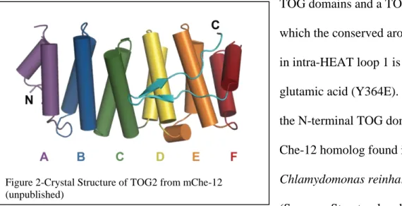
Figure 2-Crystal Structure of TOG2 from mChe-12 (unpublished)
domains promote microtubule polymerization by binding tubulin heterodimers (Slep, 2007). A TOG domain is made up of six pairs of antiparallel helices that are connected by short loops. The antiparallel helices, also known as HEAT repeats, configure themselves so that all intra-HEAT loops lie exposed on one side of the domain while all inter-intra-HEAT loops lie exposed on the opposite side of the domain. Many surface-exposed residues in the intra-HEAT loops are conserved across species and this face has been shown to bind tubulin in XMAP215-family TOG domains (Ayaz, 2012). When a conserved aromatic residue in the first intra-HEAT loop (A in Figure 2) is mutated to glutamic acid, affinity for tubulin decreases significantly which highlights the role of this exposed hydrophobic residue in the TOG domain-tubulin interaction (Slep, 2009). Knowing the structure of other TOG domain proteins allowed for identification of conserved tubulin-binding residues and similar mutations to be made in mChe-12’s predicted TOG domains.
The structure of mChe-12 TOG2 has previously been solved in our lab (Figure 2). Work I have completed in previous semesters resulted in the purification of all four wild-type predicted
TOG domains and a TOG2 mutant in which the conserved aromatic residue in intra-HEAT loop 1 is mutated to glutamic acid (Y364E). Additionally, the N-terminal TOG domain from a Che-12 homolog found in
polymerization assay in order to gain insight into the effects these proteins have on microtubule polymerization in vitro. Work also was conducted with select proteins for structural
determination using X-ray crystallography. We hypothesize that by determining the effects these TOG domains have on tubulin polymerization and determining molecular structure information regarding each protein, we will validate that these domains are indeed TOG domains, gain greater understanding of how TOG domains underlie Che-12 function, and obtain further insight into the overall role of TOG domains across protein families. Additionally, the findings from the study of both mChe-12 and the C. reinhardtii SSA6 TOG1 will help elucidate any conservation of TOG domain function in the Che-12 protein family across ciliated organisms.
METHODS
Che-12 Family TOG Domain Cloning
We have used sequence alignments to identify the presence of predicted TOG domains in mChe-12 (mouse) as well as the conserved aromatic residue in the first intra-HEAT loop of each domain (F113, Y364, W1274 and F1559), which appears to be critical for tubulin binding in other TOG domain proteins. mChe-12’s individual TOG domains (for TOG1, TOG2, TOG3 and TOG4) and SSA6’s TOG1 domain were cloned separately into pET28b vectors for Isopropyl β-D-1-thiogalactopyranoside (IPTG) inducible expression in BL21 (DE3) E. coli cells. The constructs designed have a cleavable N-terminal 6X-His tag to facilitate purification using Ni2+ -NTA affinity chromatography. The pET28b vectors include Kanamycin resistance for selection of transformed cells.
DNA encoding the before mentioned TOG domains cloned in pET28b vector were transformed into BL21(DE3) Rosetta2 cells. The transformed cells were added to a Kanamycin and Chloramphenicol containing agar plate under sterile conditions. The plate was allowed to incubate at 37˚C overnight so that resistant, transformed bacterial colonies grew up.
The plate was removed from 37˚C the next day and now had several bacterial colonies growing on it. A single colony was chosen and added to 150 mL of LB media in a flask and allowed to grow at 37˚C overnight. The next day, 10 mL of this primary inoculum was added per 1 L pre-autoclaved LB media (6 flasks of 1 L cultures in all) containing Kanamycin and Chloramphenicol (50 μg/mL each). The 1 L cultures were then allowed to grow up at 37°C until their optical density was between 0.6 and 0.8 at 600 nm. A sample of growth was isolated at this uninduced stage for later testing with sodium dodecyl sulfate- poly-acrylamide gel
electrophoresis (SDS-PAGE gel). At this point, IPTG was added to a final concentration of 0.1 mM in order to induce protein expression. The temperature of the incubator was brought down to 18˚C. The next day, the culture flasks were removed from the incubator and spun down in a Fiberlite F10 rotor at 3500 rpm for 10 minutes at 4˚C using a centrifuge. Prior to spin down, a small amount of cell culture was taken and prepared for testing with an SDS-PAGE gel to confirm protein expression. Once all cells had been spun down, the cells were resuspended in Ni2+-A buffer (for buffer ingredients see appendix) and stored at -20˚C.
Frozen cell suspension was removed from -20°C and allowed to thaw.
rotor. The supernatant containing soluble proteins was poured into a clean 500 mL beaker while the debris pellet was disposed of.
The protein sample was then allowed to run through a Ni2+-NTA column by gravity at 4˚C. A sample was taken from the flow through in order to test to make sure the protein bound and did not pass through the column. Once all protein had run onto the column, 200 mL of Ni2+ -A buffer was run through the column as well to wash off non-specifically bound impurities (a sample of the flow through was taken at this point for SDS-PAGE gel). The Ni2+-NTA column was then eluted over a linear gradient between Ni2+ buffer A and Ni2+ buffer B with an
increasing imidazole concentration on an AKTA Liquid Chromatography apparatus (ingredients for buffers found in appendix). Fractions were collected using a fraction collector.
Using the resulting chromatogram, fractions were chosen for testing by SDS-PAGE to confirm that the target protein eluted and whether there were any impurities present. The fractions containing the TOG domain were pooled and treated with bovine alpha thrombin in 2.5 mM CaCl2 buffer in order to cleave off the His tag present on the protein. The protein mix sat at
4°C until thrombin digestion was complete as signified by a 2kDa shift in the TOG domain band on an SDS PAGE gel (sample taken every 24 hours).
Once the protein was completely cleaved, the sample was added into an appropriately sized dialysis bag and placed into a larger volume of dialysis buffer (see appendix for buffer ingredients). The solutions reached equilibrium within 5 hours while sitting at 4°C. Next, the protein was filtered through 0.5 mL Benzamadine sepharose (GE Healthcare) to remove
fractions were then pooled and concentrated (see final protein concentrations in Table 1 in results). The protein was aliquoted, frozen in liquid nitrogen, and stored at -80°C.
TOG1, TOG2 Y364E, TOG3, TOG4, TOG4 SeMet SP-Sepharose Column Elution
The proteins listed above were found not to be completely pure following Ni2+-NTA purification. Therefore, these individual TOG protein constructs were loaded onto a 10 mL SP-sepharose fast flow column (GE Healthcare) following thrombin removal, washed with 200 mL of S-Sepharose Buffer A and eluted over a linear gradient between S-Sepharose Buffer A and B in the AKTA Liquid Chromatography apparatus (increasing NaCl gradient). Samples were taken to be verified using an SDS-PAGE gel in the same manner as described in the Ni2+-NTA elution protocol to track protein presence throughout the purification.
Protein fractions with the TOG domain present were pooled, concentrated, and
exchanged into 25 mM HEPES pH 7.5, 150 mM NaCl in a Millipore Ultrafree 10,000 MWCO concentrator. The protein was concentrated (see results), aliquotted, frozen in liquid nitrogen, and stored at -80°C.
Crystallization Protocol
crystals with more well-defined edges would form. These proteins were then looped after crystals grew up to optimal dimensions (100-400 μm on each side), transferred to cryoprotectant solution, and flash frozen in liquid nitrogen to be shipped for diffraction analysis.
Tubulin Polymerization Assay
The assembly of tubulin was measured by using a light scattering assay in a SPEX Fluorolog-3 system. Microtubule polymerization, with or without the addition of purified TOG domains, was monitored as an increase in observed light scattering signal at 350 nm. Each 300 μL sample was prepared on ice and contained a mixture of 15 μM tubulin, 2x polymerization buffer (see appendix), 1.5 μM TOG domain (except for tubulin controls), 1 mM GTP, and water. The sample was then moved to 37 °C to induce polymerization and relative scattering data was collected at 350 nm for 1500 seconds.
RESULTS
TOG4 mChe-12 Purification
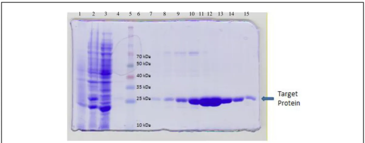
(NOTE: Results showing analysis for one TOG protein purification, mChe-12 TOG4, will be shown. Purifications for before mentioned TOG domains proceeded in the same manner but will be omitted for brevity.)
Figure 3- Chromatogram from elution on Ni2+-NTA column with increasing
imidazole concentration for mChe-12 TOG4
As seen in Figure 3, the majority of the TOG4 protein domain eluted in fractions 10 through 35. The absorbance peak shown for these fractions designated where the protein came off the Ni2+ column.
As seen in Figure 4, the TOG4 domain protein shown at 25 kDa was present in all chromatogram peak fractions. In this case, fractions 15-34 were pooled because they all showed some amount of purified protein.
Figure 4- SDS PAGE gel showing the purification steps of Ni2+ NTA column. The samples are 1)pre-IPTG cells 2)post-IPTG cells 3) Ni2+ NTA column flow through 4) Ni2+ NTA column wash 5)ladder 6-15) fractions 6/8/10/12/15/17/20/22/26/30/34/38/42 respectively
Fraction Number
Figure 5-SDS PAGE gel results from thrombin digestion.
Figure 5 shows the results of thrombin digestion following Ni2+ NTA column elution. The pooled protein fractions after elution from the Ni2+-column were incubated with Bovine α-Thrombin for 24 hours. The SDS-PAGE in Fig. 3 shows the expected size shift accompanying the cleaving of the N-terminal 6X His tag by thrombin.
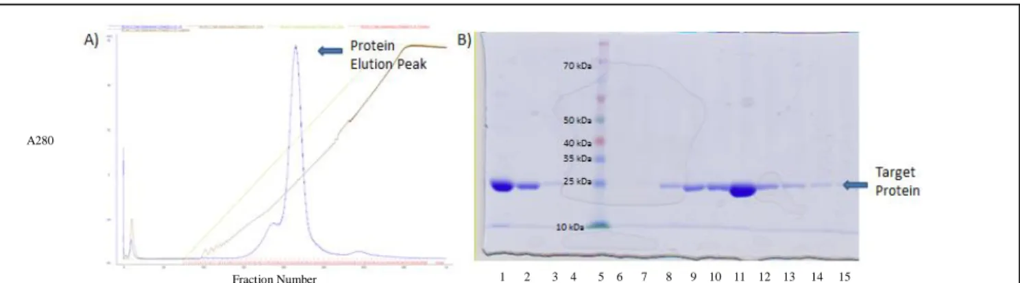
Figure 6 shows that TOG4 protein was already purified prior to being run over the SP-sepharose fast flow column. The protein band indicating TOG4 remains prevalent at around 22 kDa. Fractions 15-30 were pooled and concentrated to 15 mg/mL following the elution.
Figure 6- SP-sepharose fast flow column purification A) Chromatogram from column with increasing NaCl concentration B)SDS PAGE gel of purification steps (1-protein before column, 2-flow through S Sepharose column, 3-wash S
Sepharose column, 4-blank,5- ladder,6-15- Fractions 7/10/15/18/21/24/27/30/32/34/36 respectively.
1 2 3 4 5 6 7 8 9 10 11 12 13 14 15 Fraction Number
Summary of Proteins Purified
Protein Final Concentration Obtained Crystallization Trial Result Summary
mChe-12 TOG 1 13.5 mg/mL No Crystallization
Conditions
mChe-12 TOG2 Y364E mutant 15 mg/mL Crystallized but did not diffract adequately
mChe-12 TOG3 20 mg/mL No Crystallization
Conditions
mChe-12 TOG4 15 mg/mL Crystallized and diffracted
to 2.5 Å
mChe-12 TOG4 SeMet 13.5 mg/mL Crystallized but did not
diffract adequately
SSA6 TOG1 10 mg/mL Crystallized and diffracted
to 2.8 Å
SSA6 TOG1 SeMet 16 mg/mL Crystallized and diffracted
to 3.5 Å
Table1. Summary of purified proteins used for crystallization and/or microtubule polymerization assays
TOG Domain Crystallization Conditions
Figure 7 shows the resulting crystals after several trials of honing in on the best
conditions to crystallize TOG4. The crystals have a thin plate morphology and grew in clumps within 1 hour of setting up crystallization conditions and reached maximum size in 24 hours. Crystals were then looped, transferred to cryptoprotectants, frozen in liquid nitrogen, and sent for
remote x-ray diffraction experiments at the Advanced Photon Source at Argonne National Labs. Using these crystals, useful diffraction data for TOG4 was successfully obtained.
Figure 8 shows the resulting crystals after conducting crystallization screens for SSA6 TOG1 protein. Both types of crystals (native and SeMet) provided useful diffraction data after the same looping and freezing method described for TOG4. With both native and SeMet diffraction data, the electron density obtained for SSA6 TOG1 may allow for the SSA6 TOG1 structure to be determined.
Figure 8- A) Native SSA6 TOG1 crystals (conditions: 0.12M NH4Cl, 12% PEG3350, 7.5% Ethylene
Tubulin Polymerization Assay for TOG1-2 and TOG3-4
Figure 9: Light scattering signal of tubulin alone and in presence of TOG domains (A) TOG1, TOG2, and TOG2 Y364E(B) TOG3 and TOG4 (C) TOG1+TOG2 and TOG(1+2) (D) TOG3+TOG4 and TOG(3+4)
Figure 9 shows the results of the tubulin polymerization assay conducted. Addition of TOG domains increased microtubule assembly reactions relative to control reactions that
domains are connected by a linker region. The coupled TOG domains show more activity in the polymerization assay in comparison to adding the same two domains separately.
DISCUSSION
In this study, all of mChe-12’s TOG domains were purified. The purpose for these purifications was two-fold: 1) in order to carry out tubulin polymerization assays to gauge the different effects of each domain on the rate of tubulin polymerization and 2) in order to obtain crystal structures of each domain through the SAD method collection of both native and anomalous diffraction data using Se-Met substituted protein.
Crystallization and Structure Determination
Up to this point, constructs giving rise to crystals out of the available crystal screens include mChe-12 TOG2 Y364E, mChe-12 TOG4, and SSA6 TOG1. All constructs described previously were tested with the hopes of finding crystallization conditions. However,
modifications will have to be made to these constructs in order to make them better suited for crystallization in order for the determination of a crystal structure to be possible.
Currently, diffraction data sets have been collected for both SSA6 TOG1 native and Se-Met derivatives. Work is currently underway to solve this structure, and we hypothesize that this protein will show typical TOG domain architecture. If this hypothesis is supported, TOG
domain architecture would be conserved in Che-12 homologs in both mice (mChe-12) and single celled organism (SSA6 in C.reinhardtii). Thus, the Che-12 family could be classified as a new class of TOG domain containing proteins based on conservation of TOG domains across species.
Tubulin Polymerization Assay
Based on the tubulin polymerization assays, we found that single TOG domains increased the rate of microtubule polymerization in vitro in comparison to tubulin controls. However, TOG domains 2 and 4 showed greater tubulin polymerization activity than TOG domains 1 and 3. These results differed in comparison to Stu2p TOG domains which in a similar light scattering assay showed a decrease in microtubule assembly reactions (Ayaz, 2012). TOG domains in mChe-12 may contact tubulin in a different way in comparison to the Stu2p/XMAP215/Dis1 family such that tubulin heterodimers are not sequestered by single TOG domains. This polymerization activity was extinguished when the conserved aromatic residue in the first intra-HEAT loop of TOG2 in mChe-12 (Y364) was mutated to a glutamic acid. This further supports that the contacts being made between TOG2 and tubulin brought about this increased
REFERNCES
Ayaz, P. Rice, L.M., Pelin, A., Xuecheng, Y. (2012) A TOG:αβ-tubulin Complex Structure Reveals Conformation-Based Mechanisms for a Microtubule Polymerase. Science. 337, 857-859.
Bacaj, T., Tevlin M, Lu Y, Shaham S, (2008) The conserved proteins CHE-12 and DYF-11 are required for sensory cilium function in Caenorhabditis elegans. Genetics. 178, 989-1002. Charrasse, S.M., Schroeder, C., Gauthier-Rouviere, F., Ango, L., Cassimeris, D.L., Gard, C.
(1998) The TOG protein is a new human microtubule-associated protein homologous to the Xenopus XMAP215. J. Cell Sci., 111, 1371-1380.
Desai, A., Mitchison, T.J. (1997) Microtubule polymerization dynamics. Annu. Rev. Cell Dev. Biol. 13, 83–117.
Merchant, S.S. (2007) The Chlamydomonas genome reveals the evolution of key animal and plant functions. Science. 318, 245-250.
Perkins, L.A., Hedgecock, E.M., Thomson, J.N., Culotti, J.G. (1986) Mutant sensory cilia in the nematode Caenorhabditis elegans. Dev Biol. 117, 456-487.
Slep, K.C. (2007) Structural Basis of Microtubule Plus End Tracking by XMAP 215, CLIP-170, and EB1. Molecular Cell. 27, 976-991.
Slep, K.C. (2009) The role of TOG domains in microtubule plus end dynamics. Biochemical Transactions. 37, 1002-1006.
SUPPLEMENTAL
Purification Buffer Ingredients
-Nickel A- 25 mM Tris (pH 8.0), 300 mM NaCl, 10 mM imidazole, 0.1% βME -Nickel B-25 mM Tris (pH 8.0), 300 mM NaCl, 300 mM imidazole, 0.1% βME -S-Sepharose A- 25 mM HEPES (pH 7.0), 0.1% βME
-S-Sepharose B- 25 mM HEPES (pH 7.0), 0.1% βME, 1 M NaCl -Dialysis Buffer-25 mM HEPES (pH 7.0), 0.1% βME, 150 mM NaCl
Tubulin Polymerization Buffer Ingredients