METABOLISM AND ADVERSE EFFECTS OF ARSENIC
IN GENETICALLY DIVERSE MOUSE STRAINS
by
Jinglin Ji
Honors Thesis
Department of Nutrition
University of North Carolina
2019
Approved:
Advisor: Mirek Styblo, PhD Signature:
Abstract
Through drinking water and food, millions of people worldwide are exposed to arsenic (As), which is a naturally occurring diabetogenic metalloid [13]. Inorganic arsenic (iAs) exposure may disrupt glucose homeostasis leading to type 2 diabetes (T2D), including: (i) insulin resistance due to inhibition of insulin signaling, (ii) inhibition of insulin secretion, and (iii) aberrant micro RNA expression and activity [1, 2, 13]. Arsenic methyltransferase (AS3MT) is the key enzyme in iAs detoxification pathway that determines disease outcome [3]. Polymorphisms in AS3MT is the single most important genetic factor affecting iAs metabolism and susceptibility to adverse effects of iAs exposure, including diabetes. This study aims to examine the relationship between As3mt polymorphism and the adverse outcomes of iAs exposure in two Collaborative Cross (CC) mouse strains, CC021/Unc (CC021) and CC027/TauUnc (CC027), with different genetic
backgrounds. Metabolic phenotype and susceptibility to adverse effects of iAs exposure are hypothesized to differ in CC021 and CC027 due to the differences in their hepatic As3mt
Aims and Hypothesis
The overall hypothesis of the study is that metabolic phenotype and susceptibility to adverse effects of iAs exposure differ between CC021 and CC027 mice due to the differences in their hepatic As3mt expression and iAs metabolism in liver.
Aim 1. Evaluation of the Diabetogenic Effects of iAs Exposure
Body weight, body composition, fasting blood glucose and insulin levels, glucose tolerance,
insulin resistance and -cell function were measured in CC mice chronically exposed to iAs. Hypothesis 1: Metabolic phenotypes of the CC strains exposed to iAs will differ.
Aim 2. Evaluation of the iAs Metabolism
The proportions and concentrations of iAs and its methylated (MAs) and dimethylated (DMAs) metabolites were measured in the urine of the CC mice after chronic exposures to iAs.
Hypothesis 2: iAs metabolism will differ in CC021 and CC027 mouse strains.
Introduction
Arsenic (As) is a naturally occurring toxic metalloid. Over 100 million people worldwide drink water with inorganic As (iAs) levels higher than the US EPA and WHO maximum level of 10 μg As/L [4, 5]. More people may be exposed to unsafe levels of iAs in foods [6-10].
The International Agency for Research on Cancer defines iAs as a Group-I human carcinogen [11]. Exposure to iAs is also associated with a wide array of non-cancer effects such as
the National Toxicology Program Workshop on the environmental diabetogens and obesogens found sufficient evidence for the relationship between moderate to high levels of exposure to iAs in drinking water (>150 ppb; i.e., >150 µg As/L) and diabetes [13]. Two recent meta-analyses that included studies at lower exposure levels found pooled relative risks of developing diabetes in the presence of iAs exposure equal to 1.7 and 1.23 [14,15].
According to previous studies, the insulin producing pancreatic β-cells are among the targets for iAs exposure [1, 16]. The inhibition of glucose-stimulated insulin secretion (GSIS) by low concentrations of the methylated metabolites of iAs may be the key mechanism of iAs-induced diabetes [1]. Trivalent arsenicals interfere with mechanisms regulating packaging of the insulin transport vesicles or with translocation of these vesicles to the plasma membrane [1]. Defects in insulin secretion could also be associated with aberrant microRNA expression and activity [2]. In
addition, micromolar concentrations of inorganic trivalent arsenite (iAsIII) or its methylated
trivalent metabolites, methylarsonite (MAsIII) and dimethylarsinite (DMAsIII), inhibit the insulin-activated signal transduction pathway, resulting in insulin resistance and impaired glucose metabolism in adipocytes and hepatocytes [13, 17, 18].
Arsenic methyltransferase (AS3MT) is the key enzyme in the pathway for iAs metabolism [3]. Methylation of inorganic As determines the efficiency of iAs detoxification and the disease risk. AS3MT converts iAs to mono- and dimethylated metabolites (MAs, DMAs) that are excreted mainly in urine. The efficiency of iAs methylation can be estimated using the proportions of As metabolites in urine. A low DMAs/MAs ratio and high percentage of total urinary As
However, the methylated trivalent metabolites formed in this pathway are potent inhibitors of processes that regulate glucose metabolism, including insulin secretion by pancreatic β-cells and
insulin signaling in the liver and peripheral tissues [1]. Thus, in a counter-intuitive manner, while essential for total body clearance, the metabolism/methylation of iAs generates metabolites that likely contribute to the diabetogenic effects of iAs exposure.
Many population studies have examined the association between genetic polymorphism and the pattern of iAs metabolism. Although polymorphisms in several genes that are thought to regulate various steps in iAs metabolism have been linked with differences in the capacity to methylate iAs and in iAs toxicity, polymorphisms in AS3MT are the single most important genetic factor affecting iAs metabolism and susceptibility to adverse effects of iAs exposure, including diabetes, in population studies [24-29]. Notably, results of laboratory studies using genetically modified cells and mice, including our published and preliminary studies, support the critical role for As3mt in iAs metabolism. [19, 42]
In short, according to previous studies, iAs exposure may disrupt glucose homeostasis leading to T2D, including: (i) insulin resistance due to inhibition of insulin signaling, (ii) inhibition of insulin secretion, and (iii) aberrant micro RNA expression and activity [1, 2, 13]. AS3MT is the key enzyme in As metabolism while its polymorphisms are the most important genetic factor affecting susceptibility to As-related diabetes [3]. However, the relationship between As3mt polymorphism and the adverse outcomes of iAs exposure has never been systematically examined in animal models.
CC027/TauUnc (CC027), that have different As3mt haplotypes (non-obese diabetic
(NOD)/ShiLtJ and Watkins Star Line B (WSB)/EiJ, respectively) and different SNPs in As3mt gene. CC strains are products of an eight-way funnel breeding design involving eight genetically diverse JAX® mice founder strains: A/J (000646), C57BL/6J (000664), 129S1/SvImJ (002448)
NOD/ShiLtJ (001976) NZO/HiLtJ (002105), CAST/EiJ (000928), PWK/PhJ (003715), and WSB/EiJ (001145) [30]. Preliminary studies carried out in our laboratory showed that the hepatic expression of As3mt (Fig. 1) and patterns of iAs metabolism in these two strains, specifically the proportions of total As present as iAs (%iAs) and DMAs (%DMAs) in the liver (Fig. 2), are also significantly different.
Figure 1: Baseline expression of As3mt in livers of male CC021 and CC027 mice. (N=4)
A
s3
mt
mR
N
A
(I
n
te
n
si
ty)
Figure 2: Proportions of As species in livers of male CC021 and CC027 mice. Mice were
exposed to 50 ppm iAs (arsenite) for 2 weeks. (N=5). Total As (Mean ±SD): 1342 ±652 ng/g (CC021) and 1609 ±317 ng/g (CC027).
The current project expands our knowledge of the mechanisms of iAs related diseases, specifically diabetes, and the role of As3mt genotype as factor that modifies the disease risk. This project also has a potential to move forward the entire field of iAs toxicology by providing novel mouse models for laboratory studies, and to contribute significantly to risk assessment of iAs-associated diseases based on genetic susceptibility.
Methods
Mice
semi-purified AIN-93G diet. Mice were housed under controlled conditions with 12-h light/ dark cycle at 22 ± 1 °C and 50 ± 10% relative humidity (2–5 mice per cage).
Exposure
After acclimatization, mice from both strains were exposed to 100 ppb or 50 ppm iAs (sodium arsenite NaAsO2, ≥99% pure; Sigma-Aldrich, St. Louis, MO, USA) in drinking water for 12
weeks; control mice continued drinking DIW. Water with sodium arsenite was prepared weekly
to minimize oxidation of iAsIII to iAsV.
Metabolic Phenotype Assessment
Body weights and water and food consumption were monitored bi-weekly during exposure. Body composition, fasting blood glucose and insulin levels, and glucose tolerance were
examined at the end of exposure. Magnetic resonance imaging (MRI) (EchoMRI 3-in-1 analyzer, Echo Medical Systems, Houston, TX, USA and Labmaster version 3.2.2. Software) was used to
determine body composition, specifically grams of fat mass and grams of lean mass. For glucose and insulin analysis, mice were fasted for 6 hours. Fasting blood glucose (FBG) levels were measured in blood collected from tail cuts using OneTouch Ultra 2 glucometer (LifeScan, Milpitas, CA, USA) and OneTouch Ultra Blue test strips. Fasted mice were then injected i.p. with 2 g/ kg b.w. of D-glucose (Sigma-Aldrich) dissolved in Dulbecco’s phosphate-buffered saline (Mediatech, Manassas, VA, USA). Glucose levels were measured in blood collected by tail bleeds 15 min injection. Plasma insulin was measured in fasting and 15 min
HOMA-IR and HOMA-B were calculated from fasting blood glucose and plasma insulin values using the following formulas:
• HOMA-IR = 𝐹𝑎𝑠𝑡𝑖𝑛𝑔 𝐵𝑙𝑜𝑜𝑑 𝐺𝑢𝑐𝑜𝑠𝑒 ( 𝑚𝑔
𝑑𝐿)×𝐹𝑎𝑠𝑡𝑖𝑛𝑔 𝑃𝑙𝑎𝑠𝑚𝑎 𝐼𝑛𝑠𝑢𝑙𝑖𝑛 ( 𝜇𝑈 𝑚𝐿)
405 [44]
• HOMA-B = 360×𝐹𝑎𝑠𝑡𝑖𝑛𝑔 𝑃𝑙𝑎𝑠𝑚𝑎 𝐼𝑛𝑠𝑢𝑙𝑖𝑛 ( 𝜇𝑈 𝑚𝐿)
𝐹𝑎𝑠𝑡𝑖𝑛𝑔 𝐵𝑙𝑜𝑜𝑑 𝐺𝑙𝑢𝑐𝑜𝑠𝑒 (𝑚𝑔𝑑𝐿)−63 % [45]
𝐹𝑎𝑠𝑡𝑖𝑛𝑔 𝑃𝑙𝑎𝑠𝑚𝑎 𝐼𝑛𝑠𝑢𝑙𝑖𝑛 (𝑚𝐿𝜇𝑈)=𝐹𝑎𝑠𝑡𝑖𝑛𝑔 𝑃𝑙𝑎𝑠𝑚𝑎 𝐼𝑛𝑠𝑢𝑙𝑖𝑛 ( 𝑛𝑔 𝑚𝐿)
0.0347 (𝑛𝑔𝜇𝑈)
Analysis of As species in urine
Spot urine samples were collected at the end of the exposure. The concentrations of iAs, MAs, and DMAs were measured in spot urine samples using hydride generation-atomic absorption spectrometry coupled with a cryotrap [32, 33]. The proportions of iAs and its methylated
metabolites in urine (%iAs, %MAs, and %DMAs) were used to assess differences in methylation capacity of mice in both strains.
Statistical Analysis
Data were analyzed using ANOVA and Tukey-Kramer multiple comparisons test. Differences with P<0.05 were regarded as statistically significant. Mean ±SE were calculated and graphed for each outcome.
Results
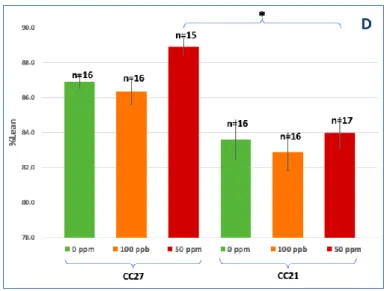
Figure 3: Average final body weight (A), weight gain (B), %body fat (C) and %lean mass (D) of
CC021 and CC027 mice exposed to 100 ppb or 50 ppm iAs and the control mice (0 ppm). Mean ±SE. * Statistically significant differences (P<0.05) using ANOVA and Tukey-Kramer multiple comparisons test.
No statistically significant difference in body weight at the end of the exposure were seen between the two strains or between different exposure levels (Fig. 3A). However, CC021 mice tended to have higher body weight while mice exposed to 50 ppm iAs in both strains tended to have lower body weight (Fig. 3A). Percent weight gain decreased significantly and in a dose-dependent manner in CC021, but not CC027 mice (Fig. 3B). CC021 mice, both iAs-exposed and controls, had significantly higher %fat mass than CC027 mice (Fig. 3C). CC027 mice had
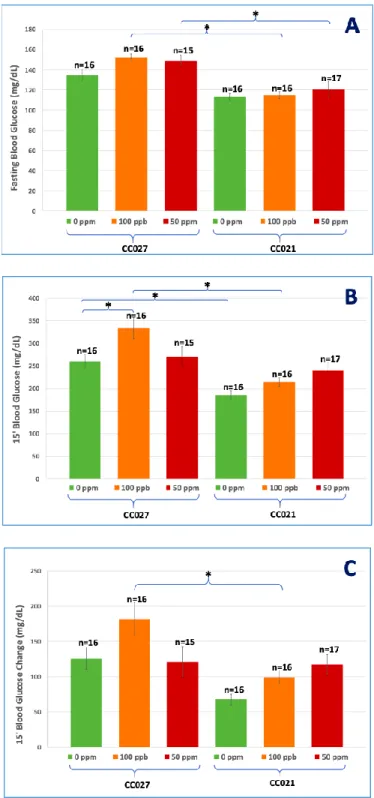
Figure 4: Blood glucose levels after fasting (A), 15 min after i.p. glucose injection (B), and the
difference (change) between fasting and 15-min glucose levels (C).
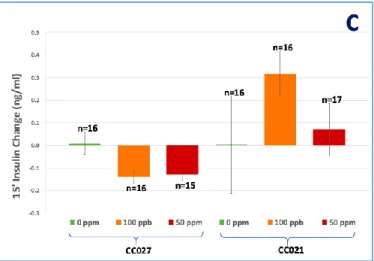
Figure 5: Plasma insulin levels after fasting (A) and 15 min after i.p. glucose injection (B), and
the difference (change) in insulin levels between fasting and 15-min after glucose injection (C). Mean ±SE. *Statistically significant differences (P<0.05) using ANOVA and Tukey-Kramer multiple comparisons test.
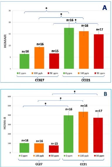
Figure 6: Insulin resistance (HOMA-IR) (A) and β-cell function (HOMA-B) (B).
*Statistically significant differences (P<0.05) using ANOVA and Tukey-Kramer multiple comparisons test.
CC021 mice (with NOD background), both iAs-exposed and controls, were more insulin
Figure 7: Proportions of As species in urine: %iAs (A) and %DMAs (B). Mean ±SE. TAs = sum
of As species. *Statistically significant differences (P<0.05) using ANOVA and Tukey-Kramer multiple comparisons test.
Discussion
Laboratory mice with varied genetic backgrounds have been extensively used in laboratory studies examining adverse effects of iAs exposure. However, it has been well documented that iAs methylation and excretion in mice is more efficient than in humans [19, 20], which likely explains the differences between these species in the responses to iAs exposure [34-36]. For instance, in all human populations studies to date, MAs and DMAs represented 10-20% and 60-70% of total urinary As, respectively, with iAs accounting for 10-30% [19, 20]. In contrast, urine of mice exposed to iAs in published studies contains more than 80% of DMAs and only traces of MAs, suggesting a more efficient conversion of MAs to DMAs [19, 20]. Thus, a better animal model is needed in which iAs metabolism would resemble the human metabolism, and in which the effects of iAs exposure found in humans can be reproduced. Strong evidence suggests that the differences in the AS3MT genotypes are in part responsible for the differences in
efficiency of iAs metabolism and in susceptibility to adverse effects of iAs exposure among humans and between mice and humans. This observation provided rationale for the present study. The goal of this study was to compare effects of iAs exposure on glucose homeostasis between 2 CC mouse strains that differ in As3mt genotype and demonstrate differences in the disposition of iAs and its metabolites in the liver.
insulin signaling, (ii) inhibition of insulin secretion, and (iii) aberrant micro RNA expression and activity [1, 2, 13].
We have previously shown that urine and livers of control mice that drank only deionized water and were fed a regular grain-based diet (pelleted 2920X Teklad rodent chow, Envigo, Madison, WI, USA) contained relatively high levels of As, suggesting that these mice were exposed to As from diet [42]. When designing the present study, we included a two-week acclimatization period to allow for clearance of As to which the mice were exposed when fed a regular grain-based diet before delivery to UNC. The methods we used for metabolic phenotype, including measures of fasting blood glucose and insulin, are widely utilized in diagnosis of diabetes in human patients and are good approximates of the diabetogenic effect of iAs exposure in mice [40, 43]. A simplified glucose tolerance test we used in this study was to assess the physiological response of mice to a glucose challenge, specifically blood glucose and plasma insulin levels at 15 minutes after injection. The homeostasis model assessments HOMA-IR and HOMA-B, based on fasting glucose and insulin measures, have been validated and widely applied for quantifying insulin resistance and β-cell function in clinical setting, and are used here as proxies of these
measure in mice [31].
(commonly called NOD) is a polygenic model for autoimmune type 1 diabetes, characterized by hyperglycemia and insulitis [30].
CC027 mice exposed to 100 ppb iAs had higher blood glucose after 6-hour fasting and 15 min after i.p. glucose injection, and higher blood glucose change between fasting and 15-min glucose levels than CC021 mice (Fig. 4A, 4B, 4C). CC027 mice exposed to 50 ppm iAs had higher blood glucose levels after 6-hour fasting than CC021 mice exposed to 50 ppm iAs (Fig. 4A). CC027 control mice had higher blood glucose levels 15 min after i.p. glucose injection than CC021 control mice (Fig. 4B).
Defects in antigen presentation, T lymphocyte repertoire, natural killer cell function, macrophage cytokine production, wound healing, and C5 complement characterize immune phenotypes in the NOD background [30]. Insulitis is a leukocytic infiltration of the pancreatic islets and should be characterized by decreased β-cell mass and insulin secretion [30]. According to our results, CC027 mice, both iAs-exposed and controls, had lower plasma insulin levels after 6-hour fasting and 15 min after i.p. glucose injection than CC021 mice (Fig. 5A, 5B). CC027 mice exposed to 100 ppb and 50 ppm iAs in drinking water had negative insulin change after glucose challenge (Fig. 5C). Lower plasma insulin level and negative insulin change in response to glucose challenge in CC027 probably represents decreased β-cell function and insulin secretion while exposure to iAs might deteriorate insulitis.
The mechanism of developing insulitis in CC021 is unclear for now. According to previous
trivalent metabolites, methylarsonite (MAsIII) and dimethylarsinite (DMAsIII) inhibit the insulin-activated signal transduction pathway, resulting in insulin resistance in adipocytes and hepatocytes [13, 17, 18]. CC021 mice, both iAs-exposed and controls, had significantly higher %fat mass (Fig. 3C) and were more insulin resistant (Fig. 5, 6) than their CC027 counterparts probably due to higher %fat mass. Insulin resistance could cause a long-term
overstimulation of β-cells which leads to inflammation and autoimmune attack on β-cells. Insulin resistance and lower insulin secretion could together induce diabetes.
Most of the phenotypic differences between the two strains were dependent on genetic background and independent of iAs exposure. Strain differences were pronounced in body composition (Fig. 3C, 3D), blood glucose (Fig. 4), plasma insulin (Fig. 5A, 5B) and insulin resistance (Fig. 6). iAs exposure had mostly minor effects on phenotype of mice in either strain. Dose dependent effects on the proportions of As species were seen in the urine of both
strains: %iAs decreased and %DMA increased with increasing exposure level (Fig. 7). This is in contrast with data from human cohorts where %DMAs has been found to decrease with
However, the fact that most of the differences between these 2 strains were independent of iAs exposure suggests that the phenotypes of CC021 and CC027 mice exposed to iAs are determined primarily by their broader genetic backgrounds rather than by As3mt polymorphism and
expression in the liver, or by the differences in iAs metabolism. Notably, the proportions of As species in urine of both strains were consistent with those reported in previous mouse studies and differed from the proportions found in human urine [19, 20]. Thus, neither CC021 nor CC027 is an ideal model to study metabolism of iAs or adverse effects associated with iAs exposure in humans.
Reference
1. Douillet, C., Currier, J.M., Saunders, J., Bodnar, W., Matoušek, T., Stýblo, M. (2013) Methylated trivalent arsenicals are potent inhibitors of glucose stimulated insulin secretion by murine pancreatic islets. Toxicol. Appl. Pharmacol. 267, 11-15.
2. Beck, Rowan, Miroslav Styblo, and Praveen Sethupathy. "Arsenic exposure and type 2 diabetes: microRNAs as mechanistic links?." Current diabetes reports 17.3 (2017): 3. Thomas DJ, Li J, Waters SB, Xing W, Adair BM, Drobna Z, Devesa V, Styblo M.
Arsenic (+3 oxidation state) methyltransferase and the methylation of arsenicals. Experimental biology and medicine. 2007;232(1):3-13. PubMed PMID: 17202581; PMCID: 2408740.
4. Who. Guidelines for Drinking Water Quality. World Health Organization (1993).
5. Arslan B, Djamgoz MB, Akün E. ARSENIC: A Review on Exposure Pathways, Accumulation, Mobility and Transmission into the Human Food Chain. Rev Environ Contam Toxicol. 2016 Dec 23. doi: 10.1007/398_2016_18.
6. Cubadda F, Jackson BP, Cottingham KL, Van Horne YO, Kurzius-Spencer M. Human exposure to dietary inorganic arsenic and other arsenic species: State of knowledge, gaps and uncertainties. Sci Total Environ. 2017 Feb 1;579:1228-1239.
7. Azam SM, Sarker TC, Naz S. Factors affecting the soil arsenic bioavailability, accumulation in rice and risk to human health: a review. Toxicol Mech Methods. 2016 Oct;26(8):565-579.
9. Environ Health Perspect. 2016 Aug 19. [Epub ahead of print] PMID: 27539714.
10.Wilson D. Arsenic Consumption in the United States. J Environ Health. 2015 Oct;78(3):8-14.
11.International Agency for Research on Cancer (IARC, 2004). Arsenic in drinking water. International agency for research on cancer monographs on the evaluation of carcinogenic risk to humans. Some drinking water disinfectants and contaminants, including arsenic. IARC Press, Lyon, vol.84, pp. 269-477.
12.Naujokas MF, Anderson B, Ahsan H et al. The broad scope of health effects from chronic arsenic exposure: update on a worldwide public health problem. Environ. Health
Perspect. 121(3), 295-302 (2013).
13.Maull EA, Ahsan H, Edwards J, Longnecker MP, Navas-Acien A, Pi J, Silbergeld EK, Styblo M, Tseng CH, Thayer KA, Loomis D. Evaluation of the association between arsenic and diabetes: a National Toxicology Program workshop review. Environmental health perspectives. 2012;120(12):1658-70. doi: 10.1289/ehp.1104579. PubMed PMID: 22889723; PMCID: 3548281.
14.Sung TC, Huang JW, Guo HR. Association between Arsenic Exposure and Diabetes: A Meta-Analysis. BioMed research international. 2015;2015:368087. doi:
10.1155/2015/368087. PubMed PMID: 26000288; PMCID: 4427062.
16.Dover EN, Beck R, Huang MC, Douillet C, Wang Z, Klett EL, Stýblo M. Arch Toxicol. Arsenite and methylarsonite inhibit mitochondrial metabolism and glucose-stimulated insulin secretion in INS-1 832/13 β cells. 2018 Feb;92(2):693-704.
17.Paul, D.S., Harmon, A.W., Devesa, V., Thomas, D.J., Styblo, M., 2007a. Molecular mechanisms of diabetogenic effects of arsenic: inhibition of insulin signaling by arsenite and methylarsonous acid. Environ. Health Perspect. 115, 734–742.
18.Zhang C, Fennel EMJ, Douillet C, Stýblo M. Exposures to arsenite and methylarsonite produce insulin resistance and impair insulin-dependent glycogen metabolism in
hepatocytes. Arch Toxicol. 2017 Dec;91(12):3811-3821. doi: 10.1007/s00204-017-2076-9. Epub 2017 Sep 26.
19.Vahter M. Genetic polymorphism in the biotransformation of inorganic arsenic and its role in toxicity. Toxicol Lett. 2000;112-113:209-17. Epub 2000/03/18. PubMed PMID: 10720733.
20.Vahter M. Methylation of inorganic arsenic in different mammalian species and population groups. Sci Prog. 1999; 82 (Pt 1):69-88.
22.Ahsan H, Chen Y, Kibriya MG, Slavkovich V, Parvez F, Jasmine F, Gamble MV, Graziano JH. Arsenic metabolism, genetic susceptibility, and risk of premalignant skin lesions in Bangladesh. Cancer Epidemiol Biomarkers Prev. 2007 Jun;16(6):1270-8.
23.Tseng CH. A review on environmental factors regulating arsenic methylation in humans. Toxicol Appl Pharmacol. 2009;235:338–50.
24.Drobna Z, Del Razo LM, Garcia-Vargas GG, Sanchez-Pena LC, Barrera-Hernandez A, Styblo M, Loomis D. Environmental exposure to arsenic, AS3MT polymorphism and prevalence of diabetes in Mexico. J Expo Sci Environ Epidemiol. 2013;23(2):151-5. Epub 2012/10/25. doi: 10.1038/jes.2012.103. PubMed PMID: 23093101; PMCID: PMC4067760.
25.Pan WC, Kile ML, Seow WJ, Lin X, Quamruzzaman Q, Rahman M, Mahiuddin G, Mostofa G, Lu Q, Christiani DC. Genetic susceptible locus in NOTCH2 interacts with arsenic in drinking water on risk of type 2 diabetes. PloS one. 2013;8(8):e70792. Epub 2013/08/24. doi: 10.1371/journal.pone.0070792. PubMed PMID: 23967108; PMCID: PMC3743824.
26.Diaz-Villasenor A, Cruz L, Cebrian A, Hernandez-Ramirez RU, Hiriart M,
Garcia-Vargas G, Bassol S, Sordo M, Gandolfi AJ, Klimecki WT, Lopez-Carillo L, Cebrian ME, Ostrosky-Wegman P. Arsenic exposure and calpain-10 polymorphisms impair the
function of pancreatic beta-cells in humans: a pilot study of risk factors for T2DM. PloS one. 2013;8(1):e51642. Epub 2013/01/26. doi: 10.1371/journal.pone.0051642. PubMed PMID: 23349674; PMCID: PMC3551951.
27.Chen JW, Wang SL, Wang YH, Sun CW, Huang YL, Chen CJ, Li WF. Arsenic
area of southwestern Taiwan. Chemosphere. 2012;88(4):432-8. Epub 2012/03/24. doi: 10.1016/j.chemosphere.2012.02.059. PubMed PMID: 22440634.
28.Escobar-Garcia DM, Del Razo LM, Sanchez-Pena LC, Mandeville PB, Lopez-Campos C, Escudero- Lourdes C. Association of glutathione S-transferase Omega 1-1
polymorphisms (A140D and E208K) with the expression of interleukin-8 (IL-8), transforming growth factor beta (TGF-beta), and apoptotic protease- activating factor 1 (Apaf-1) in humans chronically exposed to arsenic in drinking water. Archives of toxicology. 2012;86(6):857-68. Epub 2012/02/02. doi: 10.1007/s00204-012-0802-x. PubMed PMID: 22293942.
29.Hernandez A, Marcos R. Genetic variations associated with interindividual sensitivity in the response to arsenic exposure. Pharmacogenomics. 2008;9(8):1113-32. Epub
2008/08/07. doi: 10.2217/14622416.9.8.1113. PubMed PMID: 18681785.
30.“The Collaborative Cross: a Powerful Systems Genetics Tool.” The Jackson Laboratory, www.jax.org/news-and-insights/2009/april/the-collaborative-cross-a-powerful-systems-genetics-tool.
31.Song, Yiqing, et al. "Insulin sensitivity and insulin secretion determined by homeostasis model assessment and risk of diabetes in a multiethnic cohort of women: the Women's Health Initiative Observational Study." Diabetes care 30.7 (2007): 1747-1752.
32.Hernandez-Zavala A, Matousek T, Drobna Z, Paul DS, Walton F, Adair BM, Jiri D, Thomas DJ, Styblo M. Speciation analysis of arsenic in biological matrices by automated hydride generation-cryotrapping-atomic absorption spectrometry with multiple
spectrometry. 2008;23:342-51. doi: 10.1039/b706144g. PubMed PMID: 18677417; PMCID: 2493051.
33.Matousek T, Currier JM, Trojankova N, Saunders RJ, Ishida MC, Gonzalez-Horta C, Musil S, Mester Z, Styblo M, Dedina J. Selective hydride generation- cryotrapping- ICP-MS for arsenic speciation analysis at picogram levels: analysis of river and sea water reference materials and human bladder epithelial cells. Journal of analytical atomic spectrometry. 2013;28(9):1456-65. doi: 10.1039/C3JA50021G. PubMed PMID: 24014931; PMCID: 3763853.
34.Jager JW, Ostrosky-Wegman P. Arsenic: a paradoxical human carcinogen. Mutat Res. 1997;386:181–184.
35.National Science Foundation, Committee on How Toxicogenomics Could Inform Critical Issues in Carcinogenic Risk Assessment of Environmental Chemicals . Toxicogenomic technologies and risk assessment of environmental carcinogens: A workshop study. National Academies Press; Washington, D.C.: 2005. pp. 1–55.
36.States JC, Barchowsky A, Cartwright IL, Reichard JF, Futscher BW, Lantz RC. Arsenic toxicology: translating between experimental models and human pathology. Environ Health Perspect. 2011 Oct;119(10):1356-63.
37.Kuo CC, Howard BV, Umans JG, Gribble MO, Best LG, Francesconi KA, Goessler W, Lee E, Guallar E, Navas-Acien A. Arsenic Exposure, Arsenic Metabolism, and Incident Diabetes in the Strong Heart Study. Diabetes Care. 2015;38(4):620-7. Epub 2015/01/15. doi: 10.2337/dc14-1641. PubMed PMID: 25583752; PMCID: PMC4370323.
beta cells: involvement of cellular adaptive response to oxidative stress. Environmental health perspectives. 2010;118(6):864-70. Epub 2010/01/27. doi: 10.1289/ehp.0901608. PubMed PMID: 20100676; PMCID: PMC2898865.
39.Tseng CH. The potential biological mechanisms of arsenic-induced diabetes mellitus. Toxicology and applied pharmacology. 2004;197(2):67-83. Epub 2004/05/28. doi: 10.1016/j.taap.2004.02.009. PubMed PMID: 15163543.
40.Paul DS, Hernandez-Zavala A, Walton FS, Adair BM, Dedina J, Matousek T, Styblo M. Examination of the effects of arsenic on glucose homeostasis in cell culture and animal studies: development of a mouse model for arsenic-induced diabetes. Toxicology and applied pharmacology. 2007;222(3):305-14. doi: 10.1016/j.taap.2007.01.010. PubMed PMID: 17336358; PMCID: 2680915.
41.Walton FS, Harmon AW, Paul DS, Drobna Z, Patel YM, Styblo M. Inhibition of insulin-dependent glucose uptake by trivalent arsenicals: possible mechanism of arsenic-induced diabetes. Toxicology and applied pharmacology. 2004;198(3):424-33. Epub 2004/07/28. doi: 10.1016/j.taap.2003.10.026. PubMed PMID: 15276423.
42.Douillet C, Huang MC, Saunders RJ, Dover EN, Zhang C, Stýblo M. Knockout of arsenic (+3 oxidation state) methyltransferase is associated with adverse metabolic phenotype in mice: the role of sex and arsenic exposure. Arch Toxicol. 2016 Nov 15. [Epub ahead of print] PMID: 27847981
44.Huang, Madelyn C., et al. "Prenatal arsenic exposure and dietary folate and
methylcobalamin supplementation alter the metabolic phenotype of C57BL/6J mice in a sex-specific manner." Archives of toxicology 92.6 (2018): 1925-1937.
45.Imano, Hironori, et al. "Abstract P455: Insulin Resistance, Secretion and Risk of Incident Coronary Heart Disease in Non-diabetic Japanese Population: The Circulatory Risk in
Communities Study." Circulation 129.suppl_1 (2014): AP455-AP455.