© 2019 by the Serbian Biological Society How to cite this article: Li X, Li J, Yang J. Liprin-β1 is up regulated in human 469 hepatocellular carcinoma and is associated with advanced tumor stage. Arch Biol Sci. 2019;71(3):469-74.
Liprin-β1 is upregulated in human hepatocellular carcinoma and is associated with
advanced tumor stage
Xinying Li1, Jingmei Li1,* and Jiao Yang2,#
1School of Life Science and Technology, Changchun University of Science and Technology, Changchun, 130022, China
2Jiangsu Key Lab of Medical Optics, Suzhou Institute of Biomedical Engineering and Technology, Chinese Academy of Sciences,
Suzhou, 215163, China
Corresponding authors: *[email protected]; #[email protected]
Received: January 16, 2019; Revised: April 3, 2019; Accepted: April 25, 2019; Published online: May 10, 2019
Abstract: Liprin-β1 is one of the broadly-expressed liprin family members. Dysregulation of liprin-β1 has been implicated in several types of human cancers. However, the expression of liprin-β1 and its clinicopathological significance in human hepatocellular carcinoma (HCC) remains elusive. We evaluated the protein expression of liprin-β1 in HCC and non-tumor liver tissues by immunohistochemistry, and investigated the relationship between liprin-β1 expression and the clinico-pathological attributes of HCC. We found that liprin-β1 expression was significantly higher in HCC than in non-tumor liver tissues. Further analysis showed that higher levels of liprin-β1 in HCC were significantly associated with the advanced clinical stage. Interestingly, liprin-β1 was not detected in cholangiocellular carcinoma specimens. These findings suggest that an elevated expression of liprin-β1 may be involved in HCC progression, providing the rationale that upregulation of liprin-β1 may serve as a novel biomarker for human HCC.
Keywords: liprin-β1; hepatocellular carcinoma; cholangiocellular carcinoma; cirrhosis.
INTRODUCTION
Hepatocellular carcinoma (HCC) remains the lead-ing cause of cancer-related mortality worldwide, with more than half a million new cases diagnosed per year [1-3]. Because of the insidious onset of the disease, HCC tends to be diagnosed at advanced stages and responds poorly to therapy [4,5]. Adverse prognosis can be attributed to the high frequency of local recur-rence, intrahepatic invasion and distant metastases. Thus, identification of key factors implicated in HCC progression may be helpful in classifying tumor stages and predicting clinical outcomes.
The LAR protein-tyrosine phosphatase-interacting protein (liprin) family was originally found to be es-sential for the assembly of functional presynaptic active zones and postsynaptic sites in neurons [6,7]. The mam-malian liprin family includes four liprin-α (liprin-α1 to -α4) proteins and two liprin-β (β1 and β2) proteins [8], which together form heterodimers and act as scaffolds [9]. A number of recent studies have revealed broader
functions of the liprin family in regulating the develop-ment and homeostasis in other organs, including the mammary gland and lymphatic vessel [10,11]. Notably, analysis of human cancers has revealed dysfunctions of the liprin proteins, with evidence pointing to the differ-ent roles of liprin family members in distinct aspects of tumor biology. For example, overexpression of liprin-α1 promotes breast cancer cell invasion by regulating lamel-lipodia stability and integrin-mediated focal adhesions [10,12]. By contrast, liprin-β2 impairs cell motility and suppresses tumor invasion, which is indicative of its role as a tumor suppressor [10].
in these cancer types. However, the clinical relevance of liprin-β1 in HCC has not yet been investigated.
In the present study, we assessed liprin-β1 ex-pression in primary HCC specimens and evaluated its association with a series of histopathological pa-rameters. In addition, the expression of liprin-β1 in cholangiocellular carcinoma (CCC), the second most common primary liver cancer, was also determined.
MATERIALS AND METHODS
Ethics statement, patient samples, and clinical information
This study was conducted on precollected samples. A total of 48 HCC specimens, 5 CCC specimens and 12 tissues from patients with cirrhosis were collected at Tongxu People’s Hospital, Henan, China. All tissues were collected under the highest ethical standards between 2005 and 2010, and approved by the Re-search Ethics Committee of Tongxu People’s Hospital (2005DKA21300). Consent forms were signed by all patients. The study protocol was reviewed and ap-proved by the Research Ethics Committee of Tongxu People’s Hospital (BC03117). The clinicopathological characteristics of the patients are summarized in Sup-plementary Table S1, with detailed information, in-cluding patient gender, age of diagnosis, tumor grade, lymph node metastasis, and tumor-node-metastasis (TNM) stage listed in Supplementary Table S2.
Immunohistochemistry and quantification
The protein levels of liprin-β1 were determined us-ing immunohistochemistry (IHC).
Briefly, the tissue microarray sam-ples were deparaffinized in xylene and hydrated. Antigen retrieval was performed with Tris-EDTA antigenic retrieval buffer (pH 8.0), followed by 3% H2O2 treat-ment to inhibit endogenous per-oxidase activity. Tissue sections were blocked with 2% bovine se-rum albumin (Cat No. A8020; So-larbio, China) and incubated with anti-liprin-β1 (LS-B13770, 1:500)
at 4°C overnight. The EnVision kit (DAKO) was used to detect primary antibody followed by staining with DAB reagent and counterstaining with hematoxylin. The expression of liprin-β1 was scored as 0 (absent), 1 (weak), 2 (moderate) and 3 (strong) in a double-blinded manner.
Statistical analysis
Patients were stratified into liprin-β1Low (score of 0-1) versus liprin-β1High (score of 2-3) groups. The statisti-cal analyses were performed using GraphPad Prism software version 6.0. The association of categorical vari-ables was examined by Fisher’s exact test and a P-value of <0.05 was considered to be statistically significant.
RESULTS
Expression of liprin-β1 was upregulated in HCC
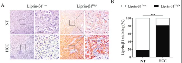
To examine whether the expression of liprin-β1 was altered in HCC, protein levels of liprin-β1 were de-tected by IHC and the staining intensity of HCC samples was compared with that of non-tumor liver tissues from patients with cirrhosis (NT). The results showed that liprin-β1 was strongly stained in HCC tissues (29/48, 60%) compared with NT (2/12, 17%), where only a smaller percentage of positivity was found (Fig. 1A and B). In addition, liprin-β1 protein was confirmed to be predominantly expressed in the parenchymal cells (Fig. 1A). These expression patterns of liprin-β1 in HCC tissues suggested that liprin-β1 might be implicated in HCC formation.
Fig. 1. Expression of liprin-β1 was upregulated in HCC. A – Expression of liprin-β1 in HCC tissues and NT tissues was determined by IHC. Tumor and NT samples were stratified into liprin-β1Low and liprin-β1High groups, and representative images are shown.
Liprin-β1 expression was positively correlated with TNM staging in HCC
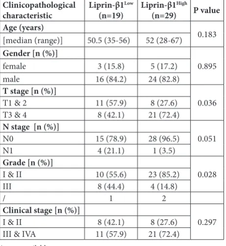
Since the expression of liprin-β1 varied from negative to strong in human HCC samples as shown above (Fig. 1), we explored wheth-er its levels wwheth-ere associated with clinicopathological features of HCC patients. Gender or age at di-agnosis, the two basic parameters related to HCC development, had no impact on liprin-β1 expression (Table 1, Fig. 2A and B). Expres-sion of liprin-β1 did not correlate with tumor differentiation status either (Table 1). Notably, a signifi-cant association between elevated liprin-β1 expression with more ad-vanced TNM stages was evident (P
=0.036). HCC patients with higher liprin-β1 expression were more prone to have TNM stages III-IV (21/29, 72%) (Fig. 3A and B). To-gether, these data demonstrated that reduced liprin-β1 may be an indicator of malignant progression of HCC.
CCC showed lower expression of liprin-β1 than HCC
CCC represents the second most common primary liver cancer and its worldwide incidence continues to rise [18]. To test whether the ex-pression levels of liprin-β1 protein were related to different subtypes of liver cancer, we compared the staining intensities of liprin-β1 in HCC tissues and CCC tissues. Contrary to the moderately strong staining in HCC samples (29/48, 60%) (Fig. 1), we barely detected liprin-β1 staining in CCC samples (0/5, 0%) (Fig. 4A). Comparison of liprin-β1 expression between HCC
Fig. 2. Liprin-β1 expression was not correlated with patients’ gender or age of diagnosis in HCC. A – Quantification of liprin-β1 expression with gender of diagnosis (P=0.895). B – Quantification of liprin-β1 expression with age of diagnosis (P=0.183).
Fig. 3. Comparison of liprin-β1 expression in HCC specimens at different TNM stages. A – Protein expression level of liprin-β1 in HCC tissues of different TNM stages. Tumor samples were stratified into liprin-β1Low and liprin-β1High groups and representative images
are shown. Scale bars, 50 μm. B – Quantification of liprin-β1 expression in HCC tissues at different TNM stages (P<0.05).
and CCC suggested a subtype-specific upregulation of liprin-β1 in liver cancers (Fig. 4B), which needs to be confirmed by a larger population-based study.
DISCUSSION
Understanding the molecular genetics and pathogen-esis of HCC is urgently needed to meet research and clinical demands. In the present study, we provide the first analysis of liprin-β1 protein expression in human liver cancer tissues. We reported that expression lev-els of liprin-β1 were enhanced in advanced HCC and was positively associated with tumor staging in HCC patients. Notably, liprin-β1 was identified as the most upregulated gene in liver tissues from patients with nonalcoholic fatty liver disease (NAFLD) compared with controls by transcriptomic profiling [19]. Con-sidering that NAFLD can progress to nonalcoholic steatohepatitis, one of the important risk factors for HCC, the data indicate that liprin-β1 may participate in a spectrum of liver pathologies, which needs to be further ascertained. Taken together, these findings may help to improve biomarker-based HCC diagnosis and patient stratification.
Protein members of the liprin family have been implicated in several human malignancies. Howev-er, evidence supports either a pro-oncogenic role or tumor-suppressive role in different cancer types. For example, liprin-α1, encoded by PPFIA1, is amplified in breast cancer and promotes tumor invasion and me-tastasis [10,20,21], while in head and neck squamous cell carcinoma, liprin-α1 exhibits anti-invasive effects [22]. Together with our findings in liver cancers, these observations strongly suggest that liprin proteins, in-cluding liprin-β1, contribute to human cancers in a tissue context-dependent manner. We propose that the functional divergence of liprin-β1 may be related to different interaction partners and subcellular localiza-tion in different cell types. For example, endogenous liprin-α1 and liprin-β1 colocalize near the protrud-ing cell front and promote cell invasion by positively regulating the organization of lamellipodia and inva-dopodia [12,21,23], whereas liprin-β1 shows a diffuse distribution in the cytoplasm and plays inhibitory roles in the invasive behavior of cancer cells [10,24,25].
Aside from forming dimers with other liprin members, liprin-β1 has been shown to interact with metastasis-associated protein S100A4 and phospho-binding protein 14-3-3ζ [26,27]. Consistent with the role of liprin-β1 in promoting cell motility and inva-sion, both S100A4 and 14-3-3ζ have been shown to facilitate epithelial-to-mesenchymal transition (EMT) and cancer metastasis in HCC [28-31], raising the possibility that these two proteins may contribute to the oncogenic role of liprin-β1 in HCC. Recent pro-teomics studies also demonstrated that liprin-β1 is present in several well-characterized structural com-plexes, including the cadherin adhesome and micro-tubule attachment complexes [32,33]. However, the biological functions of liprin-β1 in these signaling complexes have not been experimentally investigated. Further elucidation of the molecular mechanisms may explain the behavioral and functional variations of liprin-β1 in different cancer types.
Taken together, our results demonstrate an associ-ation between elevated liprin-β1 expression and tumor malignancy in liver cancer, suggesting that liprin-β1 might serve as a potential biomarker for the stratifi-cation of HCC patients. Further studies on a larger patient population and investigation of liprin-β1’s bio-logical role in HCC progression are highly warranted.
Table 1. Relationship between clinicopathological characteristics and liprin-β1 protein expression in hepatocellular carcinoma
Clinicopathological
characteristic Liprin-β1
Low
(n=19) Liprin-β1
High
(n=29) P value
Age (years)
0.183
[median (range)] 50.5 (35-56) 52 (28-67)
Gender [n (%)]
0.895
female 3 (15.8) 5 (17.2)
male 16 (84.2) 24 (82.8)
T stage [n (%)]
0.036
T1 & 2 11 (57.9) 8 (27.6)
T3 & 4 8 (42.1) 21 (72.4)
N stage [n (%)]
0.051
N0 15 (78.9) 28 (96.5)
N1 4 (21.1) 1 (3.5)
Grade [n (%)]
0.028
I & II 10 (55.6) 23 (85.2)
III 8 (44.4) 4 (14.8)
/ 1 2
Clinical stage [n (%)]
0.297
I & II 8 (42.1) 8 (27.6)
III & IVA 11 (57.9) 21 (72.4)
Acknowledgments: This work was supported by the Jiangsu Province Natural Science Foundation for Youths (BK20150358, BK20180222) and the China Postdoctoral Science Foundation Funded Project (2018M632375).
Author contributions: J.M.L. and J.Y. conceived and designed the experiments. X.Y.L. performed the experiments. X.Y.L. ana-lyzed the data. J.Y. and J.M.L. contributed the reagents/materials/ analysis tools. X.Y.L. wrote the manuscript. All authors read and approved the final manuscript.
Conflict of interest disclosure: None of the authors has a conflict of interest to disclose.
REFERENCES
1. Jemal A, Bray F, Center MM, Ferlay J, Ward E, Forman D. Global Cancer Statistics. CA Cancer J Clin. 2011;61(2):69-90. 2. Jemal A, Center MM, DeSantis C, Ward EM. Global Patterns
of Cancer Incidence and Mortality Rates and Trends. Cancer Epidemiol Biomarkers Prev. 2010;19(8):1893-907.
3. Youssef MI, Maghraby H, Youssef EA, El-Sayed MM. Expres-sion of Ki 67 in Hepatocellular Carcinoma Induced by Dieth-ylnitrosamine in Mice and its Correlation with Histopatho-logical Alterations. J Appl Pharm Sci. 2012;2(3):52-9. 4. Ferenci P, Fried M, Labrecque D, Bruix J, Sherman M, Omata
M, Heathcote J, Piratsivuth T, Kew M, Otegbayo JA, Zheng SS, Sarin S, Hamid SS, Modawi SB, Fleig W, Fedail S, Thom-son A, Khan A, Malfertheiner P, Lau G, Carillo FJ, Krabshuis J, Le Mair A. Hepatocellular Carcinoma (HCC): a Global Per-spective. Arab J Gastroenterol. 2010;11(3):174-9.
5. Ma S, Lee TK, Zheng BJ, Chan KW, Guan XY. CD133+ HCC Cancer Stem Cells Confer Chemoresistance by Preferential Expression of the Akt/PKB Survival Pathway. Oncogene. 2008;27(12):1749-58.
6. Stryker E, Johnson KG. LAR, Liprin α and the Regulation of Active Zone Morphogenesis. J Cell Sci. 2007;120(21):3723-8. 7. Spangler SA, Hoogenraad CC. Liprin-α Proteins: Scaffold
Molecules for Synapse Maturation. Biochem Soc Trans. 2007;35(5):1278-82.
8. Serrapagès C, Medley QG, Tang M, Hart A, Streuli M. Liprins, a Family of LAR Transmembrane Protein-Tyrosine Phospha-tase-Interacting Proteins. J Biol Chem. 1998;273(25):15611-20.
9. Wei Z, Zheng S, Spangler SA, Yu C, Hoogenraad CC, Zhang M. Liprin-Mediated Large Signaling Complex Organization Revealed by the Liprin-α/CASK and Liprin-α/Liprin-β Com-plex Structures. Mol Cell. 2011;43(4):586-98.
10. Chiaretti S, Astro V, Chiricozzi E, de Curtis I. Effects of the Scaffold Proteins Liprin-alpha1, beta1 and beta2 on Invasion by Breast Cancer Cells. Biol Cell. 2016;108(3):65-75. 11. Norrmen C, Vandevelde W, Ny A, Saharinen P, Gentile M,
Haraldsen G, Puolakkainen P, Lukanidin E, Dewerchin M, Alitalo K, Petrova TV. Liprin (beta)1 is Highly Expressed in Lymphatic Vasculature and is Important for Lymphatic Vessel Integrity. Blood. 2010;115(4):906-9.
12. Astro V, Tonoli D, Chiaretti S, Badanai S, Sala K, Zerial M, de Curtis I. Liprin-α1 and ERC1 Control Cell Edge Dynamics by Promoting Focal Adhesion Turnover. Sci Rep. 2016;6:33653. 13. Luo M, Mengos AE, Mandarino LJ, Sekulic A. Association of Liprin β-1 with Kank Proteins in Melanoma. Exp Dermatol. 2016;25(4):321-3.
14. Shimada Y, Kohno T, Ueno H, Ino Y, Hayashi H, Nakaoku T, Sakamoto Y, Kondo S, Morizane C, Shimada K, Okusaka T, Hiraoka N. An Oncogenic ALK Fusion and an RRAS Muta-tion in KRAS MutaMuta-tion-Negative Pancreatic Ductal Adeno-carcinoma. Oncologist. 2017;22(2):158-64.
15. Selvanathan SP, Graham GT, Erkizan HV, Dirksen U, Nata-rajan TG, Dakic A, Yu S, Liu X, Paulsen MT, Ljungman ME, Wu CH, Lawlor ER, Üren A, Toretsky JA. Oncogenic Fusion Protein EWS-FLI1 is a Network Hub that Regulates Alterna-tive Splicing. Proc Natl Acad Sci U S A. 2015;112(11):E1307-16.
16. Heidenblad M, Jonson T, Mahlamäki EH, Gorunova L, Karhu R, Johansson B, Höglund M. Detailed Genomic Mapping and Expression Analyses of 12p Amplifications in Pancreatic Car-cinomas Reveal a 3.5-Mb Target Region for Amplification. Genes Chromosomes Cancer. 2002;34(2):211-23.
17. Johansson FK, Goransson H, Westermark B. Expression Anal-ysis of Genes Involved in Brain Tumor Progression Driven by Retroviral Insertional Mutagenesis in Mice. Oncogene. 2005;24(24):3896-905.
18. Plentz RR, Malek NP. Clinical Presentation, Risk Factors and Staging Systems of Cholangiocarcinoma. Best Pract Res Clin Gastroenterol. 2015;29(2):245-52.
19. Gawrieh S, Baye TM, Carless M, Wallace J, Komorowski R, Kleiner DE, Andris D, Makladi B, Cole R, Charlton M, Cur-ran J, Dyer TD, Charlesworth J, Wilke R, Blangero J, Kissebah AH, Olivier M. Hepatic Gene Networks in Morbidly Obese Patients with Nonalcoholic Fatty Liver Disease. Obes Surg. 2010;20(12):1698-709.
20. Al-Kuraya K, Schraml P, Torhorst J, Tapia C, Zaharieva B, Novotny H, Spichtin H, Maurer R, Mirlacher M, Köchli O, Zuber M, Dieterich H, Mross F, Wilber K, Simon R, Sauter G. Prognostic Relevance of Gene Amplifications and Coamplifi-cations in Breast Cancer. Cancer Res. 2004;64(23):8534-40. 21. Astro V, Chiaretti S, Magistrati E, Fivaz M, de Curtis I. Lip-rin-alpha1, ERC1 and LL5 Define Polarized and Dynamic Structures that are Implicated in Cell Migration. J Cell Sci. 2014;127(Pt 17):3862-76.
22. Tan KD, Zhu Y, Tan HK, Rajasegaran V, Aggarwal A, Wu J, Wu HY, Hwang J, Lim DT, Soo KC, Tan P. Amplification and Overexpression of PPFIA1, a Putative 11q13 Invasion Sup-pressor Gene, in Head and Neck Squamous Cell Carcinoma. Genes Chromosomes Cancer. 2008;47(4):353-62.
23. Sala K, Raimondi A, Tonoli D, Tacchetti C, de Curtis I. Iden-tification of a Membrane-less Compartment Regulating Inva-dosome Function and Motility. Sci Rep. 2018;8(1):1164. 24. Chiaretti S, de Curtis I. Role of Liprins in the Regulation of
Tumor Cell Motility and Invasion. Curr Cancer Drug Targets. 2016;16(3):238-48.
Microenviron-ments by Suppressing Expression of Rab17 and Liprin-β2. J Cell Sci. 2012;125(6):1465-77.
26. Jin J, Smith FD, Stark C, Wells CD, Fawcett JP, Kulkarni S, Metalnikov P, O’Donnell P, Taylor P, Taylor L, Zougman A, Woodgett JR, Langeberg LK, Scott JD, Pawson T. Proteomic, Functional, and Domain-based Analysis of in vivo 14-3-3 Binding Proteins Involved in Cytoskeletal Regulation and Cellular Organization. Curr Biol. 2004;14(16):1436-50. 27. Kriajevska M, Fischer-Larsen M, Moertz E, Vorm O,
Tulchin-sky E, Grigorian M, Ambartsumian N, Lukanidin E. Liprin Beta 1, a Member of the Family of LAR Transmembrane Tyrosine Phosphatase-Interacting Proteins, is a New Target for the Metastasis-associated Protein S100A4 (Mts1). J Biol Chem. 2002;277(7):5229-35.
28. Tang Y, Liu S, Li N, Guo W, Shi J, Yu H, Zhang L, Wang K, Liu S, Cheng S. 14-3-3zeta Promotes Hepatocellular Carcinoma Venous Metastasis by Modulating Hypoxia-inducible Factor-1alpha. Oncotarget. 2016;7(13):15854-67.
29. Tang Y, Lv P, Sun Z, Han L, Zhou W. 14-3-3beta Promotes Migration and Invasion of Human Hepatocellular Car-cinoma Cells by Modulating Expression of MMP2 and MMP9 through PI3K/Akt/NF-kappaB Pathway. PLoS One. 2016;11(1):e0146070.
30. Zhang J, Zhang DL, Jiao XL, Dong Q. S100A4 Regulates Migration and Invasion in Hepatocellular Carcinoma HepG2
Cells via NF-kappaB-dependent MMP-9 Signal. Eur Rev Med Pharmacol Sci. 2013;17(17):2372-82.
31. Yan XL, Jia YL, Chen L, Zeng Q, Zhou JN, Fu CJ, Chen HX, Yuan HF, Li ZW, Shi L, Xu YC, Wang JX, Zhang XM, He LJ, Zhai C, Yue W, Pei XT. Hepatocellular Carcinoma-associated Mesenchymal Stem Cells Promote Hepatocarcinoma Progres-sion: Role of the S100A4-miR155-SOCS1-MMP9 Axis. Hepa-tology. 2013;57(6):2274-86.
32. van der Vaart B, van Riel WE, Doodhi H, Kevenaar JT, Katrukha EA, Gumy L, Bouchet BP, Grigoriev I, Spangler SA, Yu KL, Wulf PS, Wu J, Lansbergen G, van Battum EY, Paster-kamp RJ, Mimori-Kiyosue Y, Demmers J, Olieric N, Maly IV, Hoogenraad CC, Akhmanova A. CFEOM1-associated Kine-sin KIF21A is a Cortical Microtubule Growth Inhibitor. Dev Cell. 2013;27(2):145-60.
33. Guo Z, Neilson LJ, Zhong H, Murray PS, Zanivan S, Zaidel-Bar R. E-cadherin Interactome Complexity and Robust-ness Resolved by Quantitative Proteomics. Sci Signal. 2014;7(354):rs7.
Supplementary Data Supplementary Table S1 and S2.